Article

Immunomodulatory Effect of Benincasa hispida Extract Fermented by Bacillus subtilis CJH 101 on RAW 264.7 Macrophages

Haram Central Research Institute, Cheongju 28160, Republic of Korea
*
Author to whom correspondence should be addressed.
Fermentation 2023, 9(8), 701; https://doi.org/10.3390/fermentation9080701
Submission received: 30 June 2023 / Revised: 24 July 2023 / Accepted: 25 July 2023 / Published: 26 July 2023
(This article belongs to the Special Issue Fermented Foods for Boosting Health)

Abstract

:
This study aimed to confirm the immunomodulatory effects of fermented Benincasa hispida extract (HR1901-BS) on RAW 264.7 macrophages. B. hispida was fermented for 72 h using Bacillus subtilis CJH 101. To evaluate the efficacy of HR1901-BS in enhancing macrophage function, we measured and compared the levels of macrophage activation-related markers in HR1901-BS-stimulated RAW 264.7 cells. We evaluated the effects on nitric oxide (NO) production and the release of pro-inflammatory cytokines (interleukin IL-1β, IL-6, and tumor necrosis factor TNF-β) in the RAW 264.7 cell line. We confirmed that HR1901-BS affected macrophage activation by inducing a significantly high dose-dependent NO production and increasing the release of pro-inflammatory cytokines in RAW 264.7 macrophages. It also suggested that the immunomodulatory effect by HR1901-BS occurred mainly as a result of the activation of cyclooxygenase-2 (COX-2), inducible NO synthase (iNOS), and mitogen-activated protein kinase (MAPK)/nuclear factor-κB (NF-κB) pathways. Our results indicate that HR1901-BS is a promising candidate as a functional material that enhances immune responses through macrophage activation.

1. Introduction

Aging causes numerous biological changes in the immune system, generally resulting in a decline in the immunity response against pathogens as age increases. The prevalence of COVID-19 poses a threat to the health of individuals with metabolic diseases, such as decreased immune function [1]. Natural substances such as medicinal plants are gaining attention as alternatives for immune enhancement owing to their versatility, safety, and cost-effectiveness [2]. Probiotics, including the genera Lactobacillus, Bacillus, and Leuconostoc, are live bacteria that, when consumed in adequate amounts, confer health benefits to the host [3]. Probiotics act as natural immune enhancers by affecting intestinal microbial balance [4]. Thus, the interest in functional and bioactive substances from natural products and probiotics that improve immune function has increased [5,6].
The immune response is a self-defense system that protects the body from external antigens and is maintained through interactions between various immune cells. There are two basic types of immune response in humans: innate and adaptive [7]. Innate immune cells that protect the body from invading foreign antigens include phagocytes such as neutrophils, monocytes, and macrophages. They are activated early during infection by pathogenic antigens, such as viruses, fungi, and bacteria, and inhibit invasion via phagocytosis. In addition, these cells secrete immunomodulatory factors such as nitric oxide (NO), reactive oxygen species, and pro-inflammatory cytokines to induce the activation of innate and adaptive immunity by delivering external antigens [8]. Adaptive immunity is an immune response mediated by B and T cells that are activated during the mid-infection stage and present information on specific antigens to directly kill infected cells or promote the production of antibodies against pathogenic substances [9]. A normal immune system requires a limited inflammatory response to prevent disease. However, an abnormality or dysfunction of the immune system can lead to severe inflammatory diseases, including autoimmune diseases and hypersensitivity [10,11].
Benincasa hispida (Thunb.) Cogn. (Syn. Benincasa Cerifera Savi) is an annual plant of the family Cucurbitaceae that is widely distributed in Asian countries and regions, such as Korea, China, and India [12]. It is used mainly for edible and medicinal purposes in Asian countries, and currently, the Korean food ingredient list includes fruits and seeds, excluding peels [13,14]. The bioactive compounds present in B. hispida, including vitamins, essential minerals, flavonoids, phenolic compounds, and volatile oils, have been extensively studied [15]. Several studies on the functionality of B. hispida extract have reported its antioxidant, anti-inflammatory, anti-osteoporosis, anti-obesity, and cholesterol improvement effects [16,17,18,19,20,21,22]. However, no study has investigated the effects of B. hispida and fermented B. hispida on the prevention of immune-associated diseases, including the enhancement of the immune response.
In the food fermentation process, functional microorganisms are used to preserve food and improve its organoleptic properties, such as flavor, taste, texture, and nutritional value [23]. Modifying the chemical composition of natural products has the advantages of changing their nutritional and biochemical qualities, increasing food safety, and promoting health [24,25,26]. Bacillus spp. are probiotics that can survive in extreme environments by forming endospores and are stable in commercial foods. Via fermentation, Bacillus spp. produce certain essential nutrients, including lactic and amino acids and vitamins K and B12 [27,28]. These species are also known to improve intestinal microbial flora by promoting the growth of anaerobic bacteria via the consumption of free oxygen in the intestine, promoting intestinal motility, and strengthening the immune system [29,30,31,32]. Therefore, fermentation using Bacillus spp. can be considered a method for enhancing the bioactivity of plant extracts.
Therefore, in this study, the immunomodulatory effect of B. hispida extract fermented with Bacillus subtilis CJH 101 (HR1901-BS) was confirmed using RAW 264.7 macrophages and its potential as a functional food material using natural plant resources was confirmed by identifying the mechanism thereof. Here, we evaluated the effect of HR1901-BS on immunomodulatory biomarkers such as NO and cytokine production, expression of iNOS, COX-2 and MAPK/NFκB.

2. Materials and Methods

2.1. Chemicals

Trypsin-ethylenediaminetetraacetic acid (Trypsin-EDTA, 0.05%), penicillin/streptomycin (P/S), phosphate-buffered saline (PBS, pH 7.4), Dulbecco’s modified Eagle’s medium (DMEM), and fetal bovine serum (FBS) were purchased from Gibco (Waltham, MA, USA). Lipopolysaccharides (LPS) were purchased from Sigma-Aldrich (St. Louis, MO, USA). The MTT and nitric oxide (NO) assay kits were obtained from DoGenBio (Seoul, Republic of Korea) and IntronBio (Seoul, Republic of Korea), respectively. All antibodies and cytokine assay kits used for Western blotting and immunoblotting, respectively, were obtained from Cell Signaling Technology (Danvers, MA, USA), Santa Cruz Biotechnology (Santa Cruz, CA, USA), and R&D Systems (St. Louis, MO, USA). The bovine serum albumin (BSA) and enhanced chemiluminescence (ECL) kits were purchased from Bio-Rad (Hercules, CA, USA).

2.2. Sample Preparation

B. hispida cong., harvested in August 2020 in Goesan, Chungcheongbuk-do, was used. The fruit parts, except for the peel and seeds, were used. The HR1901-BS was prepared as follows, B. hispida pulp (2.4 kg) was mixed with water (2.4 kg). Then, 180 mL of B. subtilis CJH 101 (initial bacterial count: 7 × 10⁶ CFU/mL) was inoculated, cultured for 12 h, and then fermented at 33 °C for 72 h. Five times the amount of water was added to the fermented product, then the product was extracted by boiling it at 120 °C for 4 h, filtered with a 106 μm pore size test sieve (Chung Gye Inc., Seoul, Republic of Korea), and placed into a vacuum rotary evaporator (Eyela, Tokyo Rikakikai Co. Ltd., Tokyo, Japan). Thereafter, the sample was freeze-dried (Labcono, Kansas City, MO, USA) to obtain the fermented product of B. hispida (HR1901-BS).

2.3. Cell Culture and Cell Viability

RAW 264.7 macrophages were procured from the American Type Culture Collection (TIB-71, Manassas, VA, USA) and cultured in DMEM at 37 °C in a 5% CO2 incubator (Thermo Fisher Scientific, Waltham, MA, USA) with 10% FBS supplement and 100 units/mL P/S. Equal numbers of cells were plated in 96-well plates at a density of 1 × 10⁵ cells/well. The cells were incubated for 48 h following treatment with HR1901-BS. Cell viability was measured using the MTT assay kit (DoGenBio). The MTT reagents were added to the medium and incubated at 37 °C for 3 h in a 5% CO2 incubator. The absorbance of the wells was measured at 450 nm using a microplate reader (Epoch; Bitek Instruments, Inc., Winooski, VT, USA). All experiments were repeated three times.

2.4. Measurements of Nitrite (NO) Production

RAW 264.7 cells were plated in 24-well plates at a density of 5 × 10⁴ cells/well and incubated for 24 h. The medium was replaced with a fresh medium containing HR1901-BS at various doses for 48 h. Each sample culture medium was analyzed using an NO assay kit (Intron Bio, Sungnam, Republic of Korea) according to the manufacturer’s protocol. The absorbance of the reactants was measured at 540 nm. All experiments were repeated three times.

2.5. Measurement of Cytokines Secretion Analysis

RAW 264.7 cells were treated with HR1901-BS for 48 h. The cell culture mediums were centrifuged at 500× g for 5 min to obtain the supernatant. The levels of tumor necrosis factor (TNF)-α, interleukin (IL)-6, and IL-1β were determined using the supernatant and an ELISA kit (R&D systems) according to the manufacturer’s protocol. All experiments were repeated three times.

2.6. Western Blotting

We performed a Western blotting analysis on the Raw 264.7 macrophages. The RAW 264.7 cells were plated in 100 pi plates at a density of 5 × 10⁵ cells/well and incubated. After 24 h, RAW 264.7 cells were treated with HR1901-BS for 48 h. The cells were then harvested and washed with PBS. The cells were lysed with radioimmunoprecipitation assay (RIPA) buffer, and the supernatant was separated by centrifugation (15,800× g, 15 min). The Protein assay kit standardized with bovine serum albumin (Bio-Rad, Hercules, CA, USA) was used to measure the quantified protein concentration in the supernatant. Thereafter, each protein was separated by 10% sodium dodecyl sulfate-polyacrylamide gel electrophoresis (SDS-PAGE) using SDS running buffer (25 mM Tris, 190 mM glycine, 0.1% SDS, pH 8.3) for 1 h at 100 V. The separated proteins were transferred onto polyvinylidene fluoride membranes (PVDF) using transfer buffer (25 mM Tris base, 190 mM glycine, 10% methanol, pH 8.3) for 1 h and 30 min at 100 V. The membrane was blocked with 5% bovine serum albumin (BSA) and 5% skim milk at 37 °C for 1 h and subsequently washed three times for 10 min each with Tris-buffered saline containing 5% Tween-20 (TBS-T). The primary antibodies (cyclooxygenase-2, COX-2; Extracellular signal-regulated kinase 1/2, ERK; phospho-ERK; inducible NO synthase, iNOS; inhibitory kappa B, IκB; phospho-IκB; c-Jun N-terminal kinases, JNK; phospho-JNK; p38 mitogen-activated protein kinases, p38; phospho-p38, nuclear factor kappa light chain enhancer of activated B cells, NF-κB; phospho-NFκB; β-actin) were diluted 1:1000 in 1×TBS-T, reacted for 12 h at 4 °C, and were washed off thrice with 1×TBS-T. Secondary antibodies were diluted 1:5000 in 1×TBS-T, reacted for 1 h, and were washed again with TBS-T. After applying a color developing reagent of enhanced chemiluminescence (BIONOTE, Hwaseong, Korea) to the membrane, the sample was exposed to a ChemiDoc™ imaging system (Bio-Rad, Hercules, CA, USA), developed, and quantified using an imaging densitometer (ImageJ bundled with 64-bit Java 1.8.0_112, National Institutes of Health, Bethesda, Maryland, USA). All experiments were repeated thrice.

2.7. Statistical Analysis

Values for all experiments were analyzed using GraphPad Prism 8.0 (GraphPad Software, San Diego, CA, USA). The significance between each experimental group was compared using a Student’s t-test and one-way analysis of variance (ANOVA), and statistical significance was set at p < 0.05, p < 0.01, and p < 0.001. Student’s t-test and one-way analysis of variance (ANOVA) were used for the evaluation of statistical significance.

3. Results

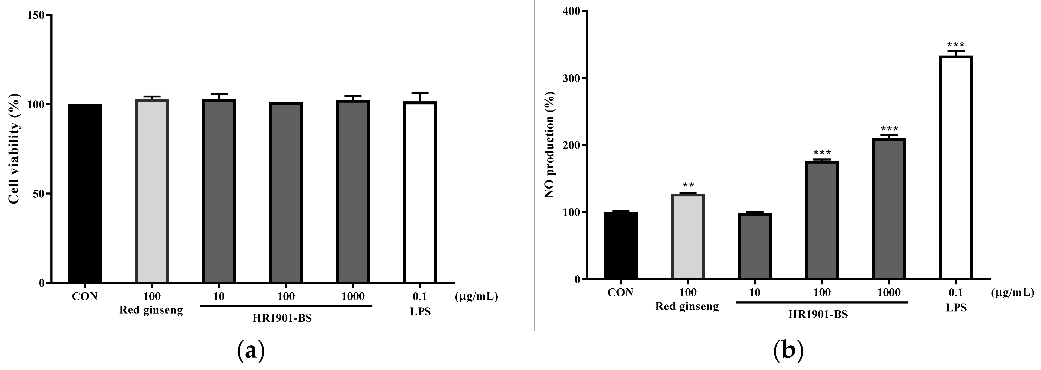
3.1. Effect of HR1901-BS on RAW 264.7 Cell Viability

The MTT assay was performed to evaluate the effect of HR1901-BS on the viability of RAW 264.7 cells. The MTT assay uses the principle that tetrazolium salt is reduced to an insoluble formazan dye by the dehydrogenase of living cells, resulting in color development [33]. As shown in Figure 1a, different concentrations of HR1901-BS (10, 100, and 1000 µg/mL) and 100 µg/mL red ginseng treatment did not affect the viability of RAW 264.7 cells compared to the control (104.33 ± 2.08%, 106.33 ± 1.15%, 103 ± 3.46%, and 107.67 ± 2.52%, respectively). Therefore, we evaluated the immunomodulatory efficacy of HR1901-BS in RAW 264.7 macrophages at concentrations of 10, 100, and 1000 μg/mL to assess the efficacy of immunomodulation.

3.2. Effect of HR1901-BS by Fermentation Time on NO Production in RAW 264.7 Cells

Effects of HR1901-BS on NO production in RAW 264.7 cells were evaluated. The NO analysis principle of the Griess reagent method is that nitrite reacts with sulfanilamide to form a diazonium salt that combines with N-1-naphylethylenediamine to measure the absorbance of the azo compound formed [34]. NO secreted by macrophages activated by the invasion of foreign antigens has various defense functions in the body, such as inhibiting the proliferation of cells or cancer cells infected with pathogenic antigens [35,36]. As shown in Figure 1b, the HR1901-BS treated groups showed significantly upregulated NO production in a dose-dependent manner. Red ginseng is a domestic functional food ingredient with immune-enhancing effects, and the red ginseng treatment group showed a significant increase in NO production. The amount of NO production upregulated to 176.05 ± 1.71%, 210.17 ± 3.42%, and 123.27 ± 1.82% in 100, 1000 μg/mL HR1901-BS and 100 µg/mL red ginseng, respectively, compared to the untreated group.

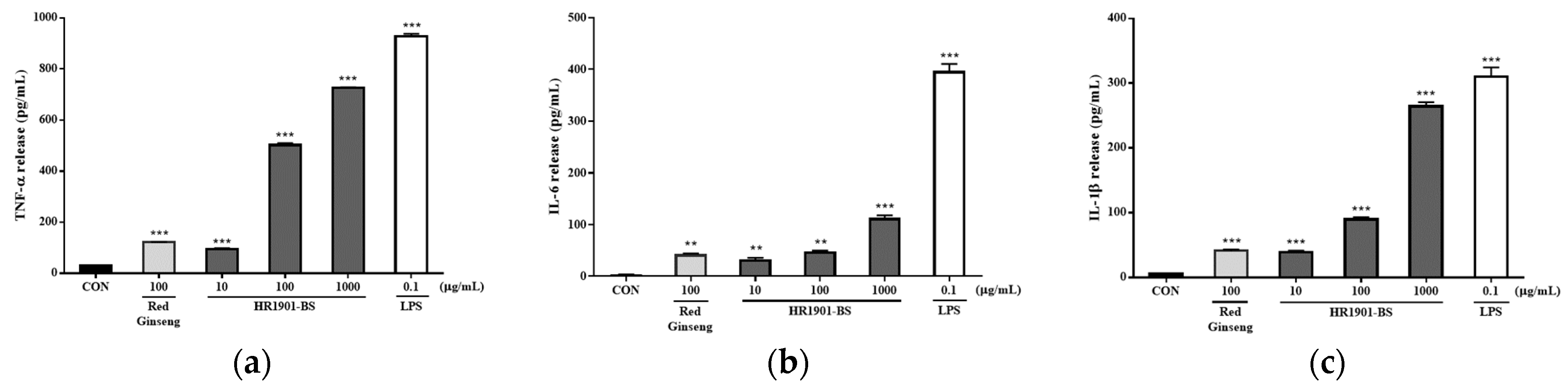
3.3. Effect of HR1901-BS on Cytokine Secretion in RAW 264.7 Cells

To determine the effect of HR1901-BS on cytokine secretion in RAW 264.7 cells, RAW 264.7 cells were incubated with HR1901-BS for 48 h, and the cytokine levels in the culture supernatant were measured. Activated macrophages can induce an immune response by secreting immune mediators such as NO and cytokines [37,38]. Representative cytokines secreted by macrophages include TNF-α, IL-6, and IL-1 β, which play key roles in suppressing excessive inflammation following the initial immune response and in promoting the differentiation of B and T cells [39,40]. As shown in Figure 2, HR1901-BS upregulated the secretion of TNF-α, IL-6, and IL-1 β in a dose-dependent manner. The 10, 100 and 1000 µg/mL HR1901-BS strongly stimulated TNF-α secretion by 3.04, 16.05, and 23.19 fold, respectively, compared with the control group. In addition, IL-6 secretion stimulated 10.87, 15.98, and 37.23 fold, and IL-1β secretion stimulated 7.54, 17.07, and 49.91 fold, respectively. Additionally, the 100 µg/mL red ginseng treatment group showed a significant increase in TNF-α, IL-6, and IL-1 β secretion (3.98, 14.19, and 7.89, respectively). These data suggested that HR1901-BS stimulated the secretion of immune-related cytokines in RAW 264.7 cells.

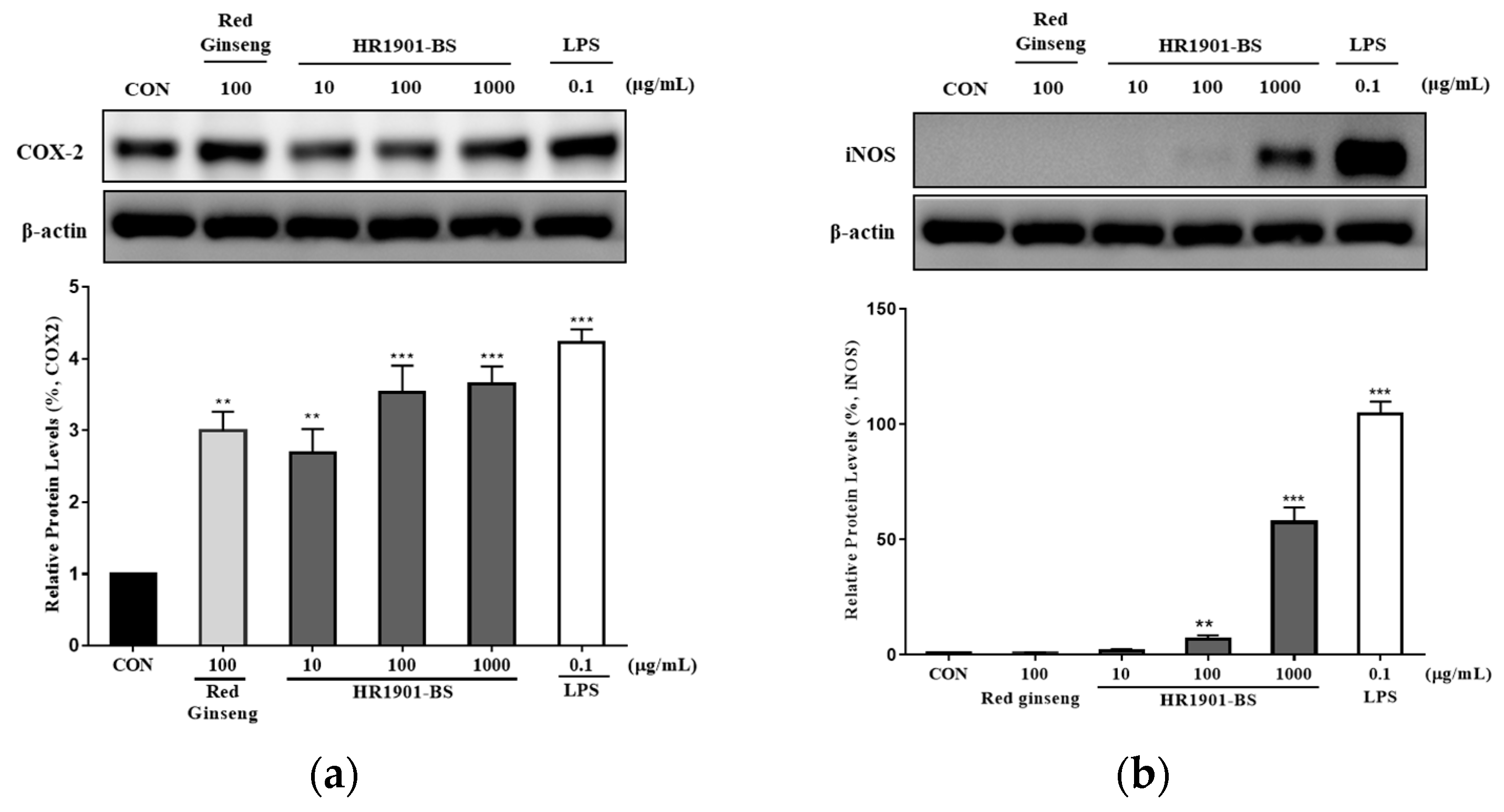
3.4. Effect of HR1901-BS on COX-2 and iNOS Protein Expression in RAW 264.7 Cells

To confirm the effect of HR1901-BS on the activation of cyclooxygenase-2 (COX-2) and inducible NO synthase (iNOS), total protein in HR1901-BS-treated RAW 264.7 cells was analyzed by immunoblotting. COX-2 is an enzyme that converts arachidonic acid into prostaglandin E2 (PGE2) and is involved in cytokine expression and immune responses [41]. iNOS is an enzyme that produces NO to maintain homeostasis of the human immune response and catalyzes the conversion of the amino acid L-arginine and molecular oxygen into L-citrulline and NO [42]. As shown in Figure 3, COX-2 and iNOS protein expressions were significantly increased by HR1901-BS treatment in a dose-dependent manner. The 10, 100 and 1000 µg/mL HR1901-BS treatment strongly stimulated COX-2 secretion by 2.68, 3.53, and 3.65 fold, respectively, compared with the control group. In addition, 1000 µg/mL HR1901-BS treatment stimulated the expression of iNOS secretion by 57.42 fold compared with the control group. In addition, the red ginseng-treated group, which was used as a positive control, was stimulated about 3.2 fold compared with the control group of COX-2. Therefore, HR1901-BS may promote immunity in RAW 264.7 cells by stimulating COX-2 and iNOS expression.

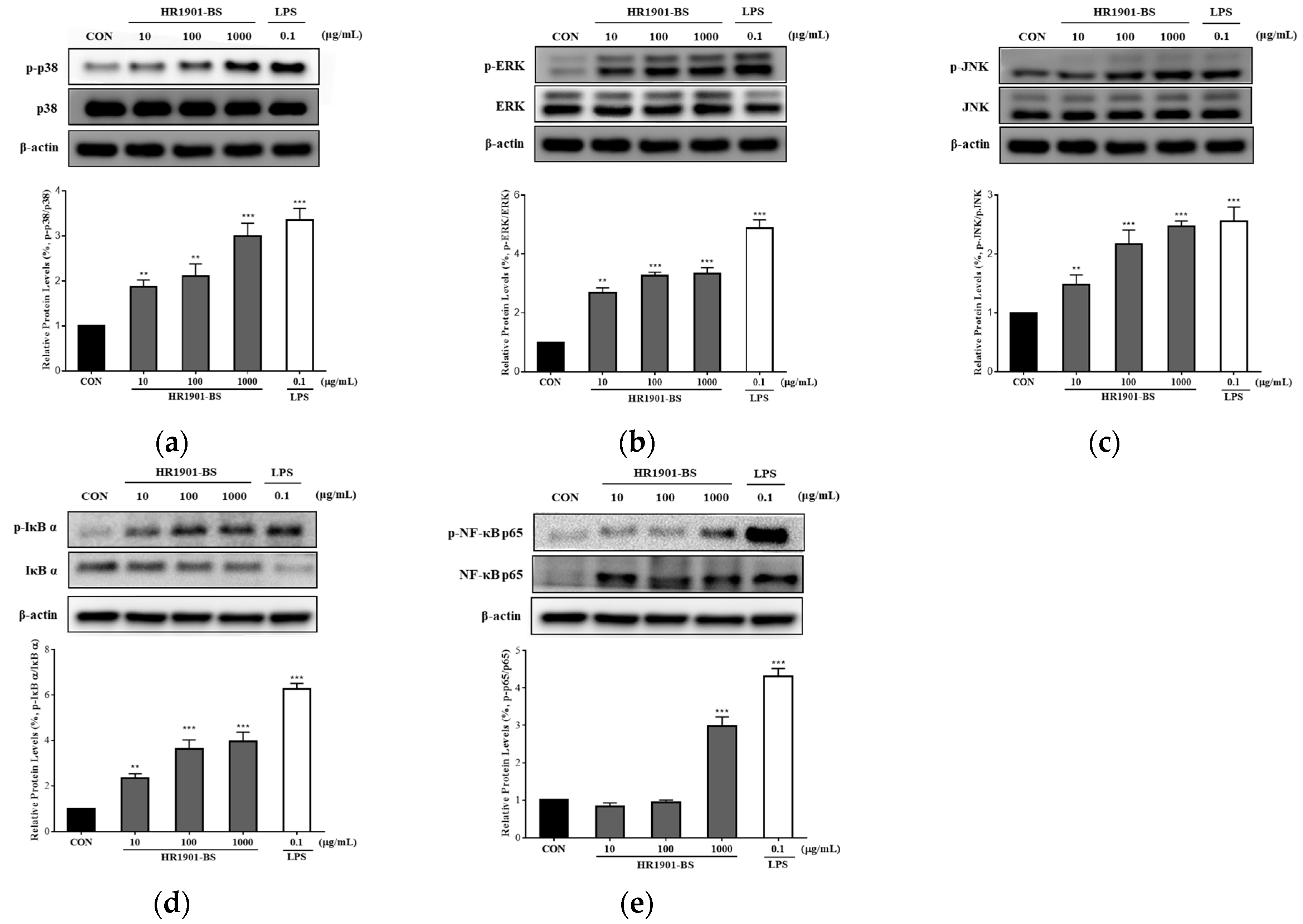
3.5. Effect of HR1901-BS on the MAPK and NF-κB Pathways in RAW 264.7 Cells

To determine the mechanisms by which HR1901-BS upregulates NO production and activates pro-inflammatory cytokines, we assessed mitogen-activated protein kinase (MAPK; p38, ERK, and JNK) and nuclear factor-κB (NF-κB; IκB and p65) pathway phosphorylation in HR1901-BS-treated RAW 264.7 cells. MAPK and NF-κB pathways are known to play key roles in mediating innate and adaptive immunities [43,44]. Activation of the MAPK pathway by immune modulators activates several transcription factors, such as NF-kB, which increases NO production, stimulates the expression of iNOS and COX-2, and increases the production of pro-inflammatory mediators, including immune-related cytokines [45,46]. As shown in Figure 4, the expressions of p38, ERK, and JNK were significantly increased by HR1901-BS treatment in a dose-dependent manner (Figure 4a–c). The 10, 100 and 1000 µg/mL HR1901-BS strongly stimulated phosphorylated p38 expression by 2.1, 2.99, and 3.35 fold, respectively, compared with the control group. In addition, phosphorylated ERK stimulated 2.69, 3.27, and 3.34 fold, and phosphorylated JNK stimulated 1.48, 2.17, and 2.47 fold, respectively. HR1901-BS also markedly increased the phosphorylation of IκBα and NF-κB (p65) (Figure 4d,e). The 10, 100 and 1000 µg/mL HR1901-BS strongly stimulated phosphorylated IκB expression by 2.35, 3.64, and 3.96 fold, respectively, compared with the control group. In addition, phosphorylated NFκB stimulated 2.98 fold in the 1000 µg/mL HR1901-BS. This suggests that HR1901-BS can enhance the immune response by stimulating the upregulated MAPK and NF-κB signaling pathways in activated macrophages.

4. Discussion

Recently, there has been growing interest in natural products for the support of the prevention and treatment of chronic diseases caused by dysfunctions of the immune system [47,48,49]. Despite the various potential pharmacological activities of B. hispida, studies targeting its immunomodulatory activity have yet to be conducted. In the current study, we evaluated the immunomodulatory potential of B.hispida (HR1901-BS) fermented with B.subtilis CJH 101 and determined its effects on NO production, iNOS-mediated COX-2 induced pathway, cytokine transcription, and the MAPK/NFκB pathway in RAW264.7 macrophages.
Fermentation is a process by which microbes produce substances that are useful to humans [50]. Recent research has reported that microbes and fermented foods are effective in modulating the immune system, resulting in health benefits. Chickens fed a diet containing B. subtilis showed significantly increased serum immunoglobulin levels and enhanced innate immune responses, which were induced by toll-like receptor 4 (TLR4) and the production of pro-inflammatory cytokines (IL-1β, IL-6, and IL-8) [31]. In addition, treatment of B. subtilis in Caco-2 cells, which are intestinal epithelial cells, induces the secretion of pro-inflammatory cytokines (IL-6 and IL-8), resulting in the activation of the intracellular NF-κB signaling pathway [51]. Bacillus spp.-fermented Dendrobium officinale polysaccharides stimulated the production of NO and IL-1β in RAW 264.7 cells, thereby showing the potential of promoting immunostimulatory activity [32]. Therefore, B. subtilis has been shown to have the potential as a functional material for enhancing immunity in the fields of single treatment and natural product fermentation.
Macrophages are present in various tissues, several body cavities, and around mucosal surfaces and are crucial in the innate immune system for host defense against many pathogens and cancers [52]. Macrophage toll-like receptors (TLRs) are pattern recognition receptors expressed on the surface of innate immune cells, including macrophages. It binds to the pathogen-associated molecular patterns (PAMPs) of invading microorganisms and activates antigen-recognizing cells, resulting in the activation of macrophages and signaling pathways, such as MAPK and NF-κB, followed by the production of pro-inflammatory cytokines and chemokines [53,54]. LPS, a type of PAMP, is a high-molecular-weight substance composed of lipids and polysaccharides found in Gram-negative bacteria and is an endotoxin that causes an inflammatory response in the human body. Macrophages are activated by LPS and release NO, pro-inflammatory cytokines, and chemokines to trigger an inflammatory response [55,56]. Therefore, we used LPS as a positive control for macrophage activation (Figure 1b).
NO is synthesized during the immune response of macrophages against infectious agents, and it serves as a mediator that regulates both innate and adaptive immune systems [57,58]. The stimulation and release of pro-inflammatory cytokines are essential for the activation of effective innate host defenses and the subsequent regulation of the adaptive immune response [59]. HR1901-BS significantly increased the NO production of RAW 264.7 macrophages. TNF-α activates the killing ability of cells contaminated by foreign antigens and can regulate the apoptotic response by inducing the NF-κB pathway [60,61]. IL-6 promotes B and T cell activation and antibody production and serves as a link between innate and adaptive immunities [62]. IL-1β plays a role in reducing the spread of the initial infection by reducing the spread of pathogenic antigens. It also activates the release of pro-inflammatory cytokines such as TNF-α and IL-6, which bind to the same receptors [63,64]. Herein, treatment of RAW 264.7 macrophages with HR1901-BS increased the release of various cytokines (TNF-α, IL-1β, and IL-6) involved in NO secretion and immune activity (Figure 2). This indicated that treatment with HR1901-BS contributed to the immune activity of macrophages. Therefore, we analyzed the mechanism underlying the immune response mediated by the treatment of HR1901-BS using Western blotting.
COX-2 participates in the immune response by stimulating the cyclooxygenase pathway, which induces PGE2 synthesis, and PGE2 promotes the recruitment of neutrophils and macrophages to the site of infection by inducing vasodilation and increasing blood flow [65]. iNOS mediates the conversion of L-arginine to L-citrulline and NO and exhibits immunomodulatory effects by regulating the differentiation and function of immune cells and inhibiting T cell activity [66,67]. The initial response of the immune system is regulated by COX-2 and iNOS and their pro-inflammatory mediators. Treatment with HR1901-BS increased COX-2 and iNOS protein expression (Figure 3). MAPK and NF-κB are central delivery cascades of the macrophage immune system, and their activation is involved in the regulation of cytokines, such as TNF-α, IL-1, and IL-6, and inducible enzymes, such as COX-2 and iNOS [68,69]. MAPK includes the ERK (p42/P44), p38, and JNK (p46/p54) pathways and is known to affect signal transduction in the innate immune response [70]. The activation of the MAPK pathway regulates several cellular processes, including oxidative stress, immune response, cell growth, differentiation, and proliferation [71]. Blockade of MAPK p38 has been found to attenuate the expression of NF-κB target genes [72,73]. IκB, an upstream kinase of NF-κB, plays an essential role in the canonical activation pathway of NF-κB. NF-κB binds to IκB in the cytoplasm and is thus inactive. When cells are stimulated by external PAMPs and TLRs, phosphorylation of IκB causes NF-κB to migrate to the nucleus, where it is activated and binds to the promoters of inflammatory-mediating genes to induce gene expression [74,75,76]. A deficiency in NF-κB p65 is known to cause impaired inflammatory responses and macrophage migration due to the invasion of foreign microorganisms [77]. HR1901-BS increases phosphorylation of ERK1/2, p38 MAPK and JNK in the MAPK pathway. In addition, HR1901-BS also increases the phosphorylation of NF-κB and IκB, the downstream signal transduction of MAPK (Figure 4).
In our study, HR1901-BS significantly stimulated the activity of NO, pro-inflammatory cytokines, and related mechanisms; however, the extent of the stimulatory effect was lower than that of LPS. Many studies have found that immune-enhancing materials activate macrophages through the MAPK pathway to exert immune-enhancing effects [78,79,80,81]. The MAPK pathway activates NF-κB, which increases the expression of iNOS, COX-2, and pro-inflammatory cytokines [82,83]. Because macrophage activation is regarded as a target for strengthening the human immune system, HR1901-BS may be used as a functional food with immune-enhancing activities. Currently, we are expecting the immunostimulatory activity by the activation of the MAPK/NF-κB pathways; however, as our in vitro studies cannot fully support the macrophage immunostimulatory activity effect of HR1901-BS, additional research is needed. Future in-depth studies should elucidate this contribution in vitro and in vivo.

5. Conclusions

In summary, this study demonstrated that HR1901-BS upregulates the immune response in RAW 264.7 macrophages. HR1901-BS increased NO production and pro-inflammatory cytokine (TNF-α, IL-1β, and IL-6) release through RAW 264.7 macrophage activation. This immunostimulatory mechanism of HR1901-BS is associated with the regulation of the iNOS-mediated COX-2 induced pathway and the MAPK/NF-kB signaling pathway. In this study, the immune-enhancing effect was evaluated in vitro using macrophages; however, follow-up studies need to reproduce this mechanism and conduct in vivo research. Taken together, our results indicate that HR1901-BS has immune-modulation activities and is a potential natural plant-based functional food for improving immune function.

Author Contributions

Conceptualization, Y.E.C.; methodology, J.M.Y. and C.W.J.; software, Y.E.C. and J.M.Y.; validation, C.W.J. and H.S.H.; formal analysis, H.S.H. and J.M.Y.; investigation, Y.E.C.; resources, J.H.C.; data curation, Y.E.C.; writing—original draft preparation, Y.E.C.; writing—review and editing, J.H.C.; visualization, Y.E.C. and C.W.J.; supervision, J.H.C.; project administration, J.H.C.; funding acquisition, J.H.C. All authors have read and agreed to the published version of the manuscript.

Funding

The research was funded by the Ministry of SMEs and Startups, grant number S3206438.

Institutional Review Board Statement

Not applicable.

Informed Consent Statement

Not applicable.

Data Availability Statement

Data sharing not applicable.

Conflicts of Interest

The authors declare no conflict of interest.

References

  1. Yang, J.; Li, H. The impact of aging and COVID-19 on our immune system: A high-resolution map from single cell analysis. Protein Cell 2020, 11, 703–706. [Google Scholar] [CrossRef]
  2. Catanzaro, M.; Corsini, E.; Rosini, M.; Racchi, M.; Lanni, C. Immunomodulators inspired by nature: A review on curcumin and echinacea. Molecules 2018, 23, 2778. [Google Scholar] [CrossRef] [PubMed] [Green Version]
  3. Guarner, F.; Schaafsma, G.J. Probiotics. Int. J. Food Microbiol. 1998, 39, 237–238. [Google Scholar] [CrossRef] [PubMed]
  4. Schrezenmeir, J.; de Vrese, M. Probiotics, prebiotics, and synbiotics—Approaching a definition. Am. J. Clin. Nutr. 2001, 73, 361s–364s. [Google Scholar] [CrossRef]
  5. Arshad, M.S.; Khan, U.; Sadiq, A.; Khalid, W.; Hussain, M.; Yasmeen, A.; Asghar, Z.; Rehana, H. Coronavirus disease (COVID-19) and immunity booster green foods: A mini review. Food Sci. Nutr. 2020, 8, 3971–3976. [Google Scholar] [CrossRef]
  6. Farzana, M.; Shahriar, S.; Jeba, F.R.; Tabassum, T.; Araf, Y.; Ullah, A.; Tasnim, J.; Chakraborty, A.; Naima, T.A.; Marma, K.K.S.; et al. Functional food: Complementary to fight against COVID-19. Beni-Suef Univ. J. Basic Appl. Sci. 2022, 11, 33. [Google Scholar] [CrossRef]
  7. Howell, M.; Shepherd, M. The immune system. Anaesth. Intensive Care Med. 2021, 22, 518–521. [Google Scholar] [CrossRef]
  8. Germic, N.; Frangez, Z.; Yousefi, S.; Simon, H.U. Regulation of the innate immune system by autophagy: Neutrophils, eosinophils, mast cells, NK cells. Cell Death Differ. 2019, 26, 703–714. [Google Scholar] [CrossRef]
  9. Esche, C.; Stellato, C.; Beck, L.A. Chemokines: Key players in innate and adaptive immunity. J. Investig. Dermatol. 2005, 125, 615–628. [Google Scholar] [CrossRef] [Green Version]
  10. Cutolo, M. Macrophages as Effectors of the Immunoendocrinologic Interactions in Autoimmune Rheumatic Diseasesa. Ann. N. Y. Acad. Sci. 1999, 876, 32–42. [Google Scholar] [CrossRef]
  11. Lesourd, B. Nutritional factors and immunological ageing. Proc. Nutr. Soc. 2006, 65, 319–325. [Google Scholar] [CrossRef] [Green Version]
  12. Mohammad, N.A.; Anwar, F.; Mehmood, T.; Hamid, A.A.; Muhammad, K.; Saari, N. Phenolic compounds, tocochromanols profile and antioxidant properties of winter melon [Benincasa hispida (Thunb.) Cogn.] seed oils. J. Food Meas. Charact. 2019, 13, 940–948. [Google Scholar] [CrossRef]
  13. Zaini, N.A.M.; Anwar, F.; Hamid, A.A.; Saari, N. Kundur [Benincasa hispida (Thunb.) Cogn.]: A potential source for valuable nutrients and functional foods. Food Res. Int. 2011, 44, 2368–2376. [Google Scholar] [CrossRef]
  14. Lim, S.J.; Jeong, J.G.; Kim, M.W.; Choi, S.S.; Han, H.K.; Park, J.E. Effects of Benincasa hispida intake on blood glucose and lipid level in streptozotocin induced diabetic rats. Korean J. Nutr. 2003, 36, 335–343. [Google Scholar]
  15. Du, Q.; Zhang, Q.; Ito, Y. Isolation and identification of phenolic compounds in the fruit of Benincasa hispida by HSCCC. J. Liq. Chromatogr. Relat. Technol. 2005, 28, 137–144. [Google Scholar] [CrossRef]
  16. Kim, M.W. Effects of Benincasa hispida seed supplementation on glycogen status and lipid peroxidation in streptozotocin-induced diabetic rats. Korean J. Nutr. 2004, 37, 865–871. [Google Scholar]
  17. Grover, J.K.; Adiga, G.; Vats, V.; Rathi, S.S. Extracts of Benincasa hispida prevent development of experimental ulcers. J. Ethnopharmacol. 2001, 78, 159–164. [Google Scholar] [CrossRef]
  18. Grover, J.K.; Rathi, S.S. Benincasa hispida: An anti-inflammatory agent with cytoprotective activity (abs). Can. J. Physiol. Pharmaol. 1994, 72, 269. [Google Scholar]
  19. Patil, R.N.; Patil, R.Y.; Ahirwar, B.; Ahirwar, D. Evaluation of antidiabetic and related actions of some Indian medicinal plants in diabetic rats. Asian Pac. J. Trop. Med. 2011, 4, 20–23. [Google Scholar] [CrossRef] [Green Version]
  20. Roy, C.; Guha, D. Role of Benincasa hispida linn. on brain electrical activity in colchicine induced experimental rat model of alzheimer’s disease: Possible involvements of antioxidants. World J. Pharm. Res 2017, 6, 1439–1443. [Google Scholar]
  21. Choi, Y.E.; Yang, J.M.; Cho, J.H. Benincasa hispida extract promotes proliferation, differentiation, and mineralization of MC3T3-E1 preosteoblasts and inhibits the differentiation of RAW 246.7 osteoclast precursors. Appl. Sci. 2022, 12, 8849. [Google Scholar] [CrossRef]
  22. Choi, S.I.; Han, X.; Men, X.; Lee, S.J.; Oh, G.; Choi, Y.E.; Yang, J.M.; Cho, J.H.; Lee, O.H. Benincasa hispida Extract Prevents Ovariectomy-Induced Osteoporosis in Female ICR Mice. Appl. Sci. 2023, 13, 832. [Google Scholar] [CrossRef]
  23. Thapa, N.; Tamang, J.P. Functionality and therapeutic values of fermented foods. Health Benefits Fermented Foods 2015, 111, 168. [Google Scholar]
  24. Martinez-Avila, G.C.G.; Aguilera, A.F.; Saucedo, S.; Rojas, R.; Rodriguez, R.; Aguilar, C.N. Fruit wastes fermentation for phenolic antioxidants production and their application in manufacture of edible coatings and films. Crit. Rev. Food Sci. Nutr. 2014, 54, 303–311. [Google Scholar] [CrossRef] [PubMed]
  25. Tamang, J.P.; Watanabe, K.; Holzapfel, W.H. Diversity of microorganisms in global fermented foods and beverages. Front. Microbiol. 2016, 7, 377. [Google Scholar] [CrossRef] [Green Version]
  26. Skowron, K.; Budzyńska, A.; Grudlewska-Buda, K.; Wiktorczyk-Kapischke, N.; Andrzejewska, M.; Wałecka-Zacharska, E.; Gospodarek-Komkowska, E. Two Faces of Fermented Foods—The Benefits and Threats of Its Consumption. Front. Microbiol. 2022, 13, 845166. [Google Scholar] [CrossRef] [PubMed]
  27. Zhou, M.; Wang, H.; Li, Z. Study on resistance to stress factors of Bacillus subtilis from swine intestinal juice. J. Anhui Agric. Univ. 2013, 40, 519–522. [Google Scholar]
  28. Sanders, M.E.; Morelli, L.; Tompkins, T.A. Sporeformers as human probiotics: Bacillus, Sporolactobacillus, and Brevibacillus. Compr. Rev. Food Sci. Food Saf. 2003, 2, 101–110. [Google Scholar] [CrossRef]
  29. Iebba, V.; Nicoletti, M.; Schippa, S. Gut microbiota and the immune system: An intimate partnership in health and disease. Int. J. Immunopathol. Pharmacol. 2012, 25, 823–833. [Google Scholar] [CrossRef] [PubMed]
  30. García-Albiach, R.; José, M.; de Felipe, P.; Angulo, S.; Morosini, M.-I.; Bravo, D.; Baquero, F.; del Campo, R. Molecular analysis of yogurt containing Lactobacillus delbrueckii subsp. bulgaricus and Streptococcus thermophilus in human intestinal microbiota. Am. J. Clin. Nutr. 2008, 87, 91–96. [Google Scholar] [CrossRef] [Green Version]
  31. Guo, M.; Li, M.; Zhang, C.; Zhang, X.; Wu, Y. Dietary administration of the Bacillus subtilis enhances immune responses and disease resistance in chickens. Front. Microbiol. 2020, 11, 1768. [Google Scholar] [CrossRef]
  32. Hosoi, T.; Hirose, R.; Saegusa, S.; Ametani, A.; Kiuchi, K.; Kaminogawa, S. Cytokine responses of human intestinal epithelial-like Caco-2 cells to the nonpathogenic bacterium Bacillus subtilis (natto). Int. J. Food Microbiol. 2003, 82, 255–264. [Google Scholar] [CrossRef]
  33. Riss, T.L.; Moravec, R.A.; Niles, A.L.; Duellman, S.; Benink, H.A.; Worzella, T.J.; Minor, L. Cell viability assays. Assay Guid. Man. 2016, 1, 1–25. [Google Scholar]
  34. Csonka, C.; Páli, T.; Bencsik, P.; Görbe, A.; Ferdinandy, P.; Csont, T. Measurement of NO in biological samples. Br. J. Pharmacol. 2015, 172, 1620–1632. [Google Scholar] [CrossRef] [PubMed] [Green Version]
  35. Alderton, W.K.; Cooper, C.E.; Knowles, R.G. Nitric oxide synthases: Structure, function and inhibition. Biochem. J. 2001, 357, 593–615. [Google Scholar] [CrossRef] [PubMed]
  36. Kim, J.Y.; Jung, K.S.; Jeong, H.G. Suppressive effects of the kahweol and cafestol on cyclooxygenase-2 expression in macrophages. FEBS Lett. 2004, 569, 321–326. [Google Scholar] [CrossRef] [Green Version]
  37. Arango Duque, G.; Descoteaux, A. Macrophage cytokines: Involvement in immunity and infectious diseases. Front. Immunol. 2014, 5, 491. [Google Scholar] [CrossRef] [PubMed] [Green Version]
  38. Hirayama, D.; Iida, T.; Nakase, H. The phagocytic function of macrophage-enforcing innate immunity and tissue homeostasis. Int. J. Mol. Sci. 2017, 19, 92. [Google Scholar] [CrossRef] [PubMed] [Green Version]
  39. Liu, Y.; Wu, X.; Jin, W.; Guo, Y. Immunomodulatory effects of a low-molecular weight polysaccharide from Enteromorpha prolifera on RAW 264.7 macrophages and cyclophosphamide-induced immunosuppression mouse models. Mar. Drugs 2020, 18, 340. [Google Scholar] [CrossRef]
  40. Murray, R.Z.; Stow, J.L. Cytokine secretion in macrophages: SNAREs, Rabs, and membrane trafficking. Front. Immunol. 2014, 5, 538. [Google Scholar] [CrossRef]
  41. Medeiros, A.; Peres-Buzalaf, C.; Verdan, F.F.; Serezani, C.H. Prostaglandin E2 and the suppression of phagocyte innate immune responses in different organs. Mediat. Inflamm. 2012, 2012, 1–13. [Google Scholar] [CrossRef] [PubMed] [Green Version]
  42. Förstermann, U.; Sessa, W.C. Nitric oxide synthases: Regulation and function. Eur. Heart J. 2012, 33, 829–837. [Google Scholar] [CrossRef] [PubMed] [Green Version]
  43. Rincón, M.; Flavell, R.A.; Davis, R.A. The Jnk and P38 MAP kinase signaling pathways in T cell–mediated immune responses. Free Radic. Biol. Med. 2000, 28, 1328–1337. [Google Scholar] [CrossRef]
  44. Dong, C.; Davis, R.J.; Flavell, R.A. MAP kinases in the immune response. Annu. Rev. Immunol. 2002, 20, 55–72. [Google Scholar] [CrossRef] [PubMed]
  45. Mi, X.J.; Tran, T.H.M.; Park, H.R.; Xu, X.Y.; Subramaniyam, S.; Choi, H.S.; Kim, J.; Koh, S.C.; Kim, Y.J. Immune-enhancing effects of postbiotic produced by Bacillus velezensis Kh2-2 isolated from Korea Foods. Food Res. Int. 2022, 152, 110911. [Google Scholar] [CrossRef] [PubMed]
  46. Hong, S.H.; Ku, J.M.; Kim, H.I.; Ahn, C.W.; Park, S.H.; Seo, H.S.; Shin, Y.C.; Ko, S.G. The immune-enhancing activity of Cervus nippon mantchuricus extract (NGE) in RAW264. 7 macrophage cells and immunosuppressed mice. Food Res. Int. 2017, 99, 623–629. [Google Scholar] [CrossRef]
  47. Trinh, T.A.; Park, J.; Oh, J.H.; Park, J.S.; Lee, D.; Kim, C.E.; Choi, H.-S.; Kim, S.-B.; Hwang, G.S.; Koo, B.A.; et al. Effect of herbal formulation on immune response enhancement in RAW 264.7 macrophages. Biomolecules 2020, 10, 424. [Google Scholar] [CrossRef] [PubMed] [Green Version]
  48. Chen, L.; Yu, J. Modulation of Toll-like receptor signaling in innate immunity by natural products. Int. Immunopharmacol. 2016, 37, 65–70. [Google Scholar] [CrossRef] [Green Version]
  49. Schultze, J.L.; Schmidt, S.V. Molecular features of macrophage activation. In Semin. Immunol. 2015, 27, 416–423. [Google Scholar] [CrossRef]
  50. Voidarou, C.; Antoniadou, M.; Rozos, G.; Tzora, A.; Skoufos, I.; Varzakas, T.; Lagiou, A.; Bezirtzoglou, E. Fermentative foods: Microbiology, biochemistry, potential human health benefits and public health issues. Foods 2020, 10, 69. [Google Scholar] [CrossRef]
  51. Tian, W.; Dai, L.; Lu, S.; Luo, Z.; Qiu, Z.; Li, J.; Li, P.; Du, B. Effect of Bacillus sp. DU-106 fermentation on Dendrobium officinale polysaccharide: Structure and immunoregulatory activities. Int. J. Biol. Macromol. 2019, 135, 1034–1042. [Google Scholar] [CrossRef] [PubMed]
  52. Pidwill, G.R.; Gibson, J.F.; Cole, J.; Renshaw, S.A.; Foster, S.J. The role of macrophages in Staphylococcus aureus infection. Front. Immunol. 2021, 11, 3506. [Google Scholar] [CrossRef] [PubMed]
  53. Takeda, K.; Kaisho, T.; Akira, S. Toll-like receptors. Annu. Rev. Immunol. 2003, 21, 335–376. [Google Scholar] [CrossRef] [PubMed]
  54. Kim, K.W.; Lee, S.H.; Cho, M.L.; Oh, H.J.; Woo, Y.J.; Kim, S.H.; Kim, H.R. IL-17 and Toll-like Receptor 2 or Toll-like Receptor 4 Combined Engagement Upregulates RANKL and IL-6 in Human Rheumatoid Synovial Fibroblasts. J. Korean Rheum. Assoc. 2010, 17, 36–45. [Google Scholar] [CrossRef] [Green Version]
  55. Medzhitov, R.; Janeway, C.A., Jr. Decoding the patterns of self and nonself by the innate immune system. Science 2002, 296, 298–300. [Google Scholar] [CrossRef] [Green Version]
  56. Horwood, N.J.; Page, T.H.; McDaid, J.P.; Palmer, C.D.; Campbell, J.; Mahon, T.; Brennan, F.M.; Webster, D.; Foxwell, B.M. Bruton’s tyrosine kinase is required for TLR2 and TLR4-induced TNF, but not IL-6, production. J. Immunol. 2006, 176, 3635–3641. [Google Scholar] [CrossRef]
  57. MacMicking, J.; Xie, Q.W.; Nathan, C. Nitric oxide and macrophage function. Annu. Rev. Immunol. 1997, 15, 323–350. [Google Scholar] [CrossRef]
  58. Palmieri, E.M.; McGinity, C.; Wink, D.A.; McVicar, D.W. Nitric oxide in macrophage immunometabolism: Hiding in plain sight. Metabolites 2020, 10, 429. [Google Scholar] [CrossRef]
  59. Booth, J.S.; Nichani, A.K.; Benjamin, P.; Dar, A.; Krieg, A.M.; Babiuk, L.A.; Mutwiri, G.K. Innate immune responses induced by classes of CpG oligodeoxynucleotides in ovine lymph node and blood mononuclear cells. Vet. Immunol. Immunopathol. 2007, 115, 24–34. [Google Scholar] [CrossRef]
  60. Oyler-Yaniv, J.; Oyler-Yaniv, A.; Maltz, E.; Wollman, R. TNF controls a speed-accuracy tradeoff in the cell death decision to restrict viral spread. Nat. Commun. 2021, 12, 2992. [Google Scholar] [CrossRef]
  61. Rahman, M.M.; McFadden, G. Modulation of tumor necrosis factor by microbial pathogens. PLoS Pathog. 2006, 2, e4. [Google Scholar] [CrossRef] [PubMed] [Green Version]
  62. Dienz, O.; Eaton, S.M.; Bond, J.P.; Neveu, W.; Moquin, D.; Noubade, R.; Briso, E.M.; Charland, C.; Leonard, W.J.; Ciliberto, G.; et al. The induction of antibody production by IL-6 is indirectly mediated by IL-21 produced by CD4+ T cells. J. Exp. Med. 2009, 206, 69–78. [Google Scholar] [CrossRef] [PubMed]
  63. Dinarello, C.A. Anti-inflammatory agents: Present and future. Cell 2010, 140, 935–950. [Google Scholar] [CrossRef] [PubMed] [Green Version]
  64. Chung, Y.; Chang, S.H.; Martinez, G.J.; Yang, X.O.; Nurieva, R.; Kang, H.S.; Ma, L.; Watowich, S.S.; Jetten, A.M.; Tian, Q.; et al. Critical regulation of early Th17 cell differentiation by interleukin-1 signaling. Immunity 2009, 30, 576–587. [Google Scholar] [CrossRef] [Green Version]
  65. Kulesza, A.; Paczek, L.; Burdzinska, A. The role of COX-2 and PGE2 in the regulation of immunomodulation and other functions of mesenchymal stromal cells. Biomedicines 2023, 11, 445. [Google Scholar] [CrossRef]
  66. Xue, Q.; Yan, Y.; Zhang, R.; Xiong, H. Regulation of iNOS on immune cells and its role in diseases. Int. J. Mol. Sci. 2018, 19, 3805. [Google Scholar] [CrossRef] [Green Version]
  67. Wang, Y.; Zhao, H.; Shao, Y.; Liu, J.; Li, J.; Xing, M. Copper or/and arsenic induce oxidative stress-cascaded, nuclear factor kappa B-dependent inflammation and immune imbalance, trigging heat shock response in the kidney of chicken. Oncotarget 2017, 8, 98103. [Google Scholar] [CrossRef] [Green Version]
  68. DiDonato, J.A.; Hayakawa, M.; Rothwarf, D.M.; Zandi, E.; Karin, M. A cytokine-responsive IκB kinase that activates the transcription factor NF-κB. Nature 1997, 388, 548–554. [Google Scholar] [CrossRef]
  69. Park, J.; Min, J.S.; Kim, B.; Chae, U.B.; Yun, J.W.; Choi, M.S.; Kong, I.K.; Chang, K.T.; Lee, D.S. Mitochondrial ROS govern the LPS-induced pro-inflammatory response in microglia cells by regulating MAPK and NF-κB pathways. Neurosci. Lett. 2015, 584, 191–196. [Google Scholar] [CrossRef]
  70. Johnson, G.L.; Lapadat, R. Mitogen-activated protein kinase pathways mediated by ERK, JNK, and p38 protein kinases. Science 2002, 298, 1911–1912. [Google Scholar] [CrossRef] [Green Version]
  71. Chardin, C.; Schenk, S.T.; Hirt, H.; Colcombet, J.; Krapp, A. Mitogen-Activated protein kinases in nutritional signaling in Arabidopsis. Plant Sci. 2017, 260, 101–108. [Google Scholar] [CrossRef] [PubMed] [Green Version]
  72. Bhat, N.R.; Shen, Q.; Fan, F. TAK1-mediated induction of nitric oxide synthase gene expression in glial cells. J. Neurochem. 2003, 87, 238–247. [Google Scholar] [CrossRef] [PubMed] [Green Version]
  73. Jana, M.; Dasgupta, S.; Saha, R.N.; Liu, X.; Pahan, K. Induction of tumor necrosis factor-α (TNF-α) by interleukin-12 p40 monomer and homodimer in microglia and macrophages. J. Neurochem. 2003, 86, 519–528. [Google Scholar] [CrossRef] [Green Version]
  74. Lin, A.; Karin, M. NF-kB in cancer: A marked target. Semin. Cancer Biol 2003, 13, 107–114. [Google Scholar] [CrossRef]
  75. Xie, Q.W.; Whisnant, R.; Nathan, C. Promoter of the mouse gene encoding calcium-independent nitric oxide synthase confers inducibility by interferon gamma and bacterial lipopolysaccharide. J. Exp. Med. 1993, 177, 1779–1784. [Google Scholar] [CrossRef]
  76. Yamamoto, Y.; Gaynor, R.B. IκB kinases: Key regulators of the NF-κB pathway. Trends Biochem. Sci. 2004, 29, 72–79. [Google Scholar] [CrossRef]
  77. Silverman, N.; Zhou, R.; Stöven, S.; Pandey, N.; Hultmark, D.; Maniatis, T. A Drosophila IκB kinase complex required for Relish cleavage and antibacterial immunity. Genes Dev. 2000, 14, 2461–2471. [Google Scholar] [CrossRef] [Green Version]
  78. Jeong, J.H.; Jang, S.; Jung, B.J.; Jang, K.S.; Kim, B.G.; Chung, D.K.; Kim, H. Differential immune-stimulatory effects of LTAs from different lactic acid bacteria via MAPK signaling pathway in RAW 264.7 cells. Immunobiology 2015, 220, 460–466. [Google Scholar] [CrossRef] [PubMed]
  79. Xie, Y.; Wang, L.; Sun, H.; Wang, Y.; Yang, Z.; Zhang, G.; Jiang, S.; Yang, W. Polysaccharide from alfalfa activates RAW 264.7 macrophages through MAPK and NF-κB signaling pathways. Int. J. Biol. Macromol. 2019, 126, 960–968. [Google Scholar] [CrossRef] [PubMed]
  80. Ko, M.N.; Hyun, S.B.; Ahn, K.J.; Hyun, C.G. Immunomodulatory effects of Abelmoschus esculentus water extract through MAPK and NF-κB signaling in RAW 264.7 cells. Biotechnol. Notes 2022, 3, 38–44. [Google Scholar] [CrossRef]
  81. Liu, X.; Chen, X.; Xie, L.; Xie, J.; Shen, M. Sulfated Chinese yam polysaccharide enhances the immunomodulatory activity of RAW 264.7 cells via the TLR4-MAPK/NF-κB signaling pathway. Food Funct. 2022, 13, 1316–1326. [Google Scholar] [CrossRef] [PubMed]
  82. Ji, C.; Zhang, Z.; Chen, J.; Song, D.; Liu, B.; Li, J.; Liu, R.; Niu, J.; Wang, D.; Ling, N.; et al. Immune-enhancing effects of a novel glucan from purple sweet potato Ipomoea batatas (L.) lam on RAW264. 7 macrophage cells via TLR2-and TLR4-mediated pathways. J. Agric. Food Chem. 2021, 69, 9313–9325. [Google Scholar] [CrossRef] [PubMed]
  83. Yang, Y.; Kim, S.C.; Yu, T.; Yi, Y.S.; Rhee, M.H.; Sung, G.H.; Yoo, B.C.; Cho, J.Y. Functional roles of p38 mitogen-activated protein kinase in macrophage-mediated inflammatory responses. Mediat. Inflamm. 2014, 2014, 352371. [Google Scholar] [CrossRef] [PubMed] [Green Version]
Figure 1. Effects of HR1901-BS on the cell viability, NO production in RAW 264.7 cells. The cells were incubated with HR1901-BS for 48 h. (a) Cell viability and (b) NO production were determined using an MTT assay and Griess reagent assay, respectively. All values are expressed as means ± SD. ** p < 0.01, and *** p < 0.001 when compared with the control. Data are representative of three independent experiments. LPS, lipopolysaccharides; HR1901-BS, B. hispida extract fermented with Bacillus subtilis CJH 101.
Fermentation 09 00701 g001
Figure 2. Effects of HR1901-BS on cytokine (TNF-α, IL-6, and IL-1β) release in RAW 264.7 cells. The cells were incubated with HR1901-BS for 48 h. (a) TNF-α, (b) IL-6, and (c) IL-1β secretion in RAW 264.7 cell culture supernatant was determined using an ELISA kit. All values are expressed as means ± SD. ** p < 0.01, and *** p < 0.001 when compared with the control. Data are representative of three independent experiments. LPS, lipopolysaccharides; HR1901-BS, B. hispida extract fermented with Bacillus subtilis CJH 101; TNF-α, tumor necrosis factor-α; IL, interleukin.
Fermentation 09 00701 g002
Figure 3. Effects of HR1901-BS on COX-2 and iNOS protein expression in RAW 264.7 cells. The cells were incubated with HR1901-BS for 48 h and harvested. The protein expression of (a) COX-2 and (b) iNOS was determined using immunoblotting. The relative protein expression levels were densitometrically quantified with β-actin. All values are expressed as means ± SD. ** p < 0.01, and *** p < 0.001 when compared with the control. Data are representative of three independent experiments. LPS, lipopolysaccharides; HR1901-BS, B. hispida extract fermented with Bacillus subtilis CJH 101; COX-2, cyclooxygenase-2; iNOS, inducible NO synthase.
Fermentation 09 00701 g003
Figure 4. Effects of HR1901-BS on protein expression in MAPK and NF-κB dependent pathways of RAW 264.7 cells. The cells were incubated with HR1901-BS for 48 h and harvested. The protein expression of (a) p38, (b) ERK, (c) JNK, (d) IκB, and (e) NF-κB were determined using immunoblotting. The relative protein expression levels were densitometrically quantified with β-actin. All values are expressed as means ± SD. ** p < 0.01, and *** p < 0.001 when compared with the control. Data are representative of three independent experiments. LPS, lipopolysaccharides; HR1901-BS, B. hispida extract fermented with Bacillus subtilis CJH 101; p38, p38 mitogen-activated protein kinases; ERK, extracellular signal-regulated kinase 1/2; JNK, c-Jun N-terminal kinase; NF-κB, nuclear factor kappa light chain enhancer of activated B cells; IκB, inhibitory kappa B.
Fermentation 09 00701 g004
Disclaimer/Publisher’s Note: The statements, opinions and data contained in all publications are solely those of the individual author(s) and contributor(s) and not of MDPI and/or the editor(s). MDPI and/or the editor(s) disclaim responsibility for any injury to people or property resulting from any ideas, methods, instructions or products referred to in the content.

