Article

Biotransformation of Acetaminophen by Ganoderma parvulum Ligninolytic Enzymes Immobilized on Chitosan Microspheres

by
María Alejandra Flórez-Restrepo
1,*,
Xiomara López-Legarda
1,*,
Magdalena de Jesús Rostro-Alanis
2,
Roberto Parra-Saldívar
3 and
Freimar Segura-Sánchez
1
1
Biopolimer Research Group, Facultad de Ciencias Farmacéuticas y Alimentarias, Universidad de Antioquia UdeA, Calle 70 No. 52-21, Medellín 050010, Colombia
2
Tecnologico de Monterrey, Escuela de Ingenieria y Ciencias, Ave. Eugenio Garza Sada 2501 Sur, Col: Tecnologico, Monterrey 64700, NL, Mexico
3
Magan Centre of Applied Mycology, Cranfield University, Cranfield MK43 0AL, UK
*
Authors to whom correspondence should be addressed.
Fermentation 2025, 11(7), 387; https://doi.org/10.3390/fermentation11070387
Submission received: 30 May 2025 / Revised: 20 June 2025 / Accepted: 1 July 2025 / Published: 5 July 2025
(This article belongs to the Special Issue Application of Fungi in Bioconversions and Mycoremediation)

Abstract

Water quality is essential for safeguarding human health and ensuring the stability of ecosystems. Nonetheless, the rising prevalence of emerging contaminants, particularly pharmaceutical compounds, has raised serious environmental concerns due to their bioactivity, widespread use, persistence, and potential toxicity. Among these, acetaminophen (paracetamol) is one of the most frequently detected pharmaceutical pollutants in aquatic environments. Among the various degradation strategies explored, biological methods, especially those involving white-rot fungi, have shown substantial promise owing to their production of ligninolytic enzymes capable of degrading complex pollutants. This study investigates the use of laccases from Ganoderma parvulum, covalently immobilized on chitosan microspheres, for acetaminophen degradation. The immobilization involved a 10% crosslinking agent, 60-min crosslinking time, and 10,000 U/L enzyme concentration, resulting in an immobilization efficiency of 123%, 203%, and 218%, respectively. The immobilized enzymes displayed enhanced stability across pH 3–8 and temperatures between 20 and 60 °C. Biodegradation assays achieved 97% acetaminophen removal within four hours. Nuclear Magnetic Resonance (1H NMR and COSY) confirmed structural transformation. The enzymes also retained over 95% catalytic activity after multiple reuse cycles. These findings highlight the novel application of laccases as efficient and reusable biocatalysts for pharmaceutical pollutant removal, providing valuable insights into the mechanisms of enzymatic environmental remediation.

1. Introduction

The an international, peer-reviewed Open Access journal. quality of water is fundamental to maintaining ecosystem balance and ensuring human well-being, serving as a cornerstone for sustainable development [1]. However, the detection of pollutants has highlighted a significant threat to water resources, posing potential negative impacts [2,3]. Among these pollutants, a subgroup of organic compounds frequently identified in aquatic environments is classified as emerging pollutants or micro-pollutants. These are chemical substances, either synthetic or of natural origin, that are typically unmonitored or unregulated in the environment and may exert harmful effects. This group covers a diverse array of substances and their derivatives, such as textile pigments, hormones, pesticides, hygiene products, and pharmaceuticals, including anti-inflammatory medications (NSAIDs), pain relievers, and antibiotics, among others [4,5,6,7,8].
Among the diverse categories of emerging contaminants, pharmaceutical compounds are particularly prevalent in aquatic environments due to their extensive use driven by population growth, along with their non-degradable and semi-persistent structure [3,7]. Despite undergoing treatment in wastewater treatment plants, these substances have been identified in the environment at very low concentrations and are often difficult to remove using conventional methods [3,6,9,10].
Among pharmaceutical contaminants, pain relief drugs are currently highlighted as among the most notable categories, because of their presence identified in multiple natural matrices. Among them, acetaminophen is widely used and falls into the category of micro-pollutants. It has been observed in different aquatic environments, exhibiting concentrations that vary from 5 ng/L to 30 mg/L in various natural aqueous samples [11,12]. While toxic effects have been observed at lower trophic levels of the food web, increased concentrations can extend across trophic levels, readily accumulating in the aquatic environment. This accumulation may eventually reach humans due to biomagnification [5,11,12,13].
The mechanism underlying acetaminophen toxicity holds significant ecological relevance, as it induces neurotoxicity and enzyme inactivation, DNA damage, peroxidative damage to lipids, and tissue accumulation, resulting in oxidative stress. These effects manifest as behavioral alterations in a wide range of aquatic organisms, including algae, micro-crustaceans, mollusks, and teleost fish [5,12].
Biodegradation processes offer several advantages over physicochemical techniques. These include enhanced safety, reduced environmental disruption, cost efficiency, lower energy consumption, and the ability to function as ecologically catalytic procedures. Additionally, they are effective for treating contaminants present in extremely low concentrations, which are often challenging to degrade using physicochemical methods [6,14]. Among the various treatments for the removal of emerging contaminants, white-rot fungi stand out as widely used microorganisms [5,15,16]. These fungi are considered a viable approach for treating contaminants, given their capacity to break down a wide range of substances, which includes emerging pharmaceutical contaminants, through their versatile enzymatic machinery [2,15,17]. The synthesis of extracellular ligninolytic enzymes, such as laccase, manganese peroxidase, and lignin peroxidase, represents a compelling area of research. The application of these enzymes as biocatalysts is recognized as an emerging technology that continues to gain prominence [15,18,19,20]. Reflecting this growing interest, projections indicate that the global enzyme market will increase from USD 9.4 billion in 2021 to USD 17.45 billion by 2026 [21].
The use of free enzymes, however, presents several limitations, including a lack of functional stability, single-use disposal, and susceptibility to conformational changes under harsh environmental conditions, which can hinder their industrial applications. These challenges can be addressed through immobilization processes on various solid supports. Immobilization offers numerous advantages, including enhanced stability and the ability to reuse and recycle enzymes, thereby improving their efficiency and cost-effectiveness in manufacturing applications [6,22,23,24].
This research aims to analyze various parameters associated with the immobilization of partially purified enzymes on chitosan and to assess their stability under different conditions. The immobilized enzyme systems are further characterized using scanning electron microscopy (SEM), Fourier-transform infrared spectroscopy (FT-IR), and enzymatic activity assays to confirm successful immobilization and structural modifications. Additionally, the study investigates the biodegradation of acetaminophen using the immobilized ligninolytic enzymes and analyzes the transformation of acetaminophen through nuclear magnetic resonance (NMR) spectroscopy.

2. Materials and Methods

2.1. Materials and Reagents

The fungus Ganoderma parvulum utilized in this research was previously acquired through the efforts of the Biopolimer Research Group [25,26] at the University of Antioquia (UdeA) from a tropical humid forest located in the municipality of Puerto Berrío, Antioquia, Colombia. This fungus is axenically preserved under controlled laboratory conditions. Its collection, preservation, and authorization for use was conducted in compliance with the Formal Contract No. 235 for Access to Genetic Resources and their Derivatives, signed by the Ministry of Environment and Sustainable Development of Colombia and the UdeA.
The fungal strain corresponds to a wild isolate of Ganoderma parvulum, previously identified through molecular analysis using ITS, RPB1, and RPB2 markers. The dry basidiocarp of the specimen was deposited in the Herbarium of Universidad de Antioquia under the voucher number HUA-191249. The strain is currently undergoing the deposit process in the certified Microbial Culture Collection of the School of Microbiology at Universidad de Antioquia. In future studies, it will be referenced under the official accession code assigned by the collection [25,26].
The reagents used in this study were disodium hydrogen phosphate (Na2HPO4, molar mass: 141.96 g/mol), J.T. Baker™ (Phillipsburg, NJ, USA); citric acid (C6H8O7•H2O, molar mass: 210.14 g/mol), PanReac AppliChem ITW Reagents (Chicago, IL, USA); chitosan, low molecular weight, Sigma-Aldrich®, St. Louis, MO, USA; 2,2′-azino-bis-(3-ethylbenzothiazoline-6-sulfonic acid) (ABTS), ≥98%, Sigma-Aldrich; acetic acid, Merck (Burlington, MA, USA); sodium hydroxide (NaOH), Emsure®, Merck; glutaraldehyde (50% aqueous solution), Sigma-Aldrich®; methanol (HPLC grade), LiChrosolv®, Merck; potassium dihydrogen phosphate (KH2PO4), CARLO ERBA Reagents (Cornaredo, MI, USA); and acetaminophen (USP grade, 99.5%), Mallinckrodt® (Dublin, Ireland).

2.2. Cultivation of Ganoderma parvulum for Laccase Production

Ganoderma parvulum was cultivated on modified Kirk agar [27]. The medium was prepared per liter with the following composition: peptone, 5 g; anhydrous glucose, 20 g; yeast extract, 2 g; KH2PO4, 1 g; MgSO4•7H2O, 0.5 g; oak sawdust, 2 g; and agar, 15 g. Cultures were incubated at 30 ± 2   ° C until complete colonization of the medium.
Subsequently, five wide-mouth Erlenmeyer flasks, each containing 100 mL of Bio medium, were inoculated with six plugs of Kirk medium colonized by the fungus. The cultures were incubated at 30 ± 2   ° C for seven days on an orbital shaker at 120 rpm. These flasks were then used as pre-inoculum for a BIOSTAT® A plus (Tampa, FL, USA) 5 L bioreactor operated under batch submerged cultivation conditions with the same lignocellulosic medium. The bioreactor, equipped with three Rushton turbines, was maintained at 30 ± 1   ° C , 300 rpm (3.02 RCF (relative centrifugal force, or g-force)), and an aeration rate of 1.5 vvm for five days [26].
The Bio medium, developed by the Biopolimer research group, is a patented culture medium specifically designed to support fungal growth. It consists of a blend of organic residues rich in carbon, nitrogen, and essential growth factors, including by-products from wheat cultivation. The composition is standardized for research applications and has been officially patented by the Superintendence of Industry and Commerce of Colombia (Rad. No. 11-160567, granted by resolution 49744, 2014). The standardized formulation of the Bio medium guarantees consistent enzyme yields across replicates and promotes the valorization of agro-industrial by-products, aligning with circular economy principles [25].

2.3. Partial Purification and Concentration of Fungal Enzymes

After incubation in the bioreactor, the fungal mycelium was removed from the liquid phase by centrifugation at 4000 rpm (1792 RCF) for 10 min. The supernatant was stored at 20   ° C and later brought to room temperature [28]. Exopolymeric substances formed a hydrogel, which was separated by decantation. The enzymatic broth was filtered through a Whatman #1 filter, (Maidstone, UK) followed by filtration through a 0.2 μm cellulose membrane. The process was completed with ultrafiltration using tangential flow diafiltration cartridges (VIVAFLOW 200, 10,000 MWCO Hydrosart®, Sartorius, Göttingen, Germany) equipped with membranes with 10 kDa pores [29,30,31].

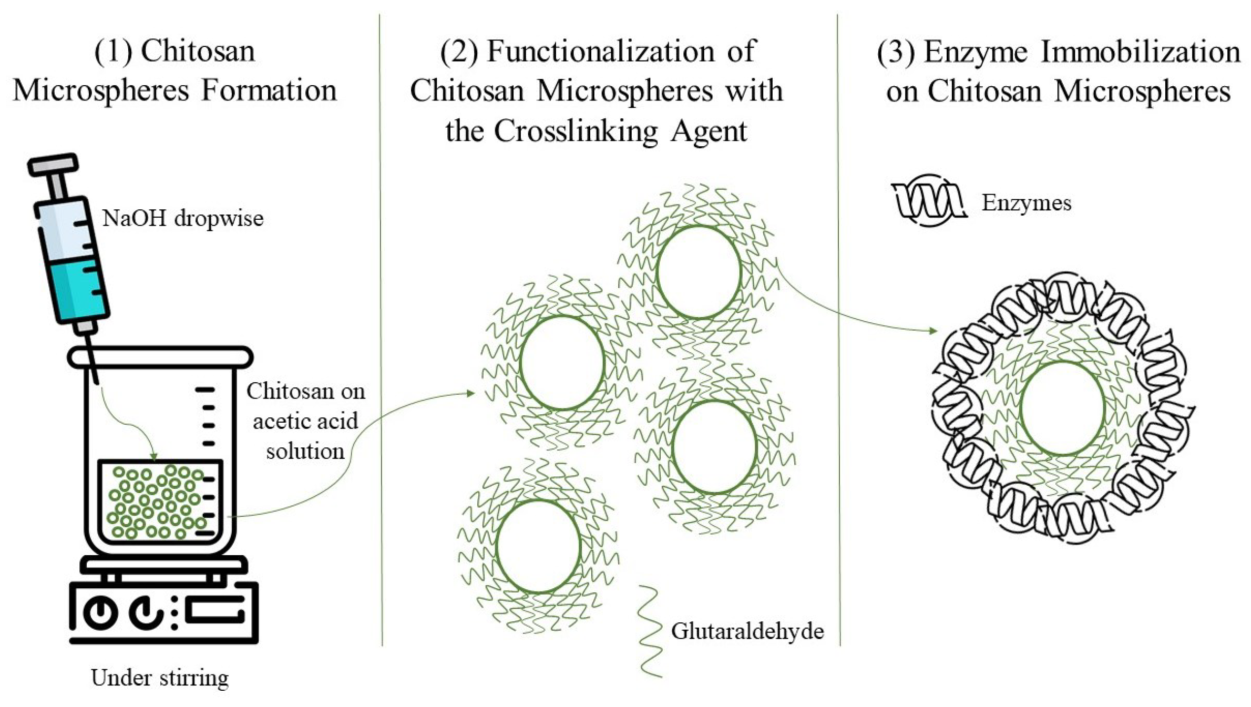
2.4. Covalent Immobilization of Enzymes on Chitosan Microspheres

To prepare chitosan microspheres, 500 mg of chitosan was added to 100 mL of a 2% acetic acid water solution for dissolution. Subsequently, 1% NaOH in water was incorporated dropwise to the solution under gentle stirring until microspheres were formed. The preparation of the crosslinking agent solution involved dissolving glutaraldehyde in a cold phosphate–citrate buffer at pH 7.0. The prepared solution was then incorporated into 5 g (wet basis) of chitosan microspheres, with both the crosslinking agent solution and the microspheres pre-refrigerated. Finally, the chitosan microspheres were combined with a phosphate–citrate buffer at pH 6.0 in a reaction flask, to which the enzymatic solution was added for conducting the immobilization.
The initial enzymatic activity of the partially purified enzymatic solution was determined to calculate the amount required for contact with the functionalized chitosan suspension. The reaction was executed under refrigeration with magnetic agitation for 24 h. After this period, the mixture was subjected to centrifugation, and the pellet formed was subjected to multiple washes with a phosphate–citrate buffer at pH 6.0. Supernatant enzymatic activity was measured after each wash by repeating the centrifugation cycles until no enzymatic activity remained in the supernatant [32,33]. A schematic representation of the enzymatic immobilization process on chitosan microspheres is illustrated in Figure 1.

2.5. Evaluation of Laccase Immobilization Efficiency Using a One-Factor-per-Time Approach

In developing the immobilization method, the response variable was the enzymatic activity of immobilized laccase. The one-factor-per-time (OFT) approach was employed, where a single variable was studied per experiment while keeping all other factors constant [35]. Three critical parameters for the immobilization process were evaluated: the concentration of the crosslinking agent (1%, 5%, and 10%), the crosslinking time (30 min, 60 min, and 90 min), and the enzyme concentration to be immobilized (10,000 U/L, 20,000 U/L, and 30,000 U/L).
The immobilization efficiency percentage (%IE) for the enzyme bound to chitosan was determined using the formula in Equation (1):
IE % = Experimental immobilized enzymatic activity Theoretical immobilized enzymatic activity × 100

2.6. pH and Temperature Stability of Free and Immobilized Laccase

Free and immobilized enzyme stability was tested under different pH conditions (3.0–8.0) through incubation in a phosphate–citrate buffer at the respective pH levels for 4 h. Laccase enzymatic activity was determined at the beginning and end of the incubation period, with the initial activity defined as 100% [22,36,37].
To evaluate thermal stability, free and immobilized ligninolytic enzymes were incubated at pH 6.0 for 4 h under varying temperatures (20–60 °C). Laccase activity was determined at the beginning and end of the procedure, with the initial enzymatic activity defined as 100% [22,36,37,38,39].

2.7. Acetaminophen Degradation by Ligninolytic Immobilized Enzymes

2.7.1. Acetaminophen Degradation Using Immobilized Laccase

A total of 500 mg of immobilized enzymes on chitosan microspheres were added to an aqueous acetaminophen solution starting with a concentration of 10 ppm, in a reaction volume of 5 mL prepared with type I water. The initial laccase enzyme activity was 20 U/mg. Drug concentration was measured hourly over a 4 h period. The mixtures were maintained at room temperature with orbital shaking at 120 rpm. Controls included chitosan microparticles without immobilized enzymes and acetaminophen solutions without chitosan microparticles. The percentage of acetaminophen transformation was calculated, taking the initial concentration as 100%. All tests were executed in triplicate.

2.7.2. Reusability of Immobilized Laccase for Acetaminophen Transformation

A total of 500 mg of immobilized enzymes on chitosan microspheres were added to an aqueous acetaminophen solution having an initial concentration of 10 ppm, in a reaction volume of 5 mL prepared with type I water. The experiment was conducted at room temperature with 120 rpm agitation for 4 h. The initial laccase enzyme activity was 20 U/mg. A control containing chitosan microspheres without immobilized enzymes was included. After the designated reaction time, to measure the acetaminophen concentration in the supernatant, the mixture was centrifuged at 4000 rpm (1792 RCF) for 10 min. The chitosan microspheres were recovered and washed three times with type I water, with each washing step involving centrifugation at 4000 rpm for 10 min to remove excess reagents and prepare the microspheres for reuse. The acetaminophen degradation percentage was determined, taking the initial concentration as 100% [36,38]. All assays were executed in triplicate.

2.8. Analytical Methods

2.8.1. Laccase Activity Determination

Free enzyme activity was measured using a modified method based on Arboleda Echavarría et al. [40], by measuring the shift in absorbance at 420 nm within 1 min. The procedure involved adding the enzyme extract to a phosphate–citrate buffer at pH 3.0 and 0.4 mM ABTS. Data acquisition was carried out with a Varian Cary 50 Bio UV-VIS spectrophotometer equipped with temperature control.
Enzyme activity values were given in international units per liter (U/L), where one unit (U) of laccase is defined as the amount of enzyme that catalyzes the oxidation of 1 μ mol of ABTS in 1 L of solution at 30 °C over the course of 1 min ( ε 420 = 36,000 M−1 cm−1) [32,40].
Immobilized Laccase Activity Determination: The reaction conditions were adapted to include constant stirring in the reaction cell, ensuring that the microparticles remained in suspension throughout the assay to directly measure enzymatic activity.
For the enzymatic activity measurement of immobilized laccase, 50 mg (wet basis) of the enzyme-loaded support was weighed and dispersed in 5 mL of a phosphate–citrate buffer at pH 5.0, with gentle magnetic stirring. Subsequently, a phosphate–citrate buffer at pH 3.0 and the homogenized enzyme-support suspension were added to a quartz cell. The suspension was thoroughly mixed before measuring the volume. The cell was then placed in a Varian Cary 50 Bio spectrophotometer equipped with temperature and magnetic stirring control for the reaction cell. The reaction was initiated by adding ABTS as a substrate, and the change in absorbance at 420 nm was recorded over 5 min. Enzymatic activity was expressed as international units per milligram (U/mg) [39].

2.8.2. SEM Analysis of Chitosan Microspheres

The surface morphologies of lyophilized chitosan microspheres without enzymes (controls) and chitosan microspheres with varying quantities of immobilized enzymes were analyzed using scanning electron microscopy (SEM) [33]. Samples were mounted on aluminum stubs with double-sided adhesive tape and coated with gold to analyze their surface morphologies and evaluate the presence of enzyme molecules within the microspheres.
The surface topography of chitosan microspheres with immobilized laccase was examined using a ZEISS EVO® (Oberkochen, Germany) MA 25 scanning electron microscope in high vacuum mode at an accelerating voltage of 10 kV. The surface morphologies of the samples were scanned, and high-resolution images were subsequently captured.

2.8.3. FT-IR Analysis of Chitosan Microspheres

Functional characterization of lyophilized chitosan microspheres was performed with Fourier transform infrared (FT-IR) analysis, with and without immobilized enzyme, using a Perkin Elmer Frontier (Waltham, MA, USA) FT-IR spectrometer in attenuated total reflectance (ATR) mode. The analysis was conducted with 32 scans at a resolution of 4 cm−1 across the spectral range of 400–4000 cm−1.

2.8.4. Quantification of Acetaminophen

Acetaminophen quantification was carried out using high-performance liquid chromatography (HPLC) on a Shimadzu (Kyoto, Japan) Prominence system equipped with an LC-20AD quaternary pump, an SPD-M20A diode array detector (DAD), and a DGU-20A5 degasser. Detection was performed at 244 nm. The analysis utilized a YMC-Triart C18 column (YMC company, Kyoto, Japan) (250 × 4.6 mm, S-5 μ m, 12 nm) with a mobile phase consisting of methanol and water (30:70 v/v), acidified with 0.5% phosphoric acid. The flow rate was set at 1 mL/min, and the injection volume was 20 μ L.

2.8.5. NMR-Based Analysis of Acetaminophen Degradation by Immobilized Enzymes

Three types of samples were prepared to evaluate the degradation of acetaminophen using immobilized ligninolytic enzymes: M1 (initial acetaminophen, untreated control), M2 (acetaminophen treated with immobilized enzymes), and M3 (acetaminophen exposed to the enzyme support material without enzymes). Each sample was prepared by dissolving acetaminophen in Type 1 water at a concentration of 1 mg/mL and exposing the drug solution to immobilized enzymes for 4 h at 120 rpm under dark conditions. After treatment, the immobilized enzymes were separated by centrifugation (4000 rpm, 10 min) and filtration through a 0.45 μ m membrane. The supernatant was concentrated using rotary evaporation, and the resulting solid residue was resuspended in deuterated methanol (Sigma-Aldrich) for nuclear magnetic resonance (NMR) analysis. 1H and COSY NMR spectra were acquired using a Bruker (Billerica, MA, USA) Avance 600 MHz spectrometer and analyzed to identify the structural modifications of acetaminophen and its potential degradation products. Results were analyzed in the software TopSpin 4.3.0. Peak assignments were performed by comparing the obtained spectra with reference data from the Spectral Database for Organic Compounds (SDBS), a freely accessible database maintained by the National Institute of Advanced Industrial Science and Technology (AIST), Japan. Specifically, the 1H NMR spectrum of acetaminophen was matched with the SDBS entry No. 3290 (Spectral code: HSP-43-745) [41].

2.9. Statistical Analysis

Data were statistically analyzed using Microsoft Excel to calculate means and standard deviations from the three replicates. Parameters investigated for the evaluation of the immobilization process, following the one-factor-per-time approach, were analyzed by one-way ANOVA with Fisher’s least significant difference (LSD) test. Results were considered statistically significant at p < 0.05 . All statistical analyses were performed using STATGRAPHICS 19 software.

3. Results and Discussion

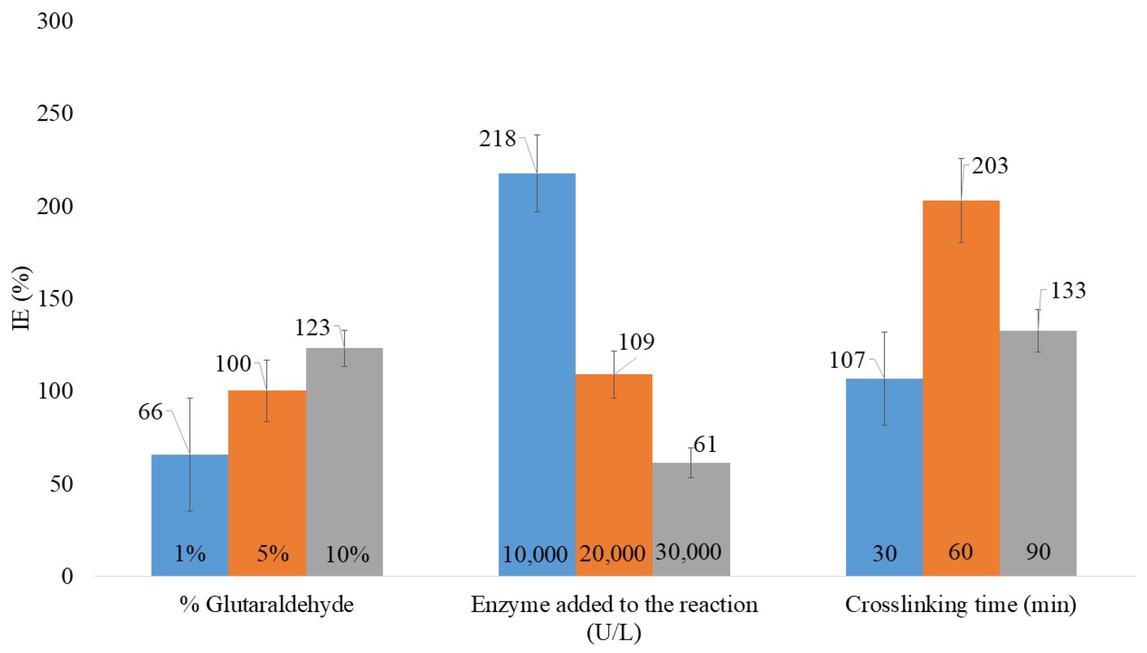
3.1. Evaluation of Laccase Immobilization Efficiency Using a One-Factor-per-Time Approach

The development of enzyme immobilization technologies and the exploration of suitable supports play a crucial role in modifying and enhancing enzyme properties [38]. In this study, critical factors in the covalent immobilization of ligninolytic enzymes onto chitosan microspheres were evaluated to achieve effective immobilization.
A one-factor-per-time approach was employed to evaluate the impact of glutaraldehyde concentration on the immobilization of enzymes onto chitosan microspheres. The ANOVA analysis yielded a p-value of 0.0864, which is higher than the significance threshold of 0.05. This indicates no statistically significant difference in mean immobilization efficiency across the tested glutaraldehyde concentrations at a 5% significance level. However, the highest laccase immobilization efficiency was observed at the highest concentration tested, specifically 10% glutaraldehyde (Figure 2).
The enhanced efficiency at higher glutaraldehyde concentrations can be attributed to the reactivity of aldehydes, chemical compounds containing a carbonyl group bonded to a hydrogen atom. This structural feature facilitates their reaction with amines, forming imine bonds. Glutaraldehyde, as a di-aldehyde, reacts with the primary amine groups in chitosan, which are abundant due to its D-glucosamine structure. This amine-rich composition makes chitosan particularly suitable for immobilization, as glutaraldehyde cross-links the chitosan microspheres, providing stability and functional support for enzyme binding [42,43].
For successful cross-linking, certain aldehyde groups must interact with the amino groups in chitosan, creating covalent imine bonds, while others stay free to bind with the enzyme amino groups. Excess glutaraldehyde is necessary to prevent all aldehyde groups from reacting solely with intra- or intermolecular amino groups of chitosan, which could lead to microsphere aggregation [42,43,44,45].
At low glutaraldehyde concentrations, reduced immobilization efficiency may result from the insufficient activation of chitosan for enzyme binding. Without adequate cross-linking, the chitosan structure lacks functional stability and robustness, leaving fewer free aldehyde groups available for enzymatic binding [32]. The presence of abundant amino groups in chitosan significantly enhances the immobilization process, but glutaraldehyde treatment is essential for achieving stable and functional enzyme binding.
The study conducted by Aslam et al. [32] investigated glutaraldehyde concentrations (1–4%) for their role as a cross-linker in the immobilization of laccase on chitosan beads. The main objective was to evaluate the efficiency of the coupling reaction between glutaraldehyde and enzyme molecules. Their results showed that optimal laccase entrapment occurred when chitosan beads were functionalized with 2% glutaraldehyde over a reaction period of 6 h. The modified chitosan beads offered an efficient and biocompatible platform for enzyme attachment, with an immobilization efficiency of 79% [32].
Unlike the findings of this study, where the highest glutaraldehyde concentration (10%) yielded the best immobilization efficiency, Aslam et al. [32] reported optimal results at a lower concentration. These differences highlight the influence of experimental conditions, such as the levels of chitosan and enzyme utilized during the reaction, which influence the amount of cross-linking agent required. Choosing the right enzyme-to-glutaraldehyde ratio and their respective concentrations is key to reducing enzyme structural alterations while retaining catalytic efficiency [43].
The crosslinking time was found to significantly influence the immobilization process. According to the ANOVA analysis, the p-value for this factor was 0.0001. Since the p-value is below the 0.05 significance threshold, there is a statistically significant difference in mean immobilization efficiency between the tested crosslinking times. As shown in Figure 2, the highest immobilization efficiency for laccase was achieved after 60 min of crosslinking, indicating that, under the study conditions, this duration is optimal for the covalent bonding of enzyme molecules to chitosan microspheres.
Properly establishing the crosslinking time is key to retaining optimal laccase activity following immobilization. Adequate exposure time is necessary to stabilize the support structure while ensuring the effective coupling of glutaraldehyde to the enzymes. However, excessive reaction times can lead to overreactivity of the aldehyde groups, which may become unstable or inactivated. This can result in aldehyde groups reacting excessively with amino groups on the chitosan, reducing the number of active aldehydes available for enzyme binding. Conversely, insufficient reaction times may prevent the glutaraldehyde from effectively coupling to the enzymes, resulting in lower immobilization efficiency [45].
Additionally, glutaraldehyde exists in aqueous solutions in multiple forms, including monomeric di-aldehyde, dimeric, trimeric, and polymeric configurations. The multiplicity of these structures, influenced by solution conditions, can affect the effectiveness of glutaraldehyde as a crosslinking agent during the immobilization process [43].
Finally, regarding the amount of immobilized active enzyme, the ANOVA analysis yielded a p-value of 0.0001. Since this value is below the 0.05 significance threshold, there is a statistically significant difference in mean immobilization efficiency across the tested enzyme concentrations. As shown in Figure 2, the highest immobilization efficiency for laccase was achieved using an enzyme solution with 10,000 U/L.
This finding may be attributed to the interaction between the amino groups of the enzyme and the aldehyde groups on the modified support. A lower enzyme concentration may result in inefficient coupling to the support, either because the enzyme is insufficient to fill the available binding sites or because multiple binding points per enzyme molecule could restrict mobility, reducing catalytic efficiency or even causing inactivation. Conversely, an excessive enzyme concentration may overload the support, leading to steric hindrance and a reduction in laccase activity [45].
The improvement and development of enzyme immobilization strategies have been a significant area of research. For example, Zheng et al. [45] investigated several factors influencing the immobilization of laccase produced by Trametes pubescens onto chitosan beads. The essential parameters investigated encompassed glutaraldehyde concentration, crosslinking duration, enzyme volume, and the immobilization period. Their study identified optimal conditions for laccase immobilization as 0.8% glutaraldehyde, a 3-h crosslinking period, and the use of 2 mL of enzyme solution (approximately 43,672 U/mL) [45].
These findings highlight how variations in particle formation methodologies, reagent concentrations, reaction times, and other variables can significantly impact the optimization of the immobilization process. Both the results of this study and those from other researchers emphasize the importance of evaluating critical parameters under specific working conditions. Factors such as temperature, pH, particle size, crosslinking and immobilization durations, and the concentrations of supports, enzymes, and reagents play essential roles in determining reactivity and immobilization efficiency. Achieving successful immobilization often requires a trial-and-error approach to balance these interdependent factors effectively [43].
It is important to note that the enzymatic extract used for immobilization was partially purified, and the reported immobilization efficiencies are based on the activity measured using ABTS as a substrate. Therefore, the yields reflect the immobilization of the pool of enzymes present in the partially purified extract, rather than a single purified laccase isoform.

3.2. Chitosan Microsphere Characterization
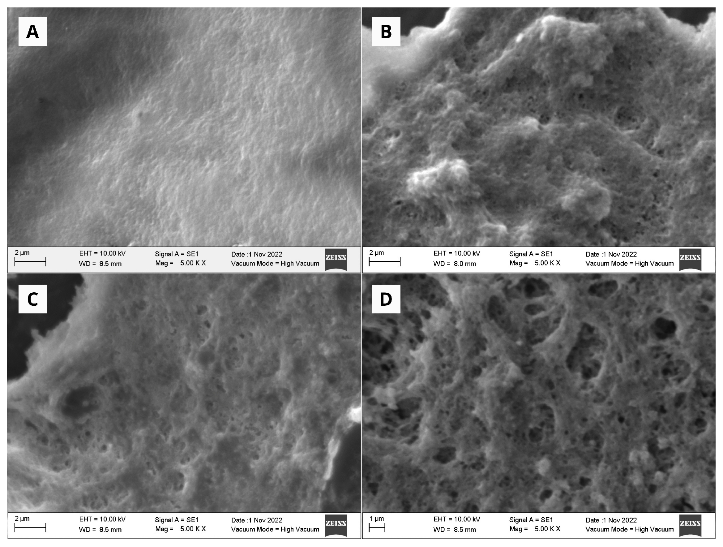
3.2.1. SEM Analysis

The surface morphology of chitosan microspheres was analyzed using SEM. High-resolution images were obtained for both the control group, consisting of microspheres without enzymes, and the experimental groups, which contained varying amounts of immobilized enzyme.
In the SEM micrographs (Figure 3), distinct surface features were observed, revealing differences between the control microspheres and those with immobilized enzymes. These differences are likely due to the functionalization process involving glutaraldehyde and subsequent enzyme immobilization. As shown in Figure 3A, the control chitosan microspheres exhibit a homogeneous surface. In contrast, the micrographs in Figure 3B–D display chitosan microspheres with immobilized enzymes, highlighting noticeable changes in surface characteristics. The contrast between untreated microspheres (Figure 3A) and treated ones (Figure 3B–D) illustrates the effect of enzyme immobilization on the morphology of the surface.
The surface of the control microspheres, without enzymes, appears smooth and uniform. In contrast, microspheres with immobilized enzymes exhibit noticeable surface modifications, including exposed aggregates and distributed pores.
In a study by Jeong & Choi [36], laccase produced by Trametes versicolor was immobilized on chitosan beads, and its surface morphology was characterized using SEM. The authors reported that the surface of the chitosan beads in the control group appeared smooth, while the beads with immobilized laccase exhibited a rougher surface texture [36]. These findings align with the results of this study, where the surface morphology of microspheres without enzymes also appeared smoother compared to that of enzyme-immobilized microspheres.
Similarly, Bilal, Jing et al. [33] immobilized laccase on chitosan beads and documented analogous SEM outcomes. According to the authors, the significant surface alterations in chitosan beads with immobilized enzymes, as opposed to control beads, serve as compelling evidence of protein attachment to the chitosan matrix. In their investigation, Bilal et al. [33] examined chitosan hydrogel beads with smooth surfaces that had not been subjected to laccase immobilization. In contrast, beads with immobilized enzymes displayed significant surface modifications, including prominent irregular aggregates and pores [33]. Similarly, as shown in the micrographs in Figure 3, these surface alterations are likely attributed to the coupling of enzymatic molecules onto the chitosan matrix.

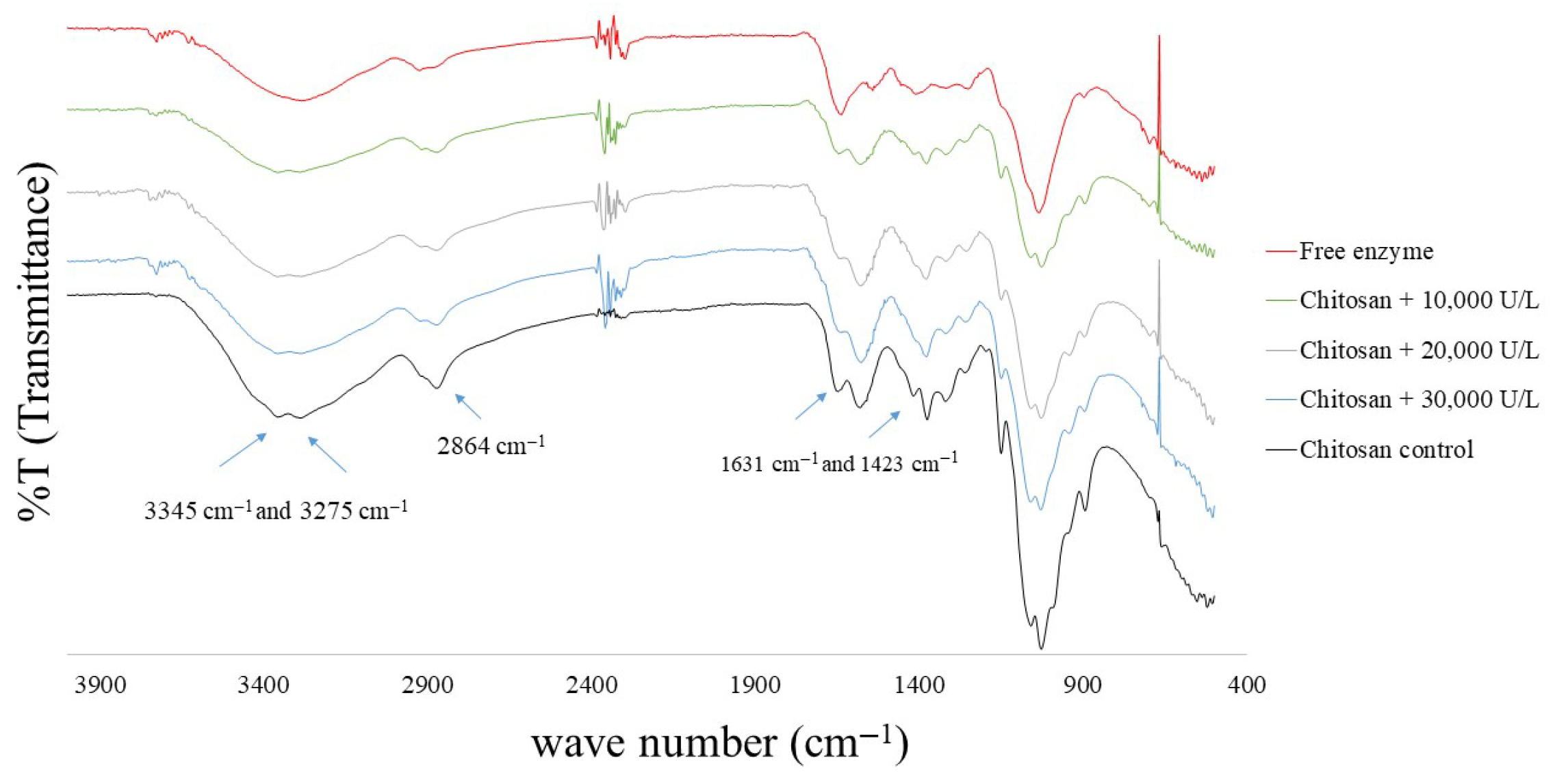
3.2.2. FT-IR Analysis of Chitosan Microspheres with Immobilized Enzymes

Transmittance spectra (Figure 4) were obtained using the FT-IR technique in ATR mode to identify the characteristic functional groups of chitosan microspheres and their variations based on the different amounts of immobilized enzymes. Figure 4 presents the infrared spectra of chitosan microspheres without enzymes (control) and those with varying amounts of immobilized enzymes (10,000, 20,000, and 30,000 U/L).
The FTIR spectrum of chitosan exhibits two prominent peaks at 3345 cm−1 and 3275 cm−1, attributed to the overlapping O–H and N–H stretching bands. Additionally, a peak at 2864 cm−1 corresponds to aliphatic C–H stretching, while peaks at 1631 cm−1 and 1423 cm−1 indicate N–H bending, associated with amino groups [46]. The immobilization of ligninolytic enzymes can be assessed through the deformation of the peaks at 1631 cm−1 and 1423 cm−1, which correspond to the deformation vibrations of chitosan amino groups, as well as the peak at 2864 cm−1, suggesting interactions between chitosan amino groups and enzyme carboxyl groups [46,47,48,49]. These findings confirm that the support retains characteristic chitosan signals and undergoes structural modifications following enzyme immobilization, particularly in its functional groups.
The covalent immobilization of enzymes on chitosan has been studied, with several works characterizing these supports using FT-IR. For instance, Aricov et al. [48] analyzed the covalent immobilization of laccase on chitosan microspheres using FT-IR [48], reporting results similar to those obtained in this study. They concluded that laccase immobilization on the chitosan surface was successfully achieved. Similarly, Nguyen et al. [49] immobilized laccase produced by Trametes versicolor on chitosan beads. Their FT-IR analysis revealed a decrease in the peak corresponding to amino groups, indicating that the enzymes occupied these functional groups, thereby confirming successful binding between the support and the enzyme [49].

3.3. Stability Assessments of Free and Immobilized Laccase
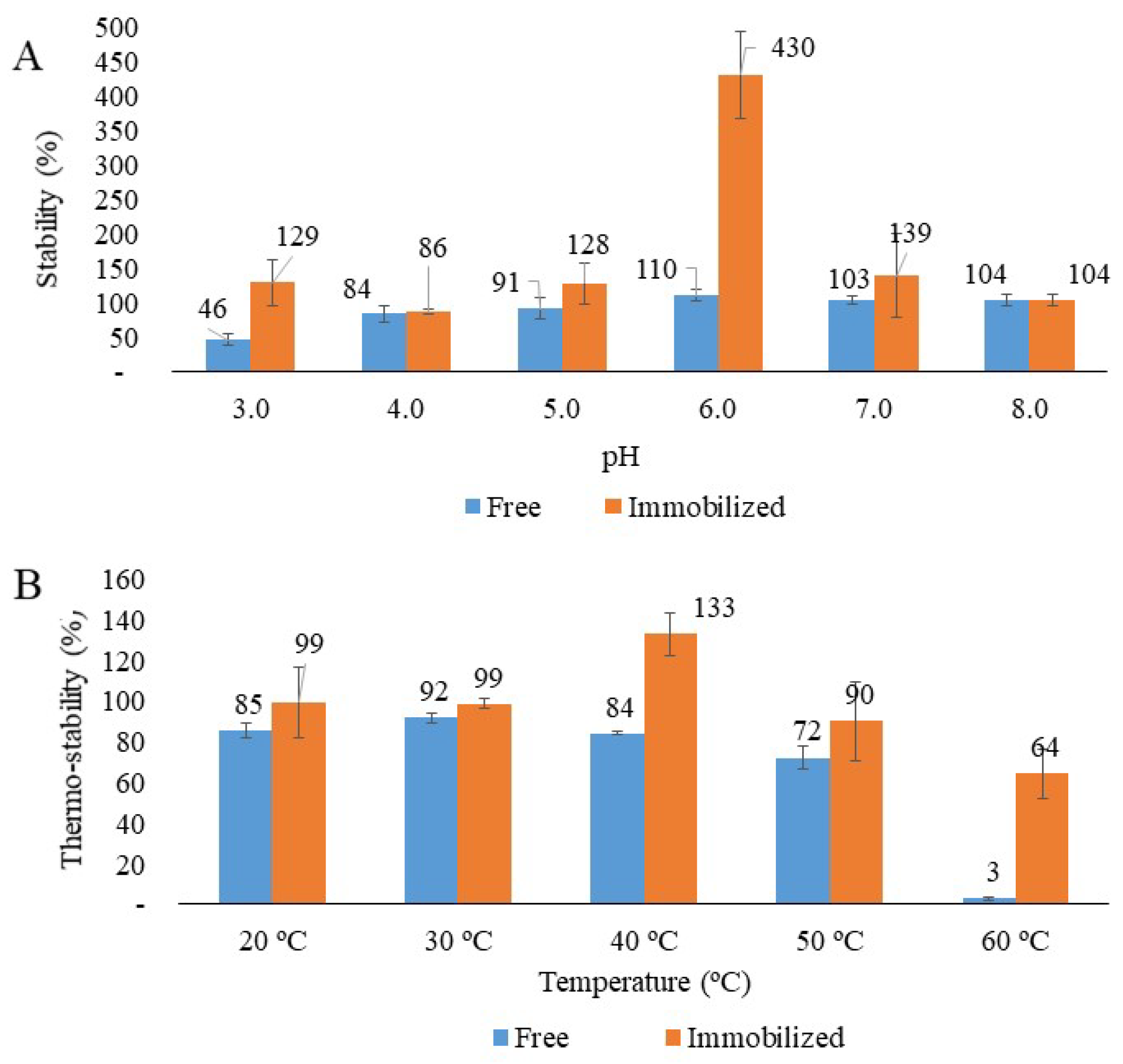
3.3.1. pH Stability

The results of the stability analysis of free and immobilized laccase under different pH conditions are presented in Figure 5A. A pH range from 3 to 8 was evaluated, revealing that free laccase was unstable at lower pH levels, losing up to 54% of its enzymatic activity at pH 3. However, when exposed to pH 5, 6, and 7 for 4 h, free laccase maintained greater stability in enzymatic activity (blue bars).
In contrast, the immobilized enzyme demonstrated significantly improved stability across the tested pH range. Notably, at pH 6, the enzymatic activity of immobilized laccase even increased (orange bars). These findings suggest that laccase produced by Ganoderma parvulum exhibits greater stability in neutral and slightly alkaline pH regions (6–8) compared to acidic conditions (pH < 4). Various factors, such as pH, significantly influence protein stability. pH affects enzyme stability by inducing the protonation or deprotonation of terminal amino and carboxylic groups, which can alter the molecular structure and contribute to the unfolding of the peptide backbone and active site [48].
A comparison of activity profiles under varying pH conditions reveals that the immobilized enzyme maintains more stable catalytic activity than the free enzyme. The observed stability can be explained by the ability of chitosan microspheres to generate a buffered microenvironment, reducing protein protonation and deprotonation. Additionally, the reduced mobility of enzymes during the immobilization process helps preserve their three-dimensional structure and prevents modifications to their active sites [48].
Some studies in the literature have evaluated the stability of free and immobilized enzymes across different pH ranges. For example, Cen et al. [50] conducted enzyme stability experiments on laccase and reported reduced stability at acidic pH levels. These findings, consistent with the results of the present study, highlight that laccase storage and application should be performed in environments with a neutral pH to ensure optimal stability [50].

3.3.2. Temperature Stability

The temperature effect on free and immobilized laccase activity using chitosan microspheres is depicted in Figure 5B. At 40 °C after 4 h, the immobilized enzyme reached maximum stability. However, stability decreased with increasing temperature for both the free and immobilized enzymes. Notably, at all evaluated temperatures, the immobilized enzymes demonstrated greater enzymatic activity than the free enzymes, highlighting its superior thermal stability, a critical property for bioprocess applications.
This higher thermal stability was particularly evident at 60 °C. While free laccase retained 3% of its activity after 4 h, the immobilized enzyme retained 64%, demonstrating a significant reduction in activity loss (97% for the free enzyme compared to 36% for the immobilized enzyme). These results underscore the enhanced thermal stability provided by immobilization, making the laccase more suitable for high-temperature bioprocesses.
Protein structural stability is closely related to thermal inactivation, as intense heat can disrupt the secondary structure and compromise enzyme stability. The activity profile of free and immobilized laccase after 4 h of exposure to different temperatures is shown in Figure 5B. The results highlight that the covalent immobilization approach adopted here is an effective strategy for enhancing enzyme stability. As suggested by Aricov et al. [48], the immobilization process may induce the enzyme to adopt a more rigid structure, improving its stability against temperature-induced denaturation and other destabilizing factors [48].
It has been reported that immobilized enzymes, such as laccase, exhibit lower sensitivity to temperature compared to free enzymes, primarily due to the protective effect of the support and conformational changes that reduce molecular mobility and enhance protein structure stability [38]. At 60 °C, free laccase experienced near-total inactivation, indicating that elevated temperatures cause significant damage to the enzyme’s molecular structure. In contrast, the properties of the support and the covalent bonds formed with the enzyme conferred greater stability to the immobilized enzyme, preserving its conformation under thermal stress.

3.4. Acetaminophen Degradation by Ligninolytic Enzymes Immobilized System

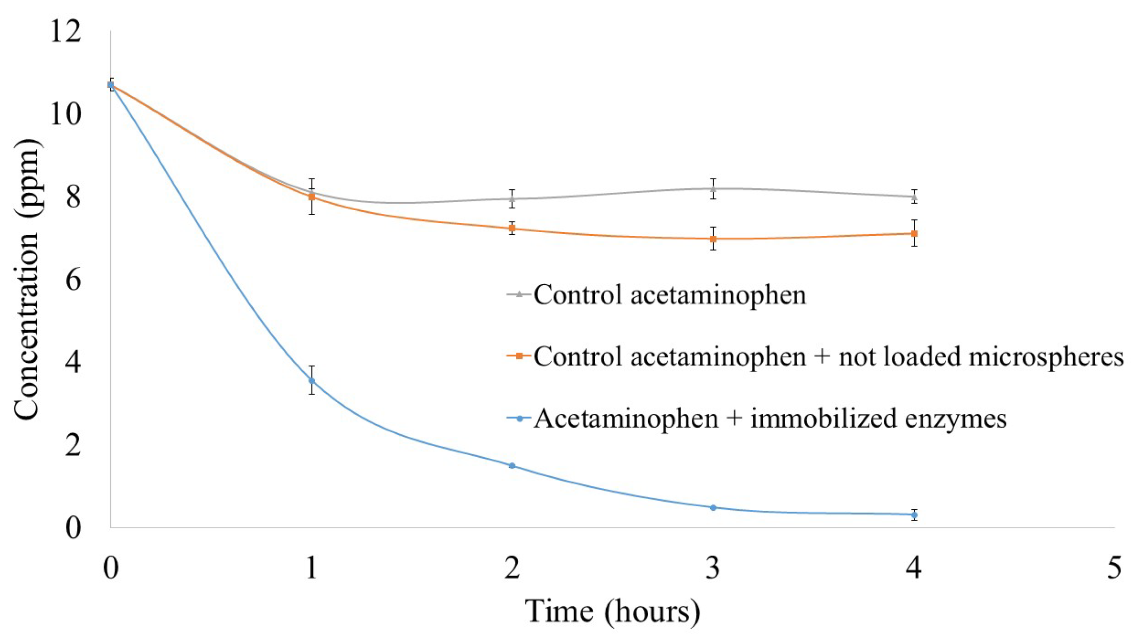
3.4.1. Enzymatic Treatment of Acetaminophen

The results of acetaminophen degradation tests using ligninolytic enzymes produced by Ganoderma parvulum immobilized on chitosan microspheres are presented in Figure 6. The control tests, which included the drug alone and the support without immobilized enzymes, showed reductions in acetaminophen concentration of 25% and 33%, respectively. The observed removal can be linked to the partial diffusion of drug molecules into chitosan pores and the stabilization of acetaminophen molecules in the water-based medium.
In contrast, treatments with microspheres containing immobilized ligninolytic enzymes achieved significantly higher acetaminophen removal, with absorption and/or biotransformation rates of up to 66% (3.5 ppm) within 1 h and 97% (0.3 ppm) after 4 h of reaction. These findings clearly indicate that the primary driver of acetaminophen biotransformation is the presence of partially purified enzymes immobilized on the chitosan support, as evidenced by the significant decrease in acetaminophen concentration only in the presence of immobilized enzymes.
Other authors have also evaluated the biodegradation of acetaminophen using ligninolytic enzymes immobilized on various supports. Hoinacki Da Silva et al. [51] studied the absorption and biotransformation of acetaminophen by a laccase produced by Trametes versicolor, covalently immobilized on avocado seed biochar. Their results indicated that supports lacking immobilized laccase adsorbed less than 20% of acetaminophen in all experiments. This minimal adsorption was linked to diffusion within biochar pores and interactions driven by van der Waals forces and hydrogen bonding between acetaminophen and the glutaraldehyde-treated biochar surface.
In contrast, biochar supports with immobilized laccase achieved 99.5% biodegradation of acetaminophen after 24 h of reaction, starting with an initial acetaminophen concentration of 30 ppm. The authors concluded that the sorption and biotransformation of acetaminophen occur primarily due to laccase activity [51]. These findings, consistent with the results of this study, highlight the effectiveness of ligninolytic enzymes in biodegradation processes, reinforcing their potential utility in environmental applications.

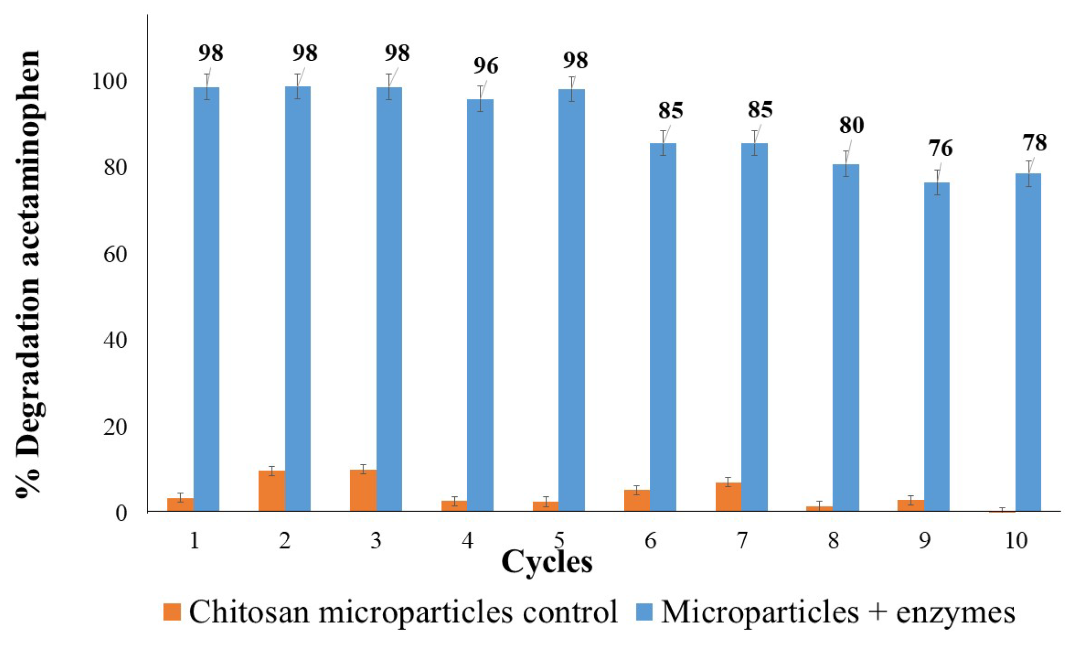
3.4.2. Repeated Use of Immobilized Enzymes in Biotransformation of Acetaminophen

Figure 7 presents the results of reusing ligninolytic enzymes produced by Ganoderma parvulum, covalently immobilized on chitosan microspheres. The biotransformation percentage of acetaminophen remained above 95% for up to five consecutive cycles, demonstrating the high immobilization efficiency of the enzymes on the chitosan microspheres. This stability indicates minimal enzyme loss during the reaction process and subsequent washing steps, enabling effective reuse.
However, from cycle 6 onward, the degradation percentage began to decrease, reaching 78% by cycle 10. This reduction can be attributed to enzyme denaturation and the loss of material mass during repeated use. These results highlight the operational stability of immobilized enzymes, demonstrating their significant potential for application in larger-scale treatment processes due to the reusability of the enzyme biocatalyst.
The study conducted by Hoinacki Da Silva et al. [51] evaluated the reusability of immobilized laccases for acetaminophen degradation over eight 24 h reaction cycles. Their findings showed that the biotransformation percentage of acetaminophen remained above 90% during the first four cycles, highlighting the high efficiency of laccase immobilization on biochar through covalent bonding. However, in subsequent cycles (5, 6, and 7), the biotransformation efficiency decreased to 80%, 65%, and 50%, respectively. The authors concluded that the proposed immobilized enzyme system offers significant potential for acetaminophen biotransformation in environmental bioremediation applications [51].

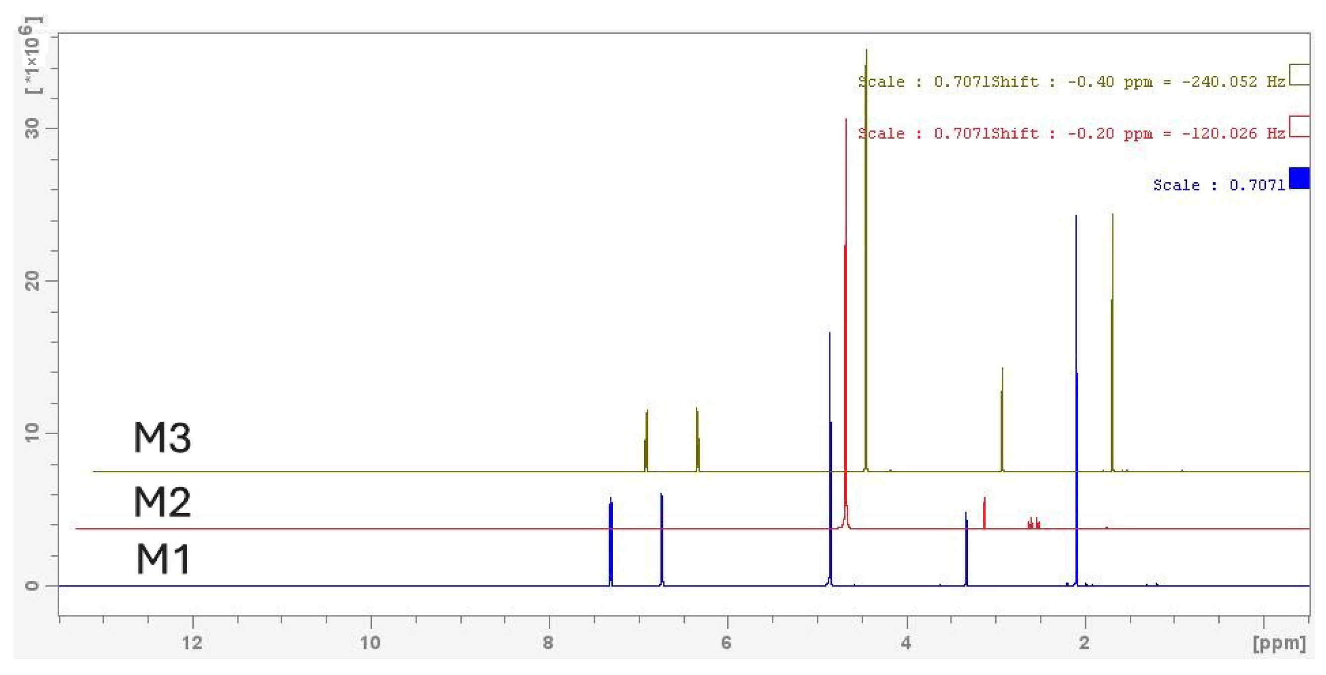
3.4.3. 1H-NMR Analysis of Acetaminophen Biotransformation

The results presented in Figure 8 focus on the 1H-NMR analysis (nuclear magnetic resonance spectroscopy) of acetaminophen and its potential chemical transformations induced by enzymatic degradation. Where M1 (Blue) (Initial Control) presents the pure, untreated acetaminophen, M2 (Red) (Treated with Immobilized Enzymes) presents the potential biotransformation of acetaminophen with the formation of secondary metabolites, and M3 (Green) (Control with Microspheres without Enzymes) looks to rule out the influence of the carrier material (chitosan) on biodegradation process.
The analysis of samples M1, M2, and M3 using 1H-NMR spectroscopy allowed for the identification and comparison of the characteristic signals of acetaminophen and the potential changes associated with enzymatic treatment. In M1, corresponding to untreated acetaminophen, signals consistent with its molecular structure were observed. In the aromatic region, a doublet at 7.3 ppm and another at 6.7 ppm were identified, corresponding to the aromatic ring protons in the ortho and meta positions relative to the hydroxyl and amide groups, respectively. These signals match the reference spectrum for acetaminophen available in the SDBS database. In the aliphatic region, a signal at 2.0 ppm was associated with the methyl group (–CH3) of acetaminophen [41]. Additionally, an intense signal at 4.8 ppm was attributed to residual water, a common feature in solvents like MeOD, while a signal at 3.3–3.5 ppm was attributed to the MeOD solvent itself [52].
In the 1H-NMR spectrum of M2, significant changes are observed, suggesting the substantial biotransformation of acetaminophen due to the action of immobilized enzymes. The characteristic signals of the aromatic ring of acetaminophen at 7.3 ppm and 6.7 ppm disappeared, indicating a possible alteration of the aromatic structure, such as oxidation or chemical substitution. Similarly, the signal of the methyl group (–CH3) at 2.1 ppm is no longer present, suggesting modifications to the acetamide group.
New signals emerged at 2.8 ppm and 2.7 ppm, which may correspond to aliphatic fragments derived from secondary metabolites, such as hydroxylated, fragmented, or deaminated products. These new signals, along with the disappearance of the original signals, confirm a structural transformation of acetaminophen by the enzymatic treatment. In contrast, the signals for the solvent (methanol at 3.3 ppm) and water (4.8 ppm) remained visible but did not provide direct information about the analyzed compound. Collectively, these findings support the hypothesis that immobilized enzymes catalyzed a biotransformation of acetaminophen, altering its aromatic ring and its acetamide functional group and generating new secondary metabolites [12,53].
On the other hand, M3, corresponding to the control with chitosan microspheres without immobilized enzymes, exhibited a spectrum very similar to that of M1. The signals of acetaminophen remained practically unchanged, indicating that the carrier material, in the absence of enzymes, does not affect the chemical structure of the compound. This result reinforces the conclusion that the changes observed in M2 are due to enzymatic action and not to the effects of chitosan as a carrier material.

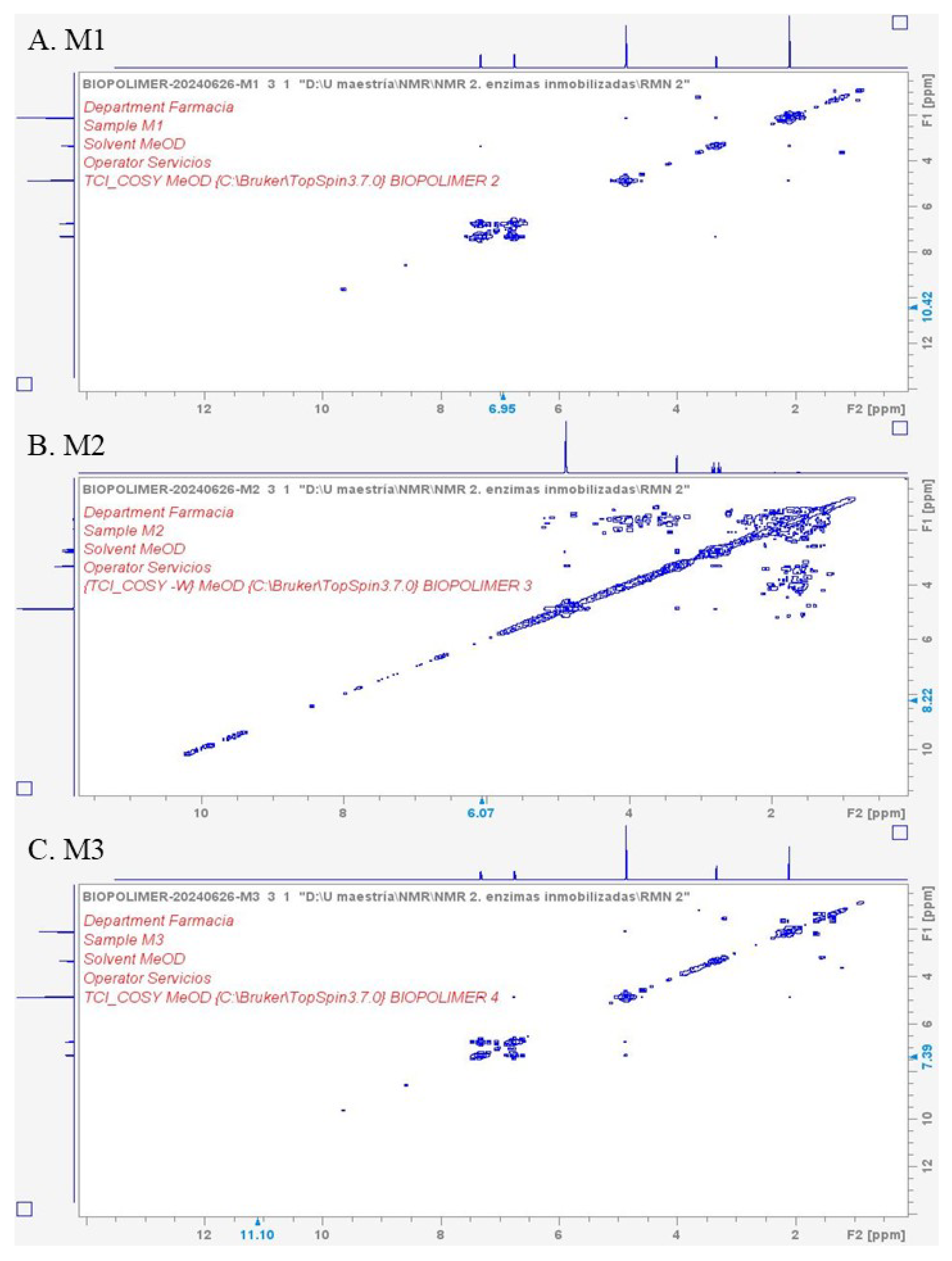
3.4.4. COSY-NMR Analysis of Acetaminophen

In the spectrum shown in Figure 9A, which presents the COSY-NMR analysis of biotransformation of acetaminophen (correlation spectroscopy), the correlation points indicate couplings between protons within the structure of pure acetaminophen. The most significant correlations correspond to the aromatic ring core and the protons of the methyl group attached to the oxygen atom. This pattern aligns with the expected signals for an unmodified acetaminophen molecule.
The aromatic ring protons exhibit correlations between protons in the ortho and meta positions on the aromatic ring, with signals around 7.0–7.5 ppm that couple with each other, reflecting interactions between H4 and H5 on the ring (a typical correlation in a monosubstituted aromatic system). Additionally, the methyl group (–CH3) displays a signal around 2.0–2.5 ppm, indirectly correlating with the ring protons through the oxygen atom of the ether group. The amide group (–NH) shows a signal near 9.0 ppm, with weak correlations to the aromatic protons due to the conjugated bond.
In contrast, the spectrum in Figure 9B, which presents the COSY spectrum of M2 (acetaminophen treated with enzymes), reveals new correlation points or changes in the intensities and positions of cross-peaks compared to M1. These changes suggest a potential biotransformation of acetaminophen, likely leading to the formation of secondary metabolites. The presence of new correlations can be attributed to protons in novel chemical environments, such as hydroxyl or carbonyl groups introduced through enzymatic action.
This spectrum exhibits significant changes compared to M1, indicating acetaminophen biotransformation. Moreover, new interactions not present in M1 are detected in the 3.5–5.0 ppm region. These interactions may correspond to the introduction of hydroxyl groups (–OH) at positions on the aromatic ring, such as the formation of catechols or hydroquinones. It is also possible that the methyl group was oxidized, producing an alcohol group (–CH2OH) that could also appear in this region. An increase in correlations is also observed in the aliphatic region (1.0–3.0 ppm), which could be associated with the formation of small aliphatic fragments or modifications to the methyl group [12,53].
Finally, the COSY spectrum of M3 in Figure 9C closely resembles that of M1, confirming that chitosan does not induce chemical transformations in the acetaminophen molecule. In this spectrum, the interactions between aromatic ring protons remain unchanged, as in M1, and no new signals are observed in aliphatic or polar regions (3.5–5.0 ppm), reinforcing the idea that the modifications observed in M2 are exclusively caused by the enzymatic treatment.
The COSY spectrum of M2, compared to M1 and M3, suggests a complex biotransformation mediated by immobilized enzymes. The main changes observed, such as the appearance of correlations in the 3.5–5.0 ppm and 1.0–3.0 ppm regions, point to the formation of more oxygenated (hydroxylation, oxidation) and possibly fragmented (ring-opening) secondary metabolites. These results highlight clear differences between the samples, underscoring the effects of enzymatic treatment.
The results obtained from the NMR spectra align in aspects with findings reported in the literature. Regarding alterations in the aromatic ring, the disappearance of the characteristic signals of this structure in the spectra of M2 (treated with immobilized enzymes) suggests significant structural degradation or modification.
This result is consistent with the findings of Qutob et al. [54], who reported that acetaminophen oxidation can induce aromatic ring cleavage, leading to the formation of compounds such as carboxylic acids or oxygenated aliphatic structures. The new signals observed in the aliphatic region of the M2 sample are consistent with the formation of aliphatic fragments, further supporting the hypothesis of biotransformation [54].
Additionally, the generation of intermediate products, such as catechols and aliphatic compounds, has been documented during acetaminophen degradation under advanced treatment processes. Complete degradation of the aromatic ring may result in simpler aliphatic compounds, such as aldehydes (e.g., formaldehyde and acetaldehyde) or ketones (e.g., acetone), which are often characteristic of advanced degradation end stages [12,53].
To complement the findings described above, 13C-NMR and HMBC spectra were also obtained and are presented in the Supplementary Materials.

4. Conclusions

The covalent immobilization of ligninolytic enzymes partially purified from Ganoderma parvulum was improved by evaluating key parameters. Optimal conditions included 10% glutaraldehyde, a 60 min crosslinking time, and an enzyme concentration of 10,000 U/L. SEM images confirmed the formation of an enzyme layer on the chitosan polymeric structure, while FT-IR analysis verified modifications to amino functional groups, providing evidence of successful immobilization.
NMR analysis provided critical insights into the structural transformations of acetaminophen following enzymatic treatment. The disappearance of characteristic aromatic signals in the 1H NMR and COSY spectra, along with the emergence of new signals in aliphatic regions, confirmed aromatic ring cleavage and the formation of hydroxylated and aliphatic metabolites. These findings directly support the enzymatic biotransformation of acetaminophen, validating the system’s efficiency. This work represents a crucial step toward the efficient bioconversion of environmental contaminants.
Immobilized enzymes exhibited enhanced thermal stability and retained enzymatic activity across a wide pH range, with remarkable stability and reusability. These properties make them highly suitable for wastewater and effluent treatment applications. Additionally, these immobilized enzymes demonstrated high efficiency in acetaminophen biotransformation within a short time frame, maintaining effectiveness across multiple reuse cycles. These findings highlight a promising strategy to address the environmental challenges posed by pharmaceutical pollutants.
Future research should prioritize the development of advanced technologies to analyze and optimize scalability processes and industrial and environmental applications. Key priorities include improving enzyme stability under varying biochemical conditions, others pollutant degradation capabilities, and applicability to diverse environmental matrices. Such advancements are critical for the successful implementation of enzyme-based technologies in environmental remediation.

Supplementary Materials

The following supporting information can be downloaded at: https://www.mdpi.com/article/10.3390/fermentation11070387/s1, Figure S1: Spectro 13C-NMR of acetaminophen. Where M1 (Blue) is the initial control of acetaminophen, M2 (Red) is the acetaminophen treated with immobilized enzymes, and M3 (Green) is the control with microspheres without enzymes. Figure S2: HMBC-NMR of acetaminophen, where A. M1 is the initial control of acetaminophen, B. M2 is the acetaminophen treated with immobilized enzymes, and C. M3 is the control with microspheres without enzymes.

Author Contributions

M.A.F.-R., conceptualization, methodology, formal analysis, investigation, resources, writing—review & editing, visualization; X.L.-L., conceptualization, formal analysis, review & editing; M.d.J.R.-A., methodology, formal analysis, resources, review & editing; R.P.-S., resources, review, project administration, funding acquisition; F.S.-S., conceptualization, methodology, formal analysis, investigation, resources, review & editing, visualization, supervision, project administration, funding acquisition. All authors have read and agreed to the published version of the manuscript.

Funding

This work was supported by the Convocatoria Programática 2019–2020: Ingeniería y Tecnología [grant numbers SIIU 2020-34952] from CODI (Comité para el Desarrollo de la Investigación) of the Universidad de Antioquia-Colombia.

Institutional Review Board Statement

Not applicable.

Informed Consent Statement

Not applicable.

Data Availability Statement

The original contributions presented in this study are included in the article/Supplementary Materials. Further inquiries can be directed to the corresponding author.

Acknowledgments

We are grateful to the Instituto Tecnológico y de Estudios Superiores de Monterrey and the Institute of Advanced Materials for Sustainable Manufacturing, to El Centro del Agua para América Latina y el Caribe, and the ‘Programa Iberoamericano de Ciencia y Tecnología para el Desarrollo’ within the Latin American development network ‘Lacasas Inmovilizadas para la Degradación de Compuestos Aromáticos en Aguas Residuales’ (LIDA, project 318RT0552). Their support included providing facilities, equipment, advice, and financing for developing certain characterization objectives in Mexico. This collaboration further strengthened the relationship between the institutions.

Conflicts of Interest

The authors declare no conflicts of interest.

References

  1. United Nations. The Sustainable Development Goals Report 2023: Special Edition. 2023. Available online: https://unstats.un.org/sdgs/report/2023/ (accessed on 13 March 2025).
  2. Hanafiah, Z.M.; Mohtar, W.H.M.W.; El Messaoudi, N.; Miyah, Y.; Maged, A.; Knani, S.; Graba, B.; Gafforov, Y.; Wan-Mohtar, W.A.A.Q.I. Wastewater treatment using Ganoderma Species via Ganoremediation Bioreactor: A Comprehensive Review. Bioresour. Technol. Rep. 2025, 30, 102105. [Google Scholar] [CrossRef]
  3. Parra Guardado, A.L. Enzymatic Degradation of Recalcitrant Pharmaceutical Micropollutants. Ph.D. Thesis, Université de Montpellier, Montpellier, France, 2019. [Google Scholar]
  4. Barrios-Estrada, C.; de Jesús Rostro-Alanis, M.; Muñoz-Gutiérrez, B.D.; Iqbal, H.M.; Kannan, S.; Parra-Saldívar, R. Emergent contaminants: Endocrine disruptors and their laccase-assisted degradation—A review. Sci. Total Environ. 2018, 612, 1516–1531. [Google Scholar] [CrossRef] [PubMed]
  5. Flórez-Restrepo, M.A.; López-Legarda, X.; Segura-Sánchez, F. Bioremediation of emerging pharmaceutical pollutants acetaminophen and ibuprofen by white-rot fungi—A review. Sci. Total Environ. 2025, 977, 179379. [Google Scholar] [CrossRef]
  6. Morsi, R.; Bilal, M.; Iqbal, H.M.; Ashraf, S.S. Laccases and peroxidases: The smart, greener and futuristic biocatalytic tools to mitigate recalcitrant emerging pollutants. Sci. Total Environ. 2020, 714, 136572. [Google Scholar] [CrossRef] [PubMed]
  7. Okoye, C.O.; Nyaruaba, R.; Ita, R.E.; Okon, S.U.; Addey, C.I.; Ebido, C.C.; Opabunmi, A.O.; Okeke, E.S.; Chukwudozie, K.I. Antibiotic resistance in the aquatic environment: Analytical techniques and interactive impact of emerging contaminants. Environ. Toxicol. Pharmacol. 2022, 96, 103995. [Google Scholar] [CrossRef]
  8. Rasheed, T.; Bilal, M.; Nabeel, F.; Adeel, M.; Iqbal, H.M. Environmentally-related contaminants of high concern: Potential sources and analytical modalities for detection, quantification, and treatment. Environ. Int. 2019, 122, 52–66. [Google Scholar] [CrossRef] [PubMed]
  9. Ratanapongleka, K.; Punbut, S. Removal of acetaminophen in water by laccase immobilized in barium alginate. Environ. Technol. 2017, 39, 336–345. [Google Scholar] [CrossRef]
  10. UNESCO. Contaminantes Emergentes en la Reutilización de Aguas Residuales en los Países en Desarrollo. 2015. Available online: https://unesdoc.unesco.org/ark:/48223/pf0000235241_spa (accessed on 4 November 2024).
  11. Chu-Wen, Y.; Yi-En, C.; Bea Ven, C. Microbial Communities Associated with Acetaminophen Biodegradation from Mangrove Sediment. Sustainability 2020, 12, 5410. [Google Scholar] [CrossRef]
  12. Żur, J.; Piński, A.; Marchlewicz, A.; Hupert-Kocurek, K.; Wojcieszyńska, D.; Guzik, U. Organic micropollutants paracetamol and ibuprofen—Toxicity, biodegradation, and genetic background of their utilization by bacteria. Environ. Sci. Pollut. Res. Int. 2018, 25, 21498–21524. [Google Scholar] [CrossRef]
  13. Poddar, K.; Sarkar, D.; Chakraborty, D.; Patil, P.B.; Maity, S.; Sarkar, A. Paracetamol biodegradation by Pseudomonas Strain PrS10 isolated from pharmaceutical effluents. Int. Biodeterior. Biodegrad. 2022, 175, 105490. [Google Scholar] [CrossRef]
  14. Unuofin, J.O.; Okoh, A.I.; Nwodo, U.U. Aptitude of oxidative enzymes for treatment of wastewater pollutants: A laccase perspective. Molecules 2019, 24, 2064. [Google Scholar] [CrossRef] [PubMed]
  15. Gugel, I.; Summa, D.; Costa, S.; Manfredini, S.; Vertuani, S.; Marchetti, F.; Tamburini, E. Mycoremediation of Synthetic Azo Dyes by White-Rot Fungi Grown on Diary Waste: A Step toward Sustainable and Circular Bioeconomy. Fermentation 2024, 10, 80. [Google Scholar] [CrossRef]
  16. Voběrková, S.; Solčány, V.; Vršanská, M.; Adam, V. Immobilization of ligninolytic enzymes from white-rot fungi in cross-linked aggregates. Chemosphere 2018, 202, 694–707. [Google Scholar] [CrossRef]
  17. Mir-Tutusaus, J.A.; Baccar, R.; Caminal, G.; Sarrà, M. Can white-rot fungi be a real wastewater treatment alternative for organic micropollutants removal? A review. Water Res. 2018, 138, 137–151. [Google Scholar] [CrossRef]
  18. Sun, X.; Lin, X.; Xian, Y.; Zhang, F.; Zhu, L.; Geng, H.; Wang, W.; Zhang, G. Engineering Bacterial Laccase with Improved Catalytic Activity and Thermostability by Rational Design. Appl. Biochem. Biotechnol. 2025, 1–17. [Google Scholar] [CrossRef] [PubMed]
  19. Suryadi, H.; Judono, J.J.; Putri, M.R.; Eclessia, A.D.; Ulhaq, J.M.; Agustina, D.N.; Sumiati, T. Biodelignification of lignocellulose using ligninolytic enzymes from white-rot fungi. Heliyon 2022, 8, e08865. [Google Scholar] [CrossRef]
  20. Zhang, H.; Zhang, X.; Geng, A. Construction of CRISPR-Cas9 genome editing platform for white-rot fungus Cerrena Unicolor BBP6 Its effects on extracellular ligninolytic enzyme biosynthesis. Biochem. Eng. J. 2022, 185, 108527. [Google Scholar] [CrossRef]
  21. GlobeNewswire. Enzymes Global Market Report 2023; GlobeNewswire: Los Angeles, CA, USA, 2023. [Google Scholar]
  22. Bagewadi, Z.K.; Mulla, S.I.; Ninnekar, H.Z. Purification and immobilization of laccase from Trichoderma Harzianum Strain HZN10 Its Appl. Dye Decolorization. J. Genet. Eng. Biotechnol. 2017, 15, 139–150. [Google Scholar] [CrossRef]
  23. Gao, W.W.; Zhang, F.X.; Zhang, G.X.; Zhou, C.H. Key factors affecting the activity and stability of enzymes in ionic liquids and novel applications in biocatalysis. Biochem. Eng. J. 2015, 99, 67–84. [Google Scholar] [CrossRef]
  24. Geor Malar, C.; Seenuvasan, M.; Kumar, K.S.; Kumar, A.; Parthiban, R. Review on surface modification of nanocarriers to overcome diffusion limitations: An enzyme immobilization aspect. Biochem. Eng. J. 2020, 158, 107574. [Google Scholar] [CrossRef]
  25. López-Legarda, X.; Arboleda-Echavarría, C.; Segura-Sanchez, F. Producción de polisacáridos a partir de Ganoderma Sp., Aisl. En La Región Andin. Rev. Colomb. Biotecnol. 2015, 17, 44–54. [Google Scholar] [CrossRef]
  26. López-Legarda, X. Producción, Caracterización y Actividad Biológica de Polisacáridos Fúngicos. Doctoral Thesis, Universidad de Antioquia, Medellín, Colombia, 2021. [Google Scholar]
  27. Kirk, T.K.; Croan, S.; Tien, M.; Murtagh, K.E.; Farrell, R.L. Production of multiple ligninases by Phanerochaete Chrysosporium: Effect of selected growth conditions and use of a mutant strain. Enzym. Microb. Technol. 1986, 8, 27–32. [Google Scholar] [CrossRef]
  28. Torrez Monroy, G.P. Producción y Purificación de Enzimas Celulolíticas, Lacasas y Manganeso Peroxidasas de Tres Cepas Fúngicas Nativas (IB-105,GL-9B Y co-1). Ph.D. Thesis, Universidad Mayor de San Andres, La Paz, Bolivia, 2015. [Google Scholar]
  29. Howlader, M.M.; Niroda, A.; Bai, R.G.; Premarathna, A.D.; Tuvikene, R. Fermentation optimization, purification and biochemical characterization of a porphyran degrading enzyme with funoran side-activity from Zobellia uliginosa. Biocatal. Agric. Biotechnol. 2022, 43, 102394. [Google Scholar] [CrossRef]
  30. Park, J.Y.; Han, S.; Kim, D.; Nguyen, T.V.T.; Nam, Y.; Kim, S.M.; Chang, R.; Kim, Y.H. Enhancing the thermostability of lignin peroxidase: Heme as a keystone cofactor driving stability changes in heme enzymes. Heliyon 2024, 10, e37235. [Google Scholar] [CrossRef] [PubMed]
  31. Rajeeva, S.; Lele, S.S. Bioprocessing of laccase produced by submerged culture of Ganoderma sp. WR-1. Sep. Purif. Technol. 2010, 76, 110–119. [Google Scholar] [CrossRef]
  32. Aslam, S.; Asgher, M.; Khan, N.A.; Bilal, M. Immobilization of Pleurotus Nebrodensis WC 850 Laccase Glutaraldehyde Cross-Linked Chitosan Beads for Enhanced Biocatalytic Degradation of Textile Dyes. J. Water Process Eng. 2021, 40, 101971. [Google Scholar] [CrossRef]
  33. Bilal, M.; Jing, Z.; Zhao, Y.; Iqbal, H.M. Immobilization of fungal laccase on glutaraldehyde cross-linked chitosan beads and its bio-catalytic potential to degrade bisphenol A. Biocatal. Agric. Biotechnol. 2019, 19, 101174. [Google Scholar] [CrossRef]
  34. Flaticon. Iconos Vectoriales y Stickers—PNG, SVG, EPS, PSD y CSS—Designed by Freepik, Wanicon and Vector Squad from Flaticon. 2025. Available online: https://www.flaticon.com/ (accessed on 18 June 2025).
  35. Suresh, G.; R, R.; Johney, J. Optimization of laccase production by Pleurotus pulmonarius through solid substrate fermentation of tender coconut fiber: Enhanced laccase production and biomass delignification. Biomass Convers. Biorefinery 2024, 15, 13769–13781. [Google Scholar] [CrossRef]
  36. Jeong, D.; Choi, K.Y. Biodegradation of Tetracycline Antibiotic by Laccase Biocatalyst Immobilized on Chitosan-Tripolyphosphate Beads. Appl. Biochem. Microbiol. 2020, 56, 306–312. [Google Scholar] [CrossRef]
  37. Naghdi, M.; Taheran, M.; Brar, S.K.; Kermanshahi-pour, A.; Verma, M.; Surampalli, R.Y. Immobilized laccase on oxygen functionalized nanobiochars through mineral acids treatment for removal of carbamazepine. Sci. Total Environ. 2017, 584–585, 393–401. [Google Scholar] [CrossRef]
  38. Qiu, X.; Wang, S.; Miao, S.; Suo, H.; Xu, H.; Hu, Y. Co-immobilization of laccase and ABTS onto amino-functionalized ionic liquid-modified magnetic chitosan nanoparticles for pollutants removal. J. Hazard. Mater. 2021, 401, 123353. [Google Scholar] [CrossRef] [PubMed]
  39. Salami, F.; Habibi, Z.; Yousefi, M.; Mohammadi, M. Covalent immobilization of laccase by one pot three component reaction and its application in the decolorization of textile dyes. Int. J. Biol. Macromol. 2018, 120, 144–151. [Google Scholar] [CrossRef]
  40. Arboleda Echavarría, C.; Mejía Gallón, A.I.; Franco Molano, A.E.; Jiménez Tobón, G.A.; Penninckx, M. Autochthonous white rot fungi from the tropical forest of Colombia for dye decolourisation and ligninolytic enzymes production. SYDOWIA Int. J. Mycol. 2008, 60, 165–180. [Google Scholar]
  41. SDBS Spectral Database for Organic Compounds. SDBS-1H NMR SDBS No.: 3290 Spectral Code: HSP-43-745. 1999. Available online: https://sdbs.db.aist.go.jp/SearchInformation.aspx (accessed on 30 March 2025).
  42. Bilal, M.; Asgher, M.; Iqbal, M.; Hu, H.; Zhang, X. Chitosan beads immobilized manganese peroxidase catalytic potential for detoxification and decolorization of textile effluent. Int. J. Biol. Macromol. 2016, 89, 181–189. [Google Scholar] [CrossRef]
  43. Migneault, I.; Dartiguenave, C.; Bertrand, M.J.; Waldron, K.C. Glutaraldehyde: Behavior in aqueous solution, reaction with proteins, and application to enzyme crosslinking. BioTechniques 2018, 37, 790–802. [Google Scholar] [CrossRef]
  44. Wahba, M.I. Chitosan-glutaraldehyde activated calcium pectinate beads as a covalent immobilization support. Biocatal. Agric. Biotechnol. 2017, 12, 266–274. [Google Scholar] [CrossRef]
  45. Zheng, F.; Cui, B.K.; Wu, X.J.; Meng, G.; Liu, H.X.; Si, J. Immobilization of laccase onto chitosan beads to enhance its capability to degrade synthetic dyes. Int. Biodeterior. Biodegrad. 2016, 110, 69–78. [Google Scholar] [CrossRef]
  46. Mehandia, S.; Ahmad, S.; Sharma, S.C.; Arya, S.K. Decolorization and detoxification of textile effluent by immobilized laccase-ACS into chitosan-clay composite beads using a packed bed reactor system: An ecofriendly approach. J. Water Process Eng. 2022, 47, 102662. [Google Scholar] [CrossRef]
  47. Alvarado Ramírez, L. Inmovilización de Lacasas en Esferas de SiO2 para la Degradación de Rojo Congo. Master’s Thesis, Universidad Autonoma de Nuevo León, Monterrey, Mexico, 2015. [Google Scholar]
  48. Aricov, L.; Leonties, A.R.; Gîfu, I.C.; Preda, D.; Raducan, A.; Anghel, D.F. Enhancement of laccase immobilization onto wet chitosan microspheres using an iterative protocol and its potential to remove micropollutants. J. Environ. Manag. 2020, 276, 111326. [Google Scholar] [CrossRef]
  49. Nguyen, T.A.; Fu, C.C.; Juang, R.S. Effective removal of sulfur dyes from water by biosorption and subsequent immobilized laccase degradation on crosslinked chitosan beads. Chem. Eng. J. 2016, 304, 313–324. [Google Scholar] [CrossRef]
  50. Cen, Q.; Wu, X.; Cao, L.; Lu, Y.; Lu, X.; Chen, J.; Fu, G.; Liu, Y.; Ruan, R. Green production of a yellow laccase by Coriolopsis gallica for phenolic pollutants removal. AMB Express 2022, 12, 96. [Google Scholar] [CrossRef] [PubMed]
  51. Hoinacki Da Silva, C.K.; Polidoro, A.S.; Cabrera Ruschel, P.M.; Thue, P.S.; Jacques, R.A.; Lima, É.C.; Bussamara, R.; Fernandes, A.N. Laccase covalently immobilized on avocado seed biochar: A high-performance biocatalyst for acetaminophen sorption and biotransformation. J. Environ. Chem. Eng. 2022, 10, 107731. [Google Scholar] [CrossRef]
  52. Merk. NMR Chemical Shifts of Impurities. Available online: https://www.sigmaaldrich.cn/CN/zh/technical-documents/technical-article/analytical-chemistry/nuclear-magnetic-resonance/1h-nmr-and-13c-nmr-chemical-shifts-of-impurities-chart?srsltid=AfmBOops5TheGGQxgV-8_FMKCJU7lkAMAiTJJ2fXmRrUg1Qr7Kc-5aHE (accessed on 6 January 2025).
  53. Schmiemann, D.; Hohenschon, L.; Bartels, I.; Hermsen, A.; Bachmann, F.; Cordes, A.; Jäger, M.; Gutmann, J.S.; Hoffmann-Jacobsen, K. Enzymatic post-treatment of ozonation: Laccase-mediated removal of the by-products of acetaminophen ozonation. Environ. Sci. Pollut. Res. 2023, 30, 53128–53139. [Google Scholar] [CrossRef] [PubMed]
  54. Qutob, M.; Hussein, M.A.; Alamry, K.A.; Rafatullah, M. A review on the degradation of acetaminophen by advanced oxidation process: Pathway, by-products, biotoxicity, and density functional theory calculation. RSC Adv. 2022, 12, 18373–18396. [Google Scholar] [CrossRef]
Figure 1. Schematic representation of the enzymatic immobilization process on chitosan microspheres. The workflow includes three main stages: (1) chitosan microsphere formation by adding NaOH dropwise into a chitosan–acetic acid solution under stirring, (2) functionalization of microspheres with the crosslinking agent glutaraldehyde, and (3) covalent immobilization of partially purified ligninolytic enzymes onto the activated chitosan support. Icons from [34].
Fermentation 11 00387 g001
Figure 2. Enzyme immobilization efficiency (IE%) on chitosan, evaluated by one-factor-per-time. Assess the factors glutaraldehyde percentage, cross-linking time (min), and amount of enzyme (U/L). Blue bars are the low level of each factor, orange bars are the medium level, and gray bars are the high level of each factor.
Fermentation 11 00387 g002
Figure 3. SEM micrographs at 500 K× magnification depict the surface of lyophilized microspheres formed with 10% crosslinking agent, 60 min crosslinking time, and different amounts of immobilized enzyme. (A) Control—chitosan beads without immobilized enzyme, (B) Chitosan beads with 10,000 U/L immobilized laccase, (C) Chitosan beads with 20,000 U/L immobilized laccase, (D) Chitosan beads with 30,000 U/L immobilized laccase.
Fermentation 11 00387 g003
Figure 4. FT-IR transmittance spectra obtained with 32 scans at 4 cm−1 resolution in the spectral region of 400–4000 cm−1 for chitosan microspheres with different amounts of immobilized enzymes, a control of chitosan without enzymes, and a control of free enzymes.
Fermentation 11 00387 g004
Figure 5. Percentages of enzyme stability on chitosan microspheres for free and immobilized laccase. (A) At different pHs after four hours. (B) At different temperatures after four hours.
Fermentation 11 00387 g005
Figure 6. Biodegradation of acetaminophen in water by ligninolytic enzymes from Ganoderma parvulum immobilized on chitosan microspheres in four hours of reaction.
Fermentation 11 00387 g006
Figure 7. Reuse cycles of immobilized enzymes for the biotransformation of acetaminophen.
Fermentation 11 00387 g007
Figure 8. Spectro 1H-NMR of acetaminophen. Where M1 (Blue) is the initial control of acetaminophen, M2 (Red) is the acetaminophen treated with immobilized enzymes, and M3 (Green) is the control with microspheres without enzymes.
Fermentation 11 00387 g008
Figure 9. COSY-NMR spectrum of acetaminophen, where (A) M1 is the initial control of acetaminophen, (B) M2 is the acetaminophen treated with immobilized enzymes, and (C) M3 is the control with microspheres without enzymes.
Fermentation 11 00387 g009
Disclaimer/Publisher’s Note: The statements, opinions and data contained in all publications are solely those of the individual author(s) and contributor(s) and not of MDPI and/or the editor(s). MDPI and/or the editor(s) disclaim responsibility for any injury to people or property resulting from any ideas, methods, instructions or products referred to in the content.

