
Phylogenetic Insights Reveal New Taxa in Thyridariaceae and Massarinaceae

 ,
, 
Abstract
1. Introduction
2. Materials and Methods
2.1. Sample Collection, Morphological Examination, and Isolation
2.2. DNA Extraction, PCR Amplification, and Sequencing
2.3. Phylogenetic Analyses
3. Results
3.1. Phylogenetic Analyses
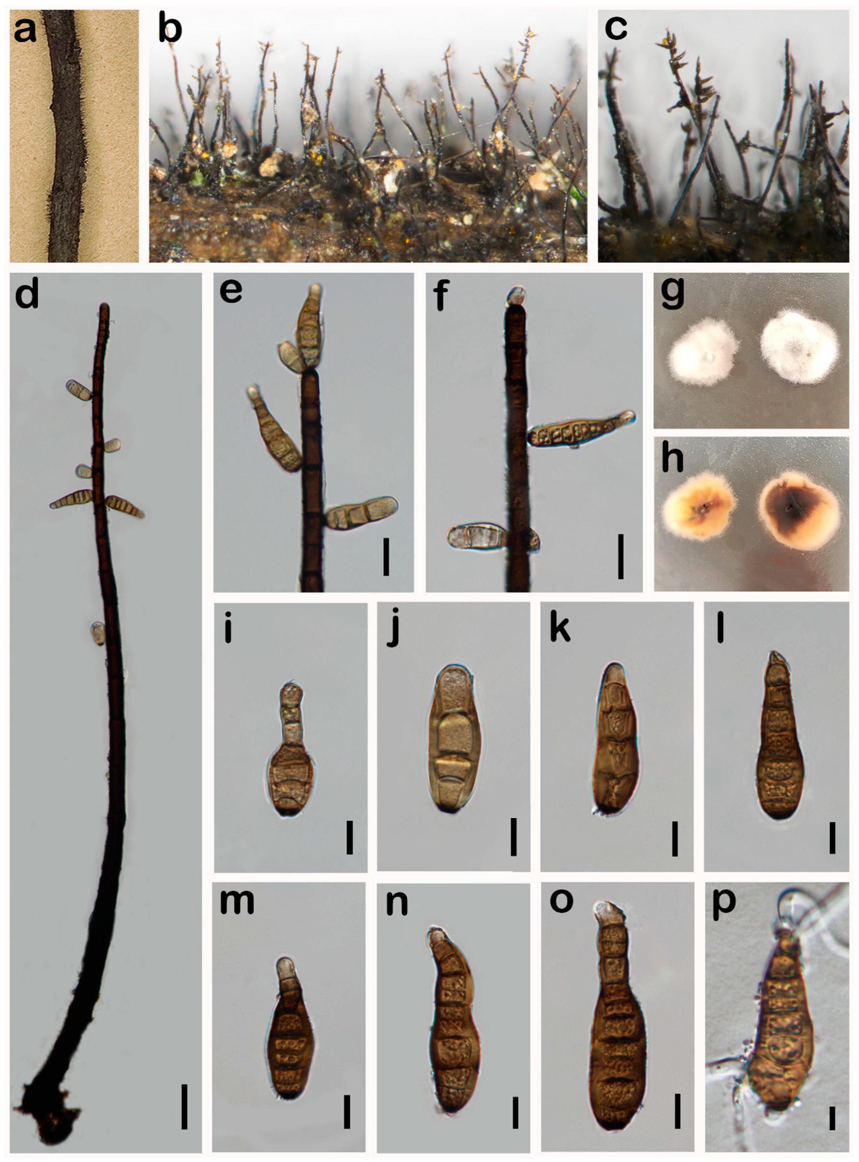
3.2. Taxonomy
4. Discussion
Author Contributions
Funding
Institutional Review Board Statement
Informed Consent Statement
Data Availability Statement
Acknowledgments
Conflicts of Interest
References
- Wijayawardene, N.N.; Hyde, K.D.; Dai, D.Q.; Sanchez-Garcia, M.; Goto, B.T.; Saxena, R.K.; Erdoğdu, M.; Selcuk, F.; Rajeshkumar, K.C.; Aptroot, A.; et al. Outline of Fungi and fungus-like taxa—2021. Mycosphere 2022, 13, 53–453. [Google Scholar] [CrossRef]
- Tian, X.G.; Bao, D.F.; Karunarathna, S.C.; Jayawardena, R.S.; Hyde, K.D.; Bhat, D.J.; Luo, Z.L.; Elgorban, A.M.; Hongsanan, S.; Maharachchikumbura, S.S.N.; et al. Taxonomy and phylogeny of ascomycetes associated with selected economically important monocotyledons in China and Thailand. Mycosphere 2024, 15, 1–274. [Google Scholar] [CrossRef]
- Câmara, M.P.; Palm, M.E.; van Berkum, P.; O’Neill, N.R. Molecular phylogeny of Leptosphaeria and Phaeosphaeria. Mycologia 2002, 94, 630–640. [Google Scholar] [CrossRef] [PubMed]
- Kodsueb, R.; Jeewon, R.; Vijaykrishna, D.; McKenzie, E.H.; Lumyong, P.; Lumyong, S.; Hyde, K.D. Systematic revision of Tubeufiaceae based on morphological and molecular data. Fungal Divers. 2006, 21, 105–130. [Google Scholar]
- Zhang, Y.; Jeewon, R.; Fournier, J.; Hyde, K.D. Multi-gene phylogeny and morphotaxonomy of Amniculicola lignicola: A novel freshwater fungus from France and its relationships to the Pleosporales. Mycol. Res. 2008, 112, 1186–1194. [Google Scholar] [CrossRef] [PubMed]
- Zhang, Y.; Fournier, J.; Crous, P.W.; Pointing, S.B.; Hyde, K.D. Phylogenetic and morphological assessment of two new species of Amniculicola and their allies (Pleosporales). Persoonia 2009, 23, 48–54. [Google Scholar] [CrossRef] [PubMed]
- Kruys, A.; Eriksson, O.E.; Wedin, M. Phylogenetic relationships of coprophilous Pleosporales (Dothideomycetes, Ascomycota), and the classification of some bitunicate taxa of unknown position. Mycol. Res. 2006, 110, 527–536. [Google Scholar] [CrossRef] [PubMed]
- Li, J.F.; Phookamsak, R.; Jeewon, R.; Bhat, D.J.; Mapook, A.; Camporesi, E.; Shang, Q.J.; Chukeatirote, E.; Bahkali, A.H.; Hyde, K.D. Molecular taxonomy and morphological characterization reveal new species and new host records of Torula species (Torulaceae, Pleosporales). Mycol. Prog. 2017, 16, 447–461. [Google Scholar] [CrossRef]
- Zhang, Y.; Crous, P.W.; Schoch, C.L.; Hyde, K.D. Pleosporales. Fungal Divers. 2012, 53, 1–221. [Google Scholar] [CrossRef] [PubMed]
- Hyde, K.D.; Jones, E.B.G.; Liu, J.K.; Ariyawansa, H.; Boehm, E.; Boonmee, S.; Braun, U.; Chomnunti, P.; Crous, P.W.; Dai, D.Q.; et al. Families of Dothideomycetes. Fungal Divers. 2013, 63, 1–313. [Google Scholar] [CrossRef]
- Hyde, K.D.; Abdel-Wahab, M.A.; Abdollahzadeh, J.; Abeywickrama, P.D.; Absalan, S.; Afshari, N.; Ainsworth, A.M.; Akulov, O.Y.; Aleoshin, V.V.; Al-Sadi, A.M.; et al. Global consortium for the classification of fungi and fungus-like taxa. Mycosphere 2023, 14, 1960–2012. [Google Scholar] [CrossRef]
- Jaklitsch, W.M.; Voglmayr, H. Hidden diversity in Thyridaria and a new circumscription of the Thyridariaceae. Stud. Mycol. 2016, 85, 35–64. [Google Scholar] [CrossRef] [PubMed]
- de Silva, N.I.; Hyde, K.D.; Lumyong, S.; Phillips, A.J.L.; Bhat, D.J.; Maharachchikumbura, S.S.N.; Thambugala, K.M.; Tennakoon, D.S.; Suwannarach, N.; Karunarathna, S.C. Morphology, phylogeny, host association and geography of fungi associated with plants of Annonaceae, Apocynaceae and Magnoliaceae. Mycosphere 2022, 13, 955–1076. [Google Scholar] [CrossRef]
- Link, H.F. Observationes in Ordines plantarum naturales. Dissertatio prima complectens Anandrarum ordines Epiphytas, Mucedines, Gastromycos et Fungos. Gesellschaft Natur. 1809, 3, 3–42. [Google Scholar]
- Konta, S.; Hyde, K.D.; Karunarathna, S.C.; Mapook, A.; Senwanna, C.; Dauner, L.A.P.; Nanayakkara, C.M.; Xu, J.; Tibpromma, S.; Lumyong, S. Multi-Gene phylogeny and morphology reveal Haplohelminthosporium gen. nov. and Helminthosporiella gen. nov. associated with Palms in Thailand and a checklist for Helminthosporium reported worldwide. Life 2021, 11, 454. [Google Scholar] [CrossRef] [PubMed]
- Chen, Y.P.; Tian, W.H.; Guo, Y.B.; Madrid, H.; Maharachchikumbura, S.S.N. Synhelminthosporium gen. et sp. nov. and two new species of Helminthosporium (Massarinaceae, Pleosporales) from Sichuan Province, China. J. Fungi 2022, 8, 712. [Google Scholar] [CrossRef] [PubMed]
- Tanaka, K.; Hirayama, K.; Yonezawa, H.; Sato, G.; Toriyabe, A.; Kudo, H.; Hashimoto, A.; Matsumura, M.; Harada, Y.; Kurihara, Y.; et al. Revision of the Massarineae (Pleosporales, Dothideomycetes). Stud. Mycol. 2015, 82, 75–136. [Google Scholar] [CrossRef] [PubMed]
- Tsuda, M.; Ueyama, A.; Nishihara, N. Pseudocochliobolus Nisikadoi, the perfect state of Helminthosporium Coicis. Mycologia 1977, 69, 1109–1120. [Google Scholar] [CrossRef]
- Hu, Y.F.; Liu, J.W.; Xu, Z.H.; Castañeda-Ruíz, R.F.; Zhang, K.; Ma, J. Morphology and multigene phylogeny revealed three new species of Helminthosporium (Massarinaceae, Pleosporales) from China. J. Fungi 2023, 9, 280. [Google Scholar] [CrossRef]
- Manawasinghe, I.S.; Calabon, M.S.; Jones, E.B.G.; Zhang, Y.X.; Liao, C.F.; Xiong, Y.R.; Chaiwan, N.; Kularathnage, N.D.; Liu, N.G.; Tang, S.M.; et al. Mycosphere notes 345–386. Mycosphere 2022, 13, 454–557. [Google Scholar] [CrossRef]
- Castañeda-Ruiz, R.F.; Li, D.W.; Zhang, X.G.; Kendrick, B.; Ramos-García, B.; Pérez-Martínez, S.; Sosa, D. Ellismarsporium gen. nov. and Stanhughesiella gen. nov. to accommodate atypical Helminthosporium and Corynesporella species. Mycotaxon 2018, 132, 759–766. [Google Scholar] [CrossRef]
- Xu, Z.H.; Qiu, L.; Kuang, W.G.; Shi, X.G.; Zhang, X.G.; Castañeda-Ruíz, R.F.; Ma, J. Varioseptispora chinensis gen. & sp. nov., V. apicalis nom. nov., V. hodgkissii comb. nov., and V. versiseptatis comb. nov. Mycotaxon 2020, 135, 753–759. [Google Scholar] [CrossRef]
- Zhang, K.; Zhang, H.; Li, D.W.; Castañeda-Ruiz, R.F. Mirohelminthosporium gen. nov. for an atypical Helminthosporium species and H. matsushimae nom. nov. Mycotaxon 2020, 135, 777–783. [Google Scholar] [CrossRef]
- Voglmayr, H.; Jaklitsch, W.M. Corynespora, Exosporium and Helminthosporium revisited—New species and generic reclassification. Stud. Mycol. 2017, 87, 43–76. [Google Scholar] [CrossRef]
- Ellis, M.B. Dematiaceous Hyphomycetes; Cabi: Wallingford, UK, 1971. [Google Scholar]
- Ellis, M.B. Dematiaceous hyphomycetes. III. Mycologia 1961, 53, 629. [Google Scholar] [CrossRef]
- Luttrell, E. Taxonomic criteria in Helminthosporium 11. Mycologia 1963, 55, 643–674. [Google Scholar] [CrossRef]
- Luttrell, E. Systematics of Helminthosporium and related genera. Mycologia 1964, 56, 119–132. [Google Scholar] [CrossRef]
- Li, C.; Adu, B.; Wu, J.; Qin, G.; Li, H.; Han, Y. Spatial and temporal variations of drought in Sichuan Province from 2001 to 2020 based on modified temperature vegetation dryness index (TVDI). Ecol. Indic. 2022, 139, 108883. [Google Scholar] [CrossRef]
- Su, P.W.; Lu, Z.H.; Tian, W.H.; Chen, Y.P.; Maharachchikumbura, S.S.N. Six additions to the genus Periconia (Dothideomycetes: Periconiaceae) from graminaceous plants in China. J. Fungi 2023, 9, 300. [Google Scholar] [CrossRef] [PubMed]
- Tian, W.H.; Chen, Y.P.; Maharachchikumbura, S.S.N. Neodigitodesmium, a novel genus of family Dictyosporiaceae from Sichuan Province, China. Phytotaxa 2022, 559, 176–184. [Google Scholar] [CrossRef]
- Tian, W.H.; Su, P.W.; Chen, Y.P.; Maharachchikumbura, S.S.N. Four new species of Torula (Torulaceae, Pleosporales) from Sichuan, China. J. Fungi 2023, 9, 150. [Google Scholar] [CrossRef]
- Chen, Y.P.; Lu, Z.H.; Faraj, T.K.; Maharachchikumbura, S.S.N. Myxospora poaceicola sp. nov. (Stachybotryaceae, Hypocreales), a novel myrothecium-like fungus from Digitaria sanguinalis (Poaceae). Phytotaxa 2023, 625, 280–288. [Google Scholar] [CrossRef]
- Su, P.W.; Chen, Y.P.; Syed, A.; Bahkali, A.H.; Maharachchikumbura, S.S.N. Taxonomic novelty in Sichuan Province, China: Veronaea polyconidia sp. nov. (Herpotrichiellaceae), a new addition to hyphomycetous fungi. Phytotaxa 2023, 632, 118–130. [Google Scholar] [CrossRef]
- Senanayake, I.C.; Rathnayaka, A.R.; Sandamali, D.S.; Calabon, M.S.; Gentekaki, E.; Lee, H.B.; Pem, D.; Dissanayake, L.S.; Wijesinghe, S.N.; Bundhun, D.; et al. Morphological approaches in studying fungi: Collection, examination, isolation, sporulation and preservation. Mycosphere 2020, 11, 2678–2754. [Google Scholar] [CrossRef]
- Wanasinghe, D.N.; Phukhamsakda, C.; Hyde, K.D.; Jeewon, R.; Lee, H.B.; Gareth Jones, E.B.; Tibpromma, S.; Tennakoon, D.S.; Dissanayake, A.J.; Jayasiri, S.C.; et al. Fungal diversity notes 709–839: Taxonomic and phylogenetic contributions to fungal taxa with an emphasis on fungi on Rosaceae. Fungal Divers. 2018, 89, 1–236. [Google Scholar] [CrossRef]
- Egger, K.N. Molecular analysis of ectomycorrhizal fungal communities. Can. J. Bot. 1995, 73, 1415–1422. [Google Scholar] [CrossRef]
- Toju, H.; Tanabe, A.S.; Yamamoto, S.; Sato, H. High-Coverage ITS primers for the DNA-based identification of Ascomycetes and Basidiomycetes in environmental samples. PLoS ONE 2012, 7, e40863. [Google Scholar] [CrossRef] [PubMed]
- Vilgalys, R.; Hester, M. Rapid genetic identification and mapping of enzymatically amplified ribosomal DNA from several Cryptococcus species. J. Bacteriol. 1990, 172, 4238–4246. [Google Scholar] [CrossRef]
- Cubeta, M.A.; Echandi, E.; Abernethy, T.; Vilgalys, R. Characterization of anastomosis groups of binucleate Rhizoctonia species using restriction analysis of an amplified ribosomal RNA gene. Phytopathology 1991, 81, 1395–1400. [Google Scholar] [CrossRef]
- Hibbett, D.S. Phylogenetic evidence for horizontal transmission of group I introns in the nuclear ribosomal DNA of mushroom-forming fungi. Mol. Biol. Evol. 1996, 13, 903–917. [Google Scholar] [CrossRef]
- Carbone, I.; Kohn, L.M. A method for designing primer sets for speciation studies in filamentous ascomycetes. Mycologia 1999, 91, 553–556. [Google Scholar] [CrossRef]
- Rehner, S.A.; Buckley, E. A Beauveria phylogeny inferred from nuclear ITS and EF1-alpha sequences: Evidence for cryptic diversification and links to Cordyceps teleomorphs. Mycologia 2005, 97, 84–98. [Google Scholar] [CrossRef] [PubMed]
- Sung, G.H.; Sung, J.M.; Hywel-Jones, N.L.; Spatafora, J.W. A multi-gene phylogeny of Clavicipitaceae (Ascomycota, Fungi): Identification of localized incongruence using a combinational bootstrap approach. Mol. Phylogenet. 2007, 44, 1204–1223. [Google Scholar] [CrossRef] [PubMed]
- Liu, Y.J.; Whelen, S.; Hall, B.D. Phylogenetic relationships among ascomycetes: Evidence from an RNA polymerse II subunit. Mol. Biol. Evol. 1999, 16, 1799–1808. [Google Scholar] [CrossRef] [PubMed]
- Paradis, E.; Schliep, K. ape 5.0: An environment for modern phylogenetics and evolutionary analyses in R. Bioinformatics 2019, 35, 526–528. [Google Scholar] [CrossRef] [PubMed]
- Katoh, K.; Misawa, K.; Kuma, K.; Miyata, T. MAFFT: A novel method for rapid multiple sequence alignment based on fast Fourier transform. Nucleic. Acids. Res. 2002, 30, 3059–3066. [Google Scholar] [CrossRef] [PubMed]
- Capella-Gutiérrez, S.; Silla-Martínez, J.M.; Gabaldón, T. trimAl: A tool for automated alignment trimming in large-scale phylogenetic analyses. Bioinformatics 2009, 25, 1972–1973. [Google Scholar] [CrossRef] [PubMed]
- Lanfear, R.; Frandsen, P.B.; Wright, A.M.; Senfeld, T.; Calcott, B. PartitionFinder 2: New methods for selecting partitioned models of evolution for molecular and morphological phylogenetic analyses. Mol. Biol. Evol. 2016, 34, 772–773. [Google Scholar] [CrossRef] [PubMed]
- Nguyen, L.T.; Schmidt, H.A.; von Haeseler, A.; Minh, B.Q. IQ-TREE: A fast and effective stochastic algorithm for estimating maximum-likelihood phylogenies. Mol. Biol. Evol. 2015, 32, 268–274. [Google Scholar] [CrossRef]
- Huelsenbeck, J.P.; Ronquist, F. MRBAYES: Bayesian inference of phylogenetic trees. Bioinformatics 2001, 17, 754–755. [Google Scholar] [CrossRef]
- Yu, G. Using ggtree to Visualize Data on Tree-Like Structures. Curr. Protoco. Bioinform. 2020, 69, e96. [Google Scholar] [CrossRef]
- Stevens, F.L. Some meliolicolous parasites and commensals from Porto Rico. Botanical Gazette 1918, 65, 227–249. [Google Scholar] [CrossRef]
- Zhang, M.; Zhang, T.Y. Taxonomic studies of Helminthosporium from China 4. Six new species and a key to Helminthosporium from China. Mycotaxon 2009, 109, 399–413. [Google Scholar] [CrossRef]
- Sydow, P.; Sydow, H. Weitere neue Micromyceten der Philippinen-Inseln. Ann. Mycol. 1920, 18, 98–104. [Google Scholar]
- Voglmayr, H.; Akulov, O.Y.; Jaklitsch, W.M. Reassessment of Allantonectria, phylogenetic position of Thyronectroidea, and Thyronectria caraganae sp. nov. Mycol. Prog. 2016, 15, 921–937. [Google Scholar] [CrossRef]
- Ciferri, R.; Fragoso, R.G. Hongos Parásitos y Saprófitos de la República Dominicana:(3a. serie); García: Woodstock, IL, USA, 1926. [Google Scholar]
- Tucker, C. A leaf, bract, and boll spot of sea-island cotton caused by Helminthosporium gossyp II n. sp. ¹. J. Agric. Res. 1926, 32, 391. [Google Scholar]
- Cooke, W.B. Western Fungi—II. Mycologia 1952, 44, 245–261. [Google Scholar] [CrossRef]
- Farr, D.F.; Rossman, A.Y. Fungal Databases; U.S. National Fungus Collections; USDA: Washingtin, DC, USA, 2022. [Google Scholar]
- Devadatha, B.; Sarma, V.V.; Jeewon, R.; Wanasinghe, D.N.; Hyde, K.D.; Gareth Jones, E.B. Thyridariella, a novel marine fungal genus from India: Morphological characterization and phylogeny inferred from multigene DNA sequence analyses. Mycol. Prog. 2018, 17, 791–804. [Google Scholar] [CrossRef]
- Mapook, A.; Hyde, K.D.; McKenzie, E.H.C.; Jones, E.B.G.; Bhat, D.J.; Jeewon, R.; Stadler, M.; Samarakoon, M.C.; Malaithong, M.; Tanunchai, B.; et al. Taxonomic and phylogenetic contributions to fungi associated with the invasive weed Chromolaena odorata (Siam weed). Fungal Divers. 2020, 101, 1–175. [Google Scholar] [CrossRef]
- Liu, J.; Hu, Y.; Luo, X.; Castañeda-Ruíz, R.F.; Ma, J. Three novel species of Helminthosporium (Massarinaceae, Pleosporales) from China. MycoKeys 2022, 94, 73–89. [Google Scholar] [CrossRef]
- Harz, I. Spondylocladium atrovirens. Bull. Soc. Imp. Mosc. 1871, 44, 42. [Google Scholar]
- Ivanyuk, V.; Zezyulina, G. Helminthosporium solani causing silver scab is reported from Belarus. Zashchita Rastenii 1991, 3, 45. [Google Scholar]
- Hermilia-Sanz, B.M. Presence of Helminthosporium solani Dur et Mont, the silver scurf fungus of potatoes, in Chile. Agricultura Tecnica (Santiago) 1976, 36, 5–44. [Google Scholar]
- El Immane-Collet, R.; Elakel, M.; Jouan, B. Comparative study of the agronomical incidence of the silver scurf disease of potato in Morocco and in France. Al Awamia 1995, 91, 1–8. [Google Scholar]
- Errampalli, D.; Saunders, J.M.; Holley, J.D. Emergence of silver scurf (Helminthosporium solani) as an economically important disease of potato. Plant Pathol. 2001, 50, 141–153. [Google Scholar] [CrossRef]
- Tian, S.M.; Chen, Y.C.; Zou, M.Q.; Xue, Q. First Report of Helminthosporium solani causing silver scurf of potato in Hebei Province, North China. Plant Dis. 2007, 91, 460. [Google Scholar] [CrossRef] [PubMed]
- Wanasinghe, D.N.; Mortimer, P.E. Taxonomic and phylogenetic insights into novel Ascomycota from forest woody litter. Biology 2022, 11, 889. [Google Scholar] [CrossRef]
- Li, W.L.; Dissanayake, A.; Liu, J.K. Mycosphere Notes 413–448: Dothideomycetes associated with woody oil plants in China. Mycosphere 2023, 14, 1436–1529. [Google Scholar] [CrossRef]


| Species | Culture/Specimen No. | GenBank Accession Numbers | ||||
|---|---|---|---|---|---|---|
| SSU | LSU | ITS | RPB2 | TEF1 | ||
| Byssothecium circinans | CBS 675.92 | GU205235 | GU205217 | OM337536 | DQ767646 | GU349061 |
| Haplohelminthosporium calami | MFLUCC 18-0074 | MT928160 | MT928156 | MT928158 | – | – |
| Helminthosporiella stilbacea | COAD 2126 | – | – | MG668862 | – | MG682500 |
| H. stilbacea | CPHmZC-01 | – | KX228355 | KX228298 | – | – |
| H. stilbacea | MFLUCC 15-0813 | MT928161 | MT928157 | MT928159 | – | MT928151 |
| Helminthosporium aquaticum | MFLUCC 15-0357 | KU697310 | KU697306 | KU697302 | – | – |
| H. austriacum | CBS 139924 | KY984420 | KY984301 | KY984301 | KY984365 | KY984437 |
| H. austriacum | CBS 14238 | – | KY984303 | KY984303 | KY984367 | KY984439 |
| H. austriacum | L137 | – | KY984302 | KY984302 | KY984366 | KY984438 |
| H. cespitosum | CBS 484.77 | KY984421 | JQ044448 | JQ044429 | KY984370 | KY984440 |
| H. cespitosum | L141 | – | KY984305 | KY984305 | KY984368 | – |
| H. cespitosum | L151 | – | KY984306 | KY984306 | KY984369 | – |
| H. chengduense | CGMCC 3.23575 | ON557757 | ON557745 | ON557751 | ON563073 | ON600598 |
| H. chengduense | UESTC 22.0025 | ON557756 | ON557744 | ON557750 | ON563072 | ON600597 |
| H. chiangraiense | MFLUCC 21-0087 | – | MZ538538 | MZ538504 | – | – |
| H. chinense | CGMCC 3.23570 | ON557760 | ON557748 | ON557754 | – | ON600601 |
| H. chlorophorae | BRIP 14521 | – | – | AF120259 | – | – |
| H. dalbergiae | MAFF 243853 | AB797231 | AB807521 | LC014555 | – | AB808497 |
| H. endiandrae | CBS 138902 | – | KP004478 | KP004450 | – | – |
| H. erythrinicola | CBS 145569 | – | MK876432 | NR_165563 | MK876486 | – |
| H. filamentosa | UESTCC 24.0132 | PP835319 | PP835316 | PP835322 | PP844886 | PP844888 |
| H. genistae | CBS 142597 | – | KY984310 | KY984310 | KY984374 | – |
| H. genistae | CBS 139922 | KY984423 | KY984309 | KY984309 | KY984373 | – |
| H. genistae | CBS 139921 | KY984422 | KY984308 | KY984308 | KY984372 | – |
| H. guanshanense | HJAUP C1022 | OQ172247 | OQ172239 | OQ172249 | OQ234978 | OQ256247 |
| H. hispanicum | CBS 136917 | KY984424 | KY984318 | KY984318 | KY984381 | KY984441 |
| H. jiulianshanense | HJAUP C1057 | – | OQ172253 | OQ172245 | OQ234979 | – |
| H. juglandinum | CBS 136922 | – | KY984321 | KY984321 | KY984384 | KY984444 |
| H. juglandinum | CBS 136911 | KY984425 | KY984322 | KY984322 | KY984385 | KY984445 |
| H. juglandinum | CBS 136912 | – | KY984319 | KY984319 | KY984382 | KY984442 |
| H. juglandinum | CBS 136913 | – | KY984320 | KY984320 | KY984383 | KY984443 |
| H. leucadendri | CBS 135133 | – | KF251654 | KF251150 | KF252159 | KF253110 |
| H. lignicola | MFLUCC 22-0118 | OP740253 | OP740252 | ON329811 | OP757656 | OP757657 |
| H. livistonae | CPC 32158 | – | NG_064539 | NR_160348 | – | – |
| H. magnisporum | MAFF 239278 | AB797232 | AB807522 | AB811452 | – | AB808498 |
| H. massarinum | CBS 139690 | AB797234 | AB807524 | AB809629 | – | AB808500 |
| H. massarinum | JCM 13094 | AB797233 | AB807523 | AB809628 | – | AB808499 |
| H. meilingense | HJAUP C1076 | OQ172246 | OQ172238 | OQ172244 | OQ234980 | OQ234981 |
| H. microsorum | CBS 136910 | KY984427 | KY984329 | KY984329 | KY984390 | KY984448 |
| H. microsorum | L94 | KY984426 | KY984327 | KY984327 | KY984388 | KY984446 |
| H. microsorum | CBS 136916 | – | KY984323 | KY984323 | KY984386 | – |
| H. microsorum | L95 | – | KY984328 | KY984328 | KY984389 | KY984447 |
| H. nabanhense | HJAUP C2054 | OP555400 | OP555398 | OP555394 | – | OP961931 |
| H. nanjingensis | HHAUF020380 | – | – | KF192322 | – | – |
| H. oligosporum | CBS 136909 | – | KY984333 | KY984333 | KY984394 | KY984451 |
| H. oligosporum | CBS 136908 | KY984428 | KY984332 | KY984332 | KY984393 | KY984450 |
| H. oligosporum | L106 | – | KY984330 | KY984330 | KY984391 | KY984449 |
| H. paraoligosporum | EI-102 | – | OM971069 | OM971061 | – | – |
| H. pini | HKAS 135177 | PP835320 | PP835317 | PP835323 | PP844887 | PP844889 |
| H. quercinum | CBS 136921 | KY984429 | KY984339 | KY984339 | KY984400 | KY984453 |
| H. quercinum | CBS 112393 | – | KY984334 | KY984334 | KY984395 | KY984452 |
| H. quercinum | CBS 136915 | – | KY984336 | KY984336 | KY984397 | – |
| H. shangrilaense | KUNCC22-12540 | OP767127 | OP767126 | OP767128 | – | OQ186449 |
| H. sinense | HJAUP C2121 | OP555399 | OP555397 | OP555393 | – | OP961932 |
| H. solani | CBS 365.75 | KY984430 | KY984341 | KY984341 | KY984402 | KY984455 |
| H. solani | CBS 640.85 | – | KY984342 | KY984342 | KY984403 | – |
| H. submersum | MFLUCC 16-1360 | MG098796 | MG098787 | – | – | MG098586 |
| H. submersum | MFLUCC 16-1290 | MG098797 | MG098788 | MG098780 | MG098592 | MG098587 |
| H. syzygii | CBS 145570 | – | MK876433 | NR_165564 | MK876487 | – |
| H. tiliae | CBS 136907 | KY984431 | KY984345 | KY984345 | KY984406 | KY984457 |
| H. tiliae | CBS 136906 | – | KY984344 | KY984344 | KY984405 | – |
| H. tiliae | L171 | – | KY984343 | KY984343 | KY984404 | KY984456 |
| H. velutinum | CBS 139923 | KY984432 | KY984352 | KY984352 | KY984413 | KY984463 |
| H. velutinum | CBS 136924 | – | KY984347 | KY984347 | KY984408 | KY984458 |
| H. velutinum | L98 | KY984433 | KY984359 | KY984359 | KY984417 | KY984466 |
| H. velutinum | L116 | – | KY984348 | KY984348 | KY984409 | KY984459 |
| H. velutinum | L117 | – | KY984349 | KY984349 | KY984410 | KY984460 |
| H. velutinum | UESTCC 24.0189 | PQ046105 | PQ038268 | PQ038261 | PQ057762 | PQ050355 |
| H. yunnanense | HJAUP C2071 | OP555392 | OP555396 | OP555395 | OP961934 | OP961933 |
| Massarina cisti | CBS 266.62 | AB521718 | AB521735 | LC014569 | – | AB808517 |
| M. eburnea | CBS 139697 | GU296170 | GU301840 | OM337528 | GU371732 | GU349040 |
| M. eburnea | CBS 473.64 | MG646979 | MG646947 | MG646958 | – | MG646986 |
| M. pandanicola | MFLUCC 17-0596 | NG_064850 | NG_059396 | NR_153490 | – | AB808540 |
| Periconia pseudodigitata | CBS 139699 | KP325438 | KP325436 | KP325434 | – | – |
| Pseudodidymosphaeria spartii | MFLUCC 13-0273 | KP325439 | KP325437 | KP325435 | – | – |
| P. spartii | MFLUCC 14-1212 | KY984434 | KY984360 | KY984360 | KY984418 | KY984467 |
| P. phorcioides | CBS 122935 | KM875455 | KM875454 | – | – | – |
| P. phorcioides | MFLUCC 13-0533 | KP683377 | KP683376 | KP683375 | – | – |
| P. phorcioides | MFLUCC 13-0611 | KP683374 | KP683373 | KP683372 | – | – |
| P. phorcioides | MFLUCC 14-0618 | – | KT950858 | KT950846 | – | KT950878 |
| Semifissispora natalis | CPC 25383 | – | KT950859 | KT950847 | – | – |
| S. rotundata | CBS 172.93 | – | NG_058526 | NR_156674 | – | – |
| S. tooloomensis | CBS 143431 | – | KF251758 | KF251255 | KF252260 | KF253205 |
| S. duoseptata | CBS 135093 | KY706138 | KY706133 | KY706143 | KY706149 | KY706146 |
| S. imperaticola | MFLUCC 15-0026 | – | NG_068239 | NR_165854 | – | – |
| S. multiseptata | MFLUCC 15-0449 | – | KF251760 | KF251257 | KF252262 | KF253207 |
| S. paludosa | CBS 135088 | – | KF251761 | KF251258 | KF252263 | – |
| S. perfecta | CBS 135099 | AB797289 | AB807579 | AB809642 | – | AB808555 |
| S. perfecta | MAFF 239609 | – | KF251763 | KF251259 | KF252265 | KF253210 |
| S. pseudocaricis | CBS 135132 | – | NG_058052 | NR_137840 | – | – |
| S. pseudopaludosa | CPC 22654 | AB797287 | AB807577 | AB809641 | – | AB808553 |
| S. pseudoperfecta | CBS 120236 | AB797290 | AB807580 | AB809643 | – | AB808556 |
| S. tainanensis | MAFF 243860 | – | NG_058081 | NR_156586 | KJ869232 | – |
| S. trichophoricola | CBS 136764 | – | KF251767 | KF251264 | KF252269 | – |
| S. uniseptata | CBS 135090 | – | KF251769 | KF251266 | KF252271 | KF253214 |
| S. uniseptata | CPC 22150 | – | KF251768 | KF251265 | KF252270 | KF253213 |
| S. uniseptata | CPC 22151 | KP842920 | KP842917 | – | – | – |
| Suttonomyces clematidis | MFLUCC 14-0240 | MG829185 | MG829085 | MG828973 | – | – |
| S. rosae | MFLUCC 15-0051 | ON557758 | ON557746 | ON557752 | ON563074 | ON600599 |
| Synhelminthosporium synnematoferum | CGMCC 3.23574 | ON557758 | ON557746 | ON557752 | ON563074 | ON600599 |
| Species | Culture/Specimen No. | GenBank Accession Numbers | ||||
|---|---|---|---|---|---|---|
| SSU | LSU | ITS | RPB2 | TEF1 | ||
| Chromolaenomyces appendiculatus | MFLUCC 17–1455 | MT214394 | NG_068705 | NR_168862 | MT235806 | MT235770 |
| Cycasicola goaensis | MFLUCC 17–0754 | MG829112 | MG829001 | MG828885 | – | MG829198 |
| C. leucaenae | MFLUCC17–0914 | MK347833 | MK347942 | NR_163322 | MK434900 | MK360046 |
| Liua muriformis | KUMCC 18–0177 | MK433595 | MK433598 | MK433599 | MK426799 | MK426798 |
| Parathyridaria clematidis | MFLUCC 17–2160 | MT226710 | MT214599 | MT310643 | MT394710 | MT394655 |
| P. clematidis | MFLUCC 17–2185 | NG_070668 | MT214598 | MT310642 | MT394709 | MT394654 |
| P. ellipsoidea | KNU–JJ–1829 | – | LC552952 | LC552950 | – | – |
| P. flabelliae | MUT 4886 | KT587317 | KP671720 | KR014358 | MN605930 | MN605910 |
| P. flabelliae | MUT 4859 | KT587315 | KP671716 | KR014355 | MN605929 | MN605909 |
| P. percutanea | CBS 128203 | KF366450 | KF366448 | KF322117 | KF366453 | KF407988 |
| P. percutanea | CBS 868.95 | KF366451 | KF366449 | KF322118 | KF366452 | KF407987 |
| P. philadelphi | CBS 143432 | – | NG_063958 | MH107905 | – | MH108023 |
| P. ramulicola | MUT 4397 | MN556311 | KF636775 | KC339235 | MN605933 | MN605913 |
| P. ramulicola | CBS 141479 | KX650514 | KX650565 | NR_147657 | KX650584 | KX650536 |
| P. robiniae | MFLUCC 14–1119 | – | KY511141 | KY511142 | – | KY549682 |
| P. rosae | MFLU 17–0623 | – | NG_059873 | NR_157530 | – | – |
| P. serratifoliae | MFLUCC 17–2210 | NG_070669 | MT214602 | MT310646 | MT394713 | MT394658 |
| P. tyrrhenica | MUT 4966 | KT587309 | KP671740 | KR014366 | MN605931 | MN605911 |
| P. tyrrhenica | MUT 5371 | KU314952 | MN556329 | KU314951 | MN605932 | MN605912 |
| P. virginianae | MFLUCC 17–2163 | NG_070670 | NG_073853 | MT310647 | MT394714 | MT394659 |
| Parathyridariella dematiacea | MUT 4419 | MN556313 | KF636786 | KC339245 | MN605925 | MN605905 |
| P. dematiacea | MUT 4884 | KT587329 | KP671726 | MN556317 | MN605926 | MN605906 |
| Pseudothyridariella chromolaenae | MFLUCC 17–1472 | MT214395 | NG_068706 | NR_168863 | MT235807 | MT235771 |
| P. idesiae | CGMCC 3.24439 | OR253216 | OR253307 | OR253148 | OR253762 | OR251154 |
| P. mahakoshae | NFCCI 4215 | MG020441 | MG020438 | MG020435 | MG020446 | MG023140 |
| Thyridaria acaciae | CBS 138873 | – | KP004497 | KP004469 | – | – |
| T. aureobrunnea | MFLUCC 21–0090 | – | MZ538562 | MZ538528 | – | – |
| T. broussonetiae | CBS 141481 | NG_063067 | – | NR_147658 | – | – |
| T. broussonetiae | TB | – | – | KX650567 | KX650585 | KX650538 |
| T. broussonetiae | TB1 | KX650515 | – | KX650568 | KX650586 | KX650539 |
| T. jonahhulmei | KUMCC 21–0816 | ON007046 | ON007037 | ON007041 | ON009135 | ON009131 |
| T. jonahhulmei | KUMCC 21–0817 | ON007047 | ON007038 | ON007042 | ON009136 | ON009132 |
| Thyridariella mangrovei | NFCCI 4214 | MG020442 | MG020439 | MG020436 | MG020447 | MG020444 |
| T. mangrovei | NFCCI 4213 | MG020440 | MG020437 | MG020434 | MG020445 | MG020443 |
| Torula herbarum | CBS 595.96 | KF443387 | KF443385 | KF443408 | KF443395 | KF443402 |
| Vaginospora sichuanensis | UESTCC 24.0191 | PQ046104 | PQ038267 | PQ038260 | PQ050358 | PQ050354 |
Disclaimer/Publisher’s Note: The statements, opinions and data contained in all publications are solely those of the individual author(s) and contributor(s) and not of MDPI and/or the editor(s). MDPI and/or the editor(s) disclaim responsibility for any injury to people or property resulting from any ideas, methods, instructions or products referred to in the content. |
© 2024 by the authors. Licensee MDPI, Basel, Switzerland. This article is an open access article distributed under the terms and conditions of the Creative Commons Attribution (CC BY) license (https://creativecommons.org/licenses/by/4.0/).
Share and Cite
Tian, W.-H.; Jin, Y.; Liao, Y.-C.; Faraj, T.K.; Guo, X.-Y.; Maharachchikumbura, S.S.N. Phylogenetic Insights Reveal New Taxa in Thyridariaceae and Massarinaceae. J. Fungi 2024, 10, 542. https://doi.org/10.3390/jof10080542
Tian W-H, Jin Y, Liao Y-C, Faraj TK, Guo X-Y, Maharachchikumbura SSN. Phylogenetic Insights Reveal New Taxa in Thyridariaceae and Massarinaceae. Journal of Fungi. 2024; 10(8):542. https://doi.org/10.3390/jof10080542
Chicago/Turabian StyleTian, Wen-Hui, Yan Jin, Yue-Chi Liao, Turki KH. Faraj, Xin-Yong Guo, and Sajeewa S. N. Maharachchikumbura. 2024. "Phylogenetic Insights Reveal New Taxa in Thyridariaceae and Massarinaceae" Journal of Fungi 10, no. 8: 542. https://doi.org/10.3390/jof10080542
APA StyleTian, W.-H., Jin, Y., Liao, Y.-C., Faraj, T. K., Guo, X.-Y., & Maharachchikumbura, S. S. N. (2024). Phylogenetic Insights Reveal New Taxa in Thyridariaceae and Massarinaceae. Journal of Fungi, 10(8), 542. https://doi.org/10.3390/jof10080542