Article

Composition and Biodiversity of Culturable Endophytic Fungi in the Roots of Alpine Medicinal Plants in Xinjiang, China

1
State Key Laboratory for Quality Ensurance and Sustainable Use of Dao-di Herbs, Institute of Medicinal Plant Development, Chinese Academy of Medical Sciences & Peking Union Medical College, No.151, Malianwa North Road, Haidian District, Beijing 100193, China
2
Xinjiang Institute of Chinese and Ethnic Medicine, Urumqi 830002, China
3
School of Pharmacy, Shandong Second Medical University, Weifang 261053, China
*
Authors to whom correspondence should be addressed.
J. Fungi 2025, 11(2), 113; https://doi.org/10.3390/jof11020113
Submission received: 11 December 2024 / Revised: 19 January 2025 / Accepted: 1 February 2025 / Published: 3 February 2025

Abstract

(1) Background: Endophytic fungi play an important role in plant growth and stress resistance. The presence of a special fungal taxon such as the dark septate endophytic (DSE) fungi in alpine environments is particularly important for plant resistance to environmental stresses. However, the composition of root endophytic fungi in different environments and between different host plants has not been well studied. (2) Results: A total of 408 culturable endophytic fungi were isolated from the roots of Saussurea involucrata and Rhodiola crenulata which were collected in 5 plots from the Tianshan and Karakoram Mountains of the Xinjiang region, belonging to 91 species, 54 genera, 31 families, and 3 phyla based on the morphological characteristics and molecular sequence. Among them, DSE fungi were the dominant group, accounting for 52.94%, and Leptodontidium orchidicola was the dominant species. In addition, we also compared the composition and diversity of root endophytic fungi from different plants and different sites, with emphasis on special fungal taxa such as DSE. (3) Conclusions: The composition and diversity of cultural endophytic fungi are significantly different in the two alpine medicinal plant species and across various locations. Some fungi showed the preferences of the host or environment. The endophytic fungal resources, especially DSE, were very rich in the two alpine medicinal plants, indicating that these fungi may play a crucial role in the ecological adaptation of host plants in harsh environments.

1. Introduction

Microbial communities respond to changing environmental conditions by regulating global biogeochemical cycles such as plant nutrient uptake and soil carbon storage [1,2]. As global climate change progresses, biodiversity is declining [3]. Alpine ecosystems face even greater challenges, as a climate shift occurs there at twice the global average rate [2]. Rising temperatures drive alpine plants to migrate to higher elevations, placing many of them at risk of “summit extinction” [4]. Mountains, covering 25% of the Earth’s land surface, are rich in biodiversity due to their complex climates and topographies. Compared to low-altitude regions, alpine areas are characterized by high elevations, hypoxia, large diurnal temperature variations, and intense radiation [5]. The mountains of Xinjiang, China, support diverse vegetation shaped by geological uplift, glaciation, and monsoonal changes, where plants often contend with various environmental challenges [6].
Xinjiang is located in the northwest of China, with a vast territory, including the Tianshan, Karakoram, Altai Mountains, and the Junggar and Tarim basins [7]. The unique topography and climate of Xinjiang region breed abundant plant resources, including valuable medicinal species like Saussurea involucrata, Rhodiola crenulata, and Lithospermum [8,9]. Saussurea involucrata (Kar. & Kir.) Sch. Bip. belonging to the Asteraceae family, is a Chinese traditional medicinal plant for regulating meridians and promoting blood circulation [10]. Pharmacological studies have shown that S. involucrata oral liquid (SIOL) can alleviate rheumatoid arthritis in clinical applications [11], while S. involucrata polysaccharide (SIP) demonstrates significant antioxidant and photoprotective effects on skin exposed to UVB radiation [12]. S. involucrata is primarily distributed in Xinjiang, Russia, and Kazakhstan, growing at altitudes of 2400–4100 m [13]. Rhodiola crenulata (Hook. f. & Thomson) H. Ohba is also a famous medicinal plant in the Crassulaceae family, which shares a similar habitat with S. involucrata and grows at altitudes of approximately 2800–5600 m, and the two species are often found coexisting [14]. According to the Chinese Pharmacopoeia, R. crenulata is used for relieving cough, strengthening the spleen, and calming the mind. Pharmacological studies have shown that R. crenulata is used to treat cardiovascular diseases, tumors, and diabetes [15]. However, due to the climate changes, over-harvesting of wild resources, and the lack of large-scale cultivation, both species are now on the brink of extinction and are listed as second-class protected wild plants in the National List of Key Protected Wild Plants [16].
In recent years, with the advancement of sequencing technology, plants have increasingly been considered as complex assemblies of plants and their associated microorganisms, of which endophytic fungi are important components [17,18]. Endophytic fungi ubiquitously colonize the internal tissues of host plants for part or all of their lifecycle without inducing apparent disease symptoms [19]. Studies have shown that the endophytic fungi can promote plant growth and development, assist in resisting environmental stresses, and facilitate the production of secondary metabolites [20]. Among the endophytic fungi, dark septate endophytic (DSE) fungi form a crucial subgroup. DSE refers to fungi that colonize plant roots with dark, septate hyphae, without causing noticeable symptoms of disease [21]. Previous studies have indicated that DSE fungi frequently colonize plants in unique environments and contribute to host plant resilience against harsh conditions, as well as promoting plant growth [22,23]. The ability of S. involucrata and R. crenulata to grow in such harsh environments may be closely related to their microbial communities, particularly DSE.
Moreover, current research on these two alpine medicinal plants primarily focuses on morphology, chemical composition, and pharmacology [24,25]. Limited studies on their endophytic fungi, especially those on the composition of DSE, the most predominant group, are unknown. In light of this, the study identifies culturable endophytic fungi from the roots of two alpine medicinal plants collected from 5 sampling sites in the Tianshan and Karakoram Mountains in Xinjiang. The study aims to understand the diversity and distribution patterns of the culturable endophytic fungi, particularly DSE, in these plants, and to lay the foundation for further analysis of the symbiotic relationship between alpine plants and fungi. At the same time, this study also aims to acquire valuable culturable endophytic fungi from these rare alpine medicinal plants, which may provide precious fungal resources for future artificial cultivation and conservation of these endangered medicinal plants, as well as the preservation of alpine biodiversity in the future by the microbiological technique.

2. Materials and Methods

2.1. Sample Collection

Xinjiang, the largest province in China, is located in the northwest (73°40′ E–96°18′ E, 34°25′ N–48°10′ N). This vast region features diverse landscapes, including mountains, basins, and deserts, with unique geomorphic and ecological characteristics. Two alpine medicinal plants, S. involucrata (Asteraceae) and R. crenulata (Crassulaceae), were selected from the Tianshan and Karakorum Mountains, two major mountain ranges in Xinjiang. The Tianshan Mountains have a temperate continental arid climate, whereas the Karakorum Mountains are situated in a transitional zone between the Qinghai-Tibet alpine region and the northwest arid region.
Root and soil samples from two alpine plants were collected from 5 sites across the Tianshan and Karakoram Mountains in Xinjiang, with their original habitat, latitude, longitude, and altitude recorded (Table 1). At each sampling site, 3 large plots (10 m × 10 m) with similar terrain were selected, each plot spaced approximately 100 m apart. Within each plot, 3 to 5 healthy plants were randomly selected, ensuring a minimum spacing of 10 m between individual plants. All the samples were collected and stored at 4 °C, and transported under low-temperature conditions to the laboratory.

2.2. Root Sample Processing, Morphological and Anatomical Observation

The root samples were washed under running water and stained using Trypan blue (Solarbio, Beijing, China) according to the steps for observing mycorrhizal fungi [26]. In brief, following the material processing method described by Phillips and Hayman (1970), the roots were cut into 1 cm segments and fixed in Formalin-Aceto-Alcohol (FAA) (Coolaber, Beijing, China) solution for 24 h, then washed with running water. The root segments were transferred to 10% KOH (Beilian, Tianjin, China) and heated in a 90 °C water bath for 40–60 min until the roots became transparent, removing the cytoplasm from the root epidermal cells. The washed root segments were immersed in a 5% lactic acid solution (XiLONG SCIENTIFIC, Shantou, China) for 5 min to facilitate staining. The acidified material was directly transferred to a 0.05% Trypan blue-lactic acid glycerol solution for 3 min of staining, then rinsed multiple times with glycerol (Yongda, Tianjin, China) and left to decolorize overnight. The root samples were placed on a glass slide and observed under the optical microscope (ZEISS Axio Imager A1, Jena, Germany), with images captured.

2.3. Isolation and Cultivation of Endophytic Fungi

Healthy, fresh, robust, and dark brown roots were selected and washed thoroughly with running water to remove soil and debris, and blotted dry. Root segments were soaked in 75% ethanol for 30 s, immersed in 5% NaClO (Beilian, Tianjin, China) for 3–5 min, rinsed with sterile water, and immersed in a 10 mL solution containing 150 µg/mL streptomycin sulfate and 150 µg/mL potassium penicillin (Gibco, Grand Island, NY, USA) for 10 min, and washed 3 times with sterile water. The solution from the last rinse was collected and 1 mL was placed on PDA agar plates, incubated at 25 °C in the dark for 3 days with 5 replicates. The absence of colony growth indicated effective sterilization of the root surfaces. The completely sterilized roots were cut into 2 mm sections and placed on 1 cm3 PDA agar blocks containing dual antibiotics (100 µg/mL streptomycin sulfate and 100 µg/mL potassium penicillin), resulting in a total of 4248 root segments. In addition, 9 PDA agar blocks were placed on each 9 cm Petri dish, with one root segment inoculated per block, resulting in a total of 472 dishes. The dishes were sealed and incubated at 25 °C in the dark for 7–10 days, with regular observations and timely treatment of contaminated plates. When colonies formed on the agar, the edges of actively growing hyphae were transferred to PDA agar for purification, and once pure colonies were obtained, they were subcultured onto PDA slants and stored in 10% glycerol tubes. The strains used in this study were stored in the strain collection of the Biotechnology Center of the Institute of Medicinal Plant Development, Chinese Academy of Medical Sciences.

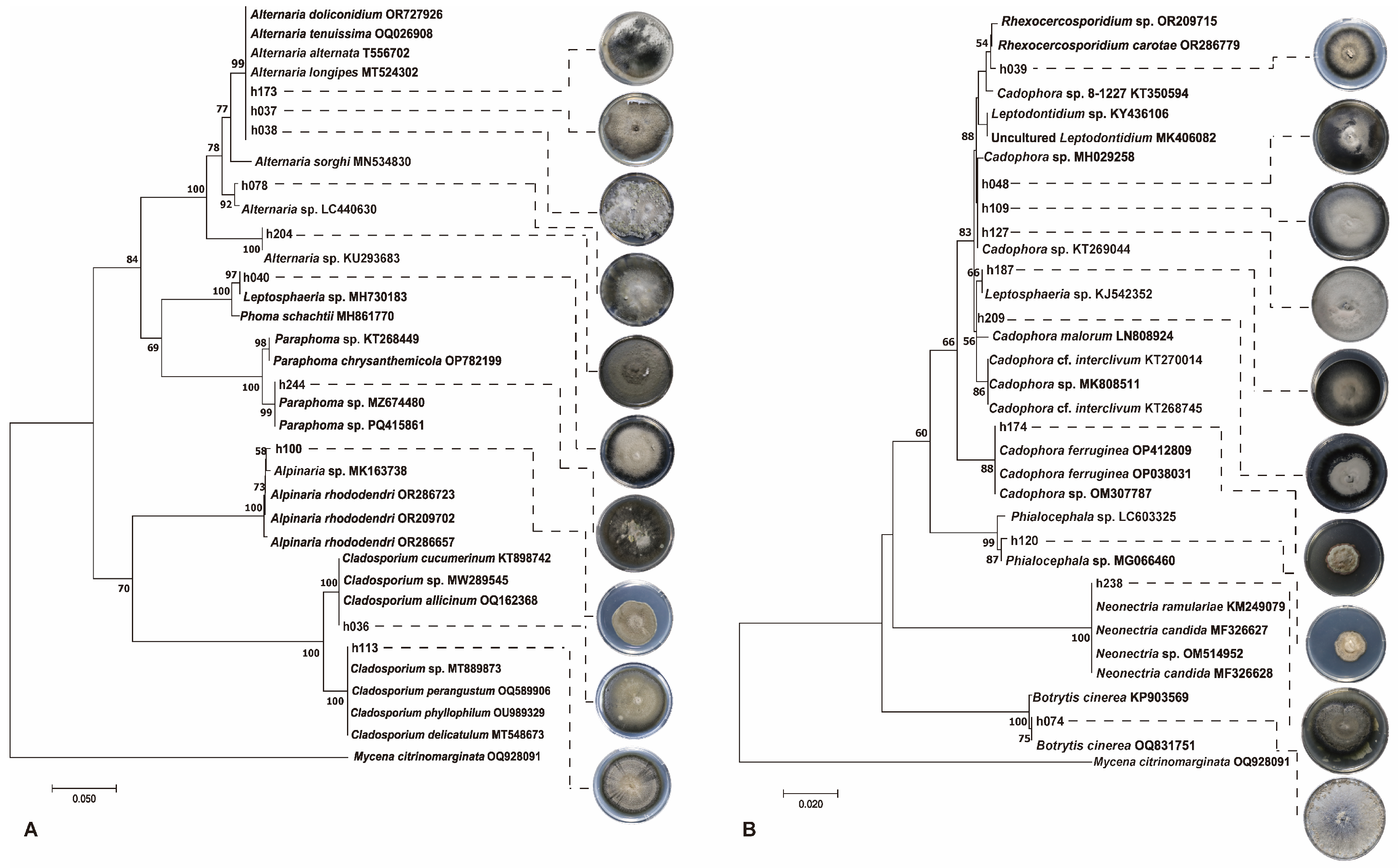
2.4. Identification of Culturable Endophytic Fungi

The strains isolated were identified using both morphological observation and molecular biology techniques. Morphological identification was carried out by observing colony characteristics, such as shape, color, and exudates, as well as microscopic features like hyphae, spore-producing structures, and spores under a ZEISS Axio Imager A1 microscope (ZEISS, Jena, Germany). The identification was based on the latest fungal classification system.
Molecular identification was performed by grinding the mycelia in liquid nitrogen, followed by DNA extraction using the CTAB method. DNA concentration was measured using Nanodrop (Thermo NanoDrop 2000, Waltham, MA, USA) and PCR amplification of (internal transcript space), and ITS was performed with ITS1 and ITS4 primers. The PCR conditions were as follows: 95 °C for 5 min (initial denaturation), 94 °C for 1 min (denaturation), 52 °C for 50 s (annealing), 72 °C for 1 min (extension), followed by a final extension at 72 °C for 7 min, with 35 cycles [27]. The amplification products were analyzed using 1% agarose (Biowest, Nuaillé, France) gel electrophoresis (BIO-RAD, Hercules, CA, USA; Agilent 2100 Bioanalyzer, Santa Clara, CA, USA) to verify their quality, and the qualified PCR products were sent to GeneWiz (Suzhou, China) for sequencing. The sequence chromatograms were analyzed using SnapGene software (4.3.6), and after manual quality control, high-quality sequences were obtained and subjected to BLAST analysis on the NCBI website (https://blast.ncbi.nlm.nih.gov/Blast.cgi (accessed on 20 November 2024)). Combining the molecular data with morphological features, a comprehensive species identification was performed.

2.5. Data Analysis

Statistical analysis was performed to determine the isolation rate and relative frequency of endophytic fungi in each plant tissue. The isolation rate (IR) was used to calculate the percentage of root segments infected by endophytic fungi, indicating the degree of infection. The relative isolation frequency (IF) was used to calculate the percentage of each fungal species relative to the total number of culturable endophytic fungi, determining the species dominance. The Richness index refers to the total number of fungal species at that site. The Shannon–Wiener and Simpson diversity indices were used to assess the diversity of endophytic fungal communities [28,29]. The Pielou evenness index was used to measure the evenness of the fungal community distribution across the samples [30]. Sorenson’s similarity coefficient (Cs) was used to compare the similarity of endophytic fungal composition between the samples [31]. Cs range from 0 to 1, with values closer to 0 indicating lower similarity between the endophytic fungal communities of the two locations. When Cs equals 0, it indicates that the endophytic fungal communities of the two locations are completely different. The formulas for these calculations are as follows: The relative abundance is calculated by dividing the number of isolated strains of a particular taxon by the total number of isolated strains across all taxa. The isolation rate (IR) is calculated by dividing the total number of root segments infected by fungi by the total number of root segments cultured.
Shannon–Wiener index: H’ = − i 1 s ( Pi)(lnPi), Pi = ni/N, N represents the total number of fungal species obtained from a particular site; ni represents the number of individuals of the i-th fungal species at the site.
Simpson index: D = 1 − i 1 s ( P i)2, Pi2 = ni(ni − 1)/N(N − 1), N is the total number of fungal species obtained from a specific site; ni is the number of individuals of the i-th fungal species at that site.
Pielou index: J = H/lnS, S represents the total number of species at the site.
Sorenson’s similarity coefficient: Cs = 2j/a + b, j represents the number of fungal species common between different sites, while a and b represent the number of fungal species at each site, respectively.

3. Results

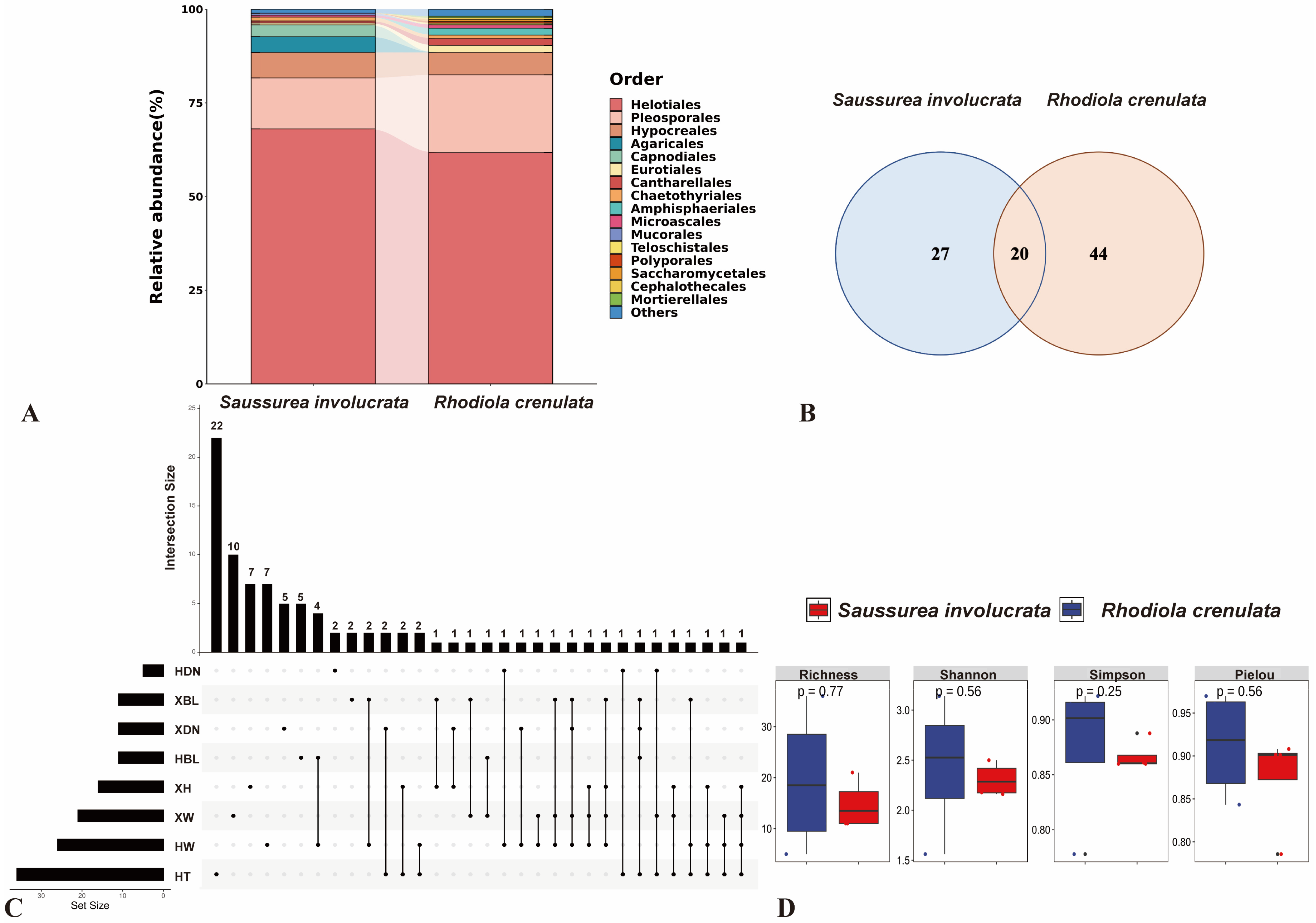
3.1. Comparison of Cultural Endophytic Fungal Community Structure and Diversity in Two Alpine Plants

Fresh root segments of two alpine medicinal plants were fixed with FAA fixative and subsequently stained with trypan blue to observe the colonization of endophytic fungi. The results revealed substantial fungal colonization in the roots of both wild S. involucrata and R. crenulata. The colonizing fungal hyphae were brown and septate, forming bead-like structures within the cells. Based on the morphological characteristics of the hyphae, these fungi were identified as belonging to the typical DSE fungi (Figure 1).
A total of 4248 root tissue blocks from two alpine medicinal plants across 5 sampling sites were used for endophytic fungal isolation and 3356 blocks produced fungal colonies. Fungi with similar morphological characters were divided into a morphological group, and molecular identification was performed on 408 endophytic fungal isolates, which were classified into 3 phyla, 9 classes, 16 orders, 31 families, 54 genera, and 91 species (Table S1). Ascomycota was the dominant phylum, with a total of 392 isolates accounting for 96.08% of the total fungal count. Helotiales was the dominant order, with 264 isolates representing 64.71% of the total fungal count and Leptodophora was the most prevalent genus, with 64 isolates constituting 15.69% of the total fungal count.
The isolation rates, abundances, and community structures of culturable endophytic fungi from the two alpine plants exhibited significant differences (Figure 2A). A total of 191 isolates belonging to 47 species of endophytic fungi were obtained from the roots of S. involucrata, while 217 isolates belonging to 64 species were obtained from the roots of R. crenulata. The composition of endophytic fungal taxa from the two medicinal plants is significantly different, with 27 species unique to S. involucrata and 44 species unique to R. crenulata. L. orchidicola and Filosporella sp. were identified as the dominant endophytic fungi in the roots of S. involucrata and R. crenulata, respectively, with relative isolation frequencies of 20.42% and 9.68% in their corresponding host root segments. The different composition of the two plants indicates that the host plants selectively recruit different fungi to form their endophytic fungal communities. Furthermore, common characteristics were observed in the structural composition of the culturable endophytic fungal communities between the two medicinal plants, with 20 species distributed in both plants (Figure 2B), including Phoma schachtii, Plenodomus meliloti, Alpinaria rhododendri, Alternaria alternata, Alternaria sp., Articulospora, Filosporella sp., L. orchidicola, Rhexocercosporidium sp., Tetracladium sp., Leptosphaeria sp., Cadophora sp., Mycochaetophora, uncultured Helotiales, Graphium basitruncatum, Dactylonectria macrodidyma, Neonectria candida, Neonectria sp., Thyridium cornearis, Monodictys arctica, etc., resulting in a Cs similarity of 36.04%.
In three collection sites, Tianshan Mountain No.1 glacier (XW and HW), Bayinbuluke, Hejing County (XBL and HBL), and Danangou Uzbek Township (XDN and HDN), which are in the Tianshan Mountains (Table 1), both medicinal alpine plants were collected, with distinct differences in the composition of their root-associated endophytic fungal communities. In these collection sites, fungi isolated from 26 S. involucrata plants belonged to 3 phyla, 8 orders, and 16 families. Similarly, in the same 3 locations, fungi isolated from 26 R. crenulata plants belonged to 3 phyla, 8 orders, and 17 families. Nine families were shared between the two plants, including Melanommataceae, Didymellaceae, Helotiaceae, Leptodontidiaceae, Ploettnerulaceae, Aspergillaceae, Pleosporaceae, Nectriaceae, and Thyridiaceae. Seven families were unique to S. involucrata, including Ceratobasidiaceae, Cladosporiaceae, Phaeosphaeriaceae, Herpotrichiellaceae, Bionectriaceae, Hypocreaceae, and Mucoraceae. Eight families were unique to R. crenulata, including Hydnaceae, Discinellaceae, Mollisiaceae, Sclerotiniaceae, Saccharomycetaceae, Amphisphaeriaceae, Bartaliniaceae, and Mortierellaceae.
Notably, a total of 208 DSE strains were isolated from the root systems of both plants (Table 2). Moreover, 110 strains and 18 species were isolated from S. involucrata, while 98 strains and 22 species were isolated from R. crenulata. These strains belonged to 4 classes, 6 orders, 12 families, 16 genera, and 31 species. Among the 16 genera of DSE, 8 genera were shared by both plants, including Phoma, Alpinaria, Alternaria, Rhexocercosporidium, Leptodophora, Leptosphaeria, Cadophora, Neonectria, etc. The DSE genera unique to S. involucrata include Cladosporium, Exophiala, and Crocicreas. The DSE genera unique to R. crenulata include Paraphoma, Cyphellophora, Leptodontidium, Phialocephala, Botrytis, and Microdochium.

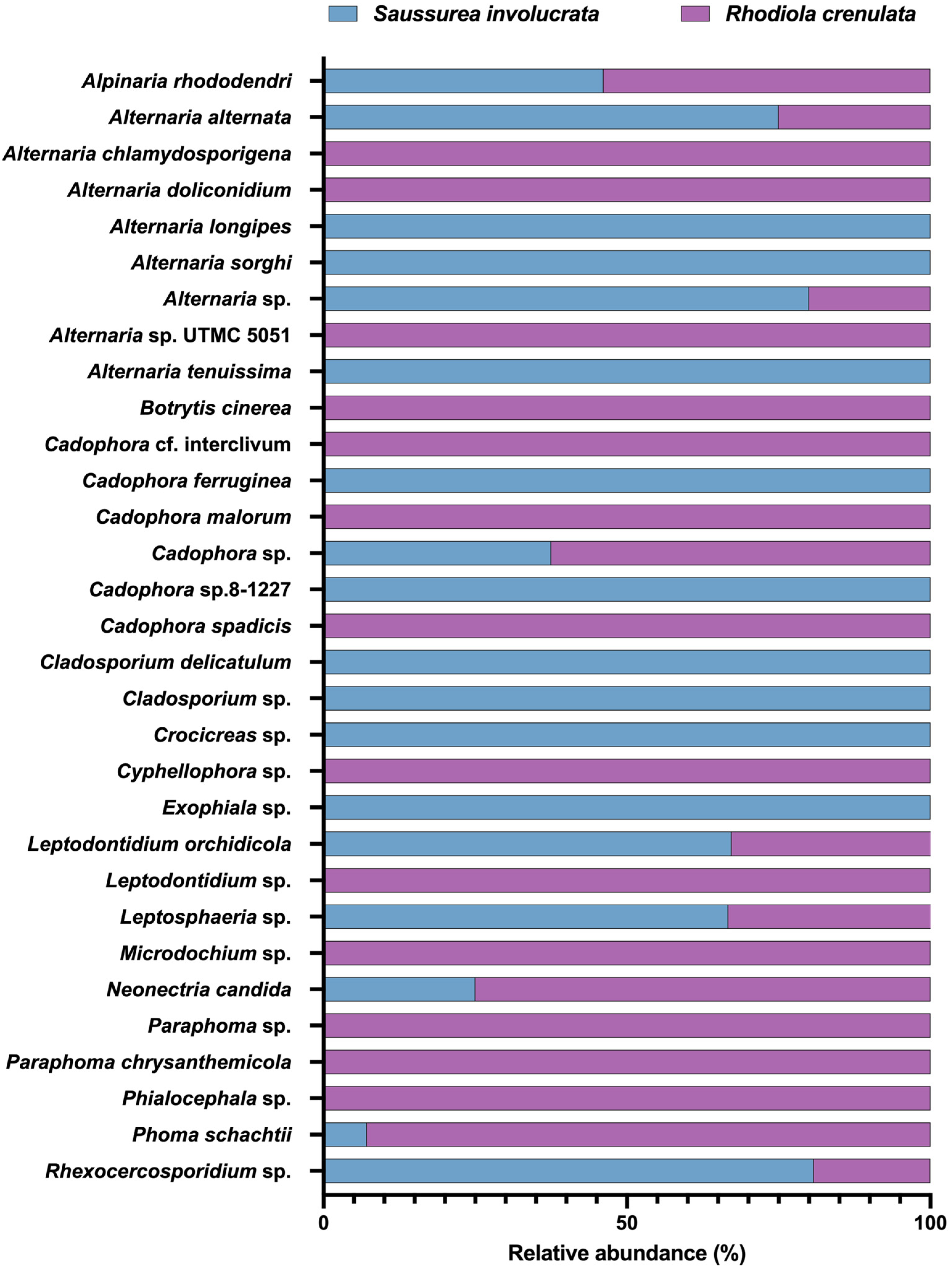
3.2. Analysis of the Community Composition and Diversity of Endophytic Fungi in Two Alpine Medicinal Plants from Different Sampling Sites

The Venn diagram analysis of culturable endophytic fungal communities in two medicinal plants revealed distinct fungal compositions across sampling sites. Ten fungi were exclusive to XW, including Cladosporium sp., C. delicatulum, Neosetophoma cerealis, Penicillium fellutanum, Exophiala sp., Rhexocercosporidium carotae, Tetracladium maxilliforme, L. sclerotioides, Cadophora sp., and Neonectria sp. Seven species were unique to XH, such as Calyptella sp., Mycena citrinomarginata, Alternaria tenuissima, Dactylaria sp., Leptodophora gamsii, Cadophora ferruginea, and Fusarium sp. Five species, including Rhizoctonia sp., Alternaria sorghi, Clonostachys rosea, Trichoderma polysporum, and Neonectria lugdunensis, were specific to XDN. XBL harbored two endemic fungi, Alternaria longipes and Crocicreas sp. A total of 22 fungi were specific to HT, including Pezicula melanigena, Porostereum spadiceum, Paraphoma chrysanthemicola, P. salicis, Paraphoma sp., Phaeosphaeria, Alternaria chlamydosporigena, A. gansuensis, Alternaria sp., Curvularia nodulosa, Pleosporales sp., Pyrenophora fugax, Dothideomycetes sp., Cyphellophora sp., Rhinocladiella similis, Xanthoria resendei, Leptodophora echinate, Cadophora cf. interclivum, Phialemonium cf., Graphium penicillioides, Fusarium solani, and Thelonectria sp. Seven fungi were unique to HW, including Sporidesmium spiraeae, Helotiales sp., Leptodontidium sp., Phialocephala sp., Microdochium sp., Fusarium venenatum, and Acremonium sclerotigenum. Five species, such as Penicillium camemberti, Penicillium glabrum, Leptodontidium, Ogataea naganishii, and Mortierella sp., were exclusive to HBL. Finally, two species, Alternaria doliconidium and F. oxysporum, were endemic to HDN (Figure 2C).
A total of 47 species of endophytic fungi were isolated from the roots of S. involucrata across four sampling sites: XW, XBL, XDN, and XH, with 21, 11, 11, and 16 species identified at each site, respectively. The fungal richness was highest at the XW site. The highest similarity (0.25) was observed between XW and XBL, with shared species including P. meliloti, Cistella sp., L. orchidicola, and Rhexocercosporidium sp. In contrast, the lowest similarity (0.0625) occurred between XW and XDN, where Rhexocercosporidium sp. was the only shared species (Table 3).
A total of 64 endophytic fungal species were isolated from the roots of R. crenulata across the 4 sampling sites HW, HBL, HDN, and HT. Among them, 26 species were isolated from HW, 11 from HBL, 5 from HDN, and 36 from HT. The highest fungal diversity was observed at the HT site, while the lowest diversity was found at the HDN site. The similarity between HW and HBL was the highest, with a similarity index of 0.2162, including Cadophora spadices, Botrytis cinerea, Seaverinia geranii, and F. acuminatum. The similarity between HDN and HBL was the lowest, with a similarity index of 0, indicating there were not any shared fungal species.
Statistical analysis of the abundance and diversity of endophytic fungi isolated from the eight sampling sites was conducted. It revealed that, except for the HDN site, R. crenulata in the HW, HBL, and HT sites exhibited a higher richness, Shannon–Wiener index, and Simpson index than S. involucrata at the XW, XBL, and XH sites. This suggests that R. crenulata has higher endophytic fungal diversity than S. involucrata at these locations. The Pielou index varied across sites for both plant species, but it was consistently above 0.75, indicating high evenness. Furthermore, XDN and HDN exhibited the highest values for both S. involucrata and R. crenulata, while XW and HW had the lowest values. Simultaneously, the richness and Simpson index were the lowest at XDN and HDN. These results indicate that the endophytic fungal community structures of the two plant species vary across different sites, with both S. involucrata and R. crenulata harboring rich endophytic fungal species across their respective sampling locations (Figure 2D).

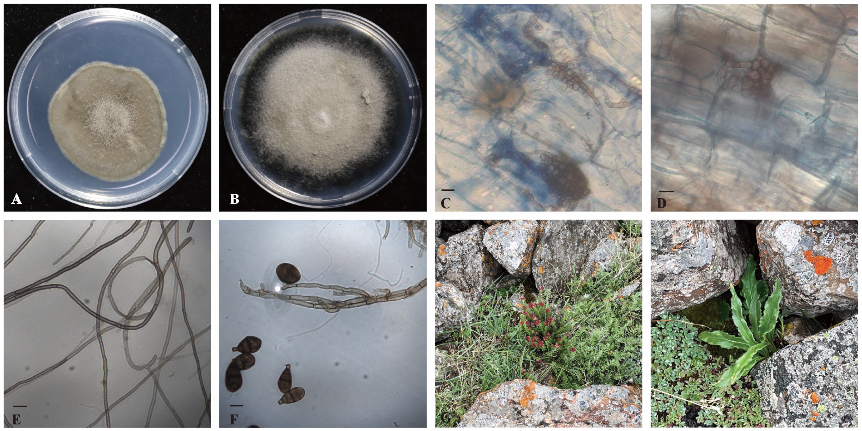
3.3. The Dominant DSE in the Culturable Endophytic Fungal Communities of Alpine Medicinal Plants

DSE represents the majority of culturable endophytic fungi in the two medicinal plants, with 208 DSE isolates accounting for 50.98% of the total culturable endophytic fungi. These include 31 species of DSE, exhibiting high diversity. Based on the morphological characteristics of the isolated DSE fungi, they were classified into 20 morphotypes (Table 4). The colonies of the isolated DSE fungi are mostly round or elliptical, with a woolly or felt-like appearance. The predominant colors are black, dark green, and gray. The edges of the colonies are relatively smooth, although a few exhibit wavy, radial, or irregular shapes. A few fungi produce red pigments in the culture medium. Among them, representative DSE fungal characteristics are shown in Figure 3.
Some common DSE fungi, such as P. schachtii, A. rhododendri, A. alternata, Alternaria sp., Rhexocercosporidium sp., L. orchidicola, Leptosphaeria sp., Cadophora sp., N. candida, etc., have colonized both of these alpine plants.
Rare DSE were also isolated, including some species reported infrequently in the literature, such as A. doliconidium, from which only a single isolate was obtained from R. crenulata, and A. sorghi, isolated as a single strain from S. involucrata. Other infrequently reported fungi isolated from R. crenulata include Cyphellophora sp., Phialocephala sp., and Cadophora cf. interclivum, while isolates from S. involucrata include A. tenuissima, Crocicreas sp., and C. ferruginea, each represented by only one isolate.

3.4. DSE Community Composition of Two Alpine Medicinal Plants in Different Sites

Analysis of the DSE community composition of 208 isolates from the two medicinal plants showed that all isolates were Ascomycota fungi, classified into 4 classes, 6 orders, 12 families, 16 genera, and 31 species. The DSE community structure exhibited some differences between the two medicinal plants (Figure 4), with 110 isolates of 18 DSE species from S. involucrata and 98 isolates of 22 DSE species from R. crenulata. Species common to both plant roots included P. schachtii, A. rhododendri, A. alternata, Alternaria sp., Rhexocercosporidium sp., L. orchidicola, Leptosphaeria sp., Cadophora sp., and N. candida, which were shared DSE fungi. L. orchidicola was the dominant species in the DSE communities of both plants, with relative isolation frequencies of 35.45% and 19.39% in S. involucrata and R. crenulata, respectively.
The host-specific DSE groups showed the distinct differences between the two plants, with 9 DSE species unique to S. involucrata, including Cladosporium sp., C. delicatulum, A. longipes, A. sorghi, A. tenuissima, Exophiala sp., Crocicreas sp., C. ferruginea, and Cadophora sp. Thirteen species were unique to R. crenulata, including P. chrysanthemicola, Paraphoma sp., A. chlamydosporigena, A. doliconidium, Alternaria sp., Cyphellophora sp., Leptodontidium sp., Phialocephala sp., Cadophora cf. interclivum, C. malorum, C. spadices, B. cinerea, and Microdochium sp. The similarity of DSE communities between the two plants was high, at 45.00%.

3.5. Analysis of DSE Diversity of the Two Species in Different Ways

A statistical analysis of DSE abundance and diversity in the two plant species across different sites (Table 5) indicated that the Shannon–Wiener index in site XW was lower than in HW, whereas both XBL and XDN were higher than HBL and HDN. Only one DSE isolate was obtained from HDN, identified as A. doliconidium. A. alternata and Rhexocercosporidium sp. were the most widely distributed, found across four sites, followed by L. orchidicola, P. schachtii, Cadophora sp., and N. candida, which were found in three sites. Except for HDN, the evenness across the other sites was consistently high. For S. involucrata, diversity was highest in XW and lowest in XDN, while for R. crenulata, it was highest in HT and lowest in HDN. Except for HDN, the diversity indices among the sites located in the Tianshan Mountains (XW, XBL, XDN, XH, HW, and HBL) were relatively similar, whereas the HT site in the Karakoram Mountains displayed significantly higher diversity and marked differences in community composition (Figure 5). These findings suggest that the DSE community structure of the two alpine medicinal plants varies between mountain ranges.

4. Discussion

4.1. Common Characteristics of the Culturable Endophytic Fungal Communities in the Roots of Both Plant Species

This study conducted molecular identification on the endophytic fungi from the roots of two alpine medicinal plants, belonging to 3 phyla, 9 classes, 16 orders, 31 families, 54 genera, and 91 species. Among them, 20 fungal species were found in both plants, including P. schachtii, P. meliloti, A. rhododendri, A. alternata, Alternaria sp., Articulospora, Filosporella sp., L. orchidicola, Rhexocercosporidium sp., Tetracladium sp., Leptosphaeria sp., Cadophora sp., Mycochaetophora, Helotiales sp., G. basitruncatum, D. macrodidyma, N. candida, Neonectria sp., T. cornearis, and M. arctica. Some species, such as Articulospora, Filosporella sp., and Tetracladium sp., are aquatic fungi, while others like Tetracladium sp., D. macrodidyma, M. arctica, and C. ferruginea are adapted to alpine environments [32,33,34,35,36,37,38]. These fungi may help plants in high-altitude, low-temperature habitats resist cold stress, enhancing their environmental adaptability.
In the Tianshan Mountain No.1 glacier (XW and HW), Bayinbuluke, Hejing County (XBL and HBL), and Danangou Uzbek Township (XDN and HDN), both plant species were collected. Shared fungal families include Melanommataceae, a common plant endophytic fungal family [39]. Members of Didymellaceae are widely distributed across ecosystems, containing important plant pathogens and species associated with endophytic, saprotrophic, and clinical conditions [40]. Helotiaceae is a dominant endophytic family in trees, while Ploettnerulaceae is found in the marine glaciers of the Qinghai-Tibet Plateau, showing high diversity [33,41]. These fungi are frequently associated with plant tissues, promoting growth and offering enzymatic resistance to cold temperatures, helping plants survive in harsh environments. Notably, although fungal species composition varies, their functional roles often converge, providing functional redundancy that stabilizes ecosystems despite potential fungal losses due to climate change [42]. For example, isolated species such as Cadophora sp. and L. orchidicola promote plant growth, while C. delicatulum, Rhexocercosporidium sp., and Cadophora sp. offer disease resistance [43,44,45,46]. Collectively, these functionally analogous fungi enhance plant health and environmental stability.

4.2. Effects of Plant Species on Endophytic Fungal Community

Although S. involucrata and R. crenulata often coexist, the species’ differences in host plants lead to variations in the composition of their root endophytic fungal communities. In the culturable endophytic fungi of the root systems of S. involucrata, the dominant species was L. orchidicola, with an isolation frequency of 20.42%, whereas the dominant fungus in the root system of R. crenulata was Filosporella sp., with an isolation frequency of 9.68%. L. orchidicola was isolated from Calypso bulbosa and is found not only in terrestrial plant roots but also in submersed plant roots [47,48]. Studies have reported that inoculation with this fungus can increase the biomass of tomato seedlings and reduce the negative impact of pathogens on plants [43]. The genus Filosporella, which includes six species (F. annelidica, F. aquatica, F. exilis, F. fistucella, F. pinguis, and F. versimorpha), comprises aquatic fungi that contribute to the decomposition of fallen leaves and other organic matter in rivers [49]. These fungi are adapted to low temperatures through the production of cold-resistant enzymes [32]. Certain fungal families were specific to one plant species. For example, Ceratobasidiaceae, Cladosporiaceae, Phaeosphaeriaceae, and Mucoraceae were isolated solely from S. involucrata. Ceratobasidiaceae, common orchid mycorrhizal fungi (OMF) [50], aid nutrient uptake and transfer carbohydrates to the soil through symbiotic relationships with orchid plants [51]. Cladosporiaceae, found in marine environments [52], produce diverse metabolites with bioactivity and hold potential for industrial and biotechnological applications, but are also associated with plant and health hazards [53,54,55]. Fungal families such as Hydnaceae, Discinellaceae, and Mollisiaceae, found exclusively in R. crenulata, are common saprobes on decaying plant material in temperate regions [56]. Sclerotiniaceae include pathogenic fungi such as Sclerotinia sclerotiorum and B. cinerea, as well as the psychrophilic S. borealis [57]. Amphisphaeriaceae members, common plant endophytes globally, secrete bioactive secondary metabolites, including chromones, spiroketals, polyketides, terpenoids, and coumarins [58]. Mortierellaceae is among the most abundant, diverse, and widely distributed soil fungi, especially in alpine and subalpine zones [59].
Overall, S. involucrata and R. crenulata host seven and eight unique fungal families, respectively, with distinct endophytic fungal community compositions indicating that different host plants exhibit varying endophytic fungal communities, even in similar habitats. This variation may be influenced by the host plant species and their exudates. Root exudates from onions release chemical signals that alter rhizosphere microbiome recruitment [60]. Studies indicated that plants selectively “recruit” specific rhizosphere microbes [61]. Additionally, according to the “Stress Gradient Hypothesis”, interspecies cooperation increases with environmental stress [62,63]. In the harsh alpine environment, beneficial microbes may migrate from non-rhizosphere soil to the plant rhizosphere, and are then recruited into plant roots to form symbiotic relationships that aid growth and stress resistance [64,65].

4.3. Effects of Sampling Sites on Endophytic Fungal Community

Our findings demonstrate that the culturable endophytic fungal community structure varies across sampling sites for the same host plant. Multiple factors shape the composition of these fungal communities, including abiotic factors such as soil physicochemical properties, pH, temperature, and precipitation [66,67,68], as well as biotic factors like habitat plant composition, plant developmental stages, rhizosphere bacterial communities, and arbuscular mycorrhizal fungi (AMF) colonization [69,70,71,72]. It has been suggested that climate and spatial factors are the primary drivers of endophytic fungal community structure, with a greater impact than soil characteristics, host genotype, or geographic distance [73]. Additional studies proposed that various endophytic fungi exhibit distinct niche preferences [67]. These findings supported the concept that different fungal lineages occupy specific ecological niches, indicating functional complementarity within endophytic fungal communities. Due to the interactions among the host identity, environmental factors, and microbial communities, distinct fungal taxa may adopt unique strategies depending on environmental context [74]. Among the five sampling sites in our study (XW and HW, XBL and HBL, XDN and HDN, XH, and HT), three of them (XW and HW, XBL and HBL, XDN and HDN, and XH) are located in the Tianshan Mountains, while HT is situated in the Karakoram Mountains. Differences in vegetation cover and climate factors across mountain ranges may contribute to variations in endophytic fungal communities within the same species in different regions [75,76]. Additionally, traditional isolation methods for endophytic fungi may not capture certain unculturable endophytic fungi, and the isolation process itself may involve certain stochastic elements, which could also contribute to variations in culturable endophytic fungal community composition.

4.4. Potential Ecological Roles of Culturable DSE

In our study, DSE accounted for 50.98% of all culturable endophytic fungi, indicating that DSE fungi represent a significant component of the endophytic fungal communities in alpine medicinal plants. This finding aligns with previous research on the endophytic fungal communities in alpine plants [77]. The primary identified DSE fungi were classified into 4 classes, 6 orders, 12 families, 16 genera, and 31 species, with high species diversity. These fungi also exhibited high functional diversity, with A. sorghi and P. chrysanthemicola showing antioxidant properties, Leptosphaeria sp. and Leptosphaeria sp. alleviating water stress, and species like C. delicatulum, Cladosporium sp., Paraphoma sp., Rhexocercosporidium sp., and Cadophora sp. displaying antimicrobial activity [78,79,80,81,82]. Other species, such as A. tenuissima, demonstrated the capacity to produce secondary metabolites like flavonoids [46]. Additionally, L. orchidicola and Microdochium sp. were found to promote plant growth [43,83].
DSE fungi are widespread across nearly all ecosystems, colonizing inside plant roots [84]. Their high tolerance to heavy metals, drought, and salinity, particularly in extreme environments such as alpine regions, subarctic zones, deserts, wetlands, saline–alkaline soils, and areas with heavy metal contamination, highlights their essential ecological role [22,23,85,86,87,88]. In this study, both alpine medicinal plants were found to share multiple DSE species, with 9 species identified in both plants. These shared DSE fungi include species common to extreme environments, such as Alternaria sp., A. alternata, L. orchidicola, and Leptosphaeria sp., which are frequently found in alpine, desert, and deep-sea habitats [43,47,78,89]. Certain DSE fungi, such as Cadophora sp., are associated with heavy metal-contaminated soils, while others, like Rhexocercosporidium sp. and Leptosphaeria sp., colonize medicinal plants. Additionally, some fungi present in both plants, such as Rhexocercosporidium sp., and N. candida, are potential pathogens [45,90,91,92]. According to the “habitat adaptation hypothesis,” plants may enhance their resistance to harsh environments through symbiosis with endophytic fungi, which can confer similar stress tolerance to phylogenetically distant plants [22,93]. The shared DSE species may aid host plant adaptation to extreme alpine environments by alleviating environmental stress, deterring pests, and providing other resistance mechanisms. This study also revealed that certain fungi co-occurred in the roots of both plants at specific sites, such as A. rhododendri at site XW and HW, and A. alternata at site XBL and HBL. A. rhododendri has rarely been reported and is identified here for the first time as a DSE in alpine medicinal plants. Notably, functional similarity extends beyond shared DSE species to include distinct DSE fungi in each plant with similar functions. For example, Cladosporium sp., isolated only from S. involucrata, and Paraphoma sp., isolated only from R. crenulata, both exhibit antibacterial properties. A. sorghi, found exclusively in S. involucrata, and P. chrysanthemicola, isolated solely from R. crenulata, both possess antioxidant properties [78,94,95,96].
The DSE community composition differs between the two medicinal plants, with the DSE diversity of R. crenulata being higher than that of S. involucrata, potentially influenced by the host plant. Additionally, a diverse DSE community may aid R. crenulata in better adapting to extreme alpine environments, which could explain its broader distribution compared to S. involucrata. Apart from the aforementioned influencing factors, variations in DSE community composition across geographical locations may also relate to stress conditions specific to each site. Our isolated DSE communities exhibit functional diversity as well, including species such as Paraphoma sp., P. chrysanthemicola, Alternaria sp., Leptosphaeria sp., Exophiala sp., and L. orchidicola, commonly found in arid regions and associated with alleviating water stress and promoting the growth of desert plants [48,78,81,97,98,99]. Similarly, in extreme environments like alpine, polar, and marine habitats, where plants endure low temperatures and oxygen, species like C. delicatulum, A. alternata, Alternaria sp., Cyphellophora sp., Phialocephala sp., C. ferruginea, and C. malorum may help plants withstand these stresses [33,45,89,98,100,101,102]. In environments contaminated by heavy metals, species like A. chlamydosporigena, Cadophora sp., and Microdochium sp. may help alleviate heavy metal stress in soils [44,103]. Thus, the distinct DSE communities in these two medicinal plants might partly explain their differing distributions. The wider distribution of R. crenulata compared to S. involucrata may be attributed not only to the inherent characteristics of the plant but also to the distinct functions of its endophytic DSE fungi. For instance, P. chrysanthemicola, unique to R. crenulata, improves the rhizosphere environment, while A. chlamydosporigena displays heavy metal tolerance [103]. Similarly, A. sorghi and A. tenuissima, present only in S. involucrata, possess antioxidant capabilities [27,46].
It is noteworthy that A. chlamydosporigena, A. doliconidium, A. sorghi, Crocicreas sp., L. orchidicola, Leptosphaeria sp., Phialocephala sp., C. spadices, N. candida, P. schachtii, and A. rhododendri, among the isolated DSE fungi in this experiment, are reported for the first time in alpine environments.
In addition, Cyphellophora sp., P. schachtii, A. rhododendri, L. orchidicol, A. chlamydosporigena, A. longipes, Crocicreas sp., C. ferruginea, C. spadices, and N. candida are reported for the first time as DSE fungi in medicinal plants.
In this study, we isolated multiple fungal strains from the roots of the alpine medicinal plants. Although limited by sample size and unable to capture the entire microbial community, our findings undeniably contribute to the diversity of microbial resources in this region. A wider range of isolation techniques (such as low-temperature and oligotrophic conditions) and culture conditions (such as varied light exposure, temperature, and nutrient availability) should be explored in future studies to obtain additional culturable strains. This experiment provides valuable fungal strains for future research, which could be selected for application in low-altitude cultivation. Moreover, future studies could focus on screening strains capable of producing secondary metabolites to enhance the quality and medicinal value of herbal materials, laying a foundation for improved cultivation and quality of medicinal plants.

5. Conclusions

Our study showed that the composition of the culturable endophytic fungal community in two alpine medicinal plants varies according to host plants and sampling sites. Among them, the dominant fungal phylum was Ascomycota. Leptodontidium and Filosporella were the predominant genera in S. involucrata and R. crenulata, respectively. L. orchidicola dominated the Tianshan Mountain No.1 glacier (XW and HW), while A. alternata and Rhexocercosporidium sp. were prevalent in the Bayinbuluke (XBL and HBL). Rhexocercosporidium sp. was dominant in the Danangou Uzbek Township (XDN and HDN), and Cadophora sp. was common in both the Houxia (XH) and Taxkorgan (HT) plots. DSE fungi were the primary group in the endophytic fungal communities of these plants. R. crenulata exhibited a higher diversity of culturable endophytic fungal communities than S. involucrata, with 20 fungal species shared between the two plant species. The community structure of endophytic fungi differed between the two plant species and across sampling sites. Among the culturable endophytic fungal communities in different sites of S. involucrata, the XW plot located in the Tianshan Mountains was the most abundant, while the HT plot, located in the Kunlun Mountains, showed the highest diversity for R. crenulata. This suggests that host plants may selectively recruit endophytic fungi and that the environment may play a role in driving endophytic fungal community composition. In addition, there were both similarities and differences in the community structure of DSE. L. orchidicola was the dominant species in the DSE communities of both plants. Apart from the 9 DSE species shared by both plants, 9 DSE species were unique to S. involucrata, and 13 DSE species were unique to R. crenulata. Understanding the diversity of endophytic fungi in S. involucrata and R. crenulata not only provides a research basis and endophytic fungal resources for revealing their potential functions in alpine medicinal plants but also contributes to the low-altitude and high-quality artificial cultivation of these two medicinal plants in the future. The current results are mainly derived from the data of culturable endophytic fungi, and their abundance needs to be further verified by high-throughput sequencing of larger sample sizes. Moreover, to determine the taxonomy of the DSE strains from our study, multi molecular genetic markers are being used to find potential new taxa. In the next step, we will screen strains that can produce secondary metabolites to improve the quality of medicinal materials and provide a basis for the low-altitude cultivation of alpine medicinal plants.

Supplementary Materials

The following supporting information can be downloaded at: https://www.mdpi.com/article/10.3390/jof11020113/s1, Table S1: Culturable endophytic fungi isolated from two alpine medicinal plants and the number of strains in different host plants.

Author Contributions

M.H. performed the experiment and wrote the manuscript draft, J.Z. and C.L. analyzed the data, X.H., M.Y., Y.Y., and Y.X. provided revisions to the work in the writing. J.C. was responsible for the experiment design, manuscript revisions, and funding. All authors have read and agreed to the published version of the manuscript.

Funding

This work was supported by the National Natural Science Foundation of China (No. 82173923), the CAMS Innovation Fund for Medical Sciences (CIFMS) (2021-I2M-1-032), and the Special Fund for Taishan Scholar Project (No. tsqn202211233).

Institutional Review Board Statement

Not applicable.

Informed Consent Statement

Not applicable.

Data Availability Statement

Data are contained within the article and Supplementary Materials.

Conflicts of Interest

The authors declare no conflicts of interest.

References

  1. Bardgett, R.D.; van der Putten, W.H. Belowground Biodiversity and Ecosystem Functioning. Nature 2014, 515, 505–511. [Google Scholar] [CrossRef] [PubMed]
  2. Broadbent, A.A.D.; Snell, H.S.K.; Michas, A.; Pritchard, W.J.; Newbold, L.; Cordero, I.; Goodall, T.; Schallhart, N.; Kaufmann, R.; Griffiths, R.I.; et al. Climate Change Alters Temporal Dynamics of Alpine Soil Microbial Functioning and Biogeochemical Cycling via Earlier Snowmelt. ISME J. 2021, 15, 2264–2275. [Google Scholar] [CrossRef] [PubMed]
  3. Legge, S.; Rumpff, L.; Garnett, S.T.; Woinarski, J.C.Z. Loss of Terrestrial Biodiversity in Australia: Magnitude, Causation, and Response. Science 2023, 381, 622–631. [Google Scholar] [CrossRef] [PubMed]
  4. Bertrand, R.; Lenoir, J.; Piedallu, C.; Riofrío-Dillon, G.; de Ruffray, P.; Vidal, C.; Pierrat, J.-C.; Gégout, J.-C. Changes in Plant Community Composition Lag behind Climate Warming in Lowland Forests. Nature 2011, 479, 517–520. [Google Scholar] [CrossRef]
  5. Parada, R.; Mendoza, L.; Cotoras, M.; Ortiz, C. Endophytic Fungi Isolated from Plants Present in a Mine Tailing Facility Show a Differential Growth Response to Lead. Lett. Appl. Microbiol. 2022, 75, 345–354. [Google Scholar] [CrossRef]
  6. Ding, W.-N.; Ree, R.H.; Spicer, R.A.; Xing, Y.-W. Ancient Orogenic and Monsoon-Driven Assembly of the World’s Richest Temperate Alpine Flora. Science 2020, 369, 578–581. [Google Scholar] [CrossRef]
  7. Wang, W.; Ding, M.; Gardner, J.D.; Wang, Y.; Miao, B.; Guo, W.; Wu, X.; Ruan, Q.; Yu, J.; Hu, X.; et al. Ancient Xinjiang Mitogenomes Reveal Intense Admixture with High Genetic Diversity. Sci. Adv. 2021, 7, eabd6690. [Google Scholar] [CrossRef]
  8. Wu, C.-L.; Lin, L.-F.; Hsu, H.-C.; Huang, L.-F.; Hsiao, C.-D.; Chou, M.-L. Saussurea involucrata (Snow Lotus) ICE1 and ICE2 Orthologues Involved in Regulating Cold Stress Tolerance in Transgenic Arabidopsis. Int. J. Mol. Sci. 2021, 22, 10850. [Google Scholar] [CrossRef]
  9. Chen, J.; Zhu, J.; Yan, B.; Li, J.M.; Guo, S.X. Preliminary Identification of Endophytic Fungi Colonized in the Root of Saussurea involucrata and Rhodiola rosea from Xinjiang Region. Mycosystema 2018, 37, 110–119. (In Chinese) [Google Scholar] [CrossRef]
  10. Gong, G.; Xie, F.; Zheng, Y.; Hu, W.; Qi, B.; He, H.; Dong, T.T.; Tsim, K.W. The Effect of Methanol Extract from Saussurea involucrata in the Lipopolysaccharide-Stimulated Inflammation in Cultured RAW 264.7 Cells. J. Ethnopharmacol. 2020, 251, 112532. [Google Scholar] [CrossRef]
  11. Cong, S.; Wang, L.; Meng, Y.; Cai, X.; Zhang, C.; Gu, Y.; Ma, X.; Luo, L. Saussurea involucrata Oral Liquid Regulates Gut Microbiota and Serum Metabolism During Alleviation of Collagen-Induced Arthritis in Rats. Phytother. Res. 2023, 37, 1242–1259. [Google Scholar] [CrossRef] [PubMed]
  12. Wang, L.; Yang, K.; Jing, R.; Zhao, W.; Guo, K.; Hu, Z.; Liu, G.; Xu, N.; Zhao, J.; Lin, L.; et al. Protective Effect of Saussurea involucrata Polysaccharide Against Skin Dryness Induced by Ultraviolet Radiation. Front. Pharmacol. 2023, 14, 1089537. [Google Scholar] [CrossRef] [PubMed]
  13. Sun, Y.; Zhang, A.; Landis, J.B.; Shi, W.; Zhang, X.; Sun, H.; Wang, H. Genome Assembly of the Snow Lotus Species Saussurea involucrata Provides Insights into Acacetin and Rutin Biosynthesis and Tolerance to an Alpine Environment. Hortic. Res. 2023, 10, uhad180. [Google Scholar] [CrossRef] [PubMed]
  14. Fu, Y.; Li, L.; Hao, S.; Guan, R.; Fan, G.; Shi, C.; Wan, H.; Chen, W.; Zhang, H.; Liu, G.; et al. Draft Genome Sequence of the Tibetan Medicinal Herb Rhodiola crenulata. Gigascience 2017, 6, gix033. [Google Scholar] [CrossRef]
  15. Pu, W.-L.; Zhang, M.-Y.; Bai, R.-Y.; Sun, L.-K.; Li, W.-H.; Yu, Y.-L.; Zhang, Y.; Song, L.; Wang, Z.-X.; Peng, Y.-F.; et al. Anti-Inflammatory Effects of Rhodiola rosea L.: A Review. Biomed. Pharmacother. 2020, 121, 109552. [Google Scholar] [CrossRef]
  16. Chen, R.; Zhang, F.; Chen, S.; Chi, X. Checklist of National Key Protected Wild Plants on the Qinghai-Tibetan Plateau. Biodivers. Data J. 2023, 11, e103289. [Google Scholar] [CrossRef]
  17. Arnold, A.E.; Lutzoni, F. Diversity and Host Range of Foliar Fungal Endophytes: Are Tropical Leaves Biodiversity Hotspots? Ecology 2007, 88, 541–549. [Google Scholar] [CrossRef]
  18. Trivedi, P.; Leach, J.E.; Tringe, S.G.; Sa, T.; Singh, B.K. Plant-Microbiome Interactions: From Community Assembly to Plant Health. Nat. Rev. Microbiol. 2020, 18, 607–621. [Google Scholar] [CrossRef]
  19. Strobel, G.; Daisy, B. Bioprospecting for Microbial Endophytes and Their Natural Products. Microbiol. Mol. Biol. Rev. 2003, 67, 491–502. [Google Scholar] [CrossRef]
  20. Wang, Z.; Wang, L.; Pan, Y.; Zheng, X.; Liang, X.; Sheng, L.; Zhang, D.; Sun, Q.; Wang, Q. Research Advances on Endophytic Fungi and Their Bioactive Metabolites. Bioprocess Biosyst. Eng. 2023, 46, 165–170. [Google Scholar] [CrossRef]
  21. Jumpponen, A.; Trappe, J.M. Dark Septate Endophytes: A Review of Facultative Biotrophic Root-colonizing Fungi. New Phytol. 1998, 140, 295–310. [Google Scholar] [CrossRef] [PubMed]
  22. Gonzalez Mateu, M.; Baldwin, A.H.; Maul, J.E.; Yarwood, S.A. Dark Septate Endophyte Improves Salt Tolerance of Native and Invasive Lineages of Phragmites australis. ISME J. 2020, 14, 1943–1954. [Google Scholar] [CrossRef] [PubMed]
  23. Netherway, T.; Bengtsson, J.; Buegger, F.; Fritscher, J.; Oja, J.; Pritsch, K.; Hildebrand, F.; Krab, E.J.; Bahram, M. Pervasive Associations Between Dark Septate Endophytic Fungi with Tree Root and Soil Microbiomes Across Europe. Nat. Commun. 2024, 15, 159. [Google Scholar] [CrossRef] [PubMed]
  24. Chik, W.-I.; Zhu, L.; Fan, L.-L.; Yi, T.; Zhu, G.-Y.; Gou, X.-J.; Tang, Y.-N.; Xu, J.; Yeung, W.-P.; Zhao, Z.-Z.; et al. Saussurea involucrata: A Review of the Botany, Phytochemistry and Ethnopharmacology of a Rare Traditional Herbal Medicine. J. Ethnopharmacol. 2015, 172, 44–60. [Google Scholar] [CrossRef] [PubMed]
  25. Hou, Y.; Fan, F.; Xie, N.; Zhang, Y.; Wang, X.; Meng, X. Rhodiola crenulata Alleviates Hypobaric Hypoxia-Induced Brain Injury by Maintaining BBB Integrity and Balancing Energy Metabolism Dysfunction. Phytomedicine 2024, 128, 155529. [Google Scholar] [CrossRef]
  26. Phillips, J.M.; Hayman, D.S. Improved Procedures for Clearing Roots and Staining Parasitic and Vesicular-Arbuscular Mycorrhizal Fungi for Rapid Assessment of Infection. Trans. Br. Mycol. Soc. 1970, 55, 158-IN18. [Google Scholar] [CrossRef]
  27. Tang, Y.-J.; Zhou, D.-Y.; Dai, J.; Li, Y.; Xing, Y.-M.; Guo, S.-X.; Chen, J. Potential Specificity Between Mycorrhizal Fungi Isolated from Widespread Dendrobium spp. and Rare D. huoshanense Seeds. Curr. Microbiol. 2022, 79, 264. [Google Scholar] [CrossRef]
  28. Hughes, C.E.; Atchison, G.W. The Ubiquity of Alpine Plant Radiations: From the Andes to the Hengduan Mountains. New Phytol. 2015, 207, 275–282. [Google Scholar] [CrossRef]
  29. Shannon, C.E. The Mathematical Theory of Communication. 1963. MD Comput 1997, 14, 306–317. [Google Scholar]
  30. Johnston, E.L.; Roberts, D.A. Contaminants Reduce the Richness and Evenness of Marine Communities: A Review and Meta-Analysis. Environ. Pollut. 2009, 157, 1745–1752. [Google Scholar] [CrossRef]
  31. Kent, M. Vegetation Description and Data Analysis: A Practical Approach; John Wiley & Sons: Hoboken, NJ, USA, 2011; ISBN 978-1-119-96239-7. [Google Scholar]
  32. Vasconcelos Rissi, D.; Ijaz, M.; Baschien, C. Comparative Genome Analysis of the Freshwater Fungus Filosporella fistucella Indicates Potential for Plant-Litter Degradation at Cold Temperatures. G3 Genes Genomes Genet. (Bethesda) 2023, 13, jkad190. [Google Scholar] [CrossRef] [PubMed]
  33. Zhang, B.; Li, X.; Li, G.; Wang, Q.-M.; Wang, M. Cadophora Species from Marine Glaciers in the Qinghai-Tibet Plateau: An Example of Unsuspected Hidden Biodiversity. IMA Fungus 2022, 13, 15. [Google Scholar] [CrossRef] [PubMed]
  34. Barros, D.; Pradhan, A.; Pascoal, C.; Cássio, F. Proteomic Responses to Silver Nanoparticles Vary with the Fungal Ecotype. Sci. Total Environ. 2020, 704, 135385. [Google Scholar] [CrossRef] [PubMed]
  35. Gonçalves, A.L.; Carvalho, A.; Bärlocher, F.; Canhoto, C. Are Fungal Strains from Salinized Streams Adapted to Salt-Rich Conditions? Philos. Trans. R. Soc. Lond. B Biol. Sci. 2018, 374, 20180018. [Google Scholar] [CrossRef]
  36. Probst, M.; Telagathoti, A.; Mandolini, E.; Peintner, U. Fungal and Bacterial Communities and Their Associations in Snow-Free and Snow Covered (Sub-)Alpine Pinus cembra Forest Soils. Environ. Microbiome 2024, 19, 20. [Google Scholar] [CrossRef]
  37. Zhao, W.; Yin, Y.-L.; Li, S.-X.; Wang, Y.-Q.; Wang, Y.-L. The Characteristics of Soil Fungal Community in Degraded Alpine Meadow in the Three Rivers Source Region, China. Ying Yong Sheng Tai Xue Bao 2021, 32, 869–877. [Google Scholar] [CrossRef]
  38. Gupta, P.; Vakhlu, J.; Sharma, Y.P.; Imchen, M.; Kumavath, R. Metagenomic Insights into the Fungal Assemblages of the Northwest Himalayan Cold Desert. Extremophiles 2020, 24, 749–758. [Google Scholar] [CrossRef]
  39. Liao, H.; Huang, L.; Li, N.; Ke, W.; Xiang, Y.; Ma, Y. Auxiliary Rapid Identification of Pathogenic and Antagonistic Microorganisms Associated with Coptis chinensis Root Rot by High-Throughput Sequencing. Sci. Rep. 2021, 11, 11141. [Google Scholar] [CrossRef]
  40. Hou, L.W.; Groenewald, J.Z.; Pfenning, L.H.; Yarden, O.; Crous, P.W.; Cai, L. The Phoma-like Dilemma. Stud. Mycol. 2020, 96, 309–396. [Google Scholar] [CrossRef]
  41. Kernaghan, G.; Patriquin, G. Host Associations Between Fungal Root Endophytes and Boreal Trees. Microb. Ecol. 2011, 62, 460–473. [Google Scholar] [CrossRef]
  42. Zeng, Q.; Lebreton, A.; Auer, L.; Man, X.; Jia, L.; Wang, G.; Gong, S.; Lombard, V.; Buée, M.; Wu, G.; et al. Stable Functional Structure despite High Taxonomic Variability across Fungal Communities in Soils of Old-Growth Montane Forests. Microbiome 2023, 11, 217. [Google Scholar] [CrossRef] [PubMed]
  43. Andrade-Linares, D.R.; Grosch, R.; Restrepo, S.; Krumbein, A.; Franken, P. Effects of Dark Septate Endophytes on Tomato Plant Performance. Mycorrhiza 2011, 21, 413–422. [Google Scholar] [CrossRef] [PubMed]
  44. Berthelot, C.; Blaudez, D.; Beguiristain, T.; Chalot, M.; Leyval, C. Co-Inoculation of Lolium perenne with Funneliformis mosseae and the Dark Septate Endophyte Cadophora sp. in a Trace Element-Polluted Soil. Mycorrhiza 2018, 28, 301–314. [Google Scholar] [CrossRef]
  45. Venkateswarulu, N.; Shameer, S.; Bramhachari, P.V.; Basha, S.K.T.; Nagaraju, C.; Vijaya, T. Isolation and Characterization of Plumbagin (5-Hydroxyl-2-Methylnaptalene-1,4-Dione) Producing Endophytic Fungi Cladosporium delicatulum from Endemic Medicinal Plants: Isolation and Characterization of Plumbagin Producing Endophytic Fungi from Endemic Medicinal Plants. Biotechnol. Rep. 2018, 20, e00282. [Google Scholar] [CrossRef]
  46. Yao, Y.Q.; Lan, F.; Qiao, Y.M.; Wei, J.G.; Huang, R.S.; Li, L.B. Endophytic Fungi Harbored in the Root of Sophora tonkinensis Gapnep: Diversity and Biocontrol Potential against Phytopathogens. Microbiologyopen 2017, 6, e00437. [Google Scholar] [CrossRef]
  47. Currah, R.S.; Hambleton, S.; Smreciu, A. Mycorrhizae and Mmcorrhizal Fungi of Calypso bulbosa. Am. J. Bot. 1988, 75, 739–752. [Google Scholar] [CrossRef]
  48. Kohout, P.; Sýkorová, Z.; Ctvrtlíková, M.; Rydlová, J.; Suda, J.; Vohník, M.; Sudová, R. Surprising Spectra of Root-Associated Fungi in Submerged Aquatic Plants. FEMS Microbiol. Ecol. 2012, 80, 216–235. [Google Scholar] [CrossRef]
  49. Jabiol, J.; Lecerf, A.; Lamothe, S.; Gessner, M.O.; Chauvet, E. Litter Quality Modulates Effects of Dissolved Nitrogen on Leaf Decomposition by Stream Microbial Communities. Microb. Ecol. 2019, 77, 959–966. [Google Scholar] [CrossRef]
  50. Freestone, M.; Reiter, N.; Swarts, N.D.; Linde, C.C. Temporal Turnover of Ceratobasidiaceae Orchid Mycorrhizal Fungal Communities with Ontogenetic and Phenological Development in Prasophyllum (Orchidaceae). Ann. Bot. 2024, 134, mcae089. [Google Scholar] [CrossRef]
  51. McCormick, M.K.; Whigham, D.F.; Canchani-Viruet, A. Mycorrhizal Fungi Affect Orchid Distribution and Population Dynamics. New Phytol. 2018, 219, 1207–1215. [Google Scholar] [CrossRef]
  52. Lee, W.; Kim, J.S.; Seo, C.W.; Lee, J.W.; Kim, S.H.; Cho, Y.; Lim, Y.W. Diversity of Cladosporium (Cladosporiales, Cladosporiaceae) Species in Marine Environments and Report on Five New Species. MycoKeys 2023, 98, 87–111. [Google Scholar] [CrossRef] [PubMed]
  53. Mohamed, G.A.; Ibrahim, S.R.M. Untapped Potential of Marine-Associated Cladosporium Species: An Overview on Secondary Metabolites, Biotechnological Relevance, and Biological Activities. Mar. Drugs 2021, 19, 645. [Google Scholar] [CrossRef] [PubMed]
  54. Prasannath, K.; Shivas, R.G.; Galea, V.J.; Akinsanmi, O.A. Novel Botrytis and Cladosporium Species Associated with Flower Diseases of Macadamia in Australia. J. Fungi 2021, 7, 898. [Google Scholar] [CrossRef] [PubMed]
  55. Wang, Y.; Li, X.; Chen, X.; Kulyar, M.F.-E.-A.; Duan, K.; Li, H.; Bhutta, Z.A.; Wu, Y.; Li, K. Gut Fungal Microbiome Responses to Natural Cryptosporidium Infection in Horses. Front. Microbiol. 2022, 13, 877280. [Google Scholar] [CrossRef]
  56. Tanney, J.B.; Seifert, K.A. Mollisiaceae: An Overlooked Lineage of Diverse Endophytes. Stud. Mycol. 2020, 95, 293–380. [Google Scholar] [CrossRef]
  57. Badet, T.; Peyraud, R.; Raffaele, S. Common Protein Sequence Signatures Associate with Sclerotinia borealis Lifestyle and Secretion in Fungal Pathogens of the Sclerotiniaceae. Front. Plant Sci. 2015, 6, 776. [Google Scholar] [CrossRef]
  58. Wang, K.; Lei, J.; Wei, J.; Yao, N. Bioactive Natural Compounds from the Plant Endophytic Fungi Pestalotiopsis spp. Mini Rev. Med. Chem. 2012, 12, 1382–1393. [Google Scholar] [CrossRef]
  59. Telagathoti, A.; Probst, M.; Peintner, U. Habitat, Snow-Cover and Soil pH, Affect the Distribution and Diversity of Mortierellaceae Species and Their Associations to Bacteria. Front. Microbiol. 2021, 12, 669784. [Google Scholar] [CrossRef]
  60. Zhou, X.; Zhang, J.; Khashi U Rahman, M.; Gao, D.; Wei, Z.; Wu, F.; Dini-Andreote, F. Interspecific Plant Interaction via Root Exudates Structures the Disease Suppressiveness of Rhizosphere Microbiomes. Mol. Plant 2023, 16, 849–864. [Google Scholar] [CrossRef]
  61. Rolfe, S.A.; Griffiths, J.; Ton, J. Crying out for Help with Root Exudates: Adaptive Mechanisms by Which Stressed Plants Assemble Health-Promoting Soil Microbiomes. Curr. Opin. Microbiol. 2019, 49, 73–82. [Google Scholar] [CrossRef]
  62. Adams, A.E.; Besozzi, E.M.; Shahrokhi, G.; Patten, M.A. A Case for Associational Resistance: Apparent Support for the Stress Gradient Hypothesis Varies with Study System. Ecol. Lett. 2022, 25, 202–217. [Google Scholar] [CrossRef] [PubMed]
  63. Dai, T.; Wen, D.; Bates, C.T.; Wu, L.; Guo, X.; Liu, S.; Su, Y.; Lei, J.; Zhou, J.; Yang, Y. Nutrient Supply Controls the Linkage Between Species Abundance and Ecological Interactions in Marine Bacterial Communities. Nat. Commun. 2022, 13, 175. [Google Scholar] [CrossRef] [PubMed]
  64. Berendsen, R.L.; Pieterse, C.M.J.; Bakker, P.A.H.M. The Rhizosphere Microbiome and Plant Health. Trends Plant Sci. 2012, 17, 478–486. [Google Scholar] [CrossRef]
  65. Yan, L.; Zhu, J.; Zhao, X.; Shi, J.; Jiang, C.; Shao, D. Beneficial Effects of Endophytic Fungi Colonization on Plants. Appl. Microbiol. Biotechnol. 2019, 103, 3327–3340. [Google Scholar] [CrossRef]
  66. Deng, Z.-S.; Liu, X.-D.; Zhang, B.-C.; Jiao, S.; Qi, X.-Y.; Sun, Z.-H.; He, X.-L.; Liu, Y.-Z.; Li, J.; Chen, K.-K.; et al. The Root Endophytic Fungi Community Structure of Pennisetum sinese from Four Representative Provinces in China. Microorganisms 2019, 7, 332. [Google Scholar] [CrossRef]
  67. Glynou, K.; Nam, B.; Thines, M.; Maciá-Vicente, J.G. Facultative Root-Colonizing Fungi Dominate Endophytic Assemblages in Roots of Nonmycorrhizal microthlaspi Species. New Phytol. 2018, 217, 1190–1202. [Google Scholar] [CrossRef]
  68. Toju, H.; Kurokawa, H.; Kenta, T. Factors Influencing Leaf- and Root-Associated Communities of Bacteria and Fungi Across 33 Plant Orders in a Grassland. Front. Microbiol. 2019, 10, 241. [Google Scholar] [CrossRef]
  69. Liu, H.; Wu, M.; Liu, J.; Qu, Y.; Gao, Y.; Ren, A. Tripartite Interactions Between Endophytic Fungi, Arbuscular Mycorrhizal Fungi, and Leymus chinensis. Microb. Ecol. 2020, 79, 98–109. [Google Scholar] [CrossRef]
  70. Mahdi, L.K.; Miyauchi, S.; Uhlmann, C.; Garrido-Oter, R.; Langen, G.; Wawra, S.; Niu, Y.; Guan, R.; Robertson-Albertyn, S.; Bulgarelli, D.; et al. The Fungal Root Endophyte Serendipita vermifera Displays Inter-Kingdom Synergistic Beneficial Effects with the Microbiota in Arabidopsis thaliana and Barley. ISME J. 2022, 16, 876–889. [Google Scholar] [CrossRef]
  71. Redkar, A.; Sabale, M.; Zuccaro, A.; Di Pietro, A. Determinants of Endophytic and Pathogenic Lifestyle in Root Colonizing Fungi. Curr. Opin. Plant Biol. 2022, 67, 102226. [Google Scholar] [CrossRef]
  72. Sun, R.; Yi, Z.; Fu, Y.; Liu, H. Dynamic Changes in Rhizosphere Fungi in Different Developmental Stages of Wheat in a Confined and Isolated Environment. Appl. Microbiol. Biotechnol. 2022, 106, 441–453. [Google Scholar] [CrossRef] [PubMed]
  73. Glynou, K.; Ali, T.; Buch, A.-K.; Haghi Kia, S.; Ploch, S.; Xia, X.; Çelik, A.; Thines, M.; Maciá-Vicente, J.G. The Local Environment Determines the Assembly of Root Endophytic Fungi at a Continental Scale. Environ. Microbiol. 2016, 18, 2418–2434. [Google Scholar] [CrossRef]
  74. Mandyam, K.G.; Jumpponen, A. Mutualism-Parasitism Paradigm Synthesized from Results of Root-Endophyte Models. Front. Microbiol. 2014, 5, 776. [Google Scholar] [CrossRef]
  75. Du, W.-B.; Jia, P.; Du, G.-Z. Current Patterns of Plant Diversity and Phylogenetic Structure on the Kunlun Mountains. Plant Divers. 2022, 44, 30–38. [Google Scholar] [CrossRef]
  76. Fan, M.; Xu, J.; Yu, W.; Chen, Y.; Wang, M.; Dai, W.; Wang, Y. Recent Tianshan Warming in Relation to Large-Scale Climate Teleconnections. Sci. Total Environ. 2023, 856, 159201. [Google Scholar] [CrossRef]
  77. Bueno de Mesquita, C.P.; Martinez Del Río, C.M.; Suding, K.N.; Schmidt, S.K. Rapid Temporal Changes in Root Colonization by Arbuscular Mycorrhizal Fungi and Fine Root Endophytes, Not Dark Septate Endophytes, Track Plant Activity and Environment in an Alpine Ecosystem. Mycorrhiza 2018, 28, 717–726. [Google Scholar] [CrossRef]
  78. Li, X.; Zhang, X.; Xu, M.; Ye, Q.; Gao, H.; He, X. Improved Tolerance of Artemisia ordosica to Drought Stress via Dark Septate Endophyte (DSE) Symbiosis. J. Fungi 2022, 8, 730. [Google Scholar] [CrossRef]
  79. Zoulikha, M.; Xiao, Q.; Boafo, G.F.; Sallam, M.A.; Chen, Z.; He, W. Pulmonary Delivery of siRNA Against Acute Lung Injury/Acute Respiratory Distress Syndrome. Acta Pharm. Sin. B 2022, 12, 600–620. [Google Scholar] [CrossRef]
  80. Chau, T.P.; Saravanan, M.; Al-Ansari, M.M.; Al-Dahmash, N.D.; Kuriakose, L.L.; Sindhu, R. Antimicrobial and Biocompatibility Nature of Methanol Extract of Lannea coromandelica Bark and Edible Coating Film Preparation for Fruit Preservation. Environ. Res. 2024, 243, 117861. [Google Scholar] [CrossRef]
  81. Li, X.; He, X.-L.; Zhou, Y.; Hou, Y.-T.; Zuo, Y.-L. Effects of Dark Septate Endophytes on the Performance of Hedysarum scoparium Under Water Deficit Stress. Front. Plant Sci. 2019, 10, 903. [Google Scholar] [CrossRef]
  82. Yakti, W.; Kovács, G.M.; Franken, P. Differential Interaction of the Dark Septate Endophyte Cadophora sp. and Fungal Pathogens In Vitro and in Planta. FEMS Microbiol. Ecol. 2019, 95, fiz164. [Google Scholar] [CrossRef] [PubMed]
  83. Parvandi, M.; Rezadoost, H.; Farzaneh, M. Introducing Alternaria tenuissima SBUp1, as an Endophytic Fungus of Ferula assa-Foetida from Iran, Which Is a Rich Source of Rosmarinic Acid. Lett. Appl. Microbiol. 2021, 73, 569–578. [Google Scholar] [CrossRef] [PubMed]
  84. Ruotsalainen, A.L.; Kauppinen, M.; Wäli, P.R.; Saikkonen, K.; Helander, M.; Tuomi, J. Dark Septate Endophytes: Mutualism from by-Products? Trends Plant Sci. 2022, 27, 247–254. [Google Scholar] [CrossRef]
  85. Zhang, Y.; Zhang, Y.; Liu, M.; Shi, X.; Zhao, Z. Dark Septate Endophyte (DSE) Fungi Isolated from Metal Polluted Soils: Their Taxonomic Position, Tolerance, and Accumulation of Heavy Metals In Vitro. J. Microbiol. 2008, 46, 624–632. [Google Scholar] [CrossRef]
  86. Fracchia, S.; Krapovickas, L.; Aranda-Rickert, A.; Valentinuzzi, V.S. Dispersal of Arbuscular Mycorrhizal Fungi and Dark Septate Endophytes by Ctenomys cf. Knighti (Rodentia) in the Northern Monte Desert of Argentina. J. Arid. Environ. 2011, 75, 1016–1023. [Google Scholar] [CrossRef]
  87. Kauppinen, M.; Raveala, K.; Wäli, P.R.; Ruotsalainen, A.L. Contrasting Preferences of Arbuscular Mycorrhizal and Dark Septate Fungi Colonizing Boreal and Subarctic Avenella flexuosa. Mycorrhiza 2014, 24, 171–177. [Google Scholar] [CrossRef]
  88. Malicka, M.; Magurno, F.; Piotrowska-Seget, Z. Plant Association with Dark Septate Endophytes: When the Going Gets Tough (and Stressful), the Tough Fungi Get Going. Chemosphere 2022, 302, 134830. [Google Scholar] [CrossRef]
  89. Li, Z.; Meng, L.; Ma, Q.; Wang, Z.; Zhao, Y.; Luo, D. Polyketides with IDH1 R132h and PTP1B Inhibitory Activities from the Desert-Plant-Derived Fungus Alternaria sp. HM 134. Front. Microbiol. 2022, 13, 975579. [Google Scholar] [CrossRef]
  90. Gao, R.; Liu, R.; Sun, C. A Marine Fungus Alternaria alternata FB1 Efficiently Degrades Polyethylene. J. Hazard. Mater. 2022, 431, 128617. [Google Scholar] [CrossRef]
  91. Berthelot, C.; Leyval, C.; Chalot, M.; Blaudez, D. Interactions between Dark Septate Endophytes, Ectomycorrhizal Fungi and Root Pathogens In Vitro. FEMS Microbiol. Lett. 2019, 366, fnz158. [Google Scholar] [CrossRef]
  92. Xie, F.; Li, H.-T.; Wang, M.; Chen, J.-Y.; Duan, H.-J.; Xia, D.-D.; Xie, T.-P.; Gao, Y.-H.; Zhou, H.; Ding, Z.-T. Phialocetones A-J, C12 Lactones from the Rhizospheric Soil-Derived Fungus Phialocephala sp. YUD18001 Associated with Gastrodia elata. Phytochemistry 2022, 202, 113359. [Google Scholar] [CrossRef] [PubMed]
  93. Rodriguez, R.J.; Henson, J.; Van Volkenburgh, E.; Hoy, M.; Wright, L.; Beckwith, F.; Kim, Y.-O.; Redman, R.S. Stress Tolerance in Plants via Habitat-Adapted Symbiosis. ISME J. 2008, 2, 404–416. [Google Scholar] [CrossRef] [PubMed]
  94. Salmi, D.; Riou, C.; Issawi, M.; Titouche, Y.; Ambrosini, V.; Smail-Saadoun, N.; Abbaci, H.; Houali, K. Antibacterial and Antioxidant Activities of Endophytic Fungi and Nettle (Urtica dioica L.) Leaves as Their Host. Cell. Mol. Biol. 2021, 67, 204–211. [Google Scholar] [CrossRef] [PubMed]
  95. Li, Y.; Wang, Y.; Wang, H.; Shi, T.; Wang, B. The Genus Cladosporium: A Prospective Producer of Natural Products. Int. J. Mol. Sci. 2024, 25, 1652. [Google Scholar] [CrossRef] [PubMed]
  96. Xu, X.; Li, J.; Zhang, K.; Wei, S.; Lin, R.; Polyak, S.W.; Yang, N.; Song, F. New Isocoumarin Analogues from the Marine-Derived Fungus Paraphoma sp. CUGBMF180003. Mar. Drugs 2021, 19, 313. [Google Scholar] [CrossRef]
  97. Chen, H.-Y.; Liu, T.-K.; Shi, Q.; Yang, X.-L. Sesquiterpenoids and Diterpenes with Antimicrobial Activity from Leptosphaeria sp. XL026, an Endophytic Fungus in Panax notoginseng. Fitoterapia 2019, 137, 104243. [Google Scholar] [CrossRef]
  98. He, C.; Wang, W.; Hou, J.; Li, X. Dark Septate Endophytes Isolated from Wild Licorice Roots Grown in the Desert Regions of Northwest China Enhance the Growth of Host Plants Under Water Deficit Stress. Front. Microbiol. 2021, 12, 522449. [Google Scholar] [CrossRef]
  99. Teixeira, M.M.; Moreno, L.F.; Stielow, B.J.; Muszewska, A.; Hainaut, M.; Gonzaga, L.; Abouelleil, A.; Patané, J.S.L.; Priest, M.; Souza, R.; et al. Exploring the Genomic Diversity of Black Yeasts and Relatives (Chaetothyriales, Ascomycota). Stud. Mycol. 2017, 86, 1–28. [Google Scholar] [CrossRef]
  100. Donalle, G.C.; Martorell, M.M.; Siless, G.E.; Ruberto, L.; Cabrera, G.M. Cyclic Heptapeptides with Metal Binding Properties Isolated from the Fungus Cadophora malorum from Antarctic Soil. Nat. Prod. Bioprospect. 2022, 12, 26. [Google Scholar] [CrossRef]
  101. Alves, I.M.S.; Gonçalves, V.N.; Oliveira, F.S.; Schaefer, C.E.G.R.; Rosa, C.A.; Rosa, L.H. The Diversity, Distribution, and Pathogenic Potential of Cultivable Fungi Present in Rocks from the South Shetlands Archipelago, Maritime Antarctica. Extremophiles 2019, 23, 327–336. [Google Scholar] [CrossRef]
  102. Li, D.-H.; Cai, S.-X.; Zhu, T.-J.; Wang, F.-P.; Xiao, X.; Gu, Q.-Q. New Cytotoxic Metabolites from a Deep-Sea-Derived Fungus, Phialocephala sp., Strain FL30r. Chem. Biodivers. 2011, 8, 895–901. [Google Scholar] [CrossRef]
  103. Mohammadian, E.; Babai Ahari, A.; Arzanlou, M.; Oustan, S.; Khazaei, S.H. Tolerance to Heavy Metals in Filamentous Fungi Isolated from Contaminated Mining Soils in the Zanjan Province, Iran. Chemosphere 2017, 185, 290–296. [Google Scholar] [CrossRef]
Figure 1. Observation of the sampling sites of two alpine medicinal plants and morphology of isolated fungi. (A,B) Typical images of isolated DSE fungi. (C,D) Microscopic observation of DSE colonized in alpine plant roots. (E,F) Microscopic view of DSE on PDA medium ((E) Cadophora sp., (F) Alternaria alternata). (G,H) Observations of sampling sites for two alpine plants ((G) R. crenulata, (H) S. involucrata). The scale bar in the legend represents 10 μm.
Jof 11 00113 g001
Figure 2. Comparison of culturable endophytic fungal communities between two alpine medicinal plants. (A) Structure of culturable endophytic fungal communities in two medicinal plants. (B) Venn diagram of the endophytic fungal community of two medicinal plants. (C) Venn diagram of culturable endophytic fungal communities of two alpine medicinal plants from different sites. XW, XBL, XDN, and XH were the sampling sites of S. involucrata, and HW, HBL, HDN, and HT were the sampling sites of R. crenulata. (D) Comparison of the diversity of culturable endophytic fungal communities in two medicinal plants.
Jof 11 00113 g002
Figure 3. Phylogenetic tree and colony morphology of typical DSE isolated in our study. (A) phylogenetic tree of Dothideomycetes, (B) phylogenetic tree of Leotiomycetes and Sordariomycetes. The phylogenetic tree was generated in MEGA 7 using the neighbor joining method, and the robustness of the tree was evaluated using bootstrapping with 1000 replicates. On the right of the figure are the corresponding colony morphologies of DSE.
Jof 11 00113 g003
Figure 4. DSE community structure of two alpine medicinal plants. The figure shows the community composition and proportion of the DSE in the two alpine medicinal plants.
Jof 11 00113 g004
Figure 5. Distribution of culturable DSE in two alpine medicinal plants. The community structure of DSE in the two alpine medicinal plants varied at different sites. XW, XBL, XDN, and XH were the sampling sites of S. involucrata, and HW, HBL, HDN, and HT were the sampling sites of R. crenulata.
Jof 11 00113 g005
Table 1. Information on the collection of S. involucrata and R. crenulata in our study.
MountainsGeographical SymbolHost Plant’s Species and NumbersSampling SiteGeographical LocationAltitude
Tianshan MountainsXW6 S. involucrataTianshan Mountain No.1 glacier42°59′23″ N 86°24′7″ E3259.2
HW11 R. crenulataTianshan Mountain No.1 glacier42°59′23″ N 86°24′7″ E3259.2
XBL6 S. involucrataBayinbuluke, Hejing County43°4′5″ N 86°44′9″ E3502.1
HBL12 R. crenulataBayinbuluke, Hejing County43°4′5″ N 86°44′9″ E3502.1
XDN14 S. involucrataDanangou Uzbek Township43°31′6″ N 90°17′19″ E3236.3
HDN3 R. crenulataDanangou Uzbek Township43°31′6″ N 90°17′19″ E3236.3
XH8 S. involucrataHouxia, Urumqi43°9′51″ N 87°11′18″ E3317.1
Kunlun MountainsHT12 R. crenulataTaxkorgan42°35′24″ N 86°32′47″ E3374.4
Table 2. DSE fungi isolated from two alpine medicinal plants.
PhylumClassOrderFamilyGenusSpecies
AscomycotaDothideomycetesCapnodialesCladosporiaceaeCladosporiumCladosporium sp.
Cladosporium delicatulum
PleosporalesDidymellaceaePhomaPhoma schachtii
Incertae sedis, PleosporalesParaphomaParaphoma chrysanthemicola
Paraphoma sp.
MelanommataceaeAlpinariaAlpinaria rhododendri
PleosporaceaeAlternariaAlternaria alternata
Alternaria chlamydosporigena
Alternaria doliconidium
Alternaria longipes
Alternaria sorghi
Alternaria sp.
Alternaria sp. UTMC 5051
Alternaria tenuissima
EurotiomycetesChaetothyrialesCyphellophoraceaeCyphellophoraCyphellophora sp.
HerpotrichiellaceaeExophialaExophiala sp.
LeotiomycetesHelotialesIncertae sedis, HelotialesCrocicreasCrocicreas sp.
RhexocercosporidiumRhexocercosporidium sp.
LeptodontidiaceaeLeptodontidiumLeptodontidium orchidicola
Leptodontidium sp.
LeptosphaeriaLeptosphaeria sp.
MollisiaceaePhialocephalaPhialocephala sp.
PloettnerulaceaeCadophoraCadophora cf. interclivum
Cadophora ferruginea
Cadophora malorum
Cadophora sp.
Cadophora spadicis
Cadophora sp. 8-1227
SclerotiniaceaeBotrytisBotrytis cinerea
SordariomycetesAmphisphaerialesAmphisphaeriaceaeMicrodochiumMicrodochium sp.
HypocrealesNectriceaeNeonectriaNeonectria candida
Table 3. Diversity and similarity of culturable endophytic fungal communities in roots of two alpine medicinal plants in different sampling sites.
Sample SitesSorenson’s Similarity CoefficientDiversity Index
XWXBLXDNXHHWHBLHDNHTRichnessShannon–WienerSimpsonPielou
XW1.0000 21.0000 2.3922 0.8600 0.7857
XBL0.2500 1.0000 11.0000 2.1609 0.8611 0.9012
XDN0.0625 0.1818 1.0000 11.0000 2.1775 0.8600 0.9081
XH0.1622 0.0741 0.0741 1.0000 16.0000 2.4992 0.8878 0.9014
HW0.2979 0.3243 0.0541 0.1429 1.0000 26.0000 2.7477 0.9142 0.8433
HBL0.0625 0.0909 0.0909 0.0000 0.2162 1.0000 11.0000 2.3035 0.8889 0.9606
HDN0.0769 0.0000 0.0000 0.0000 0.0645 0.0000 1.0000 5.0000 1.5607 0.7778 0.9697
HT0.1404 0.0851 0.1277 0.1923 0.1935 0.0426 0.0976 1.0000 36.0000 3.1403 0.9217 0.8763
Table 4. Colony characteristics of the 20 DSE morphotypes and corresponding locations.
Type of MorphologyCorresponding LicationsFungal SpeciesNo. of StrainsHOST PlantsColor of ColoniesCharacteristics of Colonies
Type 1XWCladosporium sp.h036S. involucrataOlive greenThe colony was loose and felt-like in texture, with a surface covered in villi. Some hyphae were basal, and margins were radial.
Type 2XH, XDN, HTA. alternatah038S. involucrata, R. crenulataGrayThe colony was dense and villous, with well-developed aerial structures and irregular margins.
Type 3XW, XH, HTCadophora sp.h048S. involucrata, R. crenulataOlive green, middle grayThe colony was loose with a felt-like surface, covered in villi, partially basal, with a radial margin and a slight central elevation.
Type 4XDN, HTAlternaria sp.h079S. involucrata, R. crenulataGrayThe colony was loose, felt-like, covered with white villi on the surface, with irregular margins.
Type 5HWP. schachtiih092S. involucrata, R. crenulataBlack, middle brownThe colony was relatively loose and felt-like in texture, slightly raised in the middle, part of the basal mycelium, with radial margin.
Type 6XW, HWA. rhododendrih100S. involucrata, R. crenulataGreen, middle blackThe colony was loose, intrabasal, with irregular wavy edges.
Type 7XW, HWL. orchidicolah109S. involucrata, R. crenulataDark greenThe colony was loose, felt-like, covered with villi, and some were intrabasal hyphae with irregular margins.
Type 8XWCladosporium delicatulumh113S. involucrataOlive green, middle brownThe colony was loose, felt-like, with a villous surface; colonies were flat and wrinkled, with a neat, radial edge.
Type 9HWPhialocephala sp.h120R. crenulataBrownThe colony was loose and villous, with well-developed aerial hyphae, some basal hyphae, and a neat colony edge.
Type 10HWLeptodontidium sp.h127R. crenulataGrayThe colony was dense and felt-like, with well-developed aerial mycelium, slightly raised overall.
Type 11HTParaphoma sp.h244R. crenulataOlive green, middle whiteThe colony was loose and felt-like, with a raised white central part and irregular margins.
Type 12HTA. doliconidiumh240R. crenulataOlive greenThe colony was loose and felt-like, with a predominantly intrabasal structure.
Type 13HTCadophora cf. interclivumh209R. crenulataBlack, middle whiteThe colony was dense, covered with white villi on the surface, slightly raised in the middle, with radial margins, and some were basal hyphae.
Type 14HTAlternaria sp.h208R. crenulataOlive greenThe colony was loose, producing red pigment, felt-like, slightly raised in the middle, with irregular margins.
Type 15XH, HTLeptosphaeria sp.h236S. involucrata, R. crenulataOlive greenThe colony was dense, the aerial mycelium was well developed, slightly raised overall, with an irregular edge, and some were basal mycelium.
Type 16XH, HTLeptosphaeria sp.h188S. involucrata, R. crenulataDark green, middle whiteThe colony was loose, flat, slightly raised in the middle, primarily intrabasal with neat margins.
Type 17XHCadophora sp.h176S. involucrata, R. crenulataOlive green, middle brownThe colony was loose, felt-like, and concentrically round, covered with brown villi on the surface, raised in the middle, with neat edges and exudates.
Type 18HW, HBLCadophora spadicish091R. crenulataRed brownThe colony was loose, flat, basal hyphae with red aerial hyphae in the middle, with radial margins.
Type 19XH, HTLeptosphaeria sp.h188S. involucrata, R. crenulataGreenThe colony was loose, felt, covered with white villi on the surface, raised in the middle, with neat edges and exudates.
Type 20HTCadophora malorumh230R. crenulataDark greenThe colony was loose and villous, with white surface and slightly raised brown surface and wavy margins.
Table 5. Diversity of DSE fungal communities in two alpine medicinal plants at different sites.
Sampling SitesRichness IndexShannon–Wiener IndexSimpson IndexPielou Index
XW8.0000 1.6130 0.7189 0.7757
XBL6.0000 1.6112 0.7778 0.8992
XDN4.0000 1.1622 0.6446 0.8384
XH6.0000 1.6094 0.7600 0.8982
HW11.0000 1.9307 0.8018 0.8052
HBL3.0000 1.0397 0.6250 0.9464
HDN1.0000 0.0000 0.0000 NA
HT13.0000 2.1080 0.8186 0.8219
Disclaimer/Publisher’s Note: The statements, opinions and data contained in all publications are solely those of the individual author(s) and contributor(s) and not of MDPI and/or the editor(s). MDPI and/or the editor(s) disclaim responsibility for any injury to people or property resulting from any ideas, methods, instructions or products referred to in the content.

