Next Issue
Volume 9, February
Previous Issue
Volume 8, December


J. Cardiovasc. Dev. Dis., Volume 9, Issue 1 (January 2022) – 30 articles

Cover Story (view full-size image): 
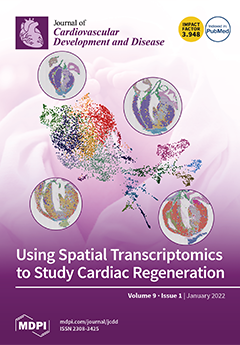
The neonatal mammalian heart exhibits remarkable regenerative potential, which includes fibrotic scar resolution and the generation of new cardiomyocytes. Here, we perform spatial transcriptomics to characterize heart repair after apical resection at near single-cell resolution. Hierarchical clustering reveals distinct domains of atrial and ventricular myocardium during postnatal heart maturation. Spatial transcriptomics also defines the resolving scar and regenerative border zone, characterized by spatially and temporally restricted programs of inflammation, epicardium expansion, extracellular matrix production, and cardiomyocyte restoration. This study provides insight into the cardiorestorative mechanisms that facilitate scar resolution and heart repair. View this paper
  • Issues are regarded as officially published after their release is announced to the table of contents alert mailing list.
  • You may sign up for e-mail alerts to receive table of contents of newly released issues.
  • PDF is the official format for papers published in both, html and pdf forms. To view the papers in pdf format, click on the "PDF Full-text" link, and use the free Adobe Reader to open them.
Order results
Result details
Section
Select all
Export citation of selected articles as:
16 pages, 4358 KiB  
Article
The Identification and Validation of Hub Genes Associated with Acute Myocardial Infarction Using Weighted Gene Co-Expression Network Analysis
by Junqiang Xue, Lu Chen, Hao Cheng, Xiaoyue Song, Yuekai Shi, Linnan Li, Rende Xu, Qing Qin, Jianying Ma and Junbo Ge
J. Cardiovasc. Dev. Dis. 2022, 9(1), 30; https://doi.org/10.3390/jcdd9010030 - 17 Jan 2022
Cited by 8 | Viewed by 5035
Abstract
Acute myocardial infarction (AMI), one of the most severe and fatal cardiovascular diseases, remains the main cause of mortality and morbidity worldwide. The objective of this study is to investigate the potential biomarkers for AMI based on bioinformatics analysis. A total of 2102 [...] Read more.
Acute myocardial infarction (AMI), one of the most severe and fatal cardiovascular diseases, remains the main cause of mortality and morbidity worldwide. The objective of this study is to investigate the potential biomarkers for AMI based on bioinformatics analysis. A total of 2102 differentially expressed genes (DEGs) were screened out from the data obtained from the gene expression omnibus (GEO) database. Weighted gene co-expression network analysis (WGCNA) explored the co-expression network of DEGs and determined the key module. The brown module was selected as the key one correlated with AMI. Gene Ontology and the Kyoto Encyclopedia of Genes and Genomes pathway enrichment analyses demonstrated that genes in the brown module were mainly enriched in ‘ribosomal subunit’ and ‘Ribosome’. Gene Set Enrichment Analysis revealed that ‘TNFA_SIGNALING_VIA_NFKB’ was remarkably enriched in AMI. Based on the protein–protein interaction network, ribosomal protein L9 (RPL9) and ribosomal protein L26 (RPL26) were identified as the hub genes. Additionally, the polymerase chain reaction (PCR) results indicated that the expression levels of RPL9 and RPL26 were both downregulated in AMI patients compared with controls, in accordance with the bioinformatics analysis. In summary, the identified DEGs, modules, pathways, and hub genes provide clues and shed light on the potential molecular mechanisms of AMI. Full article
Show Figures

Graphical abstract

8 pages, 6468 KiB  
Commentary
The Utility of Maternal Point of Care Ultrasound on Labor and Delivery Wards
by Mohammed Algodi, Diana S. Wolfe and Cynthia C. Taub
J. Cardiovasc. Dev. Dis. 2022, 9(1), 29; https://doi.org/10.3390/jcdd9010029 - 14 Jan 2022
Cited by 2 | Viewed by 2569
Abstract
Point-of-care ultrasonography (POCUS) refers to limited bedside ultrasound used to evaluate patients for conditions specific to the scope of their practice. Given the benefits of its application, interest in its use is increasing. We aimed to review the literature and assess the potential [...] Read more.
Point-of-care ultrasonography (POCUS) refers to limited bedside ultrasound used to evaluate patients for conditions specific to the scope of their practice. Given the benefits of its application, interest in its use is increasing. We aimed to review the literature and assess the potential feasibility of using POCUS of the heart and lungs in the field of obstetrics. We aim to describe its relevance and value as an adjunctive tool for critically ill obstetric patients on labor and delivery wards. Full article
(This article belongs to the Special Issue Heart Disease in Women)
Show Figures

Figure 1

11 pages, 4824 KiB  
Article
Individual Contributions of Cardiac Ion Channels on Atrial Repolarization and Reentrant Waves: A Multiscale In-Silico Study
by Henry Sutanto
J. Cardiovasc. Dev. Dis. 2022, 9(1), 28; https://doi.org/10.3390/jcdd9010028 - 14 Jan 2022
Cited by 6 | Viewed by 2718
Abstract
The excitation, contraction, and relaxation of an atrial cardiomyocyte are maintained by the activation and inactivation of numerous cardiac ion channels. Their collaborative efforts cause time-dependent changes of membrane potential, generating an action potential (AP), which is a surrogate marker of atrial arrhythmias. [...] Read more.
The excitation, contraction, and relaxation of an atrial cardiomyocyte are maintained by the activation and inactivation of numerous cardiac ion channels. Their collaborative efforts cause time-dependent changes of membrane potential, generating an action potential (AP), which is a surrogate marker of atrial arrhythmias. Recently, computational models of atrial electrophysiology emerged as a modality to investigate arrhythmia mechanisms and to predict the outcome of antiarrhythmic therapies. However, the individual contribution of atrial ion channels on atrial action potential and reentrant arrhythmia is not yet fully understood. Thus, in this multiscale in-silico study, perturbations of individual atrial ionic currents (INa, Ito, ICaL, IKur, IKr, IKs, IK1, INCX and INaK) in two in-silico models of human atrial cardiomyocyte (i.e., Courtemanche-1998 and Grandi-2011) were performed at both cellular and tissue levels. The results show that the inhibition of ICaL and INCX resulted in AP shortening, while the inhibition of IKur, IKr, IKs, IK1 and INaK prolonged AP duration (APD). Particularly, in-silico perturbations (inhibition and upregulation) of IKr and IKs only minorly affected atrial repolarization in the Grandi model. In contrast, in the Courtemanche model, the inhibition of IKr and IKs significantly prolonged APD and vice versa. Additionally, a 50% reduction of Ito density abbreviated APD in the Courtemanche model, while the same perturbation prolonged APD in the Grandi model. Similarly, a strong model dependence was also observed at tissue scale, with an observable IK1-mediated reentry stabilizing effect in the Courtemanche model but not in the Grandi atrial model. Moreover, the Grandi model was highly sensitive to a change on intracellular Ca2+ concentration, promoting a repolarization failure in ICaL upregulation above 150% and facilitating reentrant spiral waves stabilization by ICaL inhibition. Finally, by incorporating the previously published atrial fibrillation (AF)-associated ionic remodeling in the Courtemanche atrial model, in-silico modeling revealed the antiarrhythmic effect of IKr inhibition in both acute and chronic settings. Overall, our multiscale computational study highlights the strong model-dependent effects of ionic perturbations which could affect the model’s accuracy, interpretability, and prediction. This observation also suggests the need for a careful selection of in-silico models of atrial electrophysiology to achieve specific research aims. Full article
(This article belongs to the Section Electrophysiology and Cardiovascular Physiology)
Show Figures

Figure 1

14 pages, 1656 KiB  
Article
Vitamin D Inhibits IL-6 Pro-Atherothrombotic Effects in Human Endothelial Cells: A Potential Mechanism for Protection against COVID-19 Infection?
by Giovanni Cimmino, Stefano Conte, Mariarosaria Morello, Grazia Pellegrino, Laura Marra, Andrea Morello, Giuseppe Nicoletti, Gennaro De Rosa, Paolo Golino and Plinio Cirillo
J. Cardiovasc. Dev. Dis. 2022, 9(1), 27; https://doi.org/10.3390/jcdd9010027 - 13 Jan 2022
Cited by 17 | Viewed by 3057
Abstract
Background: Thrombosis with cardiovascular involvement is a crucial complication in COVID-19 infection. COVID-19 infects the host by the angiotensin converting enzyme-2 receptor (ACE2r), which is expressed in endothelial cells too. Thus, COVID-related thrombotic events might be due to endothelial dysfunction. IL-6 is one [...] Read more.
Background: Thrombosis with cardiovascular involvement is a crucial complication in COVID-19 infection. COVID-19 infects the host by the angiotensin converting enzyme-2 receptor (ACE2r), which is expressed in endothelial cells too. Thus, COVID-related thrombotic events might be due to endothelial dysfunction. IL-6 is one of the main cytokines involved in the COVID-19 inflammatory storm. Some evidence indicates that Vitamin D (VitD) has a protective role in COVID-19 patients, but the molecular mechanisms involved are still debated. Thus, we investigated the effect of VitD on Tissue Factor and adhesion molecules (CAMs) in IL-6-stimulated endothelial cells (HUVEC). Moreover, we evaluated levels of the ACE2r gene and proteins. Finally, we studied the modulation of NF-kB and STAT3 pathways. Methods: HUVEC cultivated in VitD-enriched medium were stimulated with IL-6 (0.5 ng/mL). The TF gene (RT-PCR), protein (Western blot), surface expression (FACS) and procoagulant activity (FXa generation assay) were measured. Similarly, CAMs soluble values (ELISA) and ACE2r (RT-PCR and Western blot) levels were assessed. NF-kB and STAT3 modulation (Western blot) were also investigated. Results: VitD significantly reduced TF expression at both gene and protein levels as well as TF-procoagulant activity in IL-6-treated HUVEC. Similar effects were observed for CAMs and ACE2r expression. IL-6 modulates these effects by regulating NF-κB and STAT3 pathways. Conclusions: IL-6 induces endothelial dysfunction with TF and CAMs expression via upregulation of ACE2r. VitD prevented these IL-6 deleterious effects. Thus, it might be speculated that this is one of the hypothetical mechanism(s) by which VitD exerts its beneficial effects in COVID-19 infection. Full article
(This article belongs to the Section Basic and Translational Cardiovascular Research)
Show Figures

Graphical abstract

14 pages, 1466 KiB  
Article
Cardiopulmonary Exercise Testing in Repaired Tetralogy of Fallot: Multiparametric Overview and Correlation with Cardiac Magnetic Resonance and Physical Activity Level
by Benedetta Leonardi, Federica Gentili, Marco Alfonso Perrone, Fabrizio Sollazzo, Lucia Cocomello, Stefani Silva Kikina, Rachel M. Wald, Vincenzo Palmieri, Aurelio Secinaro, Maria Giulia Gagliardi, Attilio Parisi, Attilio Turchetta, Lorenzo Galletti, Massimiliano Bianco and Fabrizio Drago
J. Cardiovasc. Dev. Dis. 2022, 9(1), 26; https://doi.org/10.3390/jcdd9010026 - 13 Jan 2022
Cited by 11 | Viewed by 3206
Abstract
Patients with repaired Tetralogy of Fallot (rToF) typically report having preserved subjective exercise tolerance. Chronic pulmonary regurgitation (PR) with varying degrees of right ventricular (RV) dilation as assessed by cardiac magnetic resonance imaging (MRI) is prevalent in rToF and may contribute to clinical [...] Read more.
Patients with repaired Tetralogy of Fallot (rToF) typically report having preserved subjective exercise tolerance. Chronic pulmonary regurgitation (PR) with varying degrees of right ventricular (RV) dilation as assessed by cardiac magnetic resonance imaging (MRI) is prevalent in rToF and may contribute to clinical compromise. Cardiopulmonary exercise testing (CPET) provides an objective assessment of functional capacity, and the International Physical Activity Questionnaire (IPAQ) can provide additional data on physical activity (PA) achieved. Our aim was to assess the association between CPET values, IPAQ measures, and MRI parameters. All rToF patients who had both an MRI and CPET performed within one year between March 2019 and June 2021 were selected. Clinical data were extracted from electronic records (including demographic, surgical history, New York Heart Association (NYHA) functional class, QRS duration, arrhythmia, MRI parameters, and CPET data). PA level, based on the IPAQ, was assessed at the time of CPET. Eighty-four patients (22.8 ± 8.4 years) showed a reduction in exercise capacity (median peak VO2 30 mL/kg/min (range 25–33); median percent predicted peak VO2 68% (range 61–78)). Peak VO2, correlated with biventricular stroke volumes (RVSV: β = 6.11 (95%CI, 2.38 to 9.85), p = 0.002; LVSV: β = 15.69 (95% CI 10.16 to 21.21), p < 0.0001) and LVEDVi (β = 8.74 (95%CI, 0.66 to 16.83), p = 0.04) on multivariate analysis adjusted for age, gender, and PA level. Other parameters which correlated with stroke volumes included oxygen uptake efficiency slope (OUES) (RVSV: β = 6.88 (95%CI, 1.93 to 11.84), p = 0.008; LVSV: β = 17.86 (95% CI 10.31 to 25.42), p < 0.0001) and peak O2 pulse (RVSV: β = 0.03 (95%CI, 0.01 to 0.05), p = 0.007; LVSV: β = 0.08 (95% CI 0.05 to 0.11), p < 0.0001). On multivariate analysis adjusted for age and gender, PA level correlated significantly with peak VO2/kg (β = 0.02, 95% CI 0.003 to 0.04; p = 0.019). We observed a reduction in objective exercise tolerance in rToF patients. Biventricular stroke volumes and LVEDVi were associated with peak VO2 irrespective of RV size. OUES and peak O2 pulse were also associated with biventricular stroke volumes. While PA level was associated with peak VO2, the incremental value of this parameter should be the focus of future studies. Full article
(This article belongs to the Special Issue Cardiovascular Effects and Benefits of Exercise)
Show Figures

Graphical abstract

9 pages, 840 KiB  
Article
Schizophrenia and Hospital Admissions for Cardiovascular Events in a Large Population: The APNA Study
by Sara Guillen-Aguinaga, Antonio Brugos-Larumbe, Laura Guillen-Aguinaga, Felipe Ortuño, Francisco Guillen-Grima, Luis Forga and Ines Aguinaga-Ontoso
J. Cardiovasc. Dev. Dis. 2022, 9(1), 25; https://doi.org/10.3390/jcdd9010025 - 13 Jan 2022
Cited by 1 | Viewed by 2408
Abstract
(1) Background: Patients with schizophrenia have higher mortality, with cardiovascular diseases being the first cause of mortality. This study aims to estimate the excess risk of hospital admission for cardiovascular events in schizophrenic patients, adjusting for comorbidity and risk factors. (2) Methods: The [...] Read more.
(1) Background: Patients with schizophrenia have higher mortality, with cardiovascular diseases being the first cause of mortality. This study aims to estimate the excess risk of hospital admission for cardiovascular events in schizophrenic patients, adjusting for comorbidity and risk factors. (2) Methods: The APNA study is a dynamic prospective cohort of all residents in Navarra, Spain. A total of 505,889 people over 18 years old were followed for five years. The endpoint was hospital admissions for a cardiovascular event. Direct Acyclic Graphs (DAG) and Cox regression were used. (3) Results: Schizophrenic patients had a Hazard Ratio (HR) of 1.414 (95% CI 1.031–1.938) of hospital admission for a cardiovascular event after adjusting for age, sex, hypertension, type 2 diabetes, dyslipidemia, smoking, low income, obesity, antecedents of cardiovascular disease, and smoking. In non-adherent to antipsychotic treatment schizophrenia patients, the HR was 2.232 (95% CI 1.267–3.933). (4) Conclusions: Patients with schizophrenia have a higher risk of hospital admission for cardiovascular events than persons with the same risk factors without schizophrenia. Primary care nursing interventions should monitor these patients and reduce cardiovascular risk factors. Full article
(This article belongs to the Section Epidemiology, Lifestyle, and Cardiovascular Health)
Show Figures

Graphical abstract

11 pages, 270 KiB  
Review
Endomyocardial Biopsy in Pediatric Myocarditis and Dilated Cardiomyopathy: A Tool in Search for a Role
by Mara Pilati, Micol Rebonato, Roberto Formigari and Gianfranco Butera
J. Cardiovasc. Dev. Dis. 2022, 9(1), 24; https://doi.org/10.3390/jcdd9010024 - 12 Jan 2022
Cited by 10 | Viewed by 2893
Abstract
Endomyocardial biopsy (EMB) is a well-known diagnostic tool for the investigation and treatment of myocardial diseases and remains the gold standard for the diagnosis of myocarditis. Due to its invasiveness, with a complication rate ranging from 1 to 15%, its role in the [...] Read more.
Endomyocardial biopsy (EMB) is a well-known diagnostic tool for the investigation and treatment of myocardial diseases and remains the gold standard for the diagnosis of myocarditis. Due to its invasiveness, with a complication rate ranging from 1 to 15%, its role in the diagnostic work-up of pediatric heart failure is not well established. The aim of this review is to define the role of EMB as diagnostic technique in the work up of children presenting with severe left ventricular dysfunction with the support of our center experience. Full article
(This article belongs to the Special Issue Pediatric Cardiomyopathies: From Genotype to Phenotype)
14 pages, 1216 KiB  
Article
Discriminative Utility of Apelin-to-NT-Pro-Brain Natriuretic Peptide Ratio for Heart Failure with Preserved Ejection Fraction among Type 2 Diabetes Mellitus Patients
by Alexander A. Berezin, Ivan M. Fushtey and Alexander E. Berezin
J. Cardiovasc. Dev. Dis. 2022, 9(1), 23; https://doi.org/10.3390/jcdd9010023 - 12 Jan 2022
Cited by 5 | Viewed by 2666
Abstract
Background: Apelin is a regulatory vasoactive peptide, which plays a pivotal role in adverse cardiac remodeling and heart failure (HF) with reduced ejection fraction. The purpose of the study was to investigate whether serum levels of apelin is associated with HF with preserved [...] Read more.
Background: Apelin is a regulatory vasoactive peptide, which plays a pivotal role in adverse cardiac remodeling and heart failure (HF) with reduced ejection fraction. The purpose of the study was to investigate whether serum levels of apelin is associated with HF with preserved election fraction (HFpEF) in patients with T2DM. Methods: The study retrospectively involved 101 T2DM patients aged 41 to 62 years (48 patients with HFpEF and 28 non-HFpEF patients). The healthy control group consisted of 25 individuals with matched age and sex. Data collection included demographic and anthropometric information, hemodynamic performances and biomarkers of the disease. Transthoracic B-mode echocardiography, Doppler and TDI were performed at baseline. Serum levels of N-terminal pro-brain natriuretic peptide (NT-proBNP) and apelin were measured by ELISA in all patients at the study entry. Results: Unadjusted multivariate logistic model yielded the only apelin to NT-proBNP ratio (OR = 1.44; p = 0.001), BMI > 34 кг/м2 (OR = 1.07; p = 0.036), NT-proBNP > 458 pmol/mL (OR = 1.17; p = 0.042), LAVI > 34 mL/m2 (OR = 1.06; p = 0.042) and E/e’ > 11 (OR = 1.04; p = 0.044) remained to be strong predictors for HFpEF. After obesity adjustment, multivariate logistic regression showed that the apelin to NT-proBNP ratio < 0.82 × 10⁻² units remained sole independent predictor for HFpEF (OR = 1.44; 95% CI: 1.18–2.77; p = 0.001) HFpEF in T2DM patients. In conclusion, we found that apelin to NT-proBNP ratio < 0.82 × 10⁻² units better predicted HFpEF in T2DM patients than apelin and NT-proBNP alone. This finding could open new approach for CV risk stratification of T2DM at higher risk of HF. Full article
(This article belongs to the Special Issue Obesity Cardiomyopathy)
Show Figures

Figure 1

18 pages, 3811 KiB  
Article
Myocardial Afterload Is a Key Biomechanical Regulator of Atrioventricular Myocyte Differentiation in Zebrafish
by Neha Ahuja, Paige Ostwald, Alex Gendernalik, Elena Guzzolino, Letizia Pitto, David Bark and Deborah M. Garrity
J. Cardiovasc. Dev. Dis. 2022, 9(1), 22; https://doi.org/10.3390/jcdd9010022 - 12 Jan 2022
Cited by 2 | Viewed by 2570
Abstract
Heart valve development is governed by both genetic and biomechanical inputs. Prior work has demonstrated that oscillating shear stress associated with blood flow is required for normal atrioventricular (AV) valve development. Cardiac afterload is defined as the pressure the ventricle must overcome in [...] Read more.
Heart valve development is governed by both genetic and biomechanical inputs. Prior work has demonstrated that oscillating shear stress associated with blood flow is required for normal atrioventricular (AV) valve development. Cardiac afterload is defined as the pressure the ventricle must overcome in order to pump blood throughout the circulatory system. In human patients, conditions of high afterload can cause valve pathology. Whether high afterload adversely affects embryonic valve development remains poorly understood. Here we describe a zebrafish model exhibiting increased myocardial afterload, caused by vasopressin, a vasoconstrictive drug. We show that the application of vasopressin reliably produces an increase in afterload without directly acting on cardiac tissue in zebrafish embryos. We have found that increased afterload alters the rate of growth of the cardiac chambers and causes remodeling of cardiomyocytes. Consistent with pathology seen in patients with clinically high afterload, we see defects in both the form and the function of the valve leaflets. Our results suggest that valve defects are due to changes in atrioventricular myocyte signaling, rather than pressure directly acting on the endothelial valve leaflet cells. Cardiac afterload should therefore be considered a biomechanical factor that particularly impacts embryonic valve development. Full article
(This article belongs to the Section Cardiac Development and Regeneration)
Show Figures

Figure 1

11 pages, 1455 KiB  
Article
Depression and Anxiety Are Associated with Physical Performance in Patients Undergoing Cardiac Rehabilitation: A Retrospective Observational Study
by Maaya Sakamoto, Yasunori Suematsu, Yuiko Yano, Koji Kaino, Reiko Teshima, Takuro Matsuda, Masaomi Fujita, Rie Tazawa, Kanta Fujimi and Shin-ichiro Miura
J. Cardiovasc. Dev. Dis. 2022, 9(1), 21; https://doi.org/10.3390/jcdd9010021 - 11 Jan 2022
Cited by 4 | Viewed by 2349
Abstract
Background: Cardiac rehabilitation (CR) combined with stress management training has been shown to be associated with fewer clinical events than CR alone. However, there have been no reports on the associations of CR with the psychological condition and detailed physical activities evaluated on [...] Read more.
Background: Cardiac rehabilitation (CR) combined with stress management training has been shown to be associated with fewer clinical events than CR alone. However, there have been no reports on the associations of CR with the psychological condition and detailed physical activities evaluated on the same day. Method: One hundred outpatients who participated in a CR program were graded on the hospital anxiety and depression scale (HADS). We divided them into a high HADS group (n = 32) and a normal HADS group (n = 68) and investigated by whole patients, ischemic heart disease (IHD) patients, and heart failure patients. Results: Overall, the patient age was 70.5 ± 9.6 years, the percentage of males was 73.0%, and the body mass index was 23.4 (21.7–26.0) kg/m2. In the high HADS group, overall functional mobility was poor and the distance in a two-minute walking test was short. Especially in IHD patients, the high HADS group showed high fat mass in body composition and low exercise tolerance and ventilator equivalents in cardiopulmonary exercise test. Conclusions: Depression and anxiety involved poor physical performance in CR outpatients and particularly involved low exercise tolerance in IHD patients. To evaluate accurate physical performance, it is necessary to investigate psychological condition. Full article
(This article belongs to the Special Issue Cardiac Rehabilitation: State of the Art and Perspectives)
Show Figures

Figure 1

14 pages, 536 KiB  
Article
Clinical Determinants and Prognosis of Left Ventricular Reverse Remodelling in Non-Ischemic Dilated Cardiomyopathy
by Carles Díez-López, Joel Salazar-Mendiguchía, Elena García-Romero, Lara Fuentes, Josep Lupón, Antoni Bayés-Genis, Nicolás Manito, Marta de Antonio, Pedro Moliner, Elisabet Zamora, Pablo Catalá-Ruiz, Miguel Caínzos-Achirica, Josep Comín-Colet and José González-Costello
J. Cardiovasc. Dev. Dis. 2022, 9(1), 20; https://doi.org/10.3390/jcdd9010020 - 11 Jan 2022
Cited by 2 | Viewed by 3538
Abstract
Aims: Non-ischaemic dilated cardiomyopathy (NIDCM) is characterized by left ventricular (LV) chamber enlargement and systolic dysfunction in the absence of coronary artery disease. Left ventricular reverse remodelling (LVRR) is the ability of a dilated ventricle to restore its normal size, shape and function. [...] Read more.
Aims: Non-ischaemic dilated cardiomyopathy (NIDCM) is characterized by left ventricular (LV) chamber enlargement and systolic dysfunction in the absence of coronary artery disease. Left ventricular reverse remodelling (LVRR) is the ability of a dilated ventricle to restore its normal size, shape and function. We sought to determine the frequency, clinical predictors and prognostic implications of LVRR, in a cohort of heart failure (HF) patients with NIDCM. Methods: We conducted a multicentre observational, retrospective cohort study of patients with NIDCM, with prospective serial echocardiography evaluations. LVRR was defined as an increase of ≥15% in left ventricular ejection fraction (LVEF) or as a LVEF increase ≥ 10% plus reduction of LV end-systolic diameter index ≥ 20%. We used multivariable logistic regression analyses to identify the baseline clinical predictors of LVRR and evaluate the prognostic impact of LVRR. Results: LVRR was achieved in 42.5% of 527 patients with NIDCM during the first year of follow-up (median LVEF 49%, median change +22%), Alcoholic aetiology, HF duration, baseline LVEF and the absence of LBBB (plus NT-proBNP levels when in the model), were the strongest predictors of LVRR. During a median follow-up of 47 months, 134 patients died (25.4%) and 7 patients (1.3%) received a heart transplant. Patients with LVRR presented better outcomes, regardless of other clinical conditions. Conclusions: In patients with NIDCM, LVRR was frequent and was associated with improved prognosis. Major clinical predictors of LVRR were alcoholic cardiomyopathy, absence of LBBB, shorter HF duration, and lower baseline LVEF and NT-proBNP levels. Our study advocates for clinical phenotyping of non-ischaemic dilated cardiomyopathy and intense gold-standard treatment optimization of patients according to current guidelines and recommendations in specialized HF units. Full article
(This article belongs to the Special Issue Understanding Heart Failure Management and Treatment)
Show Figures

Figure 1

11 pages, 1588 KiB  
Article
Impact of the COVID-19 Pandemic on Non-COVID-19 Clinical Trials
by Katia Audisio, Hillary Lia, Newell Bryce Robinson, Mohamed Rahouma, Giovanni Soletti, Gianmarco Cancelli, Roberto Perezgrovas Olaria, David Chadow, Derrick Y. Tam, Dominique Vervoort, Michael E. Farkouh, Deepak L. Bhatt, Stephen E. Fremes and Mario Gaudino
J. Cardiovasc. Dev. Dis. 2022, 9(1), 19; https://doi.org/10.3390/jcdd9010019 - 10 Jan 2022
Cited by 18 | Viewed by 3589
Abstract
Randomized controlled trials (RCT) were impacted by the COVID-19 pandemic, but no systematic analysis has evaluated the overall impact of COVID-19 on non-COVID-19-related RCTs. The ClinicalTrials.gov database was queried in February 2020. Eligible studies included all randomized trials with a start date after [...] Read more.
Randomized controlled trials (RCT) were impacted by the COVID-19 pandemic, but no systematic analysis has evaluated the overall impact of COVID-19 on non-COVID-19-related RCTs. The ClinicalTrials.gov database was queried in February 2020. Eligible studies included all randomized trials with a start date after 1 January 2010 and were active during the period from 1 January 2015 to 31 December 2020. The effect of the pandemic period on non-COVID-19 trials was determined by piece-wise regression models using 11 March 2020 as the start of the pandemic and by time series analysis (models fitted using 2015–2018 data and forecasted for 2019–2020). The study endpoints were early trial stoppage, normal trial completion, and trial activation. There were 161,377 non-COVID-19 trials analyzed. The number of active trials increased annually through 2019 but decreased in 2020. According to the piece-wise regression models, trial completion was not affected by the pandemic (p = 0.56) whereas trial stoppage increased (p = 0.001). There was a pronounced decrease in trial activation early during the pandemic (p < 0.001) which then recovered. The findings from the time series models were consistent comparing forecasted and observed results (trial completion p = 0.22; trial stoppage p < 0.01; trial activation, p = 0.01). During the pandemic, there was an increase in non-COVID-19 RCTs stoppage without changes in RCT completion. There was a sharp decline in new RCTs at the beginning of the pandemic, which later recovered. Full article
(This article belongs to the Special Issue Cardiac Surgery: Outcomes, Management and Critical Care)
Show Figures

Figure 1

4 pages, 394 KiB  
Case Report
Eradication of Ventricular Assist Device Driveline Infection in Paediatric Patients with Taurolidine
by Johannes Weichsel, Benito Baldauf, Hendrik Bonnemeier, Ernest W. Lau, Sven Dittrich and Robert Cesnjevar
J. Cardiovasc. Dev. Dis. 2022, 9(1), 18; https://doi.org/10.3390/jcdd9010018 - 10 Jan 2022
Cited by 4 | Viewed by 2446
Abstract
Ventricular assist devices (VADs) are used to provide mechanical circulatory support to patients with end-stage heart failure. The driveline connecting the external power source to the pump(s) of the intra-corporal VAD breaches the protective skin barrier and provides a track for microbes to [...] Read more.
Ventricular assist devices (VADs) are used to provide mechanical circulatory support to patients with end-stage heart failure. The driveline connecting the external power source to the pump(s) of the intra-corporal VAD breaches the protective skin barrier and provides a track for microbes to invade the interior of the patient’s body. Driveline infection constitutes a major and potentially fatal vulnerability of VAD therapy. Driveline infection cannot traditionally be salvaged and requires the extraction of the entire VAD system. We report here the successful eradication of a VAD driveline infection with a taurolidine-containing antimicrobial solution used for preventing the infection of cardiac implantable electronic devices. If replicated in more cases, the novel treatment concept described here may provide a valuable alternative management strategy of salvage rather than explantation for VAD driveline infection. Full article
(This article belongs to the Section Pediatric Cardiology and Congenital Heart Disease)
Show Figures

Figure 1

8 pages, 2602 KiB  
Article
The Use of Machine Learning Algorithms in the Evaluation of the Effectiveness of Resynchronization Therapy
by Bartosz Krzowski, Jakub Rokicki, Renata Główczyńska, Nikola Fajkis-Zajączkowska, Katarzyna Barczewska, Mariusz Mąsior, Marcin Grabowski and Paweł Balsam
J. Cardiovasc. Dev. Dis. 2022, 9(1), 17; https://doi.org/10.3390/jcdd9010017 - 10 Jan 2022
Cited by 3 | Viewed by 2717
Abstract
Background: Cardiovascular disease remains the leading cause of death in the European Union and worldwide. Constant improvement in cardiac care is leading to an increased number of patients with heart failure, which is a challenging condition in terms of clinical management. Cardiac resynchronization [...] Read more.
Background: Cardiovascular disease remains the leading cause of death in the European Union and worldwide. Constant improvement in cardiac care is leading to an increased number of patients with heart failure, which is a challenging condition in terms of clinical management. Cardiac resynchronization therapy is becoming more popular because of its grounded position in guidelines and clinical practice. However, some patients do not respond to treatment as expected. One way of assessing cardiac resynchronization therapy is with ECG analysis. Artificial intelligence is increasing in terms of everyday usability due to the possibility of everyday workflow improvement and, as a result, shortens the time required for diagnosis. A special area of artificial intelligence is machine learning. AI algorithms learn on their own based on implemented data. The aim of this study was to evaluate using artificial intelligence algorithms for detecting inadequate resynchronization therapy. Methods: A total of 1241 ECG tracings were collected from 547 cardiac department patients. All ECG signals were analyzed by three independent cardiologists. Every signal event (QRS-complex) and rhythm was manually classified by the medical team and fully reviewed by additional cardiologists. The results were divided into two parts: 80% of the results were used to train the algorithm, and 20% were used for the test (Cardiomatics, Cracow, Poland). Results: The required level of detection sensitivity of effective cardiac resynchronization therapy stimulation was achieved: 99.2% with a precision of 92.4%. Conclusions: Artificial intelligence algorithms can be a useful tool in assessing the effectiveness of resynchronization therapy. Full article
(This article belongs to the Special Issue Therapeutic Strategies for Heart Failure)
Show Figures

Figure 1

9 pages, 971 KiB  
Article
News from the Cold Chamber: Clinical Experiences of POLARx versus Arctic Front Advance for Single-Shot Pulmonary Vein Isolation
by Denise Guckel, Philipp Lucas, Khuraman Isgandarova, Mustapha El Hamriti, Leonard Bergau, Thomas Fink, Vanessa Sciacca, Guram Imnadze, Martin Braun, Moneeb Khalaph, Georg Nölker, Philipp Sommer and Christian Sohns
J. Cardiovasc. Dev. Dis. 2022, 9(1), 16; https://doi.org/10.3390/jcdd9010016 - 08 Jan 2022
Cited by 15 | Viewed by 2737
Abstract
Cryoballoon (CB)-guided pulmonary vein isolation (PVI) represents a cornerstone in the treatment of atrial fibrillation (AF). Recently, a novel balloon-guided single shot device (POLARx, Boston Scientific) was designed. Our study aimed to compare the efficacy, safety and characteristics of the novel CB system [...] Read more.
Cryoballoon (CB)-guided pulmonary vein isolation (PVI) represents a cornerstone in the treatment of atrial fibrillation (AF). Recently, a novel balloon-guided single shot device (POLARx, Boston Scientific) was designed. Our study aimed to compare the efficacy, safety and characteristics of the novel CB system with the established one (Arctic Front Advance (Pro), AFA, Medtronic). A total number of 596 patients undergoing CB-guided ablation for AF were included. 65 patients (65.0 ± 11.6, 31% female) undergoing PVI with the POLARx were compared to a cohort of 531 consecutive patients (63.0 ± 27.9, 25% female) treated with AFA. Acute PVI was achieved in all patients (n = 596, 100%). Total procedure duration (POLARx 113.3 ± 23.2 min, AFA 100.9 ± 21.3 min; p < 0.001) and fluoroscopy time (POLARx 10.5 ± 5.9 min, AFA 4.8 ± 3.6 min; p < 0.001) were significantly longer in the POLARx group. The POLARx balloon achieved significantly lower nadir temperatures (POLARx −57.7 ± 0.9 °C, AFA −45.1 ± 2.6 °C; p < 0.001) and a significantly higher percentage of pulmonary veins successfully isolated with the first freeze (p = 0.027 *). One major complication occurred in the POLARx (2%) and three (1%) in the AFA group. Both ablation systems are comparably safe and effective. AF ablation utilizing the POLARx system is associated with longer procedure and fluoroscopy times as well as lower nadir temperatures. Full article
(This article belongs to the Special Issue Catheter Ablation of Cardiac Arrhythmias: Practices and Outcomes)
Show Figures

Graphical abstract

12 pages, 1167 KiB  
Article
Renin Angiotensin System Blockers and Risk of Mortality in Hypertensive Patients Hospitalized for COVID-19: An Italian Registry
by Fabio Angeli, Paolo Verdecchia, Antonella Balestrino, Claudio Bruschi, Piero Ceriana, Luca Chiovato, Laura Adelaide Dalla Vecchia, Francesco Fanfulla, Maria Teresa La Rovere, Francesca Perego, Simonetta Scalvini, Antonio Spanevello, Egidio Traversi, Dina Visca, Michele Vitacca and Tiziana Bachetti
J. Cardiovasc. Dev. Dis. 2022, 9(1), 15; https://doi.org/10.3390/jcdd9010015 - 06 Jan 2022
Cited by 17 | Viewed by 2021
Abstract
Background: It is uncertain whether exposure to renin–angiotensin system (RAS) modifiers affects the severity of the new coronavirus disease 2019 (COVID-19) because most of the available studies are retrospective. Methods: We tested the prognostic value of exposure to RAS modifiers (either angiotensin-converting enzyme [...] Read more.
Background: It is uncertain whether exposure to renin–angiotensin system (RAS) modifiers affects the severity of the new coronavirus disease 2019 (COVID-19) because most of the available studies are retrospective. Methods: We tested the prognostic value of exposure to RAS modifiers (either angiotensin-converting enzyme inhibitors [ACE-Is] or angiotensin receptor blockers [ARBs]) in a prospective study of hypertensive patients with COVID-19. We analyzed data from 566 patients (mean age 75 years, 54% males, 162 ACE-Is users, and 147 ARBs users) hospitalized in five Italian hospitals. The study used systematic prospective data collection according to a pre-specified protocol. All-cause mortality during hospitalization was the primary outcome. Results: Sixty-six patients died during hospitalization. Exposure to RAS modifiers was associated with a significant reduction in the risk of in-hospital mortality when compared to other BP-lowering strategies (odds ratio [OR]: 0.54, 95% confidence interval [CI]: 0.32 to 0.90, p = 0.019). Exposure to ACE-Is was not significantly associated with a reduced risk of in-hospital mortality when compared with patients not treated with RAS modifiers (OR: 0.66, 95% CI: 0.36 to 1.20, p = 0.172). Conversely, ARBs users showed a 59% lower risk of death (OR: 0.41, 95% CI: 0.20 to 0.84, p = 0.016) even after allowance for several prognostic markers, including age, oxygen saturation, occurrence of severe hypotension during hospitalization, and lymphocyte count (adjusted OR: 0.37, 95% CI: 0.17 to 0.80, p = 0.012). The discontinuation of RAS modifiers during hospitalization did not exert a significant effect (p = 0.515). Conclusions: This prospective study indicates that exposure to ARBs reduces mortality in hospitalized patients with COVID-19. Full article
(This article belongs to the Special Issue Recent Advances in the Treatment of Hypertension)
Show Figures

Figure 1

8 pages, 263 KiB  
Article
Genetic Variation in LRP1 Associates with Stanford Type B Aortic Dissection Risk and Clinical Outcome
by Philipp Erhart, Daniel Körfer, Caspar Grond-Ginsbach, Jia-Lu Qiao, Moritz S. Bischoff, Maja Hempel, Christian P. Schaaf, Armin Grau and Dittmar Böckler
J. Cardiovasc. Dev. Dis. 2022, 9(1), 14; https://doi.org/10.3390/jcdd9010014 - 05 Jan 2022
Cited by 1 | Viewed by 2770
Abstract
Genetic variation in LRP1 (low-density lipoprotein receptor-related protein 1) was reported to be associated with thoracic aortic dissections and aneurysms. The aims of this study were to confirm this association in a prospective single-center patient cohort of patients with acute Stanford type B [...] Read more.
Genetic variation in LRP1 (low-density lipoprotein receptor-related protein 1) was reported to be associated with thoracic aortic dissections and aneurysms. The aims of this study were to confirm this association in a prospective single-center patient cohort of patients with acute Stanford type B aortic dissections (STBAD) and to assess the impact of LRP1 variation on clinical outcome. The single nucleotide variation (SNV) rs11172113 within the LRP1 gene was genotyped in 113 STBAD patients and 768 healthy control subjects from the same population. The T-allele of rs11172113 was more common in STBAD patients as compared to the reference group (72.6% vs. 59.6%) and confirmed to be an independent risk factor for STBAD (p = 0.002) after sex and age adjustment in a logistic regression model analyzing diabetes, smoking and hypertension as additional risk factors. Analysis of clinical follow-up (median follow-up 2.0 years) revealed that patients with the T-allele were more likely to suffer aorta-related complications (T-allele 75.6% vs. 63.8%; p = 0.022). In this study sample of STBAD patients, variation in LRP1 was an independent risk factor for STBAD and affected clinical outcome. Full article
22 pages, 1250 KiB  
Review
Modeling Human Cardiac Arrhythmias: Insights from Zebrafish
by Sébastien Gauvrit, Jaclyn Bossaer, Joyce Lee and Michelle M. Collins
J. Cardiovasc. Dev. Dis. 2022, 9(1), 13; https://doi.org/10.3390/jcdd9010013 - 05 Jan 2022
Cited by 10 | Viewed by 5087
Abstract
Cardiac arrhythmia, or irregular heart rhythm, is associated with morbidity and mortality and is described as one of the most important future public health challenges. Therefore, developing new models of cardiac arrhythmia is critical for understanding disease mechanisms, determining genetic underpinnings, and developing [...] Read more.
Cardiac arrhythmia, or irregular heart rhythm, is associated with morbidity and mortality and is described as one of the most important future public health challenges. Therefore, developing new models of cardiac arrhythmia is critical for understanding disease mechanisms, determining genetic underpinnings, and developing new therapeutic strategies. In the last few decades, the zebrafish has emerged as an attractive model to reproduce in vivo human cardiac pathologies, including arrhythmias. Here, we highlight the contribution of zebrafish to the field and discuss the available cardiac arrhythmia models. Further, we outline techniques to assess potential heart rhythm defects in larval and adult zebrafish. As genetic tools in zebrafish continue to bloom, this model will be crucial for functional genomics studies and to develop personalized anti-arrhythmic therapies. Full article
Show Figures

Figure 1

15 pages, 12612 KiB  
Review
The Role of Multimodality Imaging in Left-Sided Prosthetic Valve Dysfunction
by Manuela Muratori, Laura Fusini, Maria Elisabetta Mancini, Gloria Tamborini, Sarah Ghulam Ali, Paola Gripari, Marco Doldi, Antonio Frappampina, Giovanni Teruzzi, Gianluca Pontone, Piero Montorsi and Mauro Pepi
J. Cardiovasc. Dev. Dis. 2022, 9(1), 12; https://doi.org/10.3390/jcdd9010012 - 04 Jan 2022
Cited by 3 | Viewed by 3605
Abstract
Prosthetic valve (PV) dysfunction (PVD) is a complication of mechanical or biological PV. Etiologic mechanisms associated with PVD include fibrotic pannus ingrowth, thrombosis, structural valve degeneration, and endocarditis resulting in different grades of obstruction and/or regurgitation. PVD can be life threatening and often [...] Read more.
Prosthetic valve (PV) dysfunction (PVD) is a complication of mechanical or biological PV. Etiologic mechanisms associated with PVD include fibrotic pannus ingrowth, thrombosis, structural valve degeneration, and endocarditis resulting in different grades of obstruction and/or regurgitation. PVD can be life threatening and often challenging to diagnose due to the similarities between the clinical presentations of different causes. Nevertheless, identifying the cause of PVD is critical to treatment administration (thrombolysis, surgery, or percutaneous procedure). In this report, we review the role of multimodality imaging in the diagnosis of PVD. Specifically, this review discusses the characteristics of advanced imaging modalities underlying the importance of an integrated approach including 2D/3D transthoracic and transesophageal echocardiography, fluoroscopy, and computed tomography. In this scenario, it is critical to understand the strengths and weaknesses of each modality according to the suspected cause of PVD. In conclusion, for patients with suspected or known PVD, this stepwise imaging approach may lead to a simplified, more rapid, accurate and specific workflow and management. Full article
(This article belongs to the Special Issue Cardiac Imaging in Valvular Heart Disease)
Show Figures

Figure 1

13 pages, 1335 KiB  
Article
Electrocardiographic Characteristics and Their Correlation with Echocardiographic Alterations in Fabry Disease
by Matthew Zada, Queenie Lo, Siddharth J. Trivedi, Mehmet Harapoz, Anita C. Boyd, Kerry Devine, Norman Sadick, Michel C. Tchan and Liza Thomas
J. Cardiovasc. Dev. Dis. 2022, 9(1), 11; https://doi.org/10.3390/jcdd9010011 - 03 Jan 2022
Cited by 5 | Viewed by 1802
Abstract
Fabry disease (FD) is an X-linked disorder with α-galactosidase A deficiency. Males (>30 years) and females (>40 years) often present with cardiac manifestations, predominantly left ventricular hypertrophy (LVH). The aim of this study was to evaluate electrocardiographic (ECG) characteristics within FD patients to [...] Read more.
Fabry disease (FD) is an X-linked disorder with α-galactosidase A deficiency. Males (>30 years) and females (>40 years) often present with cardiac manifestations, predominantly left ventricular hypertrophy (LVH). The aim of this study was to evaluate electrocardiographic (ECG) characteristics within FD patients to identify gender related differences, and to additionally explore the association of ECG parameters with structural and functional alterations on transthoracic echocardiography (TTE). Retrospective cross-sectional analysis of 45 FD patients with contemporaneous ECG and TTE was performed and compared to age and gender matched healthy controls. FD patients demonstrated alterations in several ECG parameters particularly in males, including prolonged P-wave duration (91 vs. 81 ms, p = 0.022), prolonged QRS duration (96 vs. 84 ms, p < 0.001), increased R-wave amplitude in lead I (8.1 vs. 5.7 mV, p = 0.047), increased Sokolow–Lyon index (25 vs. 19 mV, p = 0.002) and were more likely to meet LVH criteria (31% vs. 7%, p = 0.006). FD patients with impaired basal longitudinal strain (LS) on TTE were more likely to meet LVH criteria (41% vs. 0%, p = 0.018). Those with more advanced FD (increased LV wall thickness on TTE) were more likely to meet LVH criteria but additionally demonstrated prolonged ventricular depolarization (QRS duration 101 vs. 88 ms, p = 0.044). Therefore, alterations on ECG demonstrating delayed atrial activation, delayed ventricular depolarization and evidence of LVH were more often seen in male FD patients. Impaired basal LS, a TTE marker of early cardiac involvement, correlated with ECG abnormalities. Increased LV wall thickness on TTE, a marker of more advanced FD, was associated with more severe ECG abnormalities. Full article
Show Figures

Graphical abstract

11 pages, 249 KiB  
Article
A Single Center Study on the Risks of Peri-Intervention Stroke in Thoracic Endovascular Aortic Repair (TEVAR) and Endovascular Abdominal Aortic Repair (EVAR)
by Jirayoot Chusooth, Chanon Kongkamol, Ruedeekorn Suwannanon, Dhanakom Premprabha, Voravit Chittithavorn, Pannawit Benjhawaleemas, Hutcha Sriplung and Pornchai Sathirapanya
J. Cardiovasc. Dev. Dis. 2022, 9(1), 10; https://doi.org/10.3390/jcdd9010010 - 03 Jan 2022
Viewed by 1206
Abstract
(1) Background: The risk factors of peri-intervention stroke (PIS) in thoracic endovascular aortic repair (TEVAR) and endovascular abdominal aortic repair (EVAR) are different. This study aimed to compare the risks of PIS in both interventions. (2) Methods: Patients who had suffered a PIS [...] Read more.
(1) Background: The risk factors of peri-intervention stroke (PIS) in thoracic endovascular aortic repair (TEVAR) and endovascular abdominal aortic repair (EVAR) are different. This study aimed to compare the risks of PIS in both interventions. (2) Methods: Patients who had suffered a PIS related to TEVAR or EVAR from January 2008 to June 2015 in Songklanagarind Hospital were selected as the cases, while patients who had not suffered PIS were randomly selected to create a 1:4 case: control ratio for analysis. The associations between the factors from pre- to post-intervention and PISs in TEVAR or EVAR cases were analyzed by univariable analysis (p < 0.1). The independent risks of PIS were identified by multivariable analysis and presented in odds ratios (p < 0.05). (3) Results: A total of 17 (2.2%) out of 777 patients who had undergone TEVAR or EVAR experienced PIS, of which 9/518 (1.7%) and 8/259 (3.1%) cases were in TEVAR and EVAR groups, respectively. PIS developed within the first 24 h in nine (52.9%) cases. Large vessel ischemic stroke or watershed infarctions were the most common etiologies of PIS. The independent risks of PIS were the volume of intra-intervention blood loss (1.99 (1.88–21.12), p < 0.001) in the TEVAR-related PIS, and intervention time (2.16 (1.95–2.37), p = 0.010) and post-intervention hyperglycemia (18.60 (1.60–216.06), p = 0.001) in the EVAR-related PIS. There were no differences in the rate of PIS among the operators, intervention techniques, and status of the interventions performed. (4) Conclusion: The risks of PIS in TEVAR or EVAR in our center were different and possibly independent of the operator expertise and intervention techniques. Full article
(This article belongs to the Section Stroke and Cerebrovascular Disease)
5 pages, 2822 KiB  
Case Report
Unique Case of Spontaneous Left Main Coronary Dissection in Second Trimester of Pregnancy Successfully Treated with Percutaneous Coronary Intervention: A Happy Ending
by Francesca Mantovani, Alessandro Navazio, Giovanni Tortorella and Vincenzo Guiducci
J. Cardiovasc. Dev. Dis. 2022, 9(1), 9; https://doi.org/10.3390/jcdd9010009 - 31 Dec 2021
Cited by 3 | Viewed by 1593
Abstract
Among pregnant women, SCAD is the most frequent etiology of non-atherosclerotic acute coronary syndrome. SCAD related to pregnancy is more frequent within the first month (especially first week) of puerperium or last trimester, or is otherwise anecdotal. The concomitance of SCAD and pregnancy [...] Read more.
Among pregnant women, SCAD is the most frequent etiology of non-atherosclerotic acute coronary syndrome. SCAD related to pregnancy is more frequent within the first month (especially first week) of puerperium or last trimester, or is otherwise anecdotal. The concomitance of SCAD and pregnancy poses many issues regarding diagnosis and treatment in respect to maternal and fetal safety and requires tailored intervention with close interaction between clinical cardiologists, interventional cardiologists, cardiothoracic surgeons, and obstetricians. We report the case of a patient, pregnant in the second trimester with a life-threatening SCAD, successfully treated with percutaneous coronary intervention with excellent outcome for mother and baby. Full article
(This article belongs to the Special Issue Heart Disease in Women)
Show Figures

Figure 1

14 pages, 1012 KiB  
Article
Athlete’s Heart in Elite Biathlon, Nordic Cross—Country and Ski-Mountaineering Athletes: Cardiac Adaptions Determined Using Echocardiographic Data
by Paul Zimmermann, Othmar Moser, Max L. Eckstein, Jan Wüstenfeld, Volker Schöffl, Lukas Zimmermann, Martin Braun and Isabelle Schöffl
J. Cardiovasc. Dev. Dis. 2022, 9(1), 8; https://doi.org/10.3390/jcdd9010008 - 29 Dec 2021
Cited by 6 | Viewed by 2461
Abstract
Twelve world elite Biathlon (Bia), ten Nordic Cross Country (NCC) and ten ski-mountaineering (Ski-Mo) athletes were evaluated for pronounced echocardiographic physiological cardiac remodeling as a primary aim of our descriptive preliminary report. In this context, sports-related cardiac remodeling was analyzed by performing two-dimensional [...] Read more.
Twelve world elite Biathlon (Bia), ten Nordic Cross Country (NCC) and ten ski-mountaineering (Ski-Mo) athletes were evaluated for pronounced echocardiographic physiological cardiac remodeling as a primary aim of our descriptive preliminary report. In this context, sports-related cardiac remodeling was analyzed by performing two-dimensional echocardiography including speckle tracking analysis as left ventricular global longitudinal strain (LV-GLS). A multicenter retrospective analysis of echocardiographic data was performed in 32 elite world winter sports athletes, which were obtained between 2020 and 2021 during the annual medical examination. The matched data of the elite world winter sports athletes (14 women, 18 male athletes, age: 18–35 years) were compared for different echocardiographic parameters. Significant differences could be revealed for left ventricular systolic function (LV-EF, p = 0.0001), left ventricular mass index (LV Mass index, p = 0.0078), left atrial remodeling by left atrial volume index (LAVI, p = 0.0052), and LV-GLS (p = 0.0003) between the three professional winter sports disciplines. This report provides new evidence that resting measures of cardiac structure and function in elite winter sport professionals can identify sport specific remodeling of the left heart, against the background of training schedule and training frequency. Full article
(This article belongs to the Special Issue Echocardiography in Cardiovascular Disease)
Show Figures

Figure 1

16 pages, 682 KiB  
Article
CD147 Levels in Blood and Adipose Tissues Correlate with Vascular Dysfunction in Obese Diabetic Adults
by Mohamed M. Ali, Imaduddin Mirza, Dina Naquiallah, Chandra Hassan, Mario Masrur, Francesco M. Bianco and Abeer M. Mahmoud
J. Cardiovasc. Dev. Dis. 2022, 9(1), 7; https://doi.org/10.3390/jcdd9010007 - 28 Dec 2021
Cited by 6 | Viewed by 1602
Abstract
CD147 is a glycoprotein that stimulates the production of matrix metalloproteinases (MMPs), known contributors to cardiovascular risk. The activity of CD147 protein depends on its glycosylation. However, it is unclear whether CD147 protein expression or glycosylation are influenced by the diabetic milieu characterized [...] Read more.
CD147 is a glycoprotein that stimulates the production of matrix metalloproteinases (MMPs), known contributors to cardiovascular risk. The activity of CD147 protein depends on its glycosylation. However, it is unclear whether CD147 protein expression or glycosylation are influenced by the diabetic milieu characterized by hyperglycemia and abundant glycation-end-products (AGEs). We examined the circulating and visceral adipose tissue (VAT) levels of CD147 and their correlation with vascular function in obese, obese diabetic, and non-obese controls (n = 40, each). The circulating levels of CD147 and the glycosylated CD147 protein in VAT were considerably higher in obese, particularly obese diabetic subjects compared to controls. Obese diabetics had the lowest brachial and arteriolar vasoreactivity and the highest carotid pulse-wave velocity (PWV, a measure of arterial stiffness) among the three groups. CD147 correlated positively with body mass index (BMI), total and visceral fat mass, PWV, and plasma levels of glucose, insulin, MMPs, and AGEs and negatively with brachial artery and VAT-arteriolar vasoreactivity and nitric oxide production. Multivariate regression revealed that BMI, body fat mass, insulin, and glucose levels significantly predicted CD147. Our data suggest that higher levels of CD147 in obese subjects, particularly those with diabetes, are linked to vascular dysfunction and several cardiometabolic risk factors. Full article
Show Figures

Figure 1

11 pages, 1891 KiB  
Systematic Review
Performance of Copeptin for Early Diagnosis of Acute Coronary Syndromes: A Systematic Review and Meta-Analysis of 14,139 Patients
by Lukasz Szarpak, Marcin Lapinski, Aleksandra Gasecka, Michal Pruc, Wiktoria L. Drela, Mariusz Koda, Andrea Denegri, Frank W. Peacock, Miłosz J. Jaguszewski and Krzysztof J. Filipiak
J. Cardiovasc. Dev. Dis. 2022, 9(1), 6; https://doi.org/10.3390/jcdd9010006 - 27 Dec 2021
Cited by 5 | Viewed by 2457
Abstract
Diagnosis of acute coronary syndrome (ACS) based on copeptin level may enable one to confirm or rule-out acute myocardial infarction (AMI) with higher sensitivity and specificity, which may in turn further reduce mortality rate and decrease the economic costs of ACS treatment. We [...] Read more.
Diagnosis of acute coronary syndrome (ACS) based on copeptin level may enable one to confirm or rule-out acute myocardial infarction (AMI) with higher sensitivity and specificity, which may in turn further reduce mortality rate and decrease the economic costs of ACS treatment. We conducted a systematic review and meta-analysis to investigate the relationship between copeptin levels and type of ACS. We searched Scopus, PubMed, Web of Science, Embase, and Cochrane to locate all articles published up to 10 October 2021. We evaluated a meta-analysis with random-effects models to evaluate differences in copeptin levels. A total of 14,139 patients (4565 with ACS) were included from twenty-seven studies. Copeptin levels in AMI and non-AMI groups varied and amounted to 68.7 ± 74.7 versus 14.8 ± 19.9 pmol/L (SMD = 2.63; 95% CI: 2.02 to 3.24; p < 0.001). Copeptin levels in the AMI group was higher than in the unstable angina (UAP) group, at 51.9 ± 52.5 versus 12.8 ± 19.7 pmol/L (SMD = 1.53; 95% CI: 0.86 to 2.20; p < 0.001). Copeptin levels in ST-elevation myocardial infarction (STEMI) versus non-ST elevation myocardial infarction (NSTEMI) patient groups were 54.8 ± 53.0 versus 28.7 ± 46.8 pmol/L, respectively (SMD = 1.69; 95% CI: = 0.70 to 4.09; p = 0.17). In summary, elevated copeptin levels were observed in patients with ACS compared with patients without ACS. Given its clinical value, copeptin levels may be included in the assessment of patients with ACS as well as for the initial differentiation of ACS. Full article
Show Figures

Figure 1

17 pages, 3770 KiB  
Review
Dissecting the Complexity of Early Heart Progenitor Cells
by Miquel Sendra, Jorge N. Domínguez, Miguel Torres and Oscar H. Ocaña
J. Cardiovasc. Dev. Dis. 2022, 9(1), 5; https://doi.org/10.3390/jcdd9010005 - 26 Dec 2021
Cited by 7 | Viewed by 4368
Abstract
Early heart development depends on the coordinated participation of heterogeneous cell sources. As pioneer work from Adriana C. Gittenberger-de Groot demonstrated, characterizing these distinct cell sources helps us to understand congenital heart defects. Despite decades of research on the segregation of lineages that [...] Read more.
Early heart development depends on the coordinated participation of heterogeneous cell sources. As pioneer work from Adriana C. Gittenberger-de Groot demonstrated, characterizing these distinct cell sources helps us to understand congenital heart defects. Despite decades of research on the segregation of lineages that form the primitive heart tube, we are far from understanding its full complexity. Currently, single-cell approaches are providing an unprecedented level of detail on cellular heterogeneity, offering new opportunities to decipher its functional role. In this review, we will focus on three key aspects of early heart morphogenesis: First, the segregation of myocardial and endocardial lineages, which yields an early lineage diversification in cardiac development; second, the signaling cues driving differentiation in these progenitor cells; and third, the transcriptional heterogeneity of cardiomyocyte progenitors of the primitive heart tube. Finally, we discuss how single-cell transcriptomics and epigenomics, together with live imaging and functional analyses, will likely transform the way we delve into the complexity of cardiac development and its links with congenital defects. Full article
Show Figures

Figure 1

32 pages, 2127 KiB  
Review
PDE-Mediated Cyclic Nucleotide Compartmentation in Vascular Smooth Muscle Cells: From Basic to a Clinical Perspective
by Margarida Lorigo, Nelson Oliveira and Elisa Cairrao
J. Cardiovasc. Dev. Dis. 2022, 9(1), 4; https://doi.org/10.3390/jcdd9010004 - 22 Dec 2021
Cited by 14 | Viewed by 4426
Abstract
Cardiovascular diseases are important causes of mortality and morbidity worldwide. Vascular smooth muscle cells (SMCs) are major components of blood vessels and are involved in physiologic and pathophysiologic conditions. In healthy vessels, vascular SMCs contribute to vasotone and regulate blood flow by cyclic [...] Read more.
Cardiovascular diseases are important causes of mortality and morbidity worldwide. Vascular smooth muscle cells (SMCs) are major components of blood vessels and are involved in physiologic and pathophysiologic conditions. In healthy vessels, vascular SMCs contribute to vasotone and regulate blood flow by cyclic nucleotide intracellular pathways. However, vascular SMCs lose their contractile phenotype under pathological conditions and alter contractility or signalling mechanisms, including cyclic nucleotide compartmentation. In the present review, we focus on compartmentalized signaling of cyclic nucleotides in vascular smooth muscle. A deeper understanding of these mechanisms clarifies the most relevant axes for the regulation of vascular tone. Furthermore, this allows the detection of possible changes associated with pathological processes, which may be of help for the discovery of novel drugs. Full article
(This article belongs to the Section Basic and Translational Cardiovascular Research)
Show Figures

Figure 1

9 pages, 808 KiB  
Article
The Influence of Cardiovascular Risk Factors and Hypogonadism on Cardiac Outcomes in an Aging Population of Beta-Thalassemia Patients
by Umberto Barbero, Matteo Ajassa, Carmen Maria Gaglioti, Antonio Piga, Giovanni Battista Ferrero and Filomena Longo
J. Cardiovasc. Dev. Dis. 2022, 9(1), 3; https://doi.org/10.3390/jcdd9010003 - 21 Dec 2021
Cited by 4 | Viewed by 2724
Abstract
Beta-thalassemia major (β-TM) is a hereditary genetic disease worsened by many comorbidities due to transfusion-related iron despite chelation therapy. Since there has recently been an increase in life expectancy of patients to up to 50 years old, which influences the prevalence of these [...] Read more.
Beta-thalassemia major (β-TM) is a hereditary genetic disease worsened by many comorbidities due to transfusion-related iron despite chelation therapy. Since there has recently been an increase in life expectancy of patients to up to 50 years old, which influences the prevalence of these diseases and the time span for traditional cardiovascular risk factors to play their role, this study aims to evaluate their distribution and prevalence in a population of thalassemia major patients and their relationship with observed cardiovascular events and potential modifying factors. One hundred and fifty-nine β-TM patients with at least 15 years of follow-up were included in this study. The mean age was 40.9 ± 8.4 years; 28% had diabetes mellitus and 62% had hypogonadism. The cardiovascular risk assessed using algorithms (CUORE and Pooled Cohort Risk Equation—PCRE) was low, but 3.8% of patients had at least one episode of heart failure, 35.9% showed early signs of heart failure, 22% received a diagnosis of diastolic dysfunction, and 21.4% showed supraventricular arrhythmias. Hypogonadism was shown to be related to the occurrence of cardiovascular events. The chronic accumulation of iron in the heart and the specific metabolic profile, mainly observed in patients with hypogonadism, allows us to define β-TM as a condition with a high level of cardiovascular risk from many points of view (iron-related myopathy, atherosclerosis and arrhythmias), which requires better stratification tools and a specific follow-up program. Full article
(This article belongs to the Section Pediatric Cardiology and Congenital Heart Disease)
Show Figures

Figure 1

15 pages, 2950 KiB  
Article
Not Only Diagnostic Yield: Whole-Exome Sequencing in Infantile Cardiomyopathies Impacts on Clinical and Family Management
by Laura Pezzoli, Lidia Pezzani, Ezio Bonanomi, Chiara Marrone, Agnese Scatigno, Anna Cereda, Maria Francesca Bedeschi, Angelo Selicorni, Serena Gasperini, Paolo Bini, Silvia Maitz, Carla Maccioni, Cristina Pedron, Lorenzo Colombo, Daniela Marchetti, Matteo Bellini, Anna Rita Lincesso, Loredana Perego, Monica Pingue, Nunzia Della Malva, Giovanna Mangili, Paolo Ferrazzi and Maria Iasconeadd Show full author list remove Hide full author list
J. Cardiovasc. Dev. Dis. 2022, 9(1), 2; https://doi.org/10.3390/jcdd9010002 - 21 Dec 2021
Cited by 12 | Viewed by 3375
Abstract
Whole-exome sequencing (WES) is a powerful and comprehensive tool for the genetic diagnosis of rare diseases, but few reports describe its timely application and clinical impact on infantile cardiomyopathies (CM). We conducted a retrospective analysis of patients with infantile CMs who had trio [...] Read more.
Whole-exome sequencing (WES) is a powerful and comprehensive tool for the genetic diagnosis of rare diseases, but few reports describe its timely application and clinical impact on infantile cardiomyopathies (CM). We conducted a retrospective analysis of patients with infantile CMs who had trio (proband and parents)-WES to determine whether results contributed to clinical management in urgent and non-urgent settings. Twenty-nine out of 42 enrolled patients (69.0%) received a definitive molecular diagnosis. The mean time-to-diagnosis was 9.7 days in urgent settings, and 17 out of 24 patients (70.8%) obtained an etiological classification. In non-urgent settings, the mean time-to-diagnosis was 225 days, and 12 out of 18 patients (66.7%) had a molecular diagnosis. In 37 out of 42 patients (88.1%), the genetic findings contributed to clinical management, including heart transplantation, palliative care, or medical treatment, independent of the patient’s critical condition. All 29 patients and families with a definitive diagnosis received specific counseling about recurrence risk, and in seven (24.1%) cases, the result facilitated diagnosis in parents or siblings. In conclusion, genetic diagnosis significantly contributes to patients’ clinical and family management, and trio-WES should be performed promptly to be an essential part of care in infantile cardiomyopathy, maximizing its clinical utility. Full article
(This article belongs to the Special Issue Pediatric Cardiomyopathies: From Genotype to Phenotype)
Show Figures

Figure 1

26 pages, 361250 KiB  
Article
Characterizing Neonatal Heart Maturation, Regeneration, and Scar Resolution Using Spatial Transcriptomics
by Adwiteeya Misra, Cameron D. Baker, Elizabeth M. Pritchett, Kimberly N. Burgos Villar, John M. Ashton and Eric M. Small
J. Cardiovasc. Dev. Dis. 2022, 9(1), 1; https://doi.org/10.3390/jcdd9010001 - 21 Dec 2021
Cited by 11 | Viewed by 5486
Abstract
The neonatal mammalian heart exhibits a remarkable regenerative potential, which includes fibrotic scar resolution and the generation of new cardiomyocytes. To investigate the mechanisms facilitating heart repair after apical resection in neonatal mice, we conducted bulk and spatial transcriptomic analyses at regenerative and [...] Read more.
The neonatal mammalian heart exhibits a remarkable regenerative potential, which includes fibrotic scar resolution and the generation of new cardiomyocytes. To investigate the mechanisms facilitating heart repair after apical resection in neonatal mice, we conducted bulk and spatial transcriptomic analyses at regenerative and non-regenerative timepoints. Importantly, spatial transcriptomics provided near single-cell resolution, revealing distinct domains of atrial and ventricular myocardium that exhibit dynamic phenotypic alterations during postnatal heart maturation. Spatial transcriptomics also defined the cardiac scar, which transitions from a proliferative to secretory phenotype as the heart loses regenerative potential. The resolving scar is characterized by spatially and temporally restricted programs of inflammation, epicardium expansion and extracellular matrix production, metabolic reprogramming, lipogenic scar extrusion, and cardiomyocyte restoration. Finally, this study revealed the emergence of a regenerative border zone defined by immature cardiomyocyte markers and the robust expression of Sprr1a. Taken together, our study defines the spatially and temporally restricted gene programs that underlie neonatal heart regeneration and provides insight into cardio-restorative mechanisms supporting scar resolution. Full article
(This article belongs to the Special Issue Model Systems for Heart Regeneration)
Show Figures

Figure 1

Previous Issue
Next Issue
Back to TopTop