Article

A Randomized, Open-Label Study to Assess Efficacy of Weekly Assumption of Cholecalciferol versus Calcifediol in Older Patients with Hypovitaminosis D

Geriatrics Unit, Department of Clinical and Experimental Medicine, University of Pisa, Via Savi 10, 56126 Pisa, Italy
*
Author to whom correspondence should be addressed.
Geriatrics 2022, 7(1), 13; https://doi.org/10.3390/geriatrics7010013
Submission received: 30 October 2021 / Revised: 23 December 2021 / Accepted: 4 January 2022 / Published: 7 January 2022

Abstract

:
The aim of this single-center, open-label, randomized controlled study was to evaluate which formulation of vitamin D—between cholecalciferol and calcifediol—is most effective in the treatment of hypovitaminosis D in older adults. Demographic characteristics, clinical history, and comprehensive geriatric assessment were recorded at admission. Eligible patients were randomly assigned an equivalent vitamin D supplement, either with cholecalciferol or calcifediol, from the time of hospital admission to three months after discharge. Among the 140 older patients included (mean age 83 ± 6.6 years, 57.8% females), 69 received cholecalciferol and 71 received calcifediol. The mean plasma values of 25-hydroxyvitamin D3 (25OH-vitamin D3) found at the time of enrollment were 16.8 ± 9.9 ng/mL in patients receiving cholecalciferol and 18.8 ± 13.3 ng/mL in those treated with calcifediol (p = 0.31). At the three month follow-up, the mean concentration of 25OH-vitamin D3 was significantly higher in patients treated with calcifediol than in those receiving cholecalciferol (30.7 ± 8.4 vs. 45.4 ± 9.8 ng/mL, respectively; p < 0.001). Supplementation with either cholecalciferol or calcifediol effectively results in reaching the optimal circulating values of 25OH-vitamin D3 in older patients suffering from hypovitaminosis D. However, supplementation with calcifediol led to average circulating values of 25OH-vitamin D3 that were significantly higher (over 50%) than those obtained with cholecalciferol.

1. Introduction

Hypovitaminosis D represents a widespread condition worldwide, particularly in the elderly population; it is estimated that about 7% of the world population is affected by severe hypovitaminosis ([25-hydroxyvitamin D3 (25OH-vitamin D3)] less than 10–12 ng/mL), while 37% of the population has moderate hypovitaminosis (25OH-vitamin D3 between 20 ng/mL and 10–12 ng/mL) [1]. There is broad consensus in the literature on how achieving sufficient levels of vitamin D plays an important role in improving not only bone homeostasis but also muscle performance and physical health [2,3,4,5]. The therapeutic strategy most often used to reach adequate levels of vitamin D is administering vitamin D supplements with a correct daily intake of calcium, the latter preferably with food [4]. Cholecalciferol (D3) and ergocalciferol (D2) have been used as supplements for a long time. Recently, calcifediol, the form activated by the hepatic enzyme 25-hydroxylase, has also been considered a valid therapeutic alternative [6].
Even though cholecalciferol and calcifediol are related molecules, they present several differences from the pharmacokinetic point of view. The first noticeable difference is represented by the half-life; in particular, the half-life of cholecalciferol is around two months, compared to the two weeks of calcifediol, due to the higher lipophilicity of the former molecule [7]. Thanks to its higher lipophilia, cholecalciferol is accumulated in the adipose tissue and is released gradually over time in a sort of self-regulation, which allows for the intermittent administration of the chosen dose and higher compliance among patients [7]. Calcifediol has a quicker elimination rate; therefore, a sufficient dosage should be administered on a daily or weekly basis [8]. Moreover, there is a different affinity for the vitamin D receptors (VDR), which is lower for cholecalciferol than calcifediol [7,9,10]. The intestinal absorption of cholecalciferol is effective among healthy subjects, while it might be severely compromised in patients with intestinal malabsorption [11]—a condition which does not spare geriatric patients [12,13]. On the contrary, calcifediol is absorbed very effectively and the difference in intestinal absorption kinetics largely explains its remarkable bioavailability [6].
Few studies have evaluated differences in the efficacy of the two compounds in correcting vitamin D deficiency among different age groups—mainly young adults and post-menopausal women [14,15,16,17,18]; however, data in the older and oldest-old population are very scarce, and only Ruggiero et al. considered a population with a mean age >80 years [19]. Given the paucity of data for individuals in this specific age range, who are often hospitalized for fragility fractures and would benefit most from vitamin D supplementation, we conducted a prospective, randomized study to evaluate which vitamin D formulation—between cholecalciferol and calcifediol—is the most effective in treating hypovitaminosis D in older adults.

2. Materials and Methods

A single-center, open-label, randomized controlled study was conducted enrolling geriatric patients consecutively hospitalized in the Geriatric Unit of the University Hospital of Pisa for acute illness from May to September 2020. No age restriction was applied, and we enrolled patients with 25OH-vitamin D3 levels < 30 ng/mL. Demographic characteristics and clinical history were collected at the time of admission. Within the first 24 h of admission, each patient underwent a comprehensive geriatric assessment (CGA), composed of the following measures: Cumulative Illness Rating Scale (CIRS) [20], Activities of Daily Living (ADL) [21], Instrumental Activities of Daily Living (IADL) [22], Short Portable Mental Status Questionnaire (SPMSQ) [23], Mini-Nutritional Assessment (MNA) [24], and Exton Smith Scale (ESS) [25]. Individuals’ body mass index (BMI) and multi-prognostic index (MPI) [26] values were also recorded. In order to investigate the presence of sarcopenia, the handgrip strength (HGS) test was performed on the dominant hand [27] using a hand dynamometer. Participants were seated with their shoulder adducted, elbow flexed to 90 degrees, and forearm and wrist neutral. The highest score out of three consecutive measurements was recorded. The study exclusion criteria were: i, having received vitamin D supplementation during the past six months; ii, stage V renal insufficiency; iii, liver failure (defined as a Child–Pugh classification of a B or C); iv, hyperparathyroidism; v, malabsorption syndromes or the long-term prescription of drugs reducing vitamin D absorption (i.e., antiepileptic drugs, long-term corticosteroids, or bisphosphonates); vi, neoplastic disease under treatment; vii, patients being unable to give informed consent. The dose of vitamin D supplementation was chosen based on current recommendations (20 mcg = 800 UI/day) [28,29,30,31,32]. Considering that calcifediol is about 3-fold more potent than cholecalciferol [10], eligible patients randomly received a bioequivalent dose of vitamin D—either with cholecalciferol (10,000 IU/mL, equivalent to 70 drops/week, 437.5 mcg/week) or calcifediol (1.5 mg/10 mL equivalent to 28 drops/week, 140 mcg/week)—once each week on the same day and at the same time (after lunch) during hospitalization and for three months after discharge. Randomization was performed by a physician using coin-flipping procedure. Before starting vitamin D supplementation, baseline blood samples were taken the first morning after admission, after an overnight fast, at 6 a.m.; 25OH-vitamin D3, parathyroid hormone, total calcium, calcium ion, phosphate, albumin, and creatinine were measured. 25OH-vitamin D3 levels were measured (blood samples were collected at the baseline evaluation, and the relative plasmas were stored at −20 °C) by tandem mass spectrometry coupled with high performances liquid chromatography (HPLC-MS-MS), using the MSMS VitD Kit from PerkinElmer (Waltham, MA, USA). A standard biochemical blood sample analysis was performed by Roche Autoanalyzer (Indianapolis, IN, USA) at the central laboratory of the University Hospital of Pisa. Three months after discharge, patients were re-evaluated at the geriatric-endocrinology outpatient clinic, where they underwent an HGS test and blood tests. The study protocol complied with the Declaration of Helsinki and was approved by the Pisa University Hospital Ethics Committee (n° protocol: CEAVNO-881/2020). Written informed consent was obtained from each enrolled patient.

Statistical Analysis

SPSS statistical software (IBM SPSS version 27.0, IBM Corporation, Chicago, IL, USA and its licensor 1989–2020) was utilized for the entire statistical analysis, whereas GraphPad Prism 9 was utilized for the graph plotting. A sample size of 58 for each group in the study achieved 90% power to detect a 15% difference among the means versus the alternative of equal means using an F test at a 0.05 significance level. The size of the variation in the means is represented by 0.25 of their standard deviation. The results were analyzed for normal distributions using a Shapiro–Wilk test, while homogeneity was tested using Levene’s test. Vitamin D levels were submitted to explanatory analysis for both a normal distribution and homoscedasticity. Continuous variables were presented as means ± standard deviations, ordinal variables were presented as medians and interquartile ranges (IQRs), and categorical variables were presented as percentages. Mann–Whitney and chi-square tests were used for multiple comparisons. Two-factor ANOVAs with repeated measures for time and the adjustment of p values using the Greenhouse–Geisser epsilon were performed in order to evaluate mean differences for the between-subjects model (factors: time and group) and the within-subjects model (factors: time and time for each group) among patients receiving vitamin D supplementation and their counterparts during the follow-up. Tests were performed considering a level of significance of 5%.

3. Results

Overall, 140 patients were included in the study (Figure 1), 69 received cholecalciferol (56.5% women, mean age 84.9 ± 6.4 years), and 71 received calcifediol (59.1% women, mean age 82.7 ± 6.7 years). As reported in Table 1, the two groups did not differ in terms of the reason for admission, comorbidities, the degree of disability [ADL median (IQR): 5(2) vs. 6(1), p = 0.42; IADL median (IQR): 4(5) vs. 5(4), p = 0.42], nutritional status [BMI median (IQR): 23.7(7.2) vs. 25(5.6), p = 0.95, MNA median (IQR): 23(8) vs. 25(6), p = 0.55], or strength as estimated through the HGS test (mean 17.5 ± 7.2 vs. 17.3 ± 7.2, p = 0.92). No statistical differences were found between the HG test and 25OH-vitamin D3 levels using the Spearman’s correlation analysis (Spearman’s rho =0.50, p = 0.30). Moreover, patients showed a similar degree of frailty as expressed using the MPI (mean 0.39 ± 0.20 vs. 0.32 ± 0.18, p = 0.37). Main acute illnesses requiring hospitalization were as follows: heart failure (10.7%: 10.1% in the cholecalciferol and 11.3% in the calcifediol group), arrhythmia (3.6%: 2.8% in the cholecalciferol and 4.2% in the calcifediol group), acute respiratory failure (12.9%: 13.0% in the cholecalciferol and 12.7% in the calcifediol group), bleeding (6.4%, 7.2% in the cholecalciferol and 5.6% in the calcifediol group), acute kidney failure (2.9%: 1.5% in the cholecalciferol and 4.2% in the calcifediol group), electrolyte disorders (2.9%: 2.9% in the cholecalciferol and 2.8% in the calcifediol group), stroke (7.9%: 7.2% in the cholecalciferol and 8.4% in the calcifediol group), decompensated diabetes (3.6%: 2.9% in the cholecalciferol and 4.2% in the calcifediol group), sepsis (10.7%: 11.5% in the cholecalciferol and 9.8% in the calcifediol group), and miscellaneous illnesses (38.6%: 37.6% in the cholecalciferol and 39.4% in the calcifediol group), with no significant differences between the two groups.
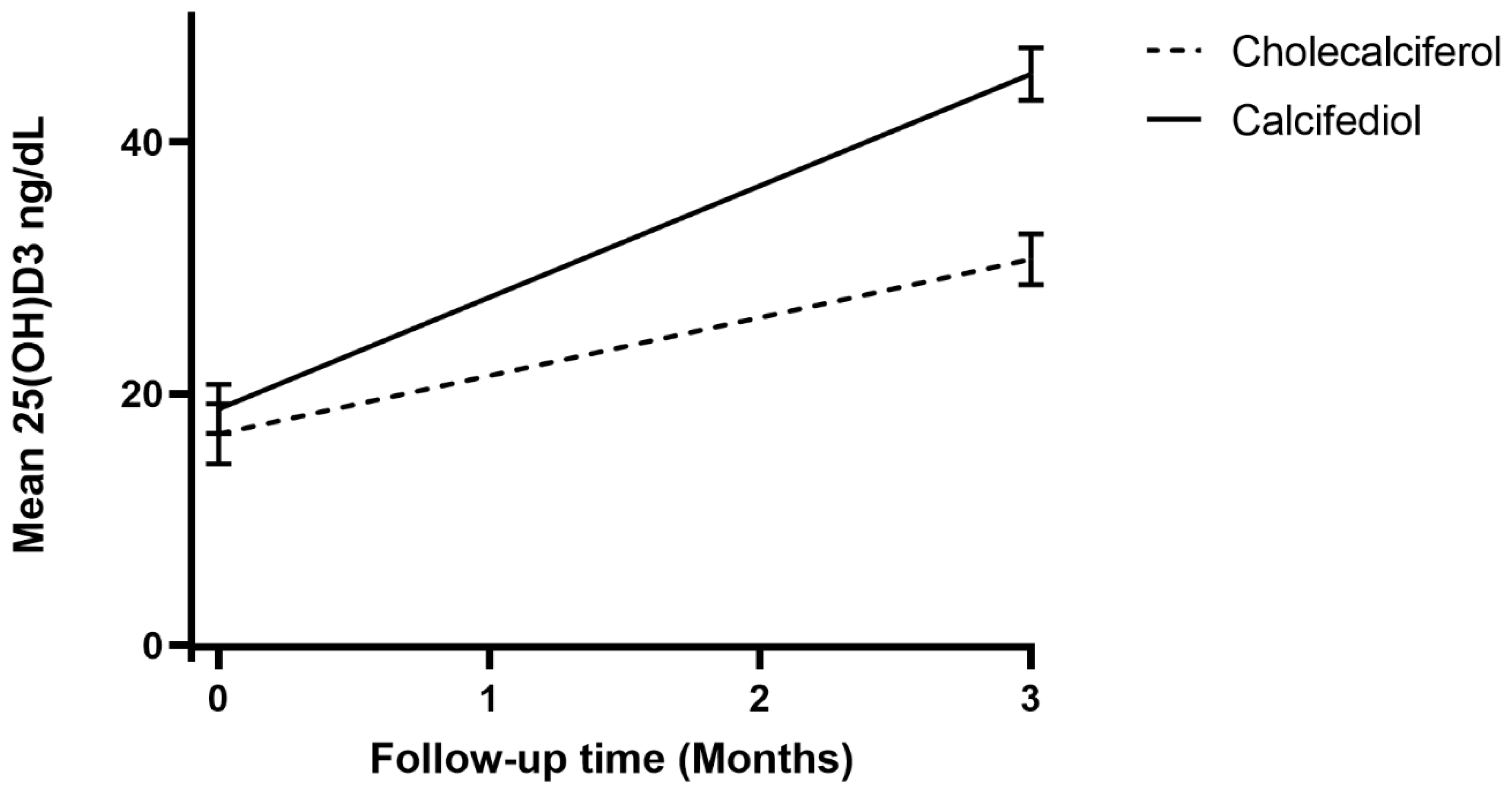
In regard to biochemistry blood exams, no significant differences were found in terms of serum creatinine concentration (1.15 ± 0.92 vs. 1.21 ± 1.02 mg/dL, p = 0.24), PTH circulating levels (48.1 ± 39.6 vs. 60.7 ± 36.9 pg/mL, p = 0.17), calcium concentration (8.8 ± 0.4 vs. 9 ± 0.4 mg/dL, p = 0.052), phosphoremia (3.2 ± 0.5 vs. 3.3 ± 0.8 mg/dL, p = 0.35), or albumin concentration (3.5 ± 0.4 vs. 3.5 ± 0.4 g/dL, p = 0.64). The mean plasma values of 25OH-vitamin D3 found during enrollment were 16.8 ± 9.9 ng/mL in patients receiving cholecalciferol and 18.8 ± 13.3 ng/mL in those treated with calcifediol (p = 0.31). At the three-month follow-up, the mean concentration of 25OH-vitamin D3 was significantly higher among patients treated with calcifediol than among those receiving cholecalciferol (30.7 ± 8.4 vs. 45.4 ± 9.8 ng/mL, respectively; p < 0.0001) (Figure 2).

4. Discussion

In the present study, we found that weekly supplementation with calcifediol appears to be more effective compared to a bioequivalent dosage of cholecalciferol in a cohort of older adults. Several studies showed that calcifediol is faster and more potent than cholecalciferol in increasing plasma 25OH-vitamin D3 levels [8,14,15,17,18,19,33,34], although most of these trials excluded the oldest-old population.
The goal of the prevention and correction of hypovitaminosis D is to achieve serum levels of 25OH-vitamin D3 ≥ 30 ng/mL (75 nmol/L), as recommended by most scientific societies [1]. The main component of the daily requirement of vitamin D derives from the endogenous synthesis in the skin following sun exposure to UVB rays. However, the latter process becomes ineffective with increasing age. The supplementation of vitamin D is the recommended therapeutic strategy, along with sufficient calcium intake [10]. Yet, hypovitaminosis D is frequent in the older and oldest-old (>85) populations [1], and reduced vitamin D levels are linked to greater vulnerability and frailty [3]. As a fact, 25OH-vitamin D3 can regulate the inflammatory response, promoting cyclin-dependent kinase (CDK) inhibitor synthesis, influencing several growth factors, and leading to the containment of systemic inflammation [35,36,37]. In a condition of 25OH-vitamin D3 deficiency, the low calcium concentration induces an increase in circulating PTH, which, through considerable renal reabsorption, increases 1,25OHD production and interaction with RANKL, restoring serum calcium values [38,39,40]. One of the strengths of the current study is that the mean age of patients was significantly higher compared to previous reports [8,14,15,17,18,33,34]; furthermore, we investigated functional status, reporting a high degree of autonomy in ADL in both groups. At baseline, no differences between the two cohorts were found in terms of BMI or MNA, confirming the homogeneity of our sample, similar to that of a previous report on a cohort of oldest-old patients [19]. In our study, although not reaching statistical significance, the finding of higher 25OH-vitamin D3 levels alongside the higher handgrip test values could support the relation between 25OH-vitamin D3 and muscle function [41,42,43].
At the 3-month follow-up, both cholecalciferol and calcifediol supplementation resulted in effectively accomplishing the 30 ng/mL threshold of patients’ 25OH-vitamin D3 values. The mean concentration of 25OH-vitamin D3 was significantly higher among patients treated with calcifediol than among those receiving cholecalciferol, further strengthening previous literature data [8,10,14,15,17,18,19,33,34]. These findings can be explained by the different intestinal absorption kinetics in older patients. Indeed, cholecalciferol is transported by chylomicrons and reaches the bloodstream via lymphatic circulation [44,45], while calcifediol is absorbed more effectively (almost 100%) [16], as it is transported directly into the bloodstream via the portal vein [46]. Furthermore, since calcifediol does not require hepatic conversion, it shows a linear relationship between the dose administered and the achieved serum levels [18]. Therefore, considering that elderly patients may experience intestinal malabsorption due to poly-therapies, gut dysbiosis caused by drugs interaction or the pathophysiological aging of the gastrointestinal tract [11,12,47], calcifediol could be more effective in reaching optimal vitamin D levels. In conclusion, the present study confirms previous findings from Ruggiero et al., but includes a larger cohort [19] and provides additional evidence regarding the oldest-old population, which is usually under-represented in clinical trials. Compared to previous findings [16,19], 25OH-vitamin D3 levels in our cohorts are higher at baseline as well as at the three-month follow-up; a possible explanation could be that we excluded all patients with malabsorption and those taking medications that could reduce vitamin D absorption.
Nonetheless, our study has some limitations. We acknowledge that the study schedule of a three-month follow-up visit is rather long; however, the enrollment window was during the COVID-19 pandemic; therefore, we tried to avoid patients’ and caregivers’ access to laboratories or outpatient clinics for as much as possible. Participants administered both of the vitamin D supplementations at home, in absence of an investigator confirmation; however, also according to hospital policy during the pandemic, we assessed patients’ adherence to the therapy as well as the possible onset of acute events via phone assessment on a monthly basis. Finally, results from our study are superimposable with previous reports carried out in similar cohorts of older patients, which underlines the reliability of our findings.

5. Conclusions

This study documents how three months of either cholecalciferol or calcifediol supplementation effectively results in reaching the optimal circulating values of 25OH-vitamin D3 in older patients suffering from hypovitaminosis D. However, supplementation with calcifediol shows average circulating values of 25OH-vitamin D3 to be significantly higher (over 50%) than those obtained with cholecalciferol. Further multi-center studies are nonetheless needed to confirm these findings.

Author Contributions

Conceptualization, C.O., V.C., F.N. and F.M.; methodology, C.O. and F.M.; validation, C.O., V.C. and F.M.; formal analysis, C.O.; data curation, G.C. and V.M.; writing—original draft preparation, R.F., S.R. and F.N.; writing—review and editing, C.O., V.C. and A.M.C.; supervision, N.C. and F.M.; project administration, C.O., V.C. and F.M. All authors have read and agreed to the published version of the manuscript.

Funding

This research received no external funding.

Institutional Review Board Statement

The study was conducted according to the guidelines of the Declaration of Helsinki, and approved by the Tuscany Regional Ethical Committee for Clinical Experimentation (n° protocol: CEAVNO-881/2020).

Informed Consent Statement

Informed consent was obtained from all subjects involved in the study.

Data Availability Statement

The data presented in this study are available upon request from the corresponding author.

Conflicts of Interest

The authors declare no conflict of interest.

References

  1. Hilger, J.; Friedel, A.; Herr, R.; Rausch, T.; Roos, F.; Wahl, D.A.; Pierroz, D.D.; Weber, P.; Hoffmann, K. A systematic review of vitamin D status in populations worldwide. Br. J. Nutr. 2013, 111, 23–45. [Google Scholar] [CrossRef] [Green Version]
  2. Bouillon, R.; Carmeliet, G.; Verlinden, L.; van Etten, E.; Verstuyf, A.; Luderer, H.F.; Lieben, L.; Mathieu, C.; DeMay, M. Vitamin D and Human Health: Lessons from Vitamin D Receptor Null Mice. Endocr. Rev. 2008, 29, 726–776. [Google Scholar] [CrossRef] [PubMed]
  3. Holick, M.F. Vitamin D Deficiency. N. Engl. J. Med. 2007, 357, 266–281. [Google Scholar] [CrossRef] [PubMed]
  4. Girgis, C.M.; Clifton-Bligh, R.; Hamrick, M.W.; Holick, M.; Gunton, J.E. The Roles of Vitamin D in Skeletal Muscle: Form, Function, and Metabolism. Endocr. Rev. 2013, 34, 33–83. [Google Scholar] [CrossRef] [Green Version]
  5. Wacker, M.; Holick, M.F. Vitamin D—Effects on Skeletal and Extraskeletal Health and the Need for Supplementation. Nutrients 2013, 5, 111–148. [Google Scholar] [CrossRef] [Green Version]
  6. Cesareo, R.; Falchetti, A.; Attanasio, R.; Tabacco, G.; Naciu, A.M.; Palermo, A. Hypovitaminosis D: Is It Time to Consider the Use of Calcifediol? Nutrients 2019, 11, 1016. [Google Scholar] [CrossRef] [Green Version]
  7. Sosa Henriquez, M.; Gomez de Tejada Romero, M.J. Cholecalciferol or calcifediol in the management of vitamin d deficiency. Nutrients 2020, 12, 1617. [Google Scholar] [CrossRef] [PubMed]
  8. Navarro-Valverde, C.; Sosa-Henríquez, M.; Alhambra-Expósito, M.R.; Quesada-Gómez, J.M. Vitamin D3 and calcidiol are not equipotent. J. Steroid Biochem. Mol. Biol. 2016, 164, 205–208. [Google Scholar] [CrossRef]
  9. Mazzaferro, S.; Goldsmith, D.; Larsson, T.E.; Massy, Z.A.; Cozzolino, M. Vitamin D metabolites and/or analogs: Which D for which patient? Curr. Vasc. Pharmacol. 2014, 12, 339–349. [Google Scholar] [CrossRef]
  10. Quesada-Gomez, J.M.; Bouillon, R. Is calcifediol better than cholecalciferol for vitamin D supplementation? Osteoporos. Int. 2018, 29, 1697–1711. [Google Scholar] [CrossRef]
  11. Pappa, H.M.; Bern, E.; Kamin, D.; Grand, R.J. Vitamin D status in gastrointestinal and liver disease. Curr. Opin. Gastroenterol. 2008, 24, 176–183. [Google Scholar] [CrossRef] [PubMed]
  12. Holt, P.R. Intestinal Malabsorption in the Elderly. Dig. Dis. 2007, 25, 144–150. [Google Scholar] [CrossRef]
  13. Montoro-Huguet, M.; Belloc, B.; Domínguez-Cajal, M. Small and Large Intestine (I): Malabsorption of Nutrients. Nutrients 2021, 13, 1254. [Google Scholar] [CrossRef]
  14. Bischoff-Ferrari, H.A.; Dawson-Hughes, B.; Stöcklin, E.; Sidelnikov, E.; Willett, W.C.; Edel, J.O.; Stähelin, H.B.; Wolfram, S.; Jetter, A.; Schwager, J.; et al. Oral supplementation with 25(OH)D3versus vitamin D3: Effects on 25(OH)D levels, lower extremity function, blood pressure, and markers of innate immunity. J. Bone Miner. Res. 2012, 27, 160–169. [Google Scholar] [CrossRef]
  15. Cashman, K.D.; Seamans, K.M.; Lucey, A.J.; Stöcklin, E.; Weber, P.; Kiely, M.; Hill, T.R. Relative effectiveness of oral 25-hydroxyvitamin D3 and vitamin D3 in raising wintertime serum 25-hydroxyvitamin D in older adults. Am. J. Clin. Nutr. 2012, 95, 1350–1356. [Google Scholar] [CrossRef] [PubMed] [Green Version]
  16. Pérez-Castrillón, J.L.; Dueñas-Laita, A.; Brandi, M.L.; Jódar, E.; del Pino-Montes, J.; Quesada-Gómez, J.M.; Castro, F.C.; Gómez-Alonso, C.; López, L.G.; Martínez, J.M.O.; et al. Calcifediol is superior to cholecalciferol in improving vitamin D status in postmenopausal women: A randomized trial. J. Bone Miner. Res. 2021, 36, 1967–1978. [Google Scholar] [CrossRef]
  17. Shieh, A.; Ma, C.; Chun, R.F.; Witzel, S.; Rafison, B.; Contreras, H.; Wittwer-Schegg, J.; Swinkels, L.; Huijs, T.; Hewison, M.; et al. Effects of Cholecalciferol vs Calcifediol on Total and Free 25-Hydroxyvitamin D and Parathyroid Hormone. J. Clin. Endocrinol. Metab. 2017, 102, 1133–1140. [Google Scholar] [CrossRef] [PubMed]
  18. Vaes, A.M.; Tieland, M.; de Regt, M.F.; Wittwer, J.; van Loon, L.J.; de Groot, L.C. Dose–response effects of supplementation with calcifediol on serum 25-hydroxyvitamin D status and its metabolites: A randomized controlled trial in older adults. Clin. Nutr. 2018, 37, 808–814. [Google Scholar] [CrossRef]
  19. Ruggiero, C.; Baroni, M.; Bini, V.; Brozzetti, A.; Parretti, L.; Zengarini, E.; Lapenna, M.; Antinolfi, P.; Falorni, A.; Mecocci, P.; et al. Effects of Weekly Supplementation of Cholecalciferol and Calcifediol among the Oldest-Old People: Findings From a Randomized Pragmatic Clinical Trial. Nutrients 2019, 11, 2778. [Google Scholar] [CrossRef] [Green Version]
  20. Parmelee, P.A.; Thuras, P.D.; Katz, I.R.; Lawton, M.P. Validation of the Cumulative Illness Rating Scale in a Geriatric Residential Population. J. Am. Geriatr. Soc. 1995, 43, 130–137. [Google Scholar] [CrossRef]
  21. Katz, S.; Ford, A.B.; Moskowitz, R.W.; Jackson, B.A.; Jaffe, M.W. Studies of Illness in the Aged. The index of Adl: A standardized measure of biological and phychological funcation. JAMA 1963, 185, 914–919. [Google Scholar] [CrossRef]
  22. Lawton, M.P.; Brody, E.M. Assessment of older people: Self-maintaining and instrumental activities of daily living. Nurs. Res. 1969, 9, 179–186. [Google Scholar] [CrossRef]
  23. Pfeiffer, E. A Short Portable Mental Status Questionnaire for the Assessment of Organic Brain Deficit in Elderly Patients. J. Am. Geriatr. Soc. 1975, 23, 433–441. [Google Scholar] [CrossRef]
  24. Guigoz, P.Y.; Vellas, M.B.; Garry, P.P.J. Assessing the Nutritional Status of the Elderly: The Mini Nutritional Assessment as Part of the Geriatric Evaluation. Nutr. Rev. 1996, 54, S59–S65. [Google Scholar] [CrossRef] [PubMed]
  25. Bliss, M.R.; McLaren, R.; Exton-Smith, A.N. Mattresses for preventing pressure sores in geriatric patients. Mon. Bull. Minist. Health Public Health Lab. Serv. 1966, 25, 238–268. [Google Scholar]
  26. Pilotto, A.; Rengo, F.; Marchionni, N.; Sancarlo, D.; Fontana, A.; Panza, F.; Ferrucci, L.; The FIRI-SIGG Study Group. Comparing the Prognostic Accuracy for All-Cause Mortality of Frailty Instruments: A Multicentre 1-Year Follow-Up in Hospitalized Older Patients. PLoS ONE 2012, 7, e29090. [Google Scholar] [CrossRef] [Green Version]
  27. Lee, S.H.; Gong, H.S. Measurement and Interpretation of Handgrip Strength for Research on Sarcopenia and Osteoporosis. J. Bone Metab. 2020, 27, 85–96. [Google Scholar] [CrossRef]
  28. Ross, A.C.; Taylor, C.L.; Yaktine, A.L.; del Valle, H.B. Dietary Reference Intakes for CALCIUM and Vitamin D; Reports funded by National Institutes of Health; The National Academies Collection: Washington, DC, USA, 2011. [Google Scholar]
  29. Bischoff-Ferrari, H.; Willett, W.C.; Orav, E.J.; Lips, P.; Meunier, P.J.; Lyons, R.A.; Flicker, L.; Wark, J.; Jackson, R.D.; Cauley, J.A.; et al. A Pooled Analysis of Vitamin D Dose Requirements for Fracture Prevention. N. Engl. J. Med. 2012, 367, 40–49. [Google Scholar] [CrossRef] [Green Version]
  30. Bischoff-Ferrari, H.A.; Willett, W.C.; Wong, J.B.; Stuck, A.E.; Staehelin, H.B.; Orav, E.J.; Thoma, A.; Kiel, D.; Henschkowski, J. Prevention of Nonvertebral Fractures with Oral Vitamin D and Dose Dependency: A meta-analysis of randomized controlled trials. Arch. Intern. Med. 2009, 169, 551–561. [Google Scholar] [CrossRef] [Green Version]
  31. Cosman, F.; De Beur, S.J.; LeBoff, M.S.; Lewiecki, E.M.; Tanner, B.; Randall, S.; Lindsay, R.; National Osteoporosis Foundation. Clinician’s Guide to Prevention and Treatment of Osteoporosis. Osteoporos. Int. 2014, 25, 2359–2381. [Google Scholar] [CrossRef] [PubMed] [Green Version]
  32. Veleva, B.I.; Chel, V.G.; Achterberg, W.P. Efficacy of daily 800 IU vitamin D supplementation in reaching vitamin D sufficiency in nursing home residents: Cross-sectional patient file study. BMC Geriatr. 2014, 14, 103. [Google Scholar] [CrossRef] [Green Version]
  33. Barger-Lux, M.J.; Heaney, R.P.; Dowell, S.; Chen, T.; Holick, M. Vitamin D and its Major Metabolites: Serum Levels after Graded Oral Dosing in Healthy Men. Osteoporos. Int. 1998, 8, 222–230. [Google Scholar] [CrossRef]
  34. Jetter, A.; Egli, A.; Dawson-Hughes, B.; Staehelin, H.B.; Stoecklin, E.; Goessl, R.; Henschkowski, J.; Bischoff-Ferrari, H.A. Pharmacokinetics of oral vitamin D(3) and calcifediol. Bone 2014, 59, 14–19. [Google Scholar] [CrossRef]
  35. Bhoora, S.; Punchoo, R. Policing Cancer: Vitamin D Arrests the Cell Cycle. Int. J. Mol. Sci. 2020, 21, 9296. [Google Scholar] [CrossRef]
  36. Liu, N.; Su, H.; Zhang, Y.; Kong, J. The protective effect of 1,25(OH)2D3 against cardiac hypertrophy is mediated by the cyclin-dependent kinase inhibitor p21. Eur. J. Pharmacol. 2020, 888, 173510. [Google Scholar] [CrossRef]
  37. Vanoirbeek, E.; Krishnan, A.; Eelen, G.; Verlinden, L.; Bouillon, R.; Feldman, D.; Verstuyf, A. The anti-cancer and anti-inflammatory actions of 1,25(OH)2D3. Best Pract. Res. Clin. Endocrinol. Metab. 2011, 25, 593–604. [Google Scholar] [CrossRef] [Green Version]
  38. Bikle, D.D. Vitamin D and Bone. Curr. Osteoporos. Rep. 2012, 10, 151–159. [Google Scholar] [CrossRef] [Green Version]
  39. Pike, J.W.; Christakos, S. Biology and Mechanisms of Action of the Vitamin D Hormone. Endocrinol. Metab. Clin. N. Am. 2017, 46, 815–843. [Google Scholar] [CrossRef]
  40. Turner, A.; Hanrath, M.A.; Morris, H.A.; Atkins, G.; Anderson, P. The local production of 1,25(OH)2D3 promotes osteoblast and osteocyte maturation. J. Steroid Biochem. Mol. Biol. 2014, 144 Pt A, 114–118. [Google Scholar] [CrossRef]
  41. Dawson-Hughes, B. Vitamin D and muscle function. J. Steroid Biochem. Mol. Biol. 2017, 173, 313–316. [Google Scholar] [CrossRef]
  42. El Hajj, C.; Fares, S.; Chardigny, J.M.; Boirie, Y.; Walrand, S. Vitamin D supplementation and muscle strength in pre-sarcopenic elderly Lebanese people: A randomized controlled trial. Arch. Osteoporos. 2018, 14, 4. [Google Scholar] [CrossRef]
  43. Manoy, P.; Yuktanandana, P.; Tanavalee, A.; Anomasiri, W.; Ngarmukos, S.; Tanpowpong, T.; Honsawek, S. Vitamin D Supplementation Improves Quality of Life and Physical Performance in Osteoarthritis Patients. Nutrients 2017, 9, 799. [Google Scholar] [CrossRef] [Green Version]
  44. Davies, M.; Mawer, E.B.; Krawitt, E.L. Comparative absorption of vitamin D3 and 25-hydroxyvitamin D3 in intestinal disease. Gut 1980, 21, 287–292. [Google Scholar] [CrossRef] [Green Version]
  45. Thompson, G.R.; Lewis, B.; Booth, C.C. Absorption of vitamin D3-3H in control subjects and patients with intestinal malabsorption. J. Clin. Investig. 1966, 45, 94–102. [Google Scholar] [CrossRef] [PubMed]
  46. Maislos, M.; Silver, J.; Fainaru, M. Intestinal absorption of vitamin D sterols: Differential absorption into lymph and portal blood in the rat. Gastroenterology 1981, 80, 1528–1534. [Google Scholar] [CrossRef]
  47. Schiller, L.R. Maldigestion Versus Malabsorption in the Elderly. Curr. Gastroenterol. Rep. 2020, 22, 33. [Google Scholar] [CrossRef]
Figure 1. Flowchart of study enrollment.
Geriatrics 07 00013 g001
Figure 2. Mean 25OH-vitamin D3 values over time, according to the type of vitamin D supplementation.
Geriatrics 07 00013 g002
Table 1. Clinical characteristics of the study population.
All Patients
N = 140
Cholecalciferol
N = 69
Calcifediol
N = 71
p-Value
Female (%)81 (57.8)39 (56.5)42 (59.1)0.75
Age (years, mean, SD)83.8 (6.6)84.9 (6.4)82.7 (6.7)0.052
BMI (median, IQR)24.4 (6.1)23.7 (7.2)25 (5.6)0.95
ADL (median, IQR)6 (2)5 (2)6 (1)0.42
IADL (median, IQR)4 (5)4 (5)5 (4)0.42
MNA (median, IQR)25 (5)23 (8)25 (6)0.55
Exton Smith Scale (median, IQR)18 (3)17 (3)18 (3)0.74
SPMSQ (median, IQR)2 (2)2 (2)2 (3)0.68
CIRS–C (median, IQR)3 (2)3 (2)3 (3)0.37
MPI (mean, SD)0.35 (0.19)0.39 (0.20)0.32 (0.18)0.37
Arterial hypertension (%)98 (70)48 (69.6) 50 (70.4)0.33
CAD (%)15 (10)8 (11.6)7 (9.9)0.32
AF (%)26 (18.6)12 (17.4)14 (19.7)0.77
Heart failure (%)57 (40.7)29 (42)28 (39.4)0.57
Diabetes (%)
CKD (%)
29 (20.7)
32 (22.8)
14 (20.3)
16 (23.2)
15 (21.1)
16 (22.5)
0.42
0.30
COPD (%)13 (9.3)6 (8.6)7 (9.8)0.54
Number of drugs (median, IQR)6 (3)7 (3)6 (3)0.65
Creatinine mg/dl (mean, SD)1.15 (0.53)1.15 (0.92)1.21 (1.02)0.24
PTH ng/dL (mean, SD)55.3 (38.3)48.1 (39.6)60.7 (36.9)0.17
Serum Calcium mg/dl (mean, SD)8.9 (0.4)8.8 (0.4)9.0 (0.4)0.052
Serum Phosphate mg/dl (mean, SD)3.25 (0.8)3.2 (0.5)3.3 (0.8)0.35
Serum Albumin g/dl (mean, SD)3.5 (0.4)3.5 (0.4)3.5 (0.4)0.64
Handgrip test (mean, SD)
Males
Females
17.4 (7.4)
25.9 (5.7)
13.9 (4.8)
17.5 (7.2)
24.3 (5.4)
13.7 (4.4)
17.3 (7.2)
27.1 (6,2)
14.1 (5.3)
0.92
0.34
0.82
25OHVitD at study enrollment (ng/mL)17.8 (11.7) 16.8 (9.9)18.8 (13.3)0.31
25OHVitD at 3-month follow-up (ng/mL)38.1 (18.3)30.7 (8.4)45.4 (9.8)<0.001
25OHVitD3 mean difference at 3 months (SEM)20.2 (+17.8; +23.2)13.7 (+11.8; +15.3)26.6 (+22.9; +30.1)<0.001
BMI, body mass index; ADL, Activities of Daily Living; IADL, Instrumental Activities of Daily Living; MNA, Mini-Nutritional Assessment; SPMSQ, Short Portable Mental Status Questionnaire; CIRS-C, Cumulative Illness Rating Scale-Comorbidity; MPI, Multi Prognostic Index; PTH, parathyroid hormone; CAD. coronary heart disease; AF, atrial fibrillation; CKD: chronic kidney disease; COPD: chronic obstructive pulmonary disease.
Publisher’s Note: MDPI stays neutral with regard to jurisdictional claims in published maps and institutional affiliations.