Prostatic Localization of a Migrating Grass Awn Foreign Body in a Dog

 ,
,  and
and 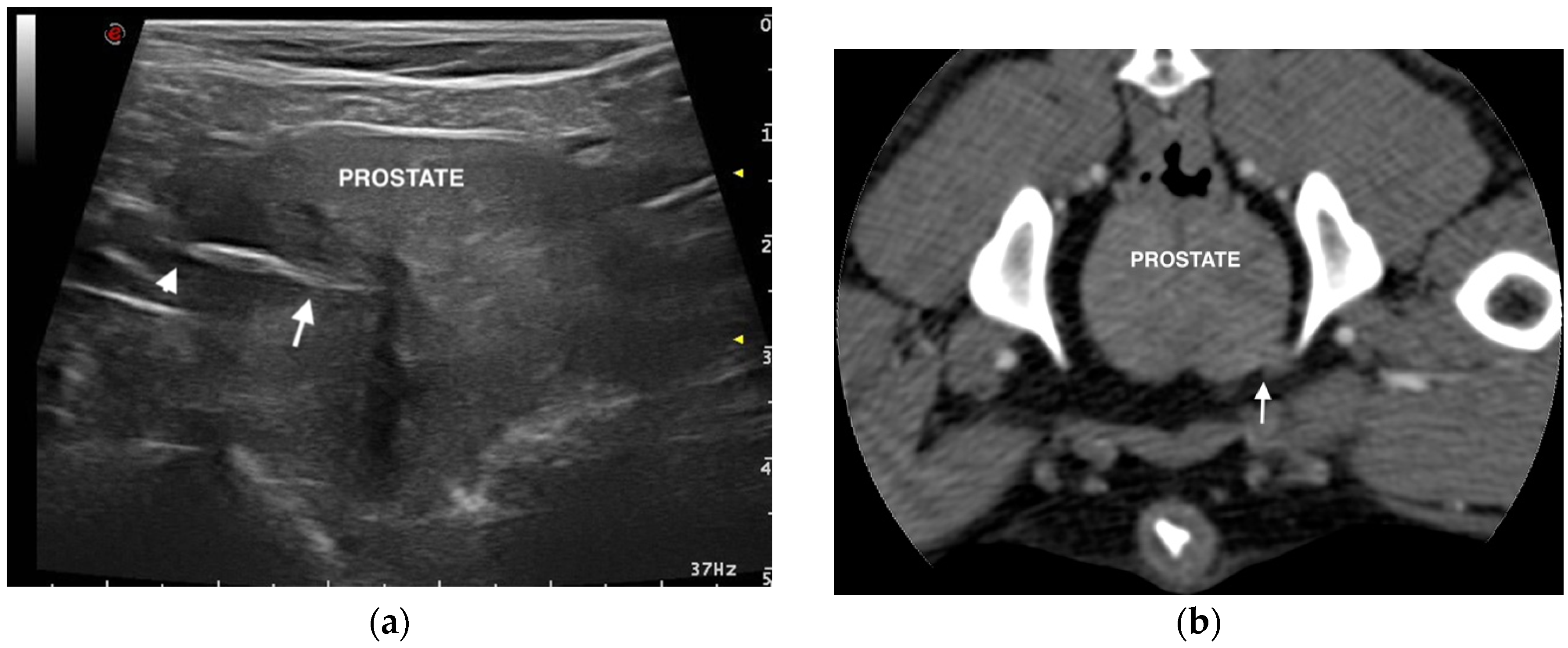
Abstract
1. Introduction
2. Case Presentation
3. Discussion
4. Conclusions
Author Contributions
Funding
Conflicts of Interest
References
- Johnston, D.E.; Summers, B.A. Osteomyelitis of the lumbar vertebrae in dogs caused by grass-seed foreign bodies. Aust. Vet. J. 1971, 47, 289–294. [Google Scholar] [CrossRef] [PubMed]
- Brennan, E.K.; Ihrke, P.J. Grass awn migration in dogs and cats: A retrospective study of 182 cases. J. Am. Vet. Med. Assoc. 1983, 182, 1201–1204. [Google Scholar] [PubMed]
- Frendin, J.; Funkquist, B.; Hansson, K.; Lönnemark, M.; Carlsten, J. Diagnostic imaging of foreign body reactions in dogs with diffuse back pain. J. Small Anim. Pract. 1999, 40, 278–285. [Google Scholar] [CrossRef] [PubMed]
- Schultz, R.M.; Zwingenberger, A. Radiographic, computed tomographic, and ultrasonographic findings with migrating intrathoracic grass awns in dogs and cats. Vet. Radiol. Ultrasound 2008, 49, 249–255. [Google Scholar] [CrossRef]
- Marchesi, M.C.; Caivano, D.; Conti, M.B.; Beccati, F.; Valli, L.; Busechian, S.; Rueca, F. A specific laryngeal finding in dogs with bronchial vegetal foreign bodies: A retrospective study of 63 cases. J. Vet. Med. Sci. 2019, 81, 213–216. [Google Scholar] [CrossRef]
- Hicks, A.; Golland, D.; Heller, J.; Malik, R.; Combs, M. Epidemiological investigation of grass seed foreign body-related disease in dogs of the Riverina District of rural Australia. Aust. Vet. J. 2016, 94, 67–75. [Google Scholar] [CrossRef]
- Cherbinsky, O.; Westropp, J.; Ting, S.; Jones, B.; Pollard, R. Ultrasonographic features of grass awns in the urinary bladder. Vet. Radiol. Ultrasound 2010, 51, 462–465. [Google Scholar] [CrossRef]
- Morshead, D. Submucosal urethral calculus secondary to foxtail awn migration in a dog. J. Am. Vet. Med. Assoc. 1983, 182, 1247–1248. [Google Scholar]
- Agut, A.; Carrillo, J.D.; Anson, A.; Belda, E.; Soler, M. Imaging diagnosis-urethrovaginal fistula caused by a migrating grass awn in the vagina. Vet. Radiol. Ultrasound 2016, 57, 30–33. [Google Scholar] [CrossRef]
- Vansteenkiste, D.P.; Lee, K.C.; Lamb, C.R. Computed tomographic findings in 44 dogs and 10 cats with grass seed foreign bodies. J. Small Anim. Pract. 2014, 55, 579–584. [Google Scholar] [CrossRef]
- Bouabdallah, R.; Moissonnier, P.; Delisle, F.; De Fornel, P.; Manassero, M.; Maaoui, M.; Fayolle, P.; Viateau, V. Use of preoperative computed tomography for surgical treatment of recurrent draining tracts. J. Small Anim. Pract. 2014, 55, 89–94. [Google Scholar] [CrossRef] [PubMed]
- Della Santa, D.; Rossi, F.; Carlucci, F.; Vignoli, M.; Kircher, P. Ultrasound-guided retrieval of plant awns. Vet. Radiol. Ultrasound 2008, 49, 484–486. [Google Scholar] [CrossRef] [PubMed]
- Staudte, K.L.; Hopper, B.J.; Gibson, N.R.; Read, R.A. Use of ultrasonography to facilitate surgical removal of non enteric foreign bodies in 17 dogs. J. Small Anim. Pract. 2004, 45, 395–400. [Google Scholar] [CrossRef] [PubMed]
- Gnudi, G.; Volta, A.; Bonazzi, M.; Gazzola, M.; Bertoni, G. Ultrasonographic features of grass awn migration in the dog. Vet. Radiol. Ultrasound 2005, 46, 423–426. [Google Scholar] [CrossRef]
- Whitty, C.; Milner, H.; Oram, B. Use of magnetic resonance imaging in the diagnosis of spinal empyema caused by a migrating grass awn in a dog. N. Z. Vet. J. 2013, 61, 115–118. [Google Scholar] [CrossRef]
- Marchegiani, A.; Fruganti, A.; Cerquetella, M.; Cassarani, M.P.; Laus, F.; Spaterna, A. Penetrating palpebral grass awn in a dog: Unusual case of a penetrating grass awn in an eyelid. J. Ultrasound 2017, 20, 81–84. [Google Scholar] [CrossRef]
- Caivano, D.; Bufalari, A.; Giorgi, M.E.; Conti, M.B.; Marchesi, M.C.; Angeli, G.; Porciello, F.; Birettoni, F. Imaging diagnosis—Transesophageal ultrasoundguided removal of a migrating grass awn foreign body in a dog. Vet. Radiol. Ultrasound 2014, 55, 561–564. [Google Scholar] [CrossRef]
- Caivano, D.; Birettoni, F.; Rishniw, M.; Bufalari, A.; De Monte, V.; Proni, A.; Giorgi, M.E.; Porciello, F. Ultrasonographic findings and outcomes of dogs with suspected migrating intrathoracic grass awns: 43 cases (2010–2013). J. Am. Vet. Med. Assoc. 2016, 248, 413–421. [Google Scholar] [CrossRef]
- Caivano, D.; Birettoni, F.; Marchesi, M.C.; Moretti, G.; Corda, A.; Petrescu, V.F.; Porciello, F.; Bufalari, A. Septic Pericarditis and Cardiac Tamponade Caused by Migrating Intrathoracic Grass Awn in an English Setter Dog. Isr. J. Vet. Med. 2019, 74, 96–101. [Google Scholar]
- Birettoni, F.; Caivano, D.; Rishniw, M.; Moretti, G.; Porciello, F.; Giorgi, M.E.; Crovace, A.; Bianchini, E.; Bufalari, A. Preoperative and intraoperative ultrasound aids removal of migrating plant material causing iliopsoas myositis via ventral midline laparotomy: A study of 22 dogs. Acta Vet. Scand. 2017, 59, 12. [Google Scholar] [CrossRef]
- Moretti, G.; Birettoni, F.; Caivano, D.; Nannarone, S.; Crovace, A.; Porciello, F.; Bufalari, A. Mini-invasive approach for removal of iliopsoas migrating grass awns with an atraumatic wound retractor. J. Small Anim. Pract. 2019, in press. [Google Scholar] [CrossRef] [PubMed]
- Bufalari, A.; Di Meo, A.; Nannarone, S.; Padua, S.; Adami, C. Fentanyl or sufentanil continuous infusion during isoflurane anaesthesia in dogs: Clinical experiences. Vet. Res. Commun. 2007, 31, 277–280. [Google Scholar] [CrossRef] [PubMed]
- Cerasoli, I.; Nannarone, S.; Schauvliege, S.; Duchateau, L.; Bufalari, A. The effects of intravenous lidocaine before propofol induction in premedicated dogs. J. Small Anim. Pract. 2016, 57, 435–440. [Google Scholar] [CrossRef] [PubMed]
- Gunzel-Apel, A.R.; Mohrke, C.; Poulsen Nautrup, C. Colour-coded pulsed Doppler sonography of the canine testis, epididymis and prostate gland: Physiological and pathologic findings. Reprod. Domest. Anim. 2001, 36, 236–240. [Google Scholar] [CrossRef]
- Cannizo, K.L.; McLoughlin, M.A.; Chew, D.J.; DiBartola, S.P. Uroendoscopy. Evaluation of the lower urinary tract. Vet. Clin. N. Am. Small Anim. Pract. 2001, 31, 789–807. [Google Scholar]
- Henry, C.J. Management of transitional cell carcinoma. Vet. Clin. N. Am. Small Anim. Pract. 2003, 33, 597–613. [Google Scholar] [CrossRef]
- Crow, S.E. Canine cancer genetics: Transitional cell carcinoma in Scottish terriers. Cancer Ther. 2008, 6, 177–180. [Google Scholar]
- Grzegory, M.; Kubiak, K.; Jankowski, M.; Spuzak, J.; Glińska-Suchocka, K.; Nicpoń, J.; Haloń, A. Endoscopic examination of the urethra and the urinary bladder in dogs—Indications, contraindications and performance technique. Pol. J. Vet. Sci. 2013, 16, 797–801. [Google Scholar] [CrossRef][Green Version]
- Nikula, K.L.; Benjamin, S.A.; Angleton, G.M.; Lee, A.C. Transitional cell carcinomas of the urinary tract in a colony of beagle dogs. Vet. Pathol. 1989, 26, 455–461. [Google Scholar] [CrossRef]


Publisher’s Note: MDPI stays neutral with regard to jurisdictional claims in published maps and institutional affiliations. |
© 2020 by the authors. Licensee MDPI, Basel, Switzerland. This article is an open access article distributed under the terms and conditions of the Creative Commons Attribution (CC BY) license (http://creativecommons.org/licenses/by/4.0/).
Share and Cite
Marchesi, M.C.; Moretti, G.; Angeli, G.; Birettoni, F.; Porciello, F.; Bufalari, A.; Caivano, D. Prostatic Localization of a Migrating Grass Awn Foreign Body in a Dog. Vet. Sci. 2020, 7, 192. https://doi.org/10.3390/vetsci7040192
Marchesi MC, Moretti G, Angeli G, Birettoni F, Porciello F, Bufalari A, Caivano D. Prostatic Localization of a Migrating Grass Awn Foreign Body in a Dog. Veterinary Sciences. 2020; 7(4):192. https://doi.org/10.3390/vetsci7040192
Chicago/Turabian StyleMarchesi, Maria Chiara, Giulia Moretti, Giovanni Angeli, Francesco Birettoni, Francesco Porciello, Antonello Bufalari, and Domenico Caivano. 2020. "Prostatic Localization of a Migrating Grass Awn Foreign Body in a Dog" Veterinary Sciences 7, no. 4: 192. https://doi.org/10.3390/vetsci7040192
APA StyleMarchesi, M. C., Moretti, G., Angeli, G., Birettoni, F., Porciello, F., Bufalari, A., & Caivano, D. (2020). Prostatic Localization of a Migrating Grass Awn Foreign Body in a Dog. Veterinary Sciences, 7(4), 192. https://doi.org/10.3390/vetsci7040192