
Antioxidant Activity, Formulation, Optimization and Characterization of an Oil-in-Water Nanoemulsion Loaded with Lingonberry (Vaccinium vitis-idaea L.) Leaves Polyphenol Extract


Abstract
:1. Introduction
2. Materials and Methods
2.1. Materials
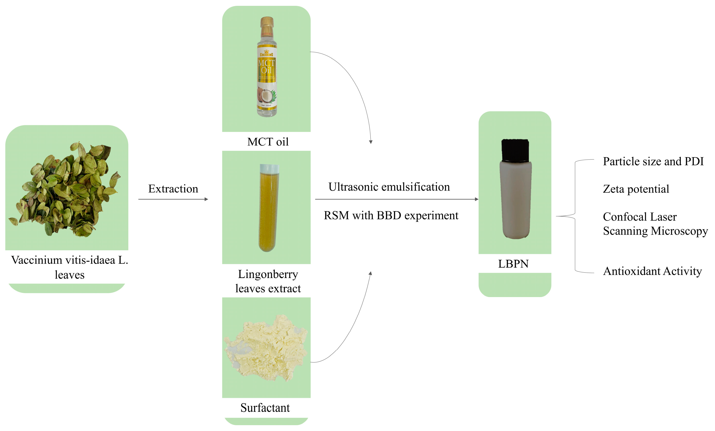
2.2. Preparation of Lingonberry Leaves Extract
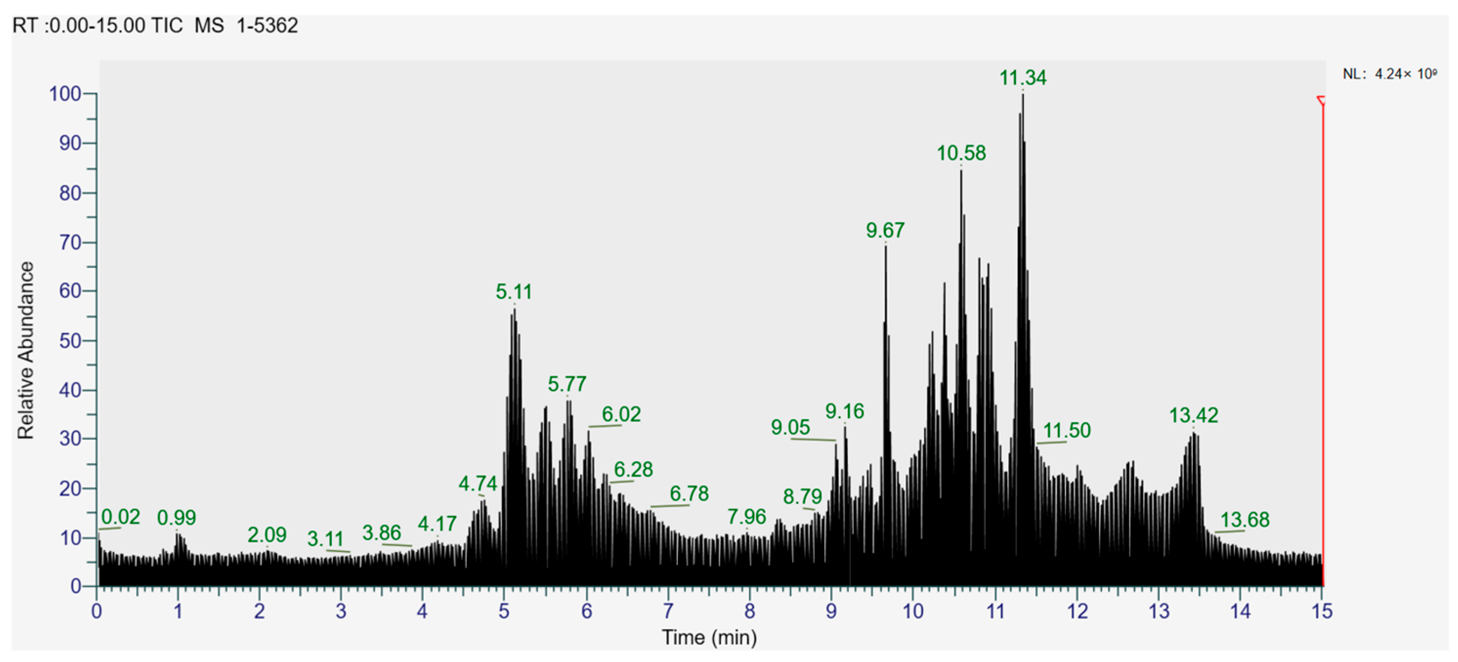
2.3. Characterization of LBPs by UPLC-TQ-MS
2.4. Preparation of the Nanoemulsion
2.5. Characterization of the W/O Nanoemulsion
2.5.1. Encapsulation Efficiency
2.5.2. Particle Size and PDI
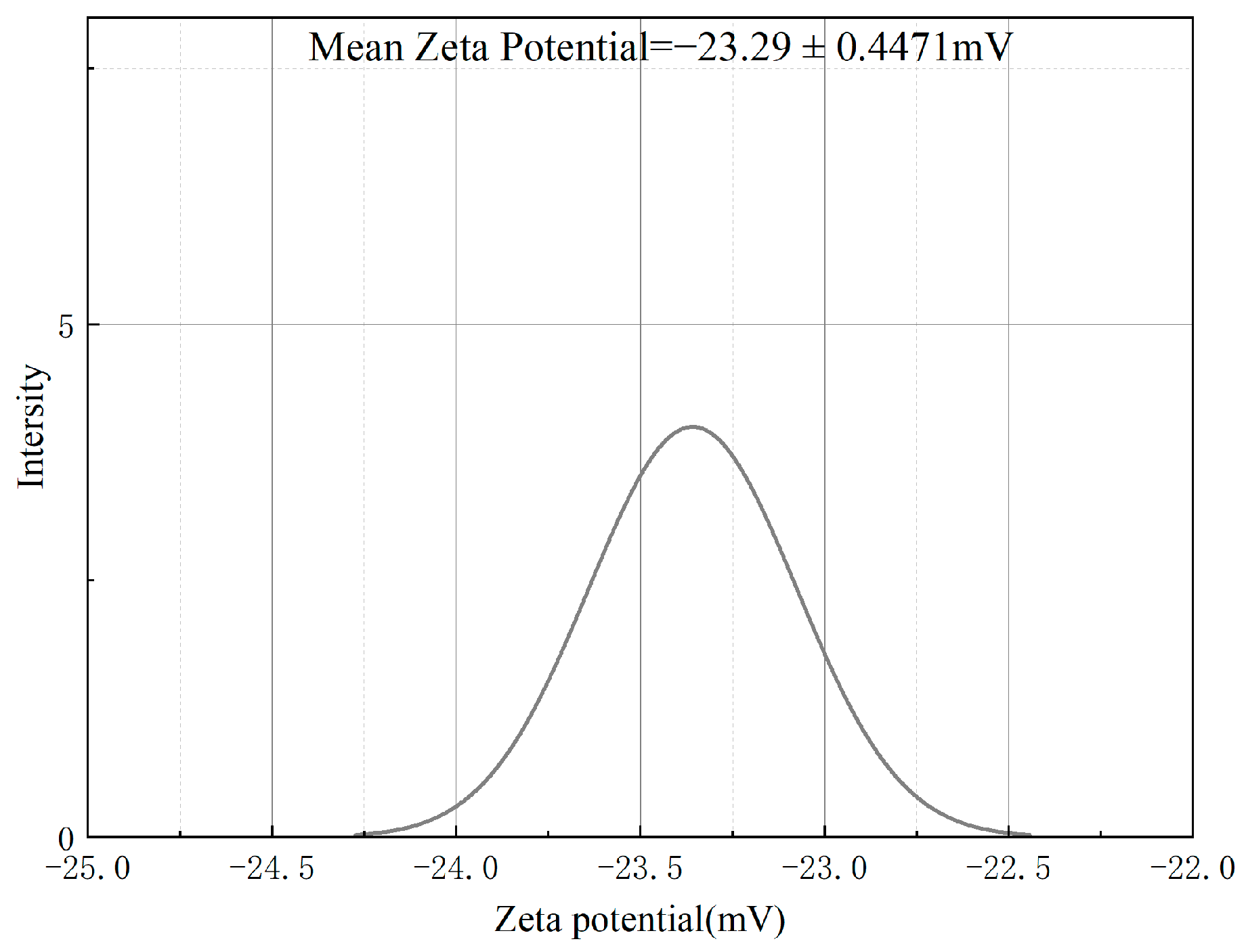
2.5.3. Zeta Potential
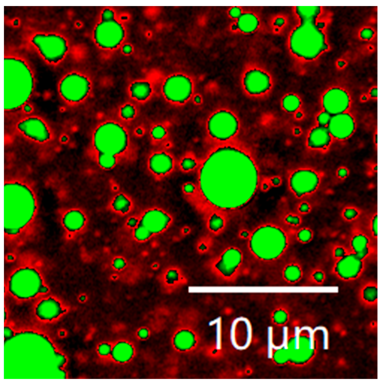
2.5.4. Confocal Laser Scanning Microscopy (CLSM)
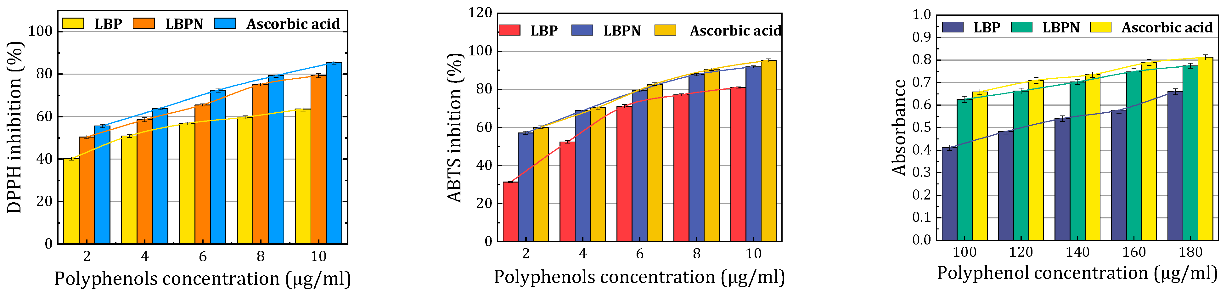
2.6. Antioxidant Activity of LBPs
2.6.1. DPPH Radical Scavenging Assay
2.6.2. ABTS•+ Radical Scavenging Assay
2.6.3. Reducing Power
2.7. Statistical Analysis
3. Results
3.1. UPLC-TQ-MS Characterization of Extract
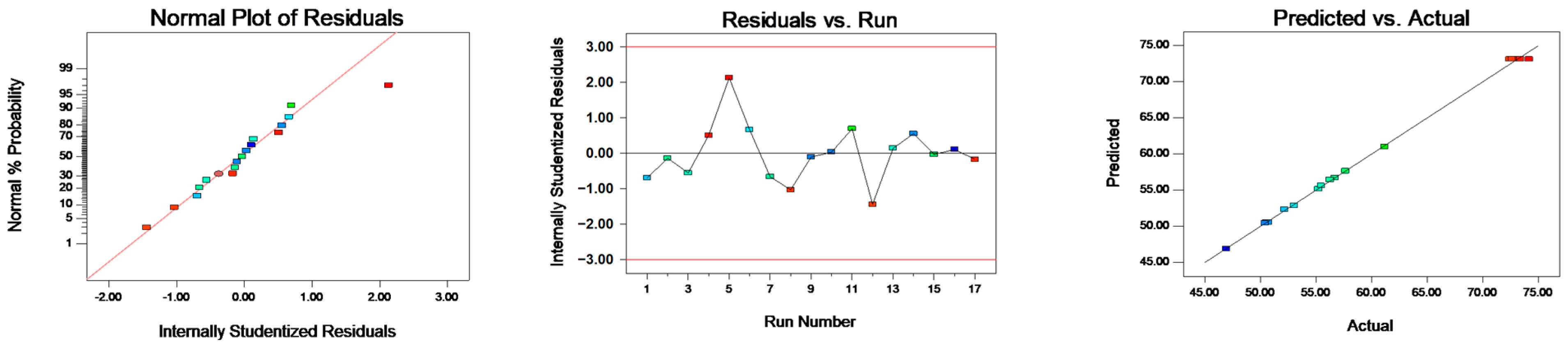
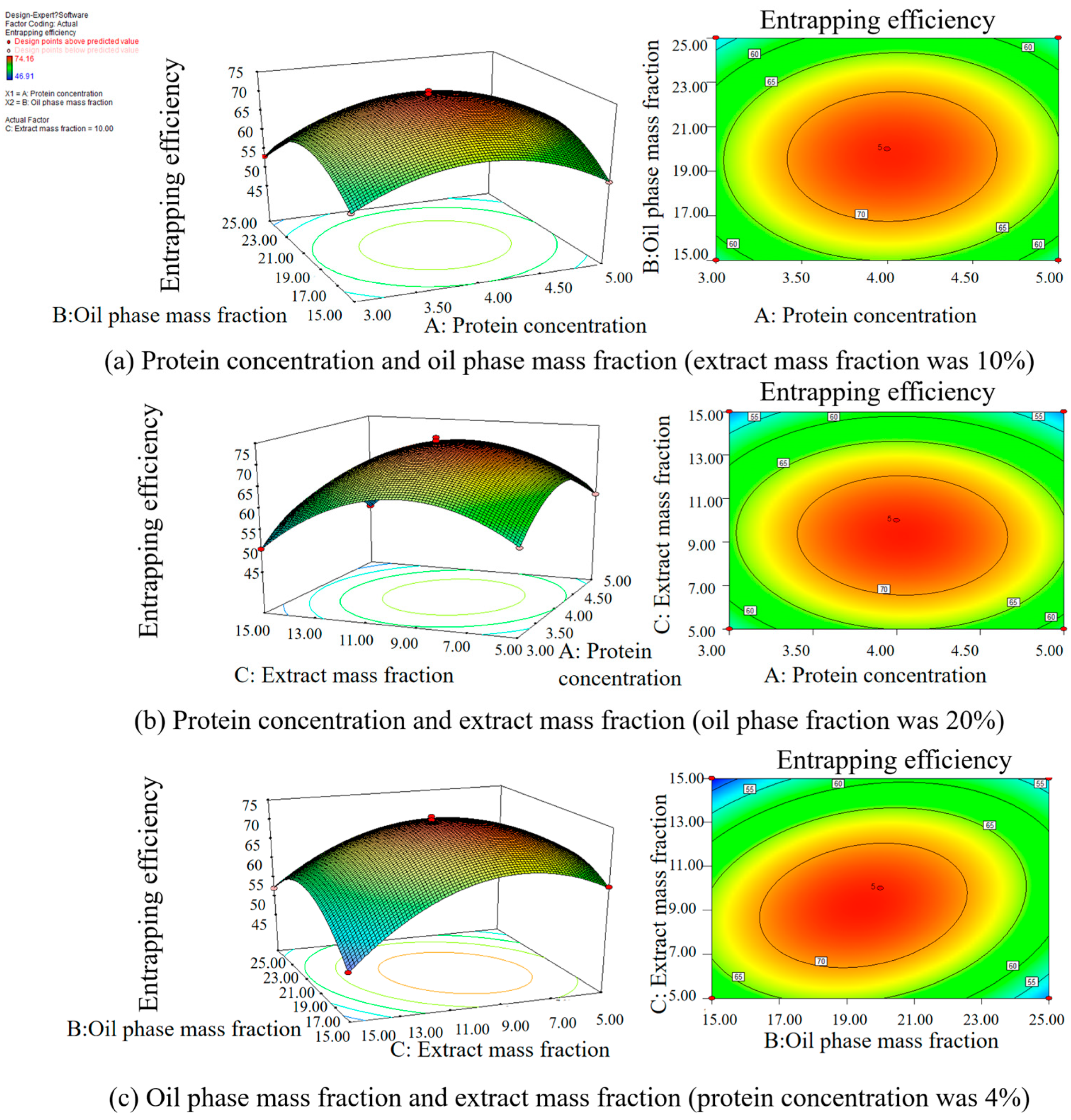
3.2. Optimization of the Lingonberry Nanoemulsion Using Response Surface Methodology
3.3. Characterization of the Optimal Lingonberry Nanoemulsion
3.3.1. Particle Size and PDI
3.3.2. Zeta Potential
3.3.3. Confocal Laser Scanning Microscopy (CLSM)
3.3.4. Antioxidant Activity
4. Conclusions and Future Research
Author Contributions
Funding
Data Availability Statement
Conflicts of Interest
References
- Plunkett, B.J.; Espley, R.V.; Dare, A.P.; Warren, B.W.; Grierson, E.R.P.; Cordiner, S.; Turner, J.L.; Allan, A.C.; Albert, N.W.; Davies, K.M.; et al. MYBA From Blueberry (Vaccinium Section Cyanococcus) Is a Subgroup 6 Type R2R3MYB Transcription Factor That Activates Anthocyanin Production. Front. Plant Sci. 2018, 9, 1300. [Google Scholar] [CrossRef] [PubMed]
- Bujor, O.C.; Ginies, C.; Popa, V.I.; Dufour, C. Phenolic compounds and antioxidant activity of lingonberry (Vaccinium vitis-idaea L.) leaf, stem and fruit at different harvest periods. Food Chem. 2018, 252, 356–365. [Google Scholar] [CrossRef] [PubMed]
- Bujor, O.C.; Tanase, C.; Popa, M.E. Phenolic Antioxidants in Aerial Parts of Wild Vaccinium Species: Towards Pharmaceutical and Biological Properties. Antioxidants 2019, 8, 649. [Google Scholar] [CrossRef] [PubMed]
- Vilkickyte, G.; Petrikaite, V.; Pukalskas, A.; Sipailiene, A.; Raudone, L. Exploring Vaccinium vitis-idaea L. as a potential source of therapeutic agents: Antimicrobial, antioxidant, and anti-inflammatory activities of extracts and fractions. J. Ethnopharmacol. 2022, 292, 115207. [Google Scholar] [CrossRef]
- Xu, J.; Yang, H.; Nie, C.D.; Wang, T.; Qin, X.Y.; Yang, J.; Chang, Y.H.; Nie, S.M.; Fu, Y.J. Comprehensive phytochemical analysis of lingonberry (Vaccinium vitis-idaea L.) from different regions of China and their potential antioxidant and antiproliferative activities. RSC Adv. 2023, 13, 29438–29449. [Google Scholar] [CrossRef]
- Vyas, P.; Curran, N.H.; Igamberdiev, A.U.; Debnath, S.C. Antioxidant properties of lingonberry (Vaccinium vitis idaea L.) leaves within a set of wild clones and cultivars. Can. J. Plant Sci. 2015, 95, 663–669. [Google Scholar] [CrossRef]
- Debnath, S.C.; Siow, Y.L.; Petkau, J.; An, D.; Bykova, N.V. Molecular markers and antioxidant activity in berry crops: Genetic diversity analysis. Can. J. Plant Sci. 2012, 92, 1121–1133. [Google Scholar] [CrossRef]
- Foley, S.L.; Debnath, S.C. Influence of in vitro and ex vitro propagation on anthocyanin content and anti-oxidant activity of lingonberries. J. Hortic. Sci. Biotechnol. 2007, 82, 114–118. [Google Scholar] [CrossRef]
- Stefanescu, B.E.; Szabo, K.; Mocan, A.; Crisan, G. Phenolic Compounds from Five Ericaceae Species Leaves and Their Related Bioavailability and Health Benefits. Molecules 2019, 24, 2046. [Google Scholar] [CrossRef]
- Li, J.; Hwang, I.C.; Chen, X.; Park, H.J. Effects of chitosan coating on curcumin loaded nano-emulsion: Study on stability and in vitro digestibility. Food Hydrocoll. 2016, 60, 138–147. [Google Scholar] [CrossRef]
- Kumar, R.; Kaur, K.; Uppal, S.; Mehta, S.K. Ultrasound processed nanoemulsion: A comparative approach between resveratrol and resveratrol cyclodextrin inclusion complex to study its binding interactions, antioxidant activity and UV light stability. Ultrason. Sonochem. 2017, 37, 478–489. [Google Scholar] [CrossRef] [PubMed]
- Zhou, H.L.; Zheng, B.J.; McClements, D.J. Encapsulation of lipophilic polyphenols in plant-based nanoemulsions: Impact of carrier oil on lipid digestion and curcumin, resveratrol and quercetin bioaccessibility. Food Funct. 2021, 12, 3420–3432. [Google Scholar] [CrossRef] [PubMed]
- Pool, H.; Mendoza, S.; Xiao, H.; McClements, D.J. Encapsulation and release of hydrophobic bioactive components in nanoemulsion-based delivery systems: Impact of physical form on quercetin bioaccessibility. Food Funct. 2013, 4, 162–174. [Google Scholar] [CrossRef]
- Huang, H.; Belwal, T.; Liu, S.B.; Duan, Z.H.; Luo, Z.S. Novel multi-phase nano-emulsion preparation for co-loading hydrophilic arbutin and hydrophobic coumaric acid using hydrocolloids. Food Hydrocoll. 2019, 93, 92–101. [Google Scholar] [CrossRef]
- Sharma, B.; Iqbal, B.; Kumar, S.; Ali, J.; Baboota, S. Resveratrol-loaded nanoemulsion gel system to ameliorate UV-induced oxidative skin damage: From in vitro to in vivo investigation of antioxidant activity enhancement. Arch. Dermatol. Res. 2019, 311, 773–793. [Google Scholar] [CrossRef]
- Ruengdech, A.; Siripatrawan, U. Application of catechin nanoencapsulation with enhanced antioxidant activity in high pressure processed catechin-fortified coconut milk. LWT-Food Sci. Technol. 2021, 140, 110594. [Google Scholar] [CrossRef]
- Pongsumpun, P.; Iwamoto, S.; Siripatrawan, U. Response surface methodology for optimization of cinnamon essential oil nanoemulsion with improved stability and antifungal activity. Ultrason. Sonochem. 2020, 60, 104604. [Google Scholar] [CrossRef]
- Niknam, S.M.; Kashaninejad, M.; Escudero, I.; Sanz, M.T.; Beltran, S.; Benito, J.M. Preparation of Water-in-Oil Nanoemulsions Loaded with Phenolic-Rich Olive Cake Extract Using Response Surface Methodology Approach. Foods 2022, 11, 279. [Google Scholar] [CrossRef]
- Fan, Z.; Chen, K.; Liu, Y.; Yang, L.; Fu, Y. Correlation between Antioxidant Activity in Vitro and Active Components of Different Solvent Extracts from Lingonberr. Food Sci. 2017, 38, 138–144. [Google Scholar]
- Tian, Y.; Puganen, A.; Alakomi, H.L.; Uusitupa, A.; Saarela, M.; Yang, B.R. Antioxidative and antibacterial activities of aqueous ethanol extracts of berries, leaves, and branches of berry plants. Food Res. Int. 2018, 106, 291–303. [Google Scholar] [CrossRef]
- Raviadaran, R.; Chandran, D.; Shin, L.H.; Manickam, S. Optimization of palm oil in water nano-emulsion with curcumin using microfluidizer and response surface methodology. LWT-Food Sci. Technol. 2018, 96, 58–65. [Google Scholar] [CrossRef]
- Yazdan-Bakhsh, M.; Nasr-Esfahani, M.; Esmaeilzadeh-Kenari, R.; Fazel-Najafabadi, M. Evaluation of antioxidant properties ofHeracleum Lasiopetalumextract in multilayer nanoemulsion with biopolymer coating to control oxidative stability of sunflower oil. J. Food Meas. Charact. 2021, 15, 1014–1023. [Google Scholar] [CrossRef]
- Wang, D.Q.; Qi, B.K.; Xu, Q.Q.; Zhang, S.; Xie, F.Y.; Li, Y. Effect of salt ions on an ultrasonically modified soybean lipophilic protein nanoemulsion. Int. J. Food Sci. Technol. 2021, 56, 6719–6731. [Google Scholar] [CrossRef]
- Nikkhah, E.; Khayami, M.; Heidari, R. In vitro antioxidant activity of berry (Morus alba var.nigra). Int. J. Plant Prod. 2009, 3, 15–18. [Google Scholar]
- Drozdz, P.; Seziene, V.; Pyrzynska, K. Phytochemical Properties and Antioxidant Activities of Extracts from Wild Blueberries and Lingonberries. Plant Foods Hum. Nutr. 2017, 72, 360–364. [Google Scholar] [CrossRef] [PubMed]
- Ștefanescu, B.-E.; Calinoiu, L.F.; Ranga, F.; Fetea, F.; Mocan, A.; Vodnar, D.C.; Crișan, G. Chemical Composition and Biological Activities of the Nord-West Romanian Wild Bilberry (Vaccinium myrtillus L.) and Lingonberry (Vaccinium vitis-idaea L.) Leaves. Antioxidants 2020, 9, 495. [Google Scholar] [CrossRef] [PubMed]
- Hajazimi, E.; Landberg, R.; Zamaratskaia, G. Simultaneous determination of flavonols and phenolic acids by HPLC-CoulArray in berries common in the Nordic diet. LWT-Food Sci. Technol. 2016, 74, 128–134. [Google Scholar] [CrossRef]
- Vilkickyte, G.; Motiekaityte, V.; Vainoriene, R.; Raudone, L. Promising cultivars and intraspecific taxa of lingonberries (Vaccinium vitis-idaea L.): Profiling of phenolics and triterpenoids. J. Food Compos. Anal. 2022, 114, 104796. [Google Scholar] [CrossRef]
- Zhang, D.; Adelina, N.M.; Fan, Z.L.; Liu, J.R. Phytochemical profile and biological activities from different parts of Vaccinium vitis-idaea. J. Berry Res. 2022, 12, 445–462. [Google Scholar] [CrossRef]
- Smiljkovic, M.; Stanisavljevic, D.; Stojkovic, D.; Petrovic, I.; Vicentic, J.M.; Popovic, J.; Grdadolnik, S.G.; Markovic, D.; Sankovic-Babic, S.; Glamoclija, J.; et al. Apigenin-7-o-glucoside versus apigenin: Insight into the modes of anticandidal and cytotoxic actions. Excli J. 2017, 16, 795–807. [Google Scholar] [CrossRef]
- Zielinska, A.; Bryk, D.; Paradowska, K.; Wawer, I. Aronia melanocarpa Leaves as a Source of Chlorogenic Acids, Anthocyanins, and Sorbitol, and Their Anti-Inflamtory Activity. Pol. J. Food Nutr. Sci. 2020, 70, 409–418. [Google Scholar] [CrossRef]
- Sato, Y.; Itagaki, S.; Kurokawa, T.; Ogura, J.; Kobayashi, M.; Hirano, T.; Sugawara, M.; Iseki, K. In vitro and in vivo antioxidant properties of chlorogenic acid and caffeic acid. Int. J. Pharm. 2011, 403, 136–138. [Google Scholar] [CrossRef]
- Kostka, T.; Ostberg-Potthoff, J.J.; Stärke, J.; Guigas, C.; Matsugo, S.; Stojanov, L.; Velickovska, S.K.; Winterhalter, P.; Esatbeyoglu, T.; Mireeski, V. Bioactive Phenolic Compounds from Lingonberry (Vaccinium vitis-idaea L.): Extraction, Chemical Characterization, Fractionation and Cellular Antioxidant Activity. Antioxidants 2022, 11, 467. [Google Scholar] [CrossRef] [PubMed]
- Stefanescu, B.E.; Nemes, S.A.; Teleky, B.E.; Calinoiu, L.F.; Mitrea, L.; Martau, G.A.; Szabo, K.; Mihai, M.; Vodnar, D.C.; Crisan, G. Microencapsulation and Bioaccessibility of Phenolic Compounds of Vaccinium Leaf Extracts. Antioxidants 2022, 11, 674. [Google Scholar] [CrossRef] [PubMed]
- Izquierdo, P.; Feng, J.; Esquena, J.; Tadros, T.F.; Dederen, J.C.; Garcia, M.J.; Azemar, N.; Solans, C. The influence of surfactant mixing ratio on nano-emulsion formation by the pit method. J. Colloid Interface Sci. 2005, 285, 388–394. [Google Scholar] [CrossRef]
- Chen, L.; Lin, X.J.; Xu, X.W.; Chen, Y.; Li, K.; Fan, X.Y.; Pang, J.; Teng, H. Self-nano-emulsifying formulation of Sonchus oleraceus Linn for improved stability: Implications for phenolics degradation under in vitro gastro-intestinal digestion Food grade drug delivery system for crude extract but not single compound. J. Funct. Foods 2019, 53, 28–35. [Google Scholar] [CrossRef]
- Ye, H.X.; Chen, T.S.; Huang, M.; Ren, G.R.; Lei, Q.F.; Fang, W.J.; Xie, H.J. Exploration of the Microstructure and Rheological Properties of Sodium Alginate-Pectin-Whey Protein Isolate Stabilized B-Carotene Emulsions: To Improve Stability and Achieve Gastrointestinal Sustained Release. Foods 2021, 10, 1991. [Google Scholar] [CrossRef] [PubMed]
- Feng, Y.Y.; Yuan, D.X.; Cao, C.A.; Kong, B.H.; Sun, F.D.; Xia, X.F.; Liu, Q. Changes of in vitro digestion rate and antioxidant activity of digestion products of ethanol-modified whey protein isolates. Food Hydrocoll. 2022, 131, 107756. [Google Scholar] [CrossRef]


| Independent Variable | Actual Levels at Coded Factor Levels | ||
|---|---|---|---|
| −1 | 0 | 1 | |
| Mass concentration of emulsifier | 3% | 4% | 5% |
| Oil phase mass fraction | 15% | 20% | 25% |
| Extract mass fraction | 5% | 10% | 15% |
| Run | Protein Concentration (X1,%) | Oil Phase Mass Fraction (X2,%) | Extract Mass Fraction (X3,%) |
|---|---|---|---|
| 1 | 4.00 (0) | 25.00 (1) | 15.00 (1) |
| 2 | 3.00 (−1) | 15.00 (−1) | 10.00 (0) |
| 3 | 3.00 (−1) | 20.00 (0) | 5.00 (−1) |
| 4 | 4.00 (0) | 20.00 (0) | 10.00 (0) |
| 5 | 4.00 (0) | 20.00 (0) | 10.00 (0) |
| 6 | 3.00 (−1) | 25.00 (1) | 10.00 (0) |
| 7 | 5.00 (1) | 15.00 (−1) | 10.00 (0) |
| 8 | 4.00 (0) | 20.00 (0) | 10.00 (0) |
| 9 | 4.00 (0) | 25.00 (1) | 5.00 (−1) |
| 10 | 3.00 (−1) | 20.00 (0) | 15.00 (1) |
| 11 | 4.00 (0) | 15.00 (−1) | 5.00 (−1) |
| 12 | 4.00 (0) | 20.00 (0) | 10.00 (0) |
| 13 | 5.00 (1) | 25.00 (1) | 10.00 (0) |
| 14 | 5.00 (1) | 20.00 (0) | 15.00 (1) |
| 15 | 5.00 (1) | 20.00 (0) | 5.00 (−1) |
| 16 | 4.00 (0) | 15.00 (−1) | 15.00 (1) |
| 17 | 4.00 (0) | 20.00 (0) | 10.00 (0) |
| Chemical Name | Area | RT/min | Molecular Weight | Molecular Formula | Molecular Structure Formula |
|---|---|---|---|---|---|
| Apigenin 7-rhamnosyl-(1->2)-galacturonide | 1.73 × 10⁹ | 12.033 | 592.14407 | C27H28O15 |  |
| Bis(4-ethylbenzylidene)sorbitol | 1.26 × 10⁹ | 9.157 | 414.19921 | C24H30O6 | 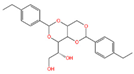 |
| Pinocembrin 7-O-neohesperidoside 3-O-acetate | 6.48 × 10⁸ | 12.596 | 606.19569 | C29H34O14 | 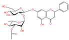 |
| Caffeic acid | 4.49 × 10⁸ | 9.662 | 162.02992 | C9H8O4 |  |
| Licochalcone A | 2.27 × 10⁸ | 6.201 | 360.13567 | C21H22O4 | 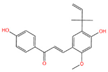 |
| Munduserone | 1.76 × 10⁸ | 0.987 | 342.11153 | C19H18O6 | 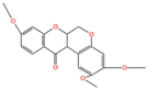 |
| Kaempferol-3-O-α-L-arabidopyranoside | 1.29 × 10⁸ | 7.316 | 418.0876 | C20H18O10 |  |
| Quercetin-3β-D-glucoside | 1.05 × 10⁸ | 7.135 | 464.08967 | C21H20O12 |  |
| Oleanolic acid | 9.03 × 10⁷ | 10.561 | 456.35435 | C30H48O3 | 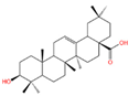 |
| Cycloartomunoxanthone | 5.21 × 10⁷ | 5.695 | 448.15239 | C26H24O7 |  |
| Quercetol B | 1.60 × 10⁷ | 10.416 | 368.19811 | C23H28O4 | 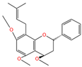 |
| Cycloartocarpin | 1.58 × 10⁷ | 6.83 | 434.17079 | C26H26O6 | 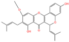 |
| Silybin | 1.39 × 10⁷ | 10.676 | 482.12114 | C25H22O10 | 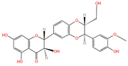 |
| 5-Hydroxy-6,6-dimethylpyrano flavone | 7.95 × 10⁶ | 0.994 | 320.10509 | C20H16O4 | 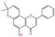 |
| Source | Sum of Squares | df | Mean Square | F-Value | p-Value |
|---|---|---|---|---|---|
| Model | 1482.84 | 9 | 164.76 | 555.31 | <0.0001 |
| A | 2.11 | 1 | 2.11 | 7.12 | 0.0321 |
| B | 13.13 | 1 | 13.13 | 44.26 | 0.0003 |
| C | 74.42 | 1 | 74.42 | 250.83 | <0.0001 |
| AB | 1.7 | 1 | 1.70 | 5.74 | 0.0478 |
| AC | 1.04 | 1 | 1.04 | 3.51 | 0.1033 |
| BC | 64.16 | 1 | 64.16 | 216.25 | <0.0001 |
| A2 | 300.80 | 1 | 300.8 | 1013.82 | <0.0001 |
| B2 | 369.85 | 1 | 369.85 | 1246.54 | <0.0001 |
| C2 | 518.29 | 1 | 518.29 | 1746.84 | <0.0001 |
| Residual | 2.08 | 7 | 0.30 | ||
| Lack of Fit | 0.19 | 3 | 0.062 | 0.13 | 0.9364 |
| Pure Error | 1.89 | 4 | 0.47 | ||
| Cor Total | 1484.92 | 16 |
Disclaimer/Publisher’s Note: The statements, opinions and data contained in all publications are solely those of the individual author(s) and contributor(s) and not of MDPI and/or the editor(s). MDPI and/or the editor(s) disclaim responsibility for any injury to people or property resulting from any ideas, methods, instructions or products referred to in the content. |
© 2023 by the authors. Licensee MDPI, Basel, Switzerland. This article is an open access article distributed under the terms and conditions of the Creative Commons Attribution (CC BY) license (https://creativecommons.org/licenses/by/4.0/).
Share and Cite
Wang, S.; Cheng, Y.; Wang, J.; Ding, M.; Fan, Z. Antioxidant Activity, Formulation, Optimization and Characterization of an Oil-in-Water Nanoemulsion Loaded with Lingonberry (Vaccinium vitis-idaea L.) Leaves Polyphenol Extract. Foods 2023, 12, 4256. https://doi.org/10.3390/foods12234256
Wang S, Cheng Y, Wang J, Ding M, Fan Z. Antioxidant Activity, Formulation, Optimization and Characterization of an Oil-in-Water Nanoemulsion Loaded with Lingonberry (Vaccinium vitis-idaea L.) Leaves Polyphenol Extract. Foods. 2023; 12(23):4256. https://doi.org/10.3390/foods12234256
Chicago/Turabian StyleWang, Siyu, Yuan Cheng, Jingyi Wang, Miao Ding, and Ziluan Fan. 2023. "Antioxidant Activity, Formulation, Optimization and Characterization of an Oil-in-Water Nanoemulsion Loaded with Lingonberry (Vaccinium vitis-idaea L.) Leaves Polyphenol Extract" Foods 12, no. 23: 4256. https://doi.org/10.3390/foods12234256
APA StyleWang, S., Cheng, Y., Wang, J., Ding, M., & Fan, Z. (2023). Antioxidant Activity, Formulation, Optimization and Characterization of an Oil-in-Water Nanoemulsion Loaded with Lingonberry (Vaccinium vitis-idaea L.) Leaves Polyphenol Extract. Foods, 12(23), 4256. https://doi.org/10.3390/foods12234256