Error in Figure
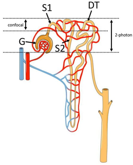
In the original publication [1], there was a mistake in Figure 2 as published. The figure reported a representative image of the renal cortex acquired in vivo by two-photon microscopy for other purposes than this review. Consequently, the image was removed and the figure legend was modified. The corrected Figure 2 and its legend appear below.

Figure 2.
Schematic representation of the renal structures identified with 2PM. The nephron segments that are visible with confocal and 2PM are shown. As indicated by the vertical arrows, 2PM allows the renal structures to be imaged about 3 times deeper compared to confocal microscopy. G = glomerulus; S1 = S1 proximal convoluted tubule; S2 = S2 proximal convoluted tubule; DT = distal tubule. This figure was drawn adapting the vector image from the Servier Medical Art bank (http://smart.servier.com/; last accessed on 22 February 2022).
In the original publication [1], there was a mistake in Figure 3 as published. The figure reported representative images of a linescan-based experiment acquired for other purposes than this review. Consequently, Figure 3, its legend, and its mention into the text were removed.
The authors apologize for any inconvenience caused and state that the scientific conclusions are unaffected. This correction was approved by the Academic Editor. The original publication has also been updated.
Reference
- Costanzo, V.; Costanzo, M. Intravital Imaging with Two-Photon Microscopy: A Look into the Kidney. Photonics 2022, 9, 294. [Google Scholar] [CrossRef]
Publisher’s Note: MDPI stays neutral with regard to jurisdictional claims in published maps and institutional affiliations. |
© 2022 by the authors. Licensee MDPI, Basel, Switzerland. This article is an open access article distributed under the terms and conditions of the Creative Commons Attribution (CC BY) license (https://creativecommons.org/licenses/by/4.0/).