Abstract
The fabrication of stable, tailored domain patterns in ferroelectric crystals has wide applications in optical and electronic industries. All-optical ferroelectric poling by pulse laser irradiation has been developed recently. In this work, we studied the creation of the domain structures in MgO-doped lithium tantalate by focused irradiation with a femtosecond near-infrared laser. Cherenkov-type second harmonic generation microscopy was used for domain imaging of the bulk. We have revealed the creation of enveloped domains around the induced microtracks under the action of the depolarization field. The domain growth is due to a pyroelectric field caused by a nonuniform temperature change. The domains in the bulk were revealed to have a three-ray star-shaped cross-section. It was shown that an increase in the field excess above the threshold leads to consequential changes in domain shape from a three-ray star to a triangular and a circular shape. The appearance of comb-like domains as a result of linear scanning was demonstrated. All effects were considered in terms of a kinetic approach, taking into account the domain wall motion by step generation and kink motion driven by excess of the local field over the threshold. The obtained knowledge is useful for the all-optical methods of domain engineering in ferroelectrics.
1. Introduction
Nonlinear photonic crystals (NPCs) represent media with spatially modulated, nonlinear susceptibility coefficients, χ(2) [1,2]. Such structures allow for frequency conversions of laser beams to be realized by means of quasi-phase matching, which is used for second-harmonic generation (SHG) [1,3] and optical parametric oscillators (OPOs) [4,5].
Owing to the reversable spontaneous polarization in ferroelectrics, it is possible to create periodical domain structures by modulation of the χ(2) sign [6]. The most popular method for creating domain patterns is electrical field poling (EFP), which has been realized in field applications using electrodes with the required geometry [3,7]. The most significant drawback of EFP is its inability to create 3D domain structures [8,9,10].
The recently developed all-optical approach of ferroelectric crystal poling by pulse laser irradiation is a promising alternative to EFP [11,12]. Usually, UV and far-IR (FIR) irradiation are strongly absorbed by the surface layer due to the strong absorption coefficient of oxide ferroelectrics [11,12]. In the case of FIR irradiation [12,13], the absorbed energy stimulates nonuniform heating and subsequent cooling, thus stimulating the appearance of a pyroelectric field which is strong enough for local polarization reversal and can be used for the formation of tailored domain structures. The method allows for the production of self-organized nanodomain structures without the ability to control the positions of the individual domains [12,13].
The spatial distribution of the pyroelectric field induced by FIR irradiation leads only to domain nucleation at the surface and their growth in a polar direction, as well as EFP, and does not allow for the creation of 3D domain structures. This drawback has been overcome recently with the use of the tightly focused irradiation of a femtosecond laser with a wavelength in the near-IR (NIR) range [14]. The main idea behind femtosecond laser application is based on the high enough multiphoton absorption of the light from the transparent spectral range due to the extremely high light intensity in the focusing region. In the beginning, this effect was successfully used for the local modification of the crystal structure in the bulk of various transparent materials [15]. In 2015, for the first time, domain switching by NIR femtosecond laser irradiation was demonstrated in ferroelectric lithium niobate (LiNbO3, LN) crystals [14]. Further works widely expanded the number of ferroelectrics used for 2D and 3D domain patterning by femtosecond laser irradiation.
A few approaches have been used for local light-only polarization reversal by NIR femtosecond laser irradiation. Initially, a focused movement along the polar axis was used for the creation of elongated domains in LN with lengths of up to 60 µm [14,16]. This method has allowed for the creation of a periodical domain structure inside a waveguide, thus realizing the SHG conversion.
The two-step approach was based on the use of additional crystal heating in MgO-doped LN [17,18]. In the first step, modified crystal channels (microtracks) with lengths of up to 200 µm were created by the movement of the laser focus in a polar direction. The subsequent crystal heating and cooling led to the formation of the through domains due to the growth of the microtracks under the action of the uniform pyroelectric field [17]. This approach can be used for the creation of 2D patterns only [18]. A similar technique has been realized in [19]. It was demonstrated that the formation of the through domains can be achieved without the movement of the laser focus.
A similar method was presented in [20], which implied the creation of microtracks (so-called markers) near the crystal surface, with the subsequent material irradiation being focused away from the microtracks. The second step led to domain growth from the irradiated area or to domain erasing, depending on the focusing depth [21].
The use of microtracks as seeds for the subsequent domain growth was presented in [22]. Moreover, in this work, scanning with a focused laser beam was used to erase the line domains that had already been obtained. The combination of domain creation and erasure led to the formation of a 3D domain structure.
In [23], the investigation of structural modifications in the bulk of LN induced by NIR femtosecond laser pulses in the nonlinear focusing regime has shown that beam propagation along the optical axis results in the production of double-track structures. It was demonstrated that the spatial positions of the laser-induced micro-tracks in crystal bulk can be directly predicted by analytical expressions under a paraxial approximation.
NPCs can be produced using one of the mentioned approaches and can be further used as elements in integral optical devices [24,25,26,27,28,29,30,31,32].
Despite the number of studies which include modeling, the physical mechanism of ferroelectric domain formation under the action of femtosecond laser irradiation is still under discussion [8]. Currently, thermoelectric and pyroelectric effects are considered as the mechanisms of electric field creation without an exact description and assessment of the quantities [14,17,18,22]. At the same time, it is clear that without a precise understanding of the switching mechanism, it is impossible to create effective optical devices.
The lithium tantalate (LiTaO3, LT) crystal, isomorphic to LN, and its Mg-doped composition (MgOCLT) open up wide perspectives for NPC production [3,33]. The equilibrium domain shape in LN is hexagonal, whereas in CLT it is triangular [34]. The nonlinear optical coefficient for LT is lower than that for LN, while record values of the frequency transformation efficiency have been obtained for periodically poled SLT crystals [35]. It was stated that MgO-doped SLT is an ideal material for high-power laser applications [36], which are based on a significant increase in the optical damage threshold [37,38,39].
The higher value of the effective pyro-coefficient in LT allows for the realization of domain creation under the action of a pyroelectric field at lower energies of laser irradiation [40,41] and increases the interest in this material for light-only domain engineering. The formation of stable domain structures with charged walls in bulk has been realized in LT crystals with a composition gradient produced by the partial vapor transport equilibration process [42]. The change in the domain wall shape under the action of the pyroelectric field was revealed.
Nevertheless, to the best of our knowledge, LT crystals have not been used for femtosecond laser domain structuring yet.
In this work, we studied the formation of the domain structures in MgO-doped LT after the irradiation of crystal plates by tightly focused femtosecond laser irradiation both locally and using linear scanning. The dependence of the domain shape on the irradiation parameters was revealed. We suppose that the domain formation near the focus point is caused by the depolarization field appearing around the microtracks and subsequent domain growth is caused by the pyroelectric field occurring during nonuniform temperature changes.
2. Materials and Methods
We have studied the light-only ferroelectric domain switching by tightly focused femtosecond laser irradiation in 8% Mg-doped congruent lithium tantalate crystal (Yamaju Ceramics, Owariasahi, Japan). The investigated samples represent 1 mm thick monodomain plates cut perpendicular to the polar axis with surface roughness below 1 nm. The local irradiation was carried out by a femtosecond regenerative amplifier (TETA-10, Avesta Project, Moscow, Russia) based on a Yb-solid state laser with the following main parameters: wavelength 1030 nm, frequency 100 kHz, pulse duration 250 fs, and energy up to 100 µJ. The samples’ positioning was realized via a 3D-motorized table representing the horizontal stage XY5050, combined with the vertical translation platform (KA050-Z, Zolix Instruments, Beijing, China). Laser irradiation, which passed through the Z− polar surface, was focused at depths of 300 and 500 µm by the objective (LMH-50X-1064, Thorlabs, Newton, NJ, USA), with 50× magnification and a numerical aperture of 0.65 (Figure 1a).
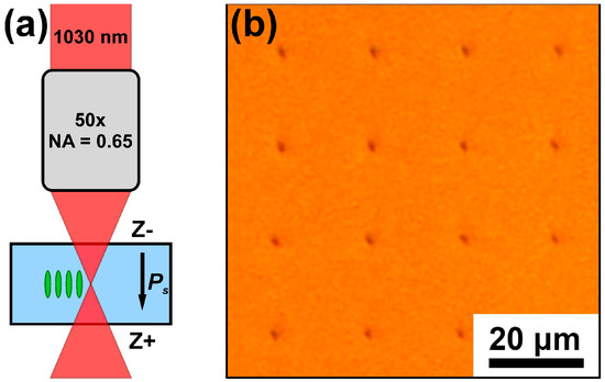
Figure 1.
(a) Scheme of the experimental setup, (b) optical image of the microtracks that appeared after the local irradiation by femtosecond laser pulses with an energy of 6.0 µJ.
Two series of experiments with local irradiation by square matrices of points were realized. The time interval between irradiations was 1 s.
In the beginning, we irradiated the samples using 10 × 10 square matrices with a 20-µm period at a 300 µm focusing depth. The number of pulses varied from 1 to 512 within each row of the matrix. The columns of the matrix correspond to various energies ranging from 1.4 to 6.7 µJ.
To study the shape of isolated domains in detail, we increased the focusing depth to 500 µm to exclude the domain from reaching the polar surface and increased the matrix period to 50 µm to avoid the domain interaction. A fixed number of 512 pulses and energy in the range from 6.1 to 8.1 µJ were used.
We also conducted an experiment with linear scanning of the MgOCLT plate with a focused laser beam at a depth of 470 below the Z− polar surface. The scanning speed was 2 mm/s, the pulse frequency was 100 kHz, and the pulse energy ranged from 0.7 to 6.7 μJ. The focus spot moved along the non-polar Y-crystal axis.
The microtracks created by the irradiation were imaged across the polar surface by an optical microscope (Olympus BX-61, Olympus, Japan). The walls of the domains created were imaged in the bulk by Cherenkov-type second harmonic generation microscopy (SHGM) [43,44] and were realized on the basis of the NTEGRA Spectra (NT-MDT, Russia). The number of 2D domain images obtained by scanning along the XY plane at a fixed depth allowed for the creation of a 3D domain image with a spatial resolution of about one micron.
3. Results
3.1. Local Irradiation
It was shown that the local irradiations across the whole ranges of energies and pulse numbers used led to the formation of optically imaged microtracks with cross-section diameters of about 1–2 μm (Figure 1b). Moreover, the SHGM imaging allowed for the domains to be reveled, which appeared around all of the microtracks, socalled “enveloped domains” (Figure 2). The length and width of the enveloped spindle-like domains, appearing after one or two pulses, essentially increased with the pulse number and energy (Figure 2).
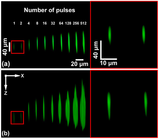
Figure 2.
SHGM side view of the domains formed by various numbers of pulses: (a) 1.4 µJ, (b) 4.0 µJ. Focusing depth was 300 µm.
The domain shape and length depend on the pulse number. The domain elongated towards both the Z− and Z+ polar surfaces in contrast to the Mg-doped lithium niobate which showed domain growth towards the Z− surface only [45]. The spindle-like shape obtained with low pulse numbers with a circular cross-section transforms to a so-called “three-wing” shape with a three-ray cross-section (Figure 3 and Figure 4). The pulse number corresponding to the qualitative shape transformation decreases with the pulse energy (Figure 3a).

Figure 3.
SHGM images of the domain XY cross-sections. (a) Domain XY cross-sections for various pulse numbers and energies. Focusing depth was 300 μm. (b) Domain XY cross-sections for different distances from the focusing point. Focusing depth was 500 µm, energy was 6.1 µJ, and pulse number was 512.
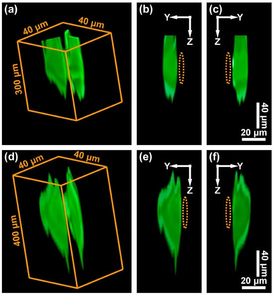
Figure 4.
SHGM images of the domains created at the various focusing depths: (a–c) 300 µm, (d,e) 500 µm. Pulse energy: (a–c) 5.4 µJ, (d,e) 6.1 µJ. Pulse number: 512. (a,d) Three-dimensional domain images, (b,c) and (e,f) two-dimensional images of the individual wings. The orange lines show the microtracks’ position.
Every domain wing was oriented strictly along the Y+ crystallographic direction (Figure 3b). The top and bottom parts of the wings have several teeth growing along the polar direction (Figure 4). For the 300 μm focusing depth, the teeth approached the Z− polar surface (Figure 4a–c), whereas for the 500 μm focusing depth, the domains were located completely within the bulk with irregular teeth in their top and bottom parts (Figure 4d–f).
The size of the wings in the polar direction (length) and along the Y+ direction (width) was measured through an analysis of the SHGM images (Figure 5). It was shown that the wing length demonstrated linear dependence on the pulse energy (Figure 5a) and logarithmic dependence on the pulse number (Figure 5b). The wing width also depended linearly on the pulse energy (Figure 5c) and logarithmically (Figure 5d) on the pulse number.
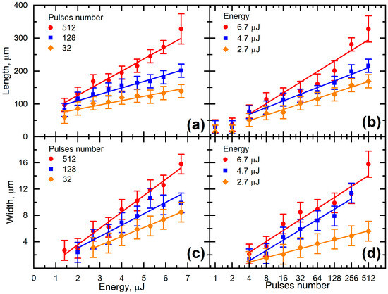
Figure 5.
Dependencies of the wing length along the polar direction (a) on the energy and (b) the pulse number and the wing width along the Y+ direction (c) on the energy and (d) the pulse number.
Such dependencies look like the ones obtained previously for the polarization switching on non-polar cuts of ferroelectric crystals. Through switching using a conductive probe in LN, it was shown that the domain base’s width and its length demonstrated root and linear dependencies on the applied voltage amplitude, respectively. The dependencies of the length and width on the duration of the applied pulses had a logarithmic character [46].
The length dependence on the pulse energy (W) was fitted by the linear function:
where Wth is the threshold energy of domain formation, and k is a parameter.
In [47], a similar dependence of the laser filament length in the focus region on energy was revealed. The filament acts as a heat source, which causes the formation of a pyroelectric field, leading, therefore, to the formation of the domain.
The linear field dependence of the domain diameter at the polar surface and logarithmic size dependence on the duration of the field pulse were obtained during local switching in various ferroelectrics [45,46,48,49,50,51]. Here, we revealed the logarithmic dependence of the domains on pulse number:
where N is the pulse number, w0 is the minimal size, N0 is the number of pulses corresponding to the appearance of the wings, and b is a parameter.
The appearance of a three-wing domain shape can be attributed to the following conditions: (1) domain growth anisotropy; (2) low field excess above the threshold value; (3) ineffective screening of the depolarization field. Such a shape was already observed in CLN and MgOCLN during the polarization reversal with screening retardation [52].
3.2. Linear Scanning
The linear scanning along the X direction by laser irradiation focused in crystal bulk, with a focusing depth of 450 μm led to the formation of the optically imaged microtracks, which look like a stripe with a width of about 1 μm (Figure 6a). The 2D SHGM imaging at the depth of 400 μm allowed a stripe domain cross-section with a thickness of about 1 μm, which was elongated in the scanning direction, to be revealed (Figure 6b). At a depth of 370 μm, the domain cross-section represented the arrays of isolated circles with a period of about 1.5 μm (Figure 6c). The SHGM imaging of the XZ cross-section showed that these domains had a double comb-like shape, localized above and below the focusing depth (Figure 6d). The width in the polar direction was about 50 μm for a pulse energy of 2.7 µJ and increased with the pulse energy. A series of measurements at different energies allowed for the discovery that scanning in the energy range from 2 to 5 µJ led to the appearance of comb-like domains only, whereas additional isolated domains appeared at higher energies.

Figure 6.
Results of the linear scanning along the X-axis. (a) Optical image of microtracks. (b–d) SHGM images of various cross-sections of comb-like domains: (b,c) XY at different depths: (b) at 400 µm, (c) at 320 µm. (d) XZ along scanning direction. Pulse energy was 2.7 µJ, focusing depth was 450 µm, and scanning rate was 2 mm/s.
The appearance of the domains growing from the surface with a comb-like shape, a stripe cross-section by the surface, and jagged charged domain walls (CDWs) were revealed by us in CLN as a result of irradiation by an FIR laser [12], in Mg-doped LN for tilt control of the CDW [53], and in thin LN films on isolator (LNOI) during local switching [54]. The formation of such comb-like domains was attributed to the creation of the quasi-regular teeth at the CDW during domain elongation.
4. Discussion
The formation of modified nonpolar volumes (microtracks) in the laser filament region as a result of crystal amorphization [55] entails the appearance of a nonpolar inclusion with a bound charge at the boundary between polar and non-polar states. The bound charge is the source of a depolarization field Edep. As a result, a local switching in the polarization is obtained in the polar region of the crystal, where the polar component of Edep.z is above the threshold value for step generation. This effect leads to the formation of an enveloped domain around microtracks. The subsequent cooling after termination of the laser pulse leads to the appearance of the pyroelectric field [12], stimulating the growth of the enveloped domains.
The formation of various domain shapes in the crystal bulk can be considered using a kinetic approach based on an analogy between the growth of ferroelectric domains and crystals [52]. The domain evolution is a result of three main processes controlled by the nucleation of different dimensionalities: (1) appearance of new domains (3D nucleation), (2) step generation at the domain walls (2D nucleation), and (3) kink motion along the domain wall (1D nucleation).
It implies that the rates of domain appearance and step generation, the probability of formation, and the growth of domains, as well as the kink motion velocity, are proportional to the excess of the polar component of the local electric field over the threshold values for proper nucleation processes:
The value of the polar component of the local field represents the sum:
where Epyr.z is the polar component of pyroelectric field, Edep.z is the polar component of depolarization field, Eb.scr.z is the polar component of the bulk screening field, and Erd.z is the polar component of the residual depolarization field.
The domain growth process contains three main stages: (1) the appearance of new domains (3D nucleation), (2) step generation at the domain walls (2D nucleation), and (3) kink motion along the domain wall (1D nucleation).
One should note the following ratio of the threshold fields for nuclei generation (Eth.n), step generation (Eth.s) and charge kink motion (Eth.k):
The screening retardation leads to a spatially nonuniform local field at the domain wall resulting in various shapes of the growing polygonal domains. The residual depolarization field Erd.z, which diminishes the local field, is spatially nonuniform for the polygonal domain and demonstrates the maxima of Eloc.z at the domain vertices [52]. This effect leads to the experimentally observed step generation at the polygon vertices and the anisotropic kink motion along the walls in the Y+ directions. At low values of ΔEloc.z the limited regions of the step generation and kink motion lead to the formation of an exotic dodecagonal domain shape as a result of the three-rays star’s growth [52].
In the previous works, it was shown that for uniaxial ferroelectrics with C3v symmetry, the hexagonal prism or pyramid domain shapes with hexagonal-shaped cross-sections at the surface were typical. The field dependence of the shape of domain cross-sections at the surface was studied experimentally and explained in terms of a kinetic approach [52]. It was revealed that an increase in the local field excess above the threshold led to consequent changes in the shape of domain cross-sections from three-ray stars (dodecagonal) to triangular and hexagonal shapes [52]. The shape change effect was attributed to the retardation of the screening of the depolarization field. The residual depolarization field Erd = Edep − Eb.scr decelerated the domain wall motion. The simulation of the Erd dependence on the wall shape demonstrated its decrease with decreasing the domain wall’s local curvature [56]. As a result, for the lowest values of ΔE, the determined nucleation with the step generation occurred at only three domain vertices, which led to the growth of domains with three-ray (dodecagonal) cross-sections [52]. The determined nucleation following the further increase in the applied field led to the growth of domains with triangular and hexagonal cross-sections. According to this approach, one can conclude that for the obtained domain shape, the ΔE in the bulk is quite low and step generation stimulates the domain growth only along the three Y+ directions.
The formation of the teeth during the domain widening, also observed for the formation of comb-like domains during linear scanning, has been attributed to the following scenario (Figure 7).
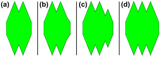
Figure 7.
Scheme of the formation of the double-sided comb-like domain during linear scanning: (a) domain edge, (b) increase in CDW tilt, (c) appearance of the additional tooth, (d) elongated domain.
The domain elongation (anisotropic growth) during linear scanning led to an increase in the CDW tilt (Figure 7b) and, consequently, to a local increase in the bound charge density at the wall. The appearance of the depolarization field in the bulk with a local value above the threshold for step generation resulted in the generation of an additional tooth at the wall (Figure 7c). This process occurred over and over, thus providing the double-sided comb-like domain with quasi-regular teeth (Figure 7d).
The obtained results differ significantly from the effects obtained by us earlier for the irradiation by focused femtosecond NIR laser irradiation of LN crystals [45]. For the first time, it was shown that the spindle-like domain shape obtained with a low pulse number and with a circular cross-section transformed into a so-called “three-wing” shape with a three-ray cross-section by increasing the pulse number. Moreover, in contrast to LN, we have revealed the formation of the double comb-like domains with quasi-regular teeth during linear scanning. The obtained difference can be attributed to an essentially higher value of the pyroelectric coefficient in LT as compared with LN which allows higher values of the pyroelectric field to be obtained with the same irradiation parameters.
5. Conclusions
In this work, we presented the results of the, to our knowledge, first investigation of the formation of domain structures in MgO-doped LT after irradiation with a focused femtosecond NIR laser, both locally and by linear scanning. We suppose that the domain appearance was caused by the depolarization field appearing around the microtrack and that the subsequent domain growth was stimulated by a pyroelectric field occurring during nonuniform temperature changes. The dependence of the domain shape on the pulse number and energy was revealed. For the first time, it was shown that the spindle-like shape obtained with a low pulse number and with a circular cross-section transformed into a so-called “three-wing” shape with a three-ray cross-section. The obtained effect was considered in terms of a kinetic approach to domain growth. The formation of the double comb-like domains during linear scanning was attributed to the creation of the quasi-regular teeth at the CDW during domain elongation. The obtained results are useful for the development of the methods of in-bulk domain engineering in ferroelectrics.
Author Contributions
Conceptualization, B.L., M.K. and V.S.; methodology, B.L. and M.K.; software, B.L.; validation, M.K. and V.S.; formal analysis, B.L., M.K. and V.S.; investigation, B.L.; resources, V.S.; data curation, B.L.; writing—original draft preparation, B.L., M.K. and V.S.; writing—review and editing, B.L., M.K. and V.S.; visualization, B.L.; supervision, M.K. and V.S.; project administration, V.S.; funding acquisition, V.S. All authors have read and agreed to the published version of the manuscript.
Funding
This research was funded by the Russian Science Foundation (Project № 24-12-00302).
Institutional Review Board Statement
Not applicable.
Informed Consent Statement
Not applicable.
Data Availability Statement
The data presented in this study are available on request from the corresponding author.
Acknowledgments
The equipment of the Ural Center for Shared Use “Modern nanotechnology” Ural Federal University (Reg.№ 2968) was used.
Conflicts of Interest
The authors declare no conflicts of interest.
References
- Fejer, M.M.; Magel, G.A.; Jundt, D.H.; Byer, R.L. Quasi-phase-matched second harmonic generation: Tuning and tolerances. IEEE J. Quantum Electron. 1992, 28, 2631–2654. [Google Scholar] [CrossRef]
- Armstrong, J.A.; Bloembergen, N.; Ducing, J.; Pershan, P.S. Interactions between light waves in a nonlinear dielectric. Phys. Rev. 1962, 127, 1918–1939. [Google Scholar] [CrossRef]
- Shur, V.Y.; Akhmatkhanov, A.R.; Baturin, I.S. Micro- and nanodomain engineering in lithium niobate. Appl. Phys. Rev. 2015, 2, 040604. [Google Scholar] [CrossRef]
- Myers, L.E.; Miller, G.D.; Eckardt, R.C.; Fejer, M.M.; Byer, R.L.; Rosenberg, W.R. Quasi-phase-matched 1.064-mm-pumped optical parametric oscillator in bulk periodically poled LiNbO3. Opt. Lett. 1995, 20, 52–54. [Google Scholar] [CrossRef] [PubMed]
- Ishizuki, H.; Taira, T. Large-aperture, axis-slant quasi-phase matching device using Mg-doped congruent LiNbO3. Opt. Mater. Express 2011, 1, 1376–1382. [Google Scholar] [CrossRef]
- Lim, E.J.; Fejer, M.M.; Byer, R.L.; Kozlovsky, W.J. Blue light generation by frequency doubling in periodically poled lithium niobate channel waveguide. Electron. Lett. 1989, 25, 731–732. [Google Scholar] [CrossRef]
- Matsumo, S.; Lim, E.J.; Hertz, H.M.; Fejer, M.M. Quasiphase-matched second harmonic generation of blue light lithium tantalate waveguides in electrically periodically-poled. Electron. Lett. 1991, 27, 2040–2041. [Google Scholar] [CrossRef]
- Sheng, Y.; Chen, X.; Xu, T.; Liu, S.; Zhao, R.; Krolikowski, W. Research progress on femtosecond laser poling of ferroelectrics. Photonics 2024, 11, 447. [Google Scholar] [CrossRef]
- Zhang, B.; Wang, L.; Chen, F. Recent advances in femtosecond laser processing of LiNbO3 crystals for photonic applications. Laser Photonics Rev. 2020, 14, 1900407. [Google Scholar] [CrossRef]
- Guo, J.; Chen, W.; Chen, H.; Zhao, Y.; Dong, F.; Liu, W.; Zhang, Y. Recent progress in optical control of ferroelectric polarization. Adv. Opt. Mater. 2021, 9, 2002146. [Google Scholar] [CrossRef]
- Sones, C.L.; Valdivia, C.E.; Scott, J.G.; Mailis, S.; Eason, R.W.; Scrymgeour, D.A.; Gopalan, V.; Jungk, T.; Soergel, E. Ultraviolet laser-induced sub-micron periodic domain formation in congruent undoped lithium niobate crystals. Appl. Phys. B 2005, 80, 341–344. [Google Scholar] [CrossRef]
- Shur, V.Y.; Kosobokov, M.S.; Makaev, A.V.; Kuznetsov, D.K.; Nebogatikov, M.S.; Chezganov, D.S.; Mingaliev, E.A. Dimensionality increase of ferroelectric domain shape by pulse laser irradiation. Acta Mater. 2021, 219, 117270. [Google Scholar] [CrossRef]
- Shur, V.Y.; Kosobokov, M.S.; Makaev, A.V.; Kuznetsov, D.K. Light-induced ordering of nanodomains in lithium tantalate as a result of multiple scanning by IR laser irradiation. J. Appl. Phys. 2023, 133, 014105. [Google Scholar] [CrossRef]
- Chen, X.; Karpinski, P.; Shvedov, V.; Koynov, K.; Wang, B.; Trull, J.; Cojocaru, C.; Krolikowski, W.; Sheng, Y. Ferroelectric domain engineering by focused infrared femtosecond pulses. Appl. Phys. Lett. 2015, 107, 141102. [Google Scholar] [CrossRef]
- Tan, D.; Sharafudeen, K.N.; Yue, Y.; Qiu, J. Femtosecond laser induced phenomena in transparent solid materials: Fundamentals and applications. Prog. Mater. Sci. 2016, 76, 154–228. [Google Scholar] [CrossRef]
- Chen, X.; Karpinski, P.; Shvedov, V.; Boes, A.; Mitchell, A.; Krolikowski, W.; Sheng, Y. Quasi-phase matching via femtosecond laser-induced domain inversion in lithium niobate waveguides. Opt. Lett. 2016, 41, 2410. [Google Scholar] [CrossRef] [PubMed]
- Imbrock, J.; Hanafi, H.; Ayoub, M.; Denz, C. Local domain inversion in MgO-doped lithium niobate by pyroelectric field-assisted femtosecond laser lithography. Appl. Phys. Lett. 2018, 113, 252901. [Google Scholar] [CrossRef]
- Imbrock, J.; Szalek, D.; Laubrock, S.; Hanafi, H.; Denz, C. Thermally assisted fabrication of nonlinear photonic structures in lithium niobate with femtosecond laser pulses. Opt. Express 2022, 30, 39340. [Google Scholar] [CrossRef]
- Lisjikh, B.I.; Kosobokov, M.S.; Efimov, A.V.; Kuznetsov, D.K.; Shur, V.Y. Thermally assisted growth of bulk domains created by femtosecond laser in magnesium doped lithium niobate. Ferroelectrics 2023, 604, 47–52. [Google Scholar] [CrossRef]
- Wang, X.; Cao, Q.; Wang, R.; Cao, X.; Liu, S. Manipulation of ferroelectric domain inversion and growth by optically induced 3D thermoelectric field in lithium niobate. Appl. Phys. Lett. 2022, 121, 181111. [Google Scholar] [CrossRef]
- Li, F.; Cao, Q.; Wang, X.; Wang, R. Nonlocal erasing and writing of ferroelectric domains using a femtosecond laser in lithium niobate. Opt. Lett. 2024, 49, 1892–1895. [Google Scholar] [CrossRef] [PubMed]
- Xu, X.; Wang, T.; Chen, P.; Zhou, C.; Ma, J.; Wei, D.; Wang, H.; Niu, B.; Fang, X.; Wu, D.; et al. Femtosecond laser writing of lithium niobate ferroelectric nanodomains. Nature 2022, 609, 496–501. [Google Scholar] [CrossRef] [PubMed]
- Guina, Y.; Zhu, J.; Gorevoy, A.; Kosobokov, M.; Turygin, A.; Lisjikh, B.; Akhmatkhanov, A.; Shur, V.; Kudryashov, S. Dimensional Analysis of Double-Track Microstructures in a Lithium Niobate Crystal Induced by Ultrashort Laser Pulses. Photonics 2023, 5, 582. [Google Scholar]
- Liu, S.; Switkowski, K.; Xu, C.; Tian, J.; Wang, B.; Lu, P.; Krolikowski, W.; Sheng, Y. Nonlinear wavefront shaping with optically induced three-dimensional nonlinear photonic crystals. Nat. Commun. 2019, 10, 3208. [Google Scholar] [CrossRef] [PubMed]
- Liu, S.; Switkowski, K.; Chen, X.; Xu, T.; Krolikowski, W.; Sheng, Y. Broadband enhancement of Čerenkov second harmonic generation in a sunflower spiral nonlinear photonic crystal. Opt. Express 2018, 26, 8628. [Google Scholar] [CrossRef]
- Liu, S.; Mazur, L.M.; Krolikowski, W.; Sheng, Y. Nonlinear volume holography in 3D nonlinear photonic crystals. Laser Photonics Rev. 2020, 14, 2000224. [Google Scholar] [CrossRef]
- Liu, D.; Liu, S.; Mazur, L.M.; Wang, B.; Lu, P.; Krolikowski, W.; Sheng, Y. Smart optically induced nonlinear photonic crystals for frequency conversion and control. Appl. Phys. Lett. 2020, 116, 051104. [Google Scholar] [CrossRef]
- Liu, S.; Wang, L.; Mazur, L.; Switkowski, K.; Wang, B.; Chen, F.; Arie, A.; Krolikowski, W.; Sheng, Y. Highly efficient 3D nonlinear photonic crystals in ferroelectrics. Adv. Opt. Mater. 2023, 18, 2300021. [Google Scholar] [CrossRef]
- Chen, Y.; Yang, C.; Liu, S.; Wang, S.; Wang, N.; Liu, Y.; Sheng, Y.; Zhao, R.; Xu, T.; Krolikowski, W. Optically induced nonlinear cubic crystal system for 3D quasi-phase matching. Adv. Photonics Res. 2022, 3, 2100268. [Google Scholar] [CrossRef]
- Chen, X.; Mazur, L.M.; Liu, D.; Liu, S.; Liu, X.; Xu, Z.; Wei, X.; Wang, J.; Sheng, Y.; Wei, Z.; et al. Quasi-phase matched second harmonic generation in PMN-38PT crystal. Opt. Lett. 2020, 47, 2056–2059. [Google Scholar] [CrossRef]
- Chen, X.; Liu, D.; Liu, S.; Mazur, L.M.; Liu, X.; Wei, X.; Xu, Z.; Wang, J.; Sheng, Y.; Wei, Z.; et al. Optical induction and erasure of ferroelectric domains in tetragonal PMN-38PT crystals. Adv. Opt. Mater. 2022, 10, 2102115. [Google Scholar] [CrossRef]
- Wei, D.; Wang, C.; Wang, H.; Hu, X.; Wei, D.; Fang, X.; Zhang, Y.; Wu, D.; Hu, Y.; Li, J.; et al. Experimental demonstration of a three-dimensional lithium niobate nonlinear photonic crystal. Nat. Photonics 2018, 12, 596–600. [Google Scholar] [CrossRef]
- Hum, D.S.; Fejer, M.M. Quasi-phasematching. C.R. Phys. 2007, 8, 180–198. [Google Scholar] [CrossRef]
- Akhamatkhanov, A.R.; Chuvakova, M.A.; Vaskina, E.M.; Shur, V.Y. Polarization reversal process in MgO doped congruent lithium tantalate single crystals. Ferroelectrics 2015, 476, 57–68. [Google Scholar] [CrossRef]
- Jia, B.S.; Zhao, Y.Q.; Zhang, X.F. Research on defects and domain characteristics of MgO-doped near-stoichiometric lithium tantalate in room-temperature polarization process. Chin. Sci. Bull. 2010, 55, 11–15. [Google Scholar] [CrossRef]
- Hu, P.; Zhang, L.; Xiong, J.; Yin, J.; Zhao, C.; He, X.; Hang, Y. Optical properties of MgO doped near-stoichiometric LiTaO3 single crystals. Opt. Mater. 2011, 33, 1677–1680. [Google Scholar] [CrossRef]
- Nakamura, M.; Higuchi, S.; Takekawa, S.; Terabe, K.; Furukawa, Y.; Kitamura, K. Optical damage resistance and refractive indices in near-stoichiometric MgO-doped LiNbO3. Jpn. J. Appl. Phys. 2002, 41, L49–L51. [Google Scholar] [CrossRef]
- Chen, S.; Liu, H.; Kong, Y.; Huang, Z. The resistance against optical damage of near-stoichiometric LiNbO3:Mg crystals prepared by vapor transport equilibration. Opt. Mater. 2007, 29, 885–888. [Google Scholar] [CrossRef]
- Yu, N.E.; Kurimura, S.; Nomura, Y.; Kitamura, K. Stable high-power green light generation with thermally conductive periodically poled stoichiometric lithium tantalate. Jpn. J. Appl. Phys. 2004, 43, L1265–L1267. [Google Scholar] [CrossRef]
- Schossig, M.; Norkus, V.; Gerlach, G. Dielectric and pyroelectric properties of ultrathin, monocrystalline lithium tantalate. Infrared Phys. Technol. 2020, 63, 35–41. [Google Scholar] [CrossRef]
- Weigel, T.; Ludt, C.; Leisegang, T.; Mehner, E.; Jachalke, S.; Stöcker, H.; Doert, T.; Meyer, D.C.; Zschornak, M. Spontaneous polarization and pyroelectric coefficient of lithium niobate and lithium tantalate determined from crystal structure data. Phys. Rev. B 2023, 108, 054105. [Google Scholar] [CrossRef]
- Greshnyakov, E.D.; Lisjikh, B.I.; Akhmatkhanov, A.R.; Shur, V.Y. Charged domain walls in lithium niobate and lithium tantalate crystals with composition gradients. Ferroelectrics 2023, 604, 32–39. [Google Scholar] [CrossRef]
- Franken, P.A.; Hill, A.E.; Peters, C.W.; Weinreich, G. Generation of optical harmonics. Phys. Rev. Lett. 1961, 7, 118–120. [Google Scholar] [CrossRef]
- Sheng, Y.; Best, A.; Butt, H.-J.; Krolikowski, W.; Arie, A.; Koynov, K. Three-dimensional ferroelectric domain visualization by Čerenkov-type second harmonic generation. Opt. Express 2010, 18, 16539–16545. [Google Scholar] [CrossRef] [PubMed]
- Lisjikh, B.; Kosobokov, M.; Turygin, A.; Efimov, A.; Shur, V. Creation of a periodic domain structure in MgOLN by femtosecond laser irradiation. Photonics 2023, 11, 1211. [Google Scholar] [CrossRef]
- Alikin, D.O.; Ievlev, A.V.; Turygin, A.P.; Lobov, A.I.; Kalinin, S.V.; Shur, V.Y. Tip-induced domain growth on the non-polar cuts of lithium niobate single-crystals. Appl. Phys. Lett. 2015, 106, 182902. [Google Scholar] [CrossRef]
- Kudryashov, S.; Rupasov, A.; Kosobokov, M.; Akhmatkhanov, A.; Krasin, G.; Danilov, D.; Lisjikh, B.; Turygin, A.; Greshnyakov, E.; Kovalev, M.; et al. Ferroelectric nanodomain engineering in bulk lithium niobate crystals in ultrashort-pulse laser nanopatterning regime. Nanomaterials 2022, 12, 4147. [Google Scholar] [CrossRef]
- Fujimoto, K.; Cho, E. High-speed switching of nanoscale ferroelectric domains in congruent single-crystal LiTaO3. Appl. Phys. Lett. 2003, 83, 5265–5267. [Google Scholar] [CrossRef]
- Kan, Y.; Lu, X.; Bo, H.; Huang, F.; Wu, X.; Zhu, J. Critical radii of ferroelectric domains for different decay processes in LiNbO3 crystals. Appl. Phys. Lett. 2007, 91, 132902. [Google Scholar] [CrossRef]
- Agronin, A.; Molotski, M.; Rosenwaks, Y.; Rosenmann, G.; Rodriguez, B.J.; Kingon, A.I.; Gruverman, A. Dynamics of ferroelectric domain growth in the field of atomic force microscopy. J. Appl. Phys. 2006, 99, 104102. [Google Scholar] [CrossRef]
- Lilienblum, M.; Soergel, E. Determination of the effective coercive field of ferroelectrics by piezoresponse force microscopy. J. Appl. Phys. 2011, 110, 052012. [Google Scholar] [CrossRef]
- Shur, V.Y.; Pelegova, E.V.; Kosobokov, M.S. Domain shapes in bulk uniaxial ferroelectrics. Ferroelectrics 2020, 569, 251–265. [Google Scholar] [CrossRef]
- Esin, A.A.; Akhmatkhanov, A.R.; Shur, V.Y. Tilt control of the charged domain walls in lithium niobate. Appl. Phys. Lett. 2019, 114, 092901. [Google Scholar] [CrossRef]
- Slautin, B.; Turygin, A.; Pashnina, E.; Slautina, A.; Chezganov, D.; Shur, V. Evolution of nanodomains and formation of self-organized structures during local switching in X-cut LNOI. Crystals 2022, 12, 659. [Google Scholar] [CrossRef]
- Wang, Z.; Zhang, B.; Wang, Z.; Zhang, J.; Kazansky, P.G.; Tan, D.; Qiu, J. 3D imprinting of voxel-level structural colors in lithium niobate crystal. Adv. Mater. 2023, 35, 2303256. [Google Scholar] [CrossRef]
- Shur, V.Y.; Mingaliev, E.A.; Kosobokov, M.S.; Nebogatikov, M.S.; Lovov, A.I.; Makaev, A.V. Self-assembled shape evolution of the domain wall and formation of nanodomain wall traces induced by multiple IR laser pulse irradiation in lithium niobate. J. Appl. Phys. 2020, 127, 094103. [Google Scholar] [CrossRef]
Disclaimer/Publisher’s Note: The statements, opinions and data contained in all publications are solely those of the individual author(s) and contributor(s) and not of MDPI and/or the editor(s). MDPI and/or the editor(s) disclaim responsibility for any injury to people or property resulting from any ideas, methods, instructions or products referred to in the content. |
© 2024 by the authors. Licensee MDPI, Basel, Switzerland. This article is an open access article distributed under the terms and conditions of the Creative Commons Attribution (CC BY) license (https://creativecommons.org/licenses/by/4.0/).