Article

Cell-Based Screening Identifies Neoblechnum brasiliense Extract as a Potent Antagonist of the Ecdysteroid Receptor in Dipteran Cells


by
Jissela Gaibor Garofalo
1,*,
Juliana Wegner
1,
Mauricio Gaibor Garofalo
2,
Guy Smagghe
3,4,
Jorge Briceño
5 and
Moises João Zotti
1,*
1
Programa de Pós-Graduação em Fitossanidade, Universidade Federal de Pelotas, Pelotas CEP 96160-000, RS, Brazil
2
Facultad de Ciencias Agropecuarias, Recursos Naturales y del Ambiente, Universidad Estatal de Bolívar, Guaranda 020102, Ecuador
3
Institute of Entomology, Guizhou University, Guiyang 550025, China
4
Department of Biology, Vrije Universiteit Brussel (VUB), 1050 Brussels, Belgium
5
Vicerrectorado de Investigación y Vinculación, Universidad Estatal de Bolívar, Guaranda 020102, Ecuador
*
Authors to whom correspondence should be addressed.
Processes 2026, 14(2), 312; https://doi.org/10.3390/pr14020312
Submission received: 3 December 2025 / Revised: 9 January 2026 / Accepted: 13 January 2026 / Published: 15 January 2026

Abstract

The ecdysteroid receptor (EcR) plays a crucial role in insect development and metamorphosis, making it a promising target for the design of novel biorational compounds. This study investigated the cytotoxicity, as well as the EcR agonist and antagonist activities, of three synthetic molecules analogous to tebufenozide and extracts from nine plant species using the dipteran S2 cell line which originates from the insect model of the fruit fly Drosophila melanogaster. Cytotoxicity assays were performed to determine appropriate concentrations of the synthetic molecules and plant extracts for cell transfection. EcR agonist and antagonist activities were evaluated using 20-hydroxyecdysone (20E) as the control hormone. The synthetic molecules analogous to tebufenozide did not activate EcR in S2 cells. In contrast, the plant extract of Neoblechnum brasiliense, commonly known as Brazilian dwarf tree fern, exhibited significant antagonistic activity at 100 µM, reducing receptor activity by 92%, likely due to its phytosteroid content, and without inducing cytotoxic effects. These findings demonstrate that certain plant extracts, particularly N. brasiliense, act as effective EcR antagonists and may represent promising natural leads for the development of environmentally compatible biorational compounds to control economically important dipteran pests, such as fruit flies and mosquitoes.

1. Introduction

Modern agriculture faces major challenges in developing pest management strategies that effectively reduce crop losses caused by arthropods while minimizing environmental damage and supporting food sovereignty [1]. Despite advances in integrated pest management, chemical control remains the predominant approach for managing insect pests. The extensive and often indiscriminate use of insecticides has led to the rapid emergence of resistant pest populations, significantly reducing the long-term efficacy of conventional chemical controls. Globally, approximately 3 billion kilograms of chemical pesticides are applied annually, placing nearly 64% of agricultural land at risk of contamination, with severe consequences for ecosystems, biodiversity, and human health [2]. These challenges underscore the urgent need for innovative, environmentally compatible pest control strategies based on rational biological targets and sustainable processes.
To reduce reliance on broad-spectrum chemical insecticides, biologically informed model systems have been increasingly adopted for pesticide discovery and screening. Drosophila melanogaster is one of the most extensively studied insect models in developmental biology and genetics, owing to its well-characterized genome, conserved endocrine signaling pathways, and the availability of powerful molecular and cellular tools [3,4]. Importantly, Drosophila serves as a model organism for the biosynthesis and regulation of steroid hormones, including ecdysone and its active metabolite 20-hydroxyecdysone (20E), which are essential regulators of insect molting, metamorphosis, and reproduction [5]. These characteristics make Drosophila-derived systems highly suitable for mechanistic and process-oriented studies targeting insect endocrine regulation.
Biorational compounds such as insect growth regulators (IGRs) have emerged as a promising class of next-generation pesticides designed to interfere selectively with insect development rather than causing acute toxicity. IGRs act by disrupting critical molecular processes during molting and metamorphosis, including ecdysteroid receptor (EcR) signaling and chitin biosynthesis, ultimately leading to developmental arrest or mortality [6,7]. However, the structural complexity of ecdysteroid hormones and their receptors has hindered the rational design and screening of effective EcR-targeting compounds, limiting the discovery pipeline for novel IGRs [8].
The EcR is a ligand-activated nuclear receptor that controls the transcription of numerous genes involved in insect development, metamorphosis, and reproduction. Structurally, EcR comprises five functional domains: the A/B domain responsible for transcriptional activation, the C domain involved in DNA binding, the D hinge domain, the E ligand-binding domain, and the F domain, which modulates receptor activity [4]. Given its central regulatory role, EcR and its associated signaling components have been identified as high-value molecular targets for the development of selective insecticides and IGRs [9,10,11]. However, there remains a need for systematic, cell-based screening platforms capable of distinguishing between agonistic and antagonistic EcR modulators while simultaneously assessing cytotoxicity.
In parallel, plant-derived extracts represent a highly diverse and renewable source of bioactive compounds with potential applications in pest control. A well-known example is azadirachtin, a terpenoid isolated from Azadirachta indica, which exhibits potent insecticidal activity by interfering with oviposition, growth, and reproduction in a wide range of pest species [12,13]. Terpenoids frequently exhibit synergistic effects arising from the combined action of multiple constituents, resulting in enhanced biological activity compared with isolated compounds [14]. Additionally, phytoecdysteroids, which are plant-derived compounds structurally similar to arthropod steroid hormones, are present in approximately 5–6% of plant species and display diverse biological effects, including disruption of insect metamorphosis, feeding inhibition through starvation, and repellent or irritant activity [3,4,15]. Despite this potential, the molecular interactions between complex plant extracts and insect hormone receptors remain insufficiently characterized.
Critically, integrative screening approaches that combine synthetic hormone analogues and complex plant extracts within mechanistically relevant biological systems are still limited. This gap restricts the identification of novel EcR modulators that could serve as environmentally safer and process-efficient insect growth regulators. Cell-based systems derived from the fruit fly D. melanogaster, such as the S2 cell line, offer a powerful platform for addressing this challenge, as they enable controlled evaluation of receptor activity, cytotoxicity, and hormone signaling dynamics within a well-defined biological process framework [16,17].
The central biological questions addressed in this study are: (i) whether synthetic molecules structurally analogous to the commercial IGR tebufenozide can function as EcR agonists or antagonists in a dipteran cellular system, and (ii) whether complex plant extracts containing natural steroid-like compounds can modulate EcR activity without inducing cytotoxic effects. We hypothesized that while simplified synthetic analogues may fail to effectively engage the EcR due to structural constraints, certain plant extracts rich in phytosteroids or terpenoids may act as potent EcR antagonists through multi-component interactions. To test these hypotheses, this study systematically evaluated the cytotoxicity, as well as the agonist and antagonist activities, of three synthetic tebufenozide analogues and extracts from nine plant species using the S2 cell line. By integrating cytotoxicity assessment with functional EcR signaling assays, this work introduces a process-oriented screening strategy for identifying novel EcR modulators and highlights plant extracts as promising natural leads for the development of environmentally compatible biorational compounds targeting economically important dipteran pests, such as fruit flies and mosquitoes.

2. Materials and Methods

Screening experiments were conducted at the Molecular Entomology Laboratory, Plant Health Department, Eliseu Maciel School of Agronomy, Federal University of Pelotas (UFPel), Pelotas, RS, Brazil (31°48′16″ S, 52°24′48″ W). The D. melanogaster S2 cell line, derived from embryonic tissues, was obtained from the Butantan Institute (São Paulo, Brazil). Cells were maintained in InsectXpress medium (Lonza Group Ltd., Basel, Switzerland) in 25-cm2 cell culture flasks (Kasvi, Curitiba, Brazil) at 27 °C. Subcultures were performed by transferring 0.5 mL of cell suspension into a new flask containing 4.5 mL of fresh medium, following methods established in previous studies [18].

2.1. Virtual Screening

For virtual screening, the following compound libraries were initially used: APExBIO (Houston, TX, USA), Asinex (Winston-Salem, NC, USA), and Life Chemicals (Niagara-on-the-Lake, ON, Canada). The OMEGA software (version 4.2.1, OpenEye, Cadence Molecular Sciences, Santa Fe, NM, USA) was used to produce high-quality en-sembles of bioactive conformations [19]. Virtual screening was performed using the ROCS software (version 3.4.3, OpenEye, Cadence Molecular Sciences, Santa Fe, NM, USA) [19,20] and the Fast Exhaustive Docking (FRED) application (version 4.0.2, OpenEye, Cadence Molecular Sciences, Santa Fe, NM, USA), following previously described protocols [21,22].
We used the ecdysone agonist insecticide Tebufenozide (Sigma-Aldrich, St. Louis, MO, USA) to search for analogs. The molecules were analyzed using the Tanimoto coefficient calculated with ROCS software (version 3.4.3, OpenEye, Cadence Molecular Sciences, Santa Fe, NM, USA), where Tanimoto combo will be equal to the sum of Tanimoto Color (chemical similarity between the reference molecules and the analyzed molecules) and Tanimoto Shape (structural similarity between the reference molecules and the analyzed molecules), among other parameters. The screening 1,276,476 compounds resulted in 140,744,768 conformers. From these resulted in 6375) analogs (hits) of Tebufenozide and 554 successfully docked in the ecdysone receptor of Drosophila melanogaster using FRED software (version 4.0.2, OpenEye, Cadence Molecular Sciences, Santa Fe, NM, USA).
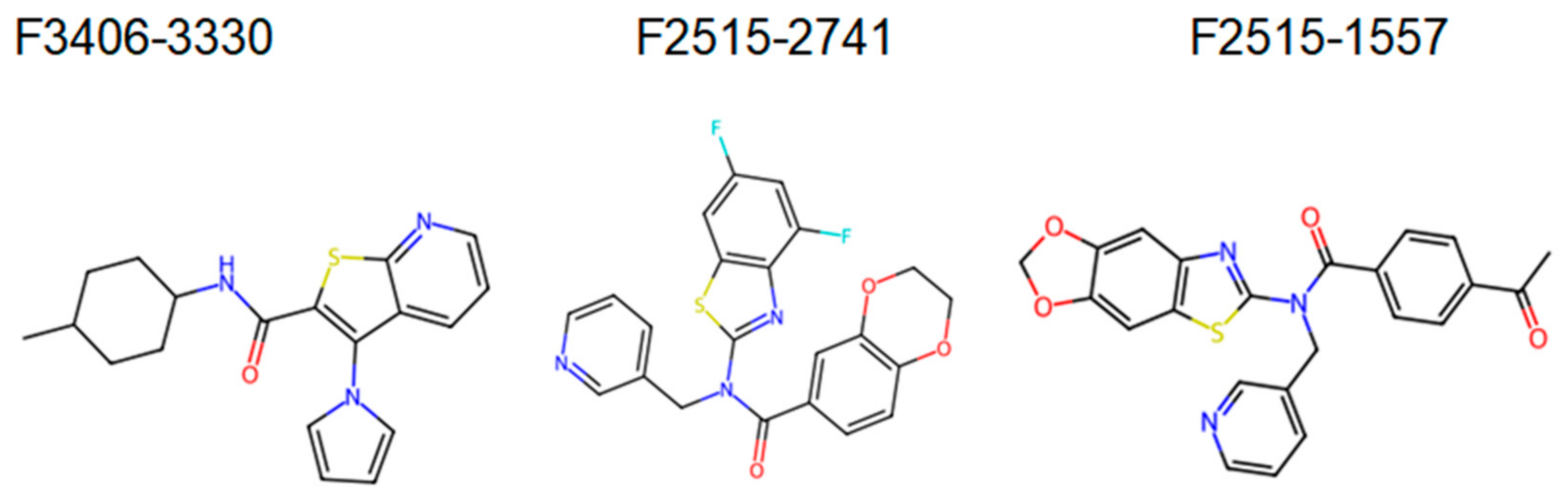
Finally, a new analysis was carried out with the compounds selected according to the best docking scores, being evaluated according to Lipinski’s Rule of Five [23]. Based on the docking score, we selected and bought the top 3 and screening with the S2 cells. The chemical structure of the 3 selected compounds is shown in Figure 1.

2.2. Synthetic Compounds

For the screening experiments, three tebufenozide analogue molecules were obtained from Life Chemicals (Kyiv, Ukraine) (Figure 1). 20-hydroxyecdysone (20E; ~95% purity; Sigma-Aldrich, Bornem, Belgium) was used as a positive control for EcR activation [24].

2.3. Plant Extracts

Nine plant extracts were evaluated in the screening experiments. Plant material was obtained from the following species: Piper hispidum (Hispid pepper, Piperaceae) and Schinus terebinthifolia (Brazilian peppertree, Anacardiaceae), collected in Tangará da Serra, MT, Brazil. Extracts of Syzygium aromaticum (clove, Myrtaceae), Azadirachta indica (neem, Meliaceae), and Tagetes sp. (marigold, Asteraceae) were provided by Federal University of Pelotas (UFPel, Pelotas, RS, Brazil). Additionally, plant material was collected from Melia azedarach (chinaberry, Meliaceae) in Herval RS, Brazil, Christella dentata (soft fern, Thelypteridaceae) in Nova Petrópolis, RS, Brazil, Neoblechnum brasiliense (Brazilian dwarf tree fern, Blechnaceae) on the Capão do Leão campus (UFPel, Pelotas, RS, Brazil), and Adiantum capillus-veneris (maidenhair fern, Pteridaceae) in Pelotas, RS, Brazil [25].
Leaf samples were dried at room temperature (≈20 °C) for one week and ground to a particle size of <0.5 mm [26]. Each powdered sample (200 mg) was macerated in methanol (5 volumes) for 5 days at 32 °C and then centrifuged at 4000× g for 15 min [27]. The resulting supernatants were filtered through Whatman No. 1 filter paper (Cytiva, Marlborough, MA, USA) and concentrated under reduced pressure using a rotary evaporator at 60 °C to remove the solvent.
Crude extracts were dissolved in absolute ethanol (Sigma-Aldrich, St. Louis, MO, USA) to obtain stock solutions of 200 mg/mL and stored in Eppendorf tubes (Eppendorf SE, Hamburg, Germany) [28]. For cytotoxicity assays, stock solutions were further diluted to 20 mg/mL, 0.2 mg/mL, and 0.002 mg/mL [29]. These concentrations were selected based on preliminary experiments that demonstrated receptor modulation without inducing excessive cytotoxicity, while also allowing the detection of dose-dependent trends.
To facilitate the interpretation of potential non-specific effects and differences in cell permeability, the basic physicochemical properties of each synthetic analogue, including estimated lipophilicity, qualitative solubility, key functional groups, and structural features relevant to stability, are summarized in Table 1.

2.4. Cytotoxicity Assays

Preliminary cytotoxicity studies of the synthetic molecules were performed to identify concentrations that maintained cell viability above 85%, which were subsequently used for in vitro experiments.
Cell viability was assessed by homogenizing the S2 cell culture and transferring 50 µL of the suspension into a 1.5 mL microcentrifuge tube (Eppendorf, Hamburg, Germany), followed by the addition of 50 µL of 0.4% Trypan Blue (TB) dye (C14H16N2; Sigma-Aldrich, St. Louis, MO, USA) [40]. TB penetrates the membranes of dead cells, staining them blue, while live cells remain unstained, allowing differentiation and counting of viable versus non-viable cells.
A 10 µL aliquot of the cell-dye mixture was loaded into each quadrant of a hemocytometer (Hausser Scientific, Horsham, PA, USA), and cells were counted under an inverted microscope (Leica DMi1, 10× objective; Leica Microsystems, Wetzlar, Germany) [41].
Stock solutions of the synthetic molecules were prepared by dissolving 1 mg of compound in dimethyl sulfoxide (DMSO; Sigma-Aldrich, St. Louis, MA, USA). 20E (≥93% purity; Sigma-Aldrich) was used as a reference compound. 20E was diluted in absolute ethanol to 1 µM and serially diluted to 1 µM, 100 nM, 10 nM, 1 nM, 100 pM, 10 pM and 1 pM to generate dose–response curves [42].
For the cytotoxicity assays, cells were seeded at ~500,000 cells per mL, with 500 µL of cell suspension added per well of a culture plate. Subsequently, 5 µL of each molecule dilution was added, and plates were incubated at 27 °C for 24 h. After incubation, cell viability was assessed using the Trypan Blue protocol described above.

2.5. Molecular Screening Assay

2.5.1. Plasmid Preparation

Reporter plasmids for luciferase assays were cloned using Escherichia coli. A single colony was inoculated into 5 mL of LB broth containing 5 µL of ampicillin and incubated at 37 °C, 200 rpm for 16 h [43]. Plasmid DNA was purified using the Wizard Plus SV Minipreps DNA Purification System (Promega, Madison, WI, USA), and integrity was confirmed via electrophoresis on 1% agarose gels (Sigma-Aldrich, St. Louis, MA, USA) [44]. All experiments were performed in triplicate.

2.5.2. Cell Transfection

S2 cells were seeded into 24-well plates at 500,000 cells per well in 500 µL of culture medium and allowed to adhere [45]. Transfection medium for each well was prepared by combining 497 µL of InsectXpress culture medium, 3 µL of Escort IV Transfection Reagent (Sigma-Aldrich, St. Louis, MA, USA), and 100 ng of reporter plasmid in a 1.5 mL Eppendorf tube. The mixture was incubated for 30 min at 22 ± 2 °C to facilitate complex formation [46].
Culture medium was removed, and the transfection medium was added to each well. Plates were incubated at 27 °C for 5 h, after which the transfection medium was replaced with 500 µL of fresh medium containing 5 µL of the test molecule. DMSO (Sigma-Aldrich, St. Louis, MO, USA) served as the vehicle control. Plates were sealed with Parafilm M (Bemis Company Inc., Neenah, WI, USA) and incubated at 27 °C for 24 h.
Luciferase expression was measured by transferring 100 µL of cell suspension into a white 96-well plate, adding an equal volume of Steady-Glo Luciferase reagent (Promega, Madison, WI, USA), and incubating for 5 min at 25 °C. Luminescence was quantified using a SpectraMax M3 microplate reader (Molecular Devices, San Jose, CA, USA).

2.5.3. Antagonist Assays

Antagonist assays followed the same protocol as agonist assays with an additional step: after the initial 24 h incubation with the test molecule, 5 µL of 20E was added to each well, and plates were incubated for a second 24 h period at 27 °C. Luminescence was measured as above to assess antagonist activity in the presence of the hormone [47].

2.6. Statistical Analysis

Differences in mean cytotoxicity, agonist, and antagonist activities were evaluated using ANOVA with the F-test, treating experimental repetitions as a random factor. Homoscedasticity and normality of residuals were assessed using Levene’s test and the Shapiro–Wilk test, respectively, at a 95% confidence level [48,49].
Dose–response relationships were modeled using nonlinear logarithmic regression [50], with luminescence ( y ) expressed as a function of 20E concentration ( x ) according to the following equation:
y = c + { d c 1 + e x p ( b ( l o g ( x ) l o g ( e ) ) ) }
where c and d represent the lower and upper response limits, b is the slope, and e corresponds to the inflection point (IC50). IC50 values were calculated using the ED function from the CKD package [51]. All statistical analyses were performed in R (version 4.0.2).

3. Results

3.1. Screening Experiments of Synthetic Molecules

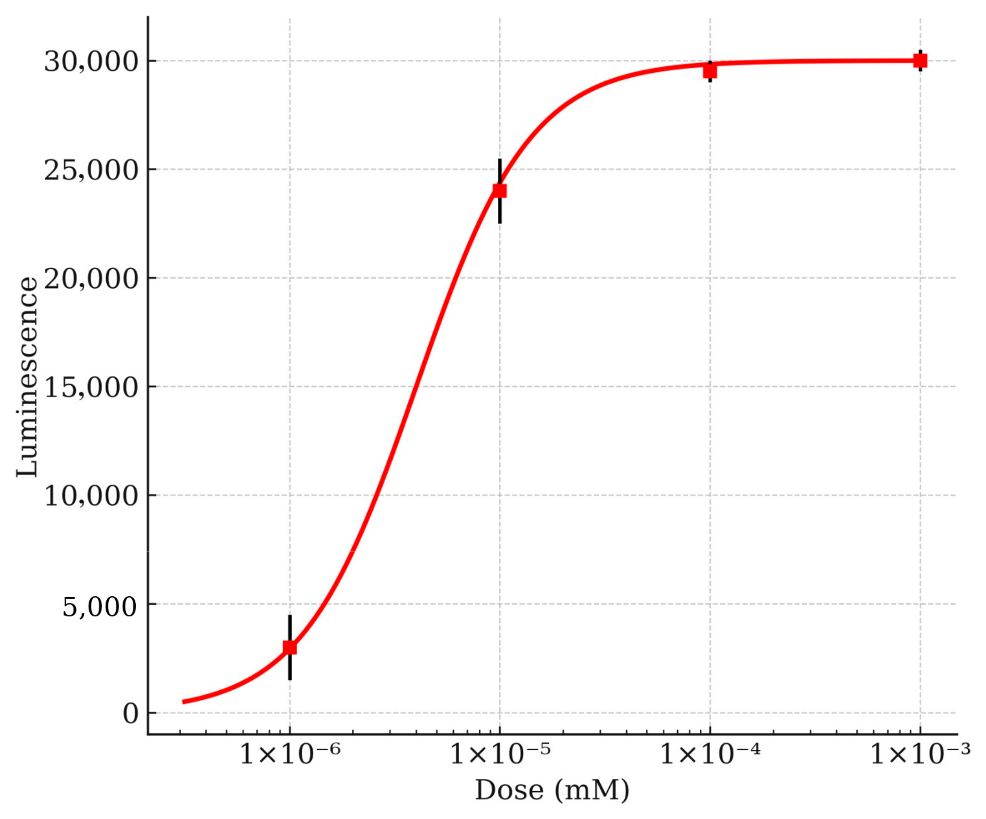
Reporter bioassays assessing ecdysteroid agonist activity demonstrated a significant, dose-dependent induction of luciferase expression in response to 20E, validating the functional responsiveness of the EcR signaling system in D. melanogaster S2 cells (Table 2). Statistical analysis using the F-test confirmed that the observed effects were significant (p < 0.05).
Nonlinear log-logistic modeling estimated an EC50 value for 20E of 4.1 nM, with a 95% confidence interval ranging from 3.3 to 4.8 nM. Maximum receptor activation corresponded to a luminescence signal of approximately 29,339 relative luminescence units (RLU). At the EC50 concentration, luminescence reached 17,453 RLU, representing 50% of the maximal EcR-mediated transcriptional response (Figure 2).
These results confirm the sensitivity and reliability of the reporter assay as a process-oriented screening platform for detecting EcR agonist activity and establish 20E as a robust positive control for subsequent comparative analyses involving synthetic analogues and plant-derived extracts.
20E exhibited activity patterns consistent with previous reports in D. melanogaster S2 dipteran cells and Sf9 lepidopteran cell lines. As an insect-specific ecdysteroid, 20E induced robust responses in both dipteran and lepidopteran cellular models, further validating its role as a reliable positive control for EcR activation.
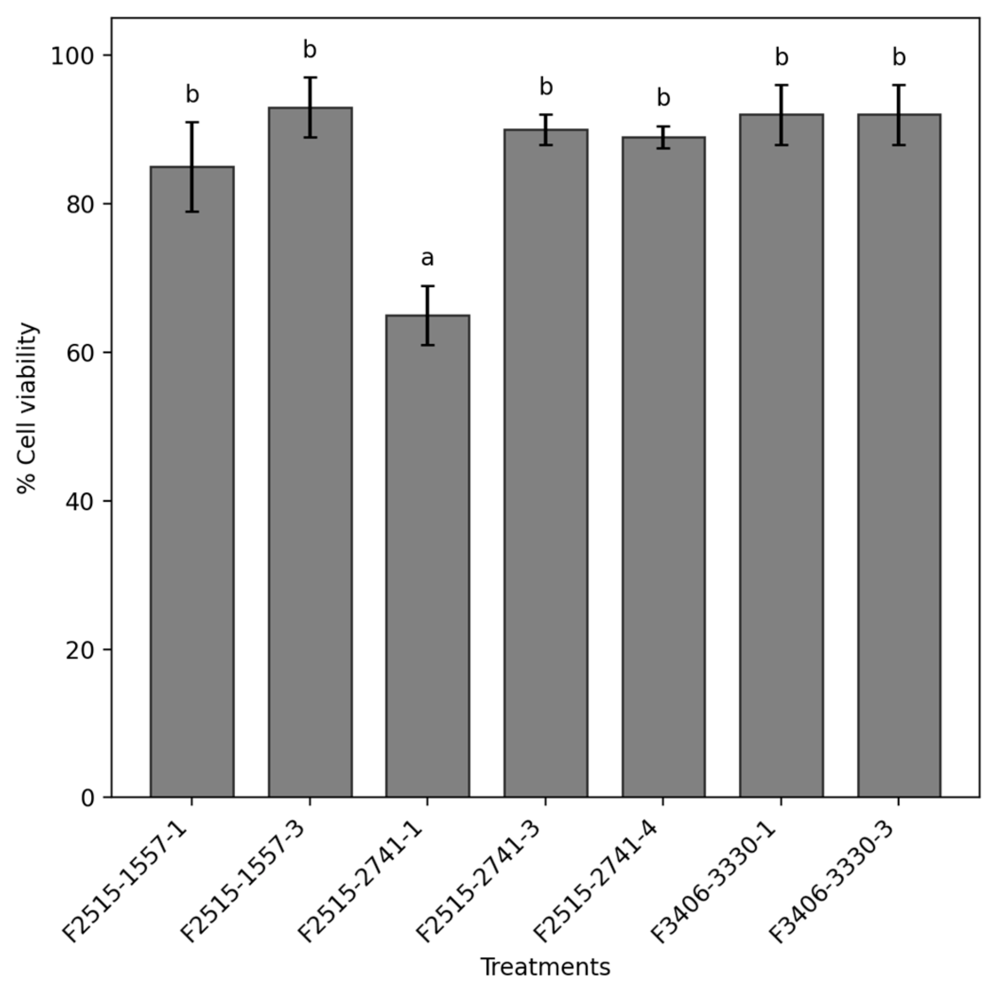
Cytotoxicity assays conducted in D. melanogaster S2 cells demonstrated that synthetic molecule concentrations of 1 µM maintained cell viability at or above 85%. In contrast, the compound F2515-2741 at a concentration of 100 µM significantly reduced cell viability to 66% (Figure 3). These findings underscore the necessity of evaluating compound cytotoxicity across a range of concentrations to distinguish receptor-mediated effects from non-specific cellular toxicity and to ensure the biological relevance of screening outcomes.
The compounds were tested at non-toxic millimolar concentrations in cell culture. Table 3 summarizes the cytotoxicity results for the evaluated concentrations.
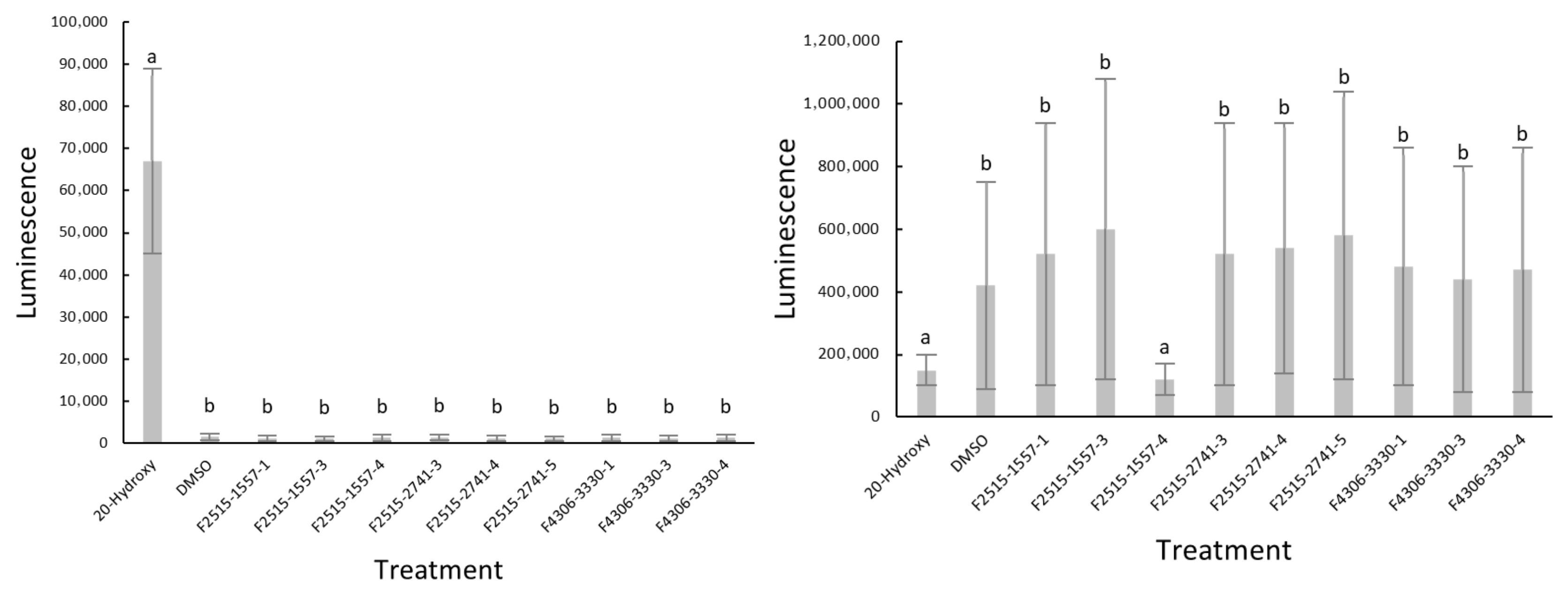
In the antagonist assay (Figure 4, left panel), treatment with 20E produced a significantly higher luminescence signal than all other treatments. In contrast, DMSO and all tested compounds exhibited uniformly low luminescence levels and did not differ significantly from one another. This pattern indicates an absence of detectable antagonistic activity among the evaluated compounds, as none significantly interfered with or reduced the 20E-mediated EcR response under the experimental conditions.
In the agonist assay (Figure 4, right panel), the compound F2515-1557-4 at 0.1 µM exhibited a low luminescence signal comparable to that of the 20E control. In contrast, DMSO and most of the tested compounds produced markedly higher luminescence signals, indicative of agonist-like activity. These results suggest differential modulation of EcR signaling among the tested molecules, with F2515-1557-4 showing a distinct response profile relative to the other compounds.
The comparable responses observed for DMSO and most tested compounds suggest a high basal or solvent-associated level of activation in this assay, against which only 20E and F2515-1557-4 exhibited a significantly reduced luminescence response.

3.2. Evaluation of Plant Extracts for EcR Activity

Cytotoxicity assays in D. melanogaster S2 cells demonstrated that all tested plant extracts maintained cell viability above 90%. The concentrations selected for subsequent EcR activity assays are summarized in Table 4, confirming that these extracts can be investigated further without compromising cell health. These results establish a safe and biologically relevant range for screening potential EcR modulators from plant-derived compounds.
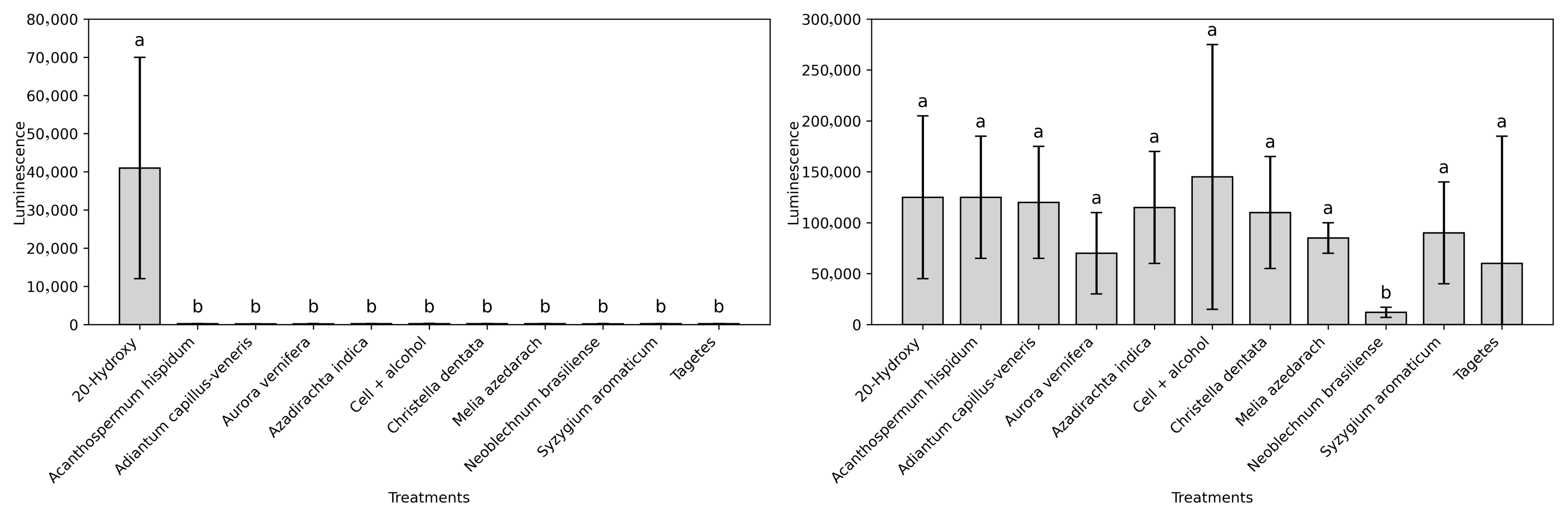
In agonist activity assays, none of the tested plant extracts at the selected concentrations elicited a significant increase in EcR-mediated luciferase expression compared to the 20E control (Figure 5, left panel). In contrast, the extract of N. brasiliense exhibited pronounced antagonist activity at 0.1 mg/mL, reducing receptor activity by 92% in the presence of 20E (Figure 5, right panel). The other extracts showed no significant effect on EcR modulation, indicating a specific inhibitory interaction between the N. brasiliense extract and the EcR. These results highlight the potential of N. brasiliense as a source of natural EcR antagonists for the development of environmentally compatible IGRs.

4. Discussion

The present study evaluated the cytotoxicity and EcR-modulating activity of three synthetic tebufenozide-analogue molecules and nine plant extracts in D. melanogaster S2 cells, providing novel insights into the potential of natural and synthetic compounds to modulate insect developmental pathways. The strong, dose-dependent luciferase response elicited by 20E confirmed the robustness and sensitivity of the cell-based reporter system. The dose–response curve and EC50 values estimated here for 20E align with previous reports in dipteran S2 and lepidopteran Sf9 cell lines [16,52], which is consistent with the insect-specific activity of 20E across these taxa. This reproducibility reinforces the suitability of S2 cells as a sensitive platform for EcR-ligand interaction screening, complementing recent advances that underscore the value of insect cell models in mechanistic mode of action studies for IGRs [53,54].
Toxicological assessments using in vitro assays provide a valuable alternative for understanding the compound’s mode of action, integrating genetic, metabolic, and immunological factors [55]. Within this framework, the present study demonstrates that S2 cells can effectively discriminate between active and inactive EcR modulators, facilitating the early-stage prioritization of compounds for further development. Importantly, preliminary cytotoxicity screening allowed the identification of biologically meaningful concentration ranges, ensuring that subsequent receptor assays were not confounded by non-specific cell stress or viability loss [40]. For instance, molecule F2515-2741 reduced cell viability at 100 µM, highlighting the necessity of careful dose selection to avoid artifacts during functional screening.
Despite their structural similarity to tebufenozide, none of the synthetic analogues displayed significant agonist or antagonist activity on dipteran EcR. Tebufenozide is a potent agonist in lepidopterans, inducing premature larval molting by activating EcR signaling [56,57]. Structural and functional analyses suggest that even modest modifications of the hydrazide scaffold can markedly alter receptor affinity and selectivity, particularly due to subtle differences in the ligand-binding domain among insect orders [9,58,59]. These differences likely explain the inactivity observed in S2 cells and indicate that dipteran EcRs may require distinct structural features for effective ligand engagement [11,60]. Comparisons with prior studies on EcR interactions in other screening systems support this conclusion. Specifically, lepidopteran Sf9 cells respond robustly to tebufenozide analogues, whereas dipteran models often display limited sensitivity, reflecting inherent receptor divergence [6,16,18]. Future efforts to optimize synthetic analogues should incorporate computational approaches, including in silico docking, molecular dynamics simulations, and receptor modeling, to refine scaffolds for improved compatibility with dipteran EcR isoforms.
Among the plant extracts evaluated, N. brasiliense exhibited remarkable antagonist activity, reducing 20E-mediated EcR activation by 92% at 0.1 mg/mL. This contrasts sharply with other extracts tested, including A. indica, M. azedarach, and P. hispidum, which showed no significant effect. The selective inhibition observed suggests a specific molecular interaction between bioactive compounds in N. brasiliense and the receptor, rather than generalized cytotoxicity or non-specific interference. Members of the Blechnaceae family are documented producers of structurally diverse phytoecdysteroids, triterpenoids, and other steroid-like metabolites [14,26], which may act as competitive ligands or allosteric inhibitors of EcR. While the precise mechanism remains to be determined, potential modes of antagonism include competitive binding at the ligand-binding pocket, allosteric conformational interference preventing EcR/USP complex formation, or modulation of intracellular ecdysteroid trafficking and metabolism, consistent with recent studies highlighting transporter-dependent regulation of hormone availability [9].
The discovery of a potent fern-derived EcR antagonist represents an important innovation in the search for potent and environmentally compatible IGRs. It demonstrates the value of integrating insect cell-based screening with natural product libraries, enabling the identification of novel modulators with distinct mechanisms of action. This compared to previous screening efforts, which have largely focused on synthetic analogues or well-known botanical compounds such as azadirachtin [6].
These findings also point to clear avenues for future research. Bioactivity-guided fractionation, combined with chromatographic and spectroscopic techniques (HPLC, NMR, mass spectrometry), will be essential to isolate and structurally characterize the active compound(s) in N. brasiliense. Subsequent structure-activity relationship (SAR) studies could elucidate the chemical features responsible for receptor antagonism and guide the rational design of optimized natural-product-derived scaffolds. Additionally, cross-order comparisons of EcR binding and functional activity in dipteran versus lepidopteran cell lines would clarify species-specific differences in ligand sensitivity, informing the design of broadly applicable IGRs.
While S2 cells provide a reliable and experimentally tractable model, limitations exist. Responses may be cell line- or species-specific, and the current assays do not fully capture in vivo pharmacokinetics, tissue distribution, or metabolic transformation of compounds. Furthermore, the lack of activity of certain synthetic analogues underscores the complexity of dipteran EcR ligand recognition and highlights gaps in our understanding of receptor–ligand interactions across insect orders. Integrating omics-based approaches, such as transcriptomics or proteomics, could reveal downstream effects of receptor modulation, providing a more comprehensive picture of compound action and off-target effects.
Overall, this study provides new insights into the potential of both natural and synthetic compounds to modulate insect endocrine pathways. It validates S2 cells as a process-oriented screening platform capable of distinguishing agonist and antagonist effects while ensuring cytotoxicity is minimized. The identification of a fern-derived EcR antagonist represents a notable innovation, offering a foundation for environmentally safer pest control strategies and expanding the chemical space for IGR discovery. Future work should focus on chemical isolation, mechanistic elucidation, SAR analysis, computational modeling, and cross-species validation to fully exploit these findings for applied entomology and sustainable agriculture.

5. Conclusions

This study highlights the value of cell-based screening assays for identifying modulators of ecdysteroid signaling and accelerating the discovery of novel insect growth regulators. Although the tested tebufenozide analogues exhibited no detectable activity at the ecdysteroid receptor in D. melanogaster S2 cells, the robust and dose-dependent response to 20E confirmed the reliability and sensitivity of the assay system. Significantly, the extract of N. brasiliense displayed strong antagonist activity, providing the first evidence of EcR inhibition by this species and underscoring ferns as an underexplored source of bioactive metabolites for insect endocrine modulation. These results establish a foundation for the isolation and structural characterization of the active compounds and support further in vivo evaluation to determine their potential as environmentally sustainable and selective pest-control agents. Overall, the integration of natural product libraries with sensitive insect cell-based assays offers a promising strategy for discovering next-generation IGRs with novel mechanisms of action, contributing to more targeted and ecologically compatible approaches to control important pest insects.

Author Contributions

Conceptualization: J.W., G.S. and M.J.Z.; Funding acquisition: M.J.Z., J.W. and J.B.; Investigation: J.G.G. and M.G.G.; Methodology: J.W., G.S., M.J.Z. and M.G.G.; Project administration: M.J.Z., and J.W.; Resources: J.G.G.; Supervision: J.W., M.J.Z. and J.B.; Validation: J.W., G.S. and M.J.Z.; Writing-original draft: J.G.G. and M.G.G.; Writing-review and editing: J.B., J.W., M.G.G., G.S., M.J.Z. and J.G.G. All authors have read and agreed to the published version of the manuscript.

Funding

This research was funded by the Coordination for Coordenação de Aperfeiçoamento de Pessoal de Nivel Superior (CAPES), Brazil and by the Universidad Estatal de Bolívar through projects PIV-66-2021 and PIV-15-2024.

Data Availability Statement

The original contributions presented in this study are included in the article. Further inquiries can be directed to the corresponding author.

Conflicts of Interest

The authors declare no conflicts of interest.

References

  1. Barragán-Fonseca, K.B.; Ortiz, J.E.; García-Arteaga, J.D.; Giron, D. The Role of Insects in Agri-Food Sustainability: Taking Advantage of Ecosystem Services to Achieve Integrated Insect Management. Insects 2025, 16, 866. [Google Scholar] [CrossRef]
  2. Food and Agriculture Organization of the United Nations (FAO). Pesticides Use and Trade, 1990–2022; Food and Agriculture Organization of the United Nations (FAO): Rome, Italy, 2024. [Google Scholar]
  3. Lafont, R.; Horn, D.H.S. Phytoecdysteroids: Structures and Occurrence. In Ecdysone: From Chemistry to Mode of Action; Koolman, J., Ed.; Georg Thieme Verlag: Stuttgart, Germany, 1989; pp. 39–64. ISBN 978-0-86577-328-8. [Google Scholar]
  4. Smagghe, G. (Ed.) Ecdysone: Structures and Functions; Springer: Dordrecht, The Netherlands, 2009; ISBN 978-1-4020-9111-7. [Google Scholar]
  5. Harmatha, J.; Dinan, L.; Lafont, R. Biological Activities of a Specific Ecdysteroid Dimer and of Selected Monomeric Structural Analogues in the BII Bioassay. Insect Biochem. Mol. Biol. 2002, 32, 181–185. [Google Scholar] [CrossRef]
  6. Swevers, L.; Kravariti, L.; Ciolfi, S.; Xenou-Kokoletsi, M.; Ragoussis, N.; Smagghe, G.; Nakagawa, Y.; Mazomenos, B.; Iatrou, K. A Cell-based High-throughput Screening System for Detecting Ecdysteroid Agonists and Antagonists in Plant Extracts and Libraries of Synthetic Compounds. FASEB J. 2004, 18, 134–136. [Google Scholar] [CrossRef]
  7. Jindra, M. New Ways and New Hopes for IGR Development. J. Pestic. Sci. 2021, 46, 3–6. [Google Scholar] [CrossRef] [PubMed]
  8. Indu; Bera, K.; Hritz, J.; Brázda, V.; Kumar, R. New Insecticide Designed to Target Ecdysone Receptors of Bemisia tabaci. ACS Agric. Sci. Technol. 2025, 5, 1096–1104. [Google Scholar] [CrossRef]
  9. Okamoto, N.; Yamanaka, N. Transporter-Mediated Ecdysteroid Trafficking across Cell Membranes: A Novel Target for Insect Growth Regulators. J. Pestic. Sci. 2021, 46, 23–28. [Google Scholar] [CrossRef] [PubMed]
  10. Hu, X.; Yin, B.; Cappelle, K.; Swevers, L.; Smagghe, G.; Yang, X.; Zhang, L. Identification of Novel Agonists and Antagonists of the Ecdysone Receptor by Virtual Screening. J. Mol. Graph. Model. 2018, 81, 77–85. [Google Scholar] [CrossRef]
  11. Dhadialla, T.S.; Retnakaran, A.; Smagghe, G. Insect Growth- and Development-Disrupting Insecticides. In Insect Control: Biological and Synthetic Agents; Gilbert, L.I., Gill, S., Eds.; Academic Press: New York, NY, USA, 2010; pp. 121–181. ISBN 978-0-12-381449-4. [Google Scholar]
  12. Ameer, H.; Qasim, S.; Fiaz, M.; Ali, S.; Noor Shah, S.; Zaheer, A.; Nawaz, B.; Ali, M.; Ahmad, Y. Efficacy of Botanical Plant Extracts on the Population Dynamics of Cotton Aphid, Aphis Gossypii Glover (Hemiptera; Aphididae). J. Bioresour. Manag. 2022, 9, 97–108. [Google Scholar]
  13. Magierowicz, K.; Górska-Drabik, E.; Sempruch, C. The Effect of Tanacetum Vulgare Essential Oil and Its Main Components on Some Ecological and Physiological Parameters of Acrobasis advenella (Zinck.) (Lepidoptera: Pyralidae). Pestic. Biochem. Physiol. 2020, 162, 105–112. [Google Scholar] [CrossRef]
  14. Jeevitha, M.; Sripathi, S.K. A Systematic Documentation on a Rare Medicinal Plant Acacia ferruginea: A Ready Reckoner. Adv. Tradit. Med. 2023, 23, 1065–1089. [Google Scholar] [CrossRef]
  15. Valverde-Rodriguez, A.; Campos-Albornoz, M.E. Plant Extracts in Reducing the Infestations of Dasiops spp. in the Cultivation of Passion Fruit. Manglar 2021, 18, 15–20. [Google Scholar] [CrossRef]
  16. Soin, T.; Swevers, L.; Kotzia, G.; Iatrou, K.; Janssen, C.R.; Rougé, P.; Harada, T.; Nakagawa, Y.; Smagghe, G. Comparison of the Activity of Non-steroidal Ecdysone Agonists between Dipteran and Lepidopteran Insects, Using Cell-based EcR Reporter Assays. Pest Manag. Sci. 2010, 66, 1215–1229. [Google Scholar] [CrossRef] [PubMed]
  17. Poels, J.; Martinez, A.; Suner, M.-M.; De Loof, A.; Dunbar, S.J.; Vanden Broeck, J. Functional and Comparative Analysis of Two Distinct Ecdysteroid-Responsive Gene Expression Constructs in Drosophila S2 Cells. Insect Biochem. Mol. Biol. 2004, 34, 451–458. [Google Scholar] [CrossRef] [PubMed]
  18. Pinto, C.P.G.; Rickes, L.N.; Zotti, M.J.; Grutzmacher, A.D. Compared Activity of Agonist Molecules towards Ecdysone Receptor in Insect Cell-Based Screening System. Arq. Inst. Biológico 2019, 86, e0312019. [Google Scholar] [CrossRef]
  19. Hawkins, P.C.D.; Skillman, A.G.; Warren, G.L.; Ellingson, B.A.; Stahl, M.T. Conformer Generation with OMEGA: Algorithm and Validation Using High Quality Structures from the Protein Databank and Cambridge Structural Database. J. Chem. Inf. Model. 2010, 50, 572–584. [Google Scholar] [CrossRef]
  20. Rush, T.S.; Grant, J.A.; Mosyak, L.; Nicholls, A. A Shape-Based 3-D Scaffold Hopping Method and Its Application to a Bacterial Protein−Protein Interaction. J. Med. Chem. 2005, 48, 1489–1495. [Google Scholar] [CrossRef]
  21. McGann, M. FRED and HYBRID Docking Performance on Standardized Datasets. J. Comput. Aided Mol. Des. 2012, 26, 897–906. [Google Scholar] [CrossRef]
  22. McGann, M.R.; Almond, H.R.; Nicholls, A.; Grant, J.A.; Brown, F.K. Gaussian Docking Functions. Biopolymers 2003, 68, 76–90. [Google Scholar] [CrossRef]
  23. Tice, C.M. Selecting the Right Compounds for Screening: Does Lipinski’s Rule of 5 for Pharmaceuticals Apply to Agrochemicals? Pest. Manag. Sci. 2001, 57, 3–16. [Google Scholar] [CrossRef]
  24. Ascunce Elizaga, N. Screening: Why and How. An. Sist. Sanit. Navar. 2015, 38, 5–7. [Google Scholar] [CrossRef]
  25. Pang, H.; Wu, L.; Tang, Y.; Zhou, G.; Qu, C.; Duan, J. Chemical Analysis of the Herbal Medicine Salviae miltiorrhizae Radix et Rhizoma (Danshen). Molecules 2016, 21, 51. [Google Scholar] [CrossRef]
  26. Martínez-Inda, B.; Simón, O.; Jiménez-Moreno, N.; Esparza, I.; Moler, J.A.; Caballero, P.; Ancín-Azpilicueta, C. Vegetable Waste Extracts as Enhancers of Baculovirus Infections. Ann. Agric. Sci. 2023, 68, 96–107. [Google Scholar] [CrossRef]
  27. Sánchez-Gómez, T.; Santamaría, Ó.; Martín-García, J.; Poveda, J. Seed Extracts as an Effective Strategy in the Control of Plant Pathogens: Scalable Industry Bioactive Compounds for Sustainable Agriculture. Biocatal. Agric. Biotechnol. 2024, 60, 103332. [Google Scholar] [CrossRef]
  28. Cámara, J.S.; Perestrelo, R.; Berenguer, C.V.; Andrade, C.F.P.; Gomes, T.M.; Olayanju, B.; Kabir, A.; Rocha, C.M.R.; Teixeira, J.A.; Pereira, J.A.M. Green Extraction Techniques as Advanced Sample Preparation Approaches in Biological, Food, and Environmental Matrices: A Review. Molecules 2022, 27, 2953. [Google Scholar] [CrossRef] [PubMed]
  29. Zia, S.; Khan, M.R.; Shabbir, M.A.; Aslam Maan, A.; Khan, M.K.I.; Nadeem, M.; Khalil, A.A.; Din, A.; Aadil, R.M. An Inclusive Overview of Advanced Thermal and Nonthermal Extraction Techniques for Bioactive Compounds in Food and Food-Related Matrices. Food Rev. Int. 2022, 38, 1166–1196. [Google Scholar] [CrossRef]
  30. Santana, A.; Vila, R.; Cañigueral, S.; Gupta, M. Chemical Composition and Biological Activity of Essential Oils from Different Species of Piper from Panama. Planta Med. 2016, 82, 986–991. [Google Scholar] [CrossRef]
  31. Nopkuesuk, N.; Klamrak, A.; Nabnueangsap, J.; Narkpuk, J.; Rahman, S.S.; Saengkun, Y.; Janpan, P.; Soonkum, T.; Sitthiwong, P.; Jangpromma, N.; et al. Hydroethanolic Extract of Schinus Terebinthifolia as a Promising Source of Anti-Influenza Agents: Phytochemical Profiling, Cheminformatics, Molecular Docking and Dynamics Simulations. PLoS ONE 2025, 20, e0324990. [Google Scholar] [CrossRef]
  32. Haro-González, J.N.; Castillo-Herrera, G.A.; Martínez-Velázquez, M.; Espinosa-Andrews, H. Clove Essential Oil (Syzygium Aromaticum L. Myrtaceae): Extraction, Chemical Composition, Food Applications, and Essential Bioactivity for Human Health. Molecules 2021, 26, 6387. [Google Scholar] [CrossRef]
  33. Mohanasundaram, P.; Antoneyraj, M.S. A Systematic Review of Neem Flower (Azadirachta indica): A Promising Source of Bioactive Compounds with Pharmacological and Immunomodulating Properties. Tradit. Med. Res. 2025, 10, 41. [Google Scholar] [CrossRef]
  34. Cen-Pacheco, F.; Ortiz-Celiseo, A.; Peniche-Cardeña, A.; Bravo-Ruiz, O.; López-Fentanes, F.C.; Valerio-Alfaro, G.; Fernández, J.J. Studies on the Bioactive Flavonoids Isolated from Azadirachta indica. Nat. Prod. Res. 2020, 34, 3483–3491. [Google Scholar] [CrossRef]
  35. Tudora, C.; Nenciu, F.; Muscalu, A.; Burnichi, F.; Gatea, F.; Boiu-Sicuia, O.A.; Israel-Roming, F. Pesticidal Potential of Essential Oil Obtained from a New Variety of Marigold (Tagetes patula L., fam. Asteraceae). Appl. Sci. 2024, 14, 3159. [Google Scholar] [CrossRef]
  36. Hieu, T.T.; Chung, N.T.; Dung, V.C.; Duc, D.X. Chemical Composition and Bioactivities of Melia Azedarach (Meliaceae): A ComprehensiveReview. Curr. Org. Chem. 2023, 26, 2160–2187. [Google Scholar] [CrossRef]
  37. Singha, R.; Sharma, D.; Saha, A.K.; Das, P. Foliar Phenols and Flavonoids Level in Pteridophytes: An Insight to Culturable Fungal Endophyte Colonisation. Arch. Microbiol. 2024, 206, 170. [Google Scholar] [CrossRef]
  38. Fasolo, J.M.M.A.; Vizuete, A.F.K.; Rico, E.P.; Rambo, R.B.S.; Toson, N.S.B.; Santos, E.; De Oliveira, D.L.; Gonçalves, C.A.S.; Schapoval, E.E.S.; Heriques, A.T. Anti-Inflammatory Effect of Rosmarinic Acid Isolated from Blechnum brasiliense in Adult Zebrafish Brain. Comp. Biochem. Physiol. Part C Toxicol. Pharmacol. 2021, 239, 108874. [Google Scholar] [CrossRef] [PubMed]
  39. Qadir, S.A.; Awlqadr, F.H.; Qadir, M.H.; Qadir, A.M.; Faraj, A.M.; Salih, S.A.; Saeed, M.N.; Arab, K.; Hosseini, S.M.N. Adiantum capillus-veneris: A Comprehensive Review of Its Medicinal Properties, Bioactive Compounds, and Advanced Extraction Techniques. Food Sci. Nutr. 2025, 13, e71118. [Google Scholar] [CrossRef] [PubMed]
  40. Käßer, L.; Harnischfeger, J.; Salzig, D.; Czermak, P. The Effect of Different Insect Cell Culture Media on the Efficiency of Protein Production by Spodoptera frugiperda Cells. Electron. J. Biotechnol. 2022, 56, 54–64. [Google Scholar] [CrossRef]
  41. Strober, W. Trypan Blue Exclusion Test of Cell Viability. Curr. Protoc. Immunol. 2015, 111, A3.B.1–A3.B.3. [Google Scholar] [CrossRef]
  42. Tian, M.; Ma, Y.; Lin, W. Fluorescent Probes for the Visualization of Cell Viability. Acc. Chem. Res. 2019, 52, 2147–2157. [Google Scholar] [CrossRef]
  43. Zhou, S.; Zhao, L.; Zuo, W.; Zheng, Y.; Zhang, P.; Sun, Y.; Wang, Y.; Du, G.; Kang, Z. Minimizing Endogenous Cryptic Plasmids to Construct Antibiotic-Free Expression Systems for Escherichia coli Nissle 1917. Synth. Syst. Biotechnol. 2024, 9, 165–175. [Google Scholar] [CrossRef]
  44. Da Silva, F.; Andrade, M.; Alexandre, F. A New Quantitative Gel Electrophoresis Method with Image-Based Detection for the Determination of Food Dyes and Metallic Ions. Talanta 2021, 221, 121602. [Google Scholar] [CrossRef]
  45. Chong, Z.X.; Yeap, S.K.; Ho, W.Y. Transfection Types, Methods and Strategies: A Technical Review. PeerJ 2021, 9, e11165. [Google Scholar] [CrossRef]
  46. Fus-Kujawa, A.; Prus, P.; Bajdak-Rusinek, K.; Teper, P.; Gawron, K.; Kowalczuk, A.; Sieron, A.L. An Overview of Methods and Tools for Transfection of Eukaryotic Cells in Vitro. Front. Bioeng. Biotechnol. 2021, 9, 701031. [Google Scholar] [CrossRef]
  47. Pang, J.; Shen, C.; Zhou, W.; Wang, Y.; Shan, L.; Chai, X.; Shao, Y.; Hu, X.; Zhu, F.; Zhu, D.; et al. Discovery of Novel Antagonists Targeting the DNA Binding Domain of Androgen Receptor by Integrated Docking-Based Virtual Screening and Bioassays. Acta Pharmacol. Sin. 2022, 43, 229–239. [Google Scholar] [CrossRef] [PubMed]
  48. Fox, J.; Weisberg, S. An R Companion to Applied Regression, 3rd ed.; Sage Publications: Thousand Oaks, CA, USA, 2018; ISBN 978-1-5443-3647-3. [Google Scholar]
  49. Gross, J.; Ligges, U. Package “Nortest”: Tests for Normality. In CRAN: Contributed Packages; CRAN: Vienna, Austria, 2015. [Google Scholar] [CrossRef]
  50. Knezevic, S.Z.; Streibig, J.C.; Ritz, C. Utilizing R Software Package for Dose-Response Studies: The Concept and Data Analysis. Weed Technol. 2007, 21, 840–848. [Google Scholar] [CrossRef]
  51. Ritz, C.; Baty, F.; Streibig, J.C.; Gerhard, D. Dose-Response Analysis Using R. PLoS ONE 2015, 10, e0146021. [Google Scholar] [CrossRef] [PubMed]
  52. Zotti, M.J.; De Geyter, E.; Swevers, L.; Braz, A.S.K.; Scott, L.P.B.; Rougé, P.; Coll, J.; Grutzmacher, A.D.; Lenardão, E.J.; Smagghe, G. A Cell-Based Reporter Assay for Screening for EcR Agonist/Antagonist Activity of Natural Ecdysteroids in Lepidoptera (Bm5) and Diptera (S2) Cell Cultures, Followed by Modeling of Ecdysteroid-EcR Interactions and Normal Mode Analysis. Pestic. Biochem. Physiol. 2013, 107, 309–320. [Google Scholar] [CrossRef]
  53. Fu, B.; Ma, R.; Liu, F.; Chen, X.; Teng, X.; Yang, P.; Liu, J.; Zhao, D.; Sun, L. Ginsenosides Improve Reproductive Capability of Aged Female Drosophila through Mechanism Dependent on Ecdysteroid Receptor (ECR) and Steroid Signaling Pathway. Front. Endocrinol. 2022, 13, 964069. [Google Scholar] [CrossRef]
  54. Hecker, F.A.; Leggio, B.; König, T.; Kim, V.; Osterland, M.; Gnutt, D.; Niehaus, K.; Geibel, S. Cell Painting Unravels Insecticidal Modes of Action on Spodoptera frugiperda Insect Cells. Pestic. Biochem. Physiol. 2024, 203, 105983. [Google Scholar] [CrossRef]
  55. Melanie, M.; Miranti, M.; Kasmara, H.; Malini, D.M.; Husodo, T.; Panatarani, C.; Joni, I.M.; Hermawan, W. Nanotechnology-Based Bioactive Antifeedant for Plant Protection. Nanomaterials 2022, 12, 630. [Google Scholar] [CrossRef]
  56. Smagghe, G.; Degheele, D. Action of a Novel Nonsteroidal Ecdysteroid Mimic, Tebufenozide (RH-5992), on Insects of Different Orders. Pestic. Sci. 1994, 42, 85–92. [Google Scholar] [CrossRef]
  57. Smagghe, G.; Eelen, H.; Verschelde, E.; Richter, K.; Degheele, D. Differential Effects of Nonsteroidal Ecdysteroid Agonists in Coleoptera and Lepidoptera: Analysis of Evagination and Receptor Binding in Imaginal Discs. Insect Biochem. Mol. Biol. 1996, 26, 687–695. [Google Scholar] [CrossRef]
  58. Feng, Y.; Cui, J.; Jin, B.; Li, X.; Zhang, X.; Liu, L.; Zhang, L. In Vitro Binding Effects of the Ecdysone Receptor-Binding Domain and PonA in Plutella xylostella. Molecules 2023, 28, 1426. [Google Scholar] [CrossRef]
  59. Hormann, R.E.; Smagghe, G.; Nakagawa, Y. Multidimensional Quantitative Structure-Activity Relationships of Diacylhydrazine Toxicity in Spodoptera exigua, Chilo suppressalis, and Leptinotarsa decemlinata. Qsar Comb. Sci. 2008, 27, 1098–1112. [Google Scholar] [CrossRef]
  60. Morou, E.; Lirakis, M.; Pavlidi, N.; Zotti, M.; Nakagawa, Y.; Smagghe, G.; Vontas, J.; Swevers, L. A New Dibenzoylhydrazine with Insecticidal Activity against Anopheles Mosquito Larvae. Pest Manag. Sci. 2013, 69, 827–833. [Google Scholar] [CrossRef]
Figure 1. Chemical structures of the three tebufenozide-analogous molecules evaluated for EcR activity in Drosophila melanogaster S2 cells [18].
Processes 14 00312 g001
Figure 2. Sigmoid dose-response curve illustrating the agonist activity of 20-hydroxyecdysone (20E) on ecdysteroid receptor (EcR)-mediated luciferase expression in D. melanogaster S2 cells (red). Bars represent mean values ± standard deviation (SD), black line.
Processes 14 00312 g002
Figure 3. Cell viability of the three synthetic molecules at different millimolar concentrations in D. melanogaster S2 cells. Different letters indicate statistically significant differences among treatments according to Tukey’s multiple comparison test (p < 0.05). Bars represent mean values ± standard deviation (SD).
Processes 14 00312 g003
Figure 4. Agonist (left panel) and antagonist (right panel) activities of the three synthetic molecules in D. melanogaster S2 cells, compared with the hormone 20E and the DMSO vehicle control. Error bars indicate confidence intervals. Different letters indicate statistically significant differences among treatments according to Tukey’s multiple comparison test (p < 0.05).
Processes 14 00312 g004
Figure 5. Agonist (left panel) and antagonist (right panel) activities of plant extracts on the ecdysteroid receptor in D. melanogaster S2 cells. 20E served as the positive control. Data are presented as mean ± standard error of the mean (SEM). Different letters indicate statistically significant differences among treatments according to Tukey’s multiple comparison test (p < 0.05).
Processes 14 00312 g005
Table 1. Chemical and structural properties of the synthetic tebufenozide analogues and the plant extracts used in this study, including estimated lipophilicity, qualitative solubility, key functional groups, and structural features relevant to stability and bioactivity.
Compound/ExtractAnalogue/Common NameSource/Plant FamilyKey Structural Features/Notes
F3406-3330Tebufenozide analogue ASyntheticCyclohexyl amide linked to a thiazole + substituted pyridine-like heterocycle
Solubility: Low; Lipophilicity: Moderate;
Permeability: Moderate
F2515-2741Tebufenozide analogue BSyntheticDifluoro aromatic ring + thiazole + morpholine moiety
Solubility: Very low; Lipophilicity: High;
Permeability: High
F2515-1557Tebufenozide analogue CSyntheticFuran/benzofuran core + thiazole-like linker + substituted phenyl ketone
Solubility: Low; Lipophilicity: Moderate;
Permeability: Moderate
Piper hispidumHispid pepperPiperaceaeComplex mixture; phenylpropanoids, alkaloids [30]
Schinus terebinthifoliaBrazilian peppertreeAnacardiaceaeRich in terpenoids and flavonoids [31]
Syzygium aromaticumCloveMyrtaceaeHigh eugenol content (phenolic) [32]
Azadirachta indicaNeemMeliaceaeTriterpenoids (azadirachtin) [33,34]
Tagetes sp.MarigoldAsteraceaeThiophenes and polyacetylenes [35]
Melia azedarachChinaberryMeliaceaeLimonoids and sesquiterpenes [36]
Christella dentataSoft fernThelypteridaceaeFlavonoids and phenols [37]
Neoblechnum brasilienseBrazilian dwarf fernBlechnaceaePhytosterols, flavonols, flavones, diterpenes and others bioactive compounds [38]
Adiantum capillus-venerisMaidenhair fernPteridaceaeFlavonoids & terpenoids [39]
Table 2. Estimated parameters of the log-logistic dose–response model describing EcR-mediated luciferase induction by 20-hydroxyecdysone (20E) in Drosophila melanogaster S2 cells, including EC50 values and associated confidence intervals. * significant different with p < 0.05; ns = not significant different.
ParametersDefinition (Equation (1))EstimateStandard Errort Valuep Value
bSlope of the log-logistic curve: determines the steepness of the dose–response transition around the inflection point.−1.581.14 × 10−1−13.87<0.001 *
cLower asymptote: the minimum predicted luminescence response at high doses relative to baseline.9.07 × 1014.84 × 1020.190.8547 ns
dUpper asymptote: the maximum predicted luminescence response in the absence of treatment.3.00 × 1043.55 × 10284.51<0.001 *
eInflection point: the dose at which 50% of the maximal response is reached; corresponds to EC50.4.05 × 10−63.29 × 10−712.32<0.001 *
Table 3. Cytotoxic activity of the synthetic molecules evaluated in bioassays using the D. melanogaster S2 cell line at the tested concentrations.
CompoundsActivityConcentration (mM)
F2515-2741Inactive1 × 10−1, 1 × 10⁻³, 1 × 10⁻⁴
F2515-1557Inactive1 × 10−1, 1 × 10⁻³
F3406-3330Inactive1 × 10−1, 1 × 10⁻³
Table 4. Cell viability of D. melanogaster S2 cells treated with plant extracts at the concentrations selected for evaluation of EcR activity.
Plant SpeciesConcentration (mg/mL)Cell Viability * (%)
Piper hispidum1 × 10−696.3 ± 1.5
Syzygium aromaticum1 × 10−398.0 ± 0.4
Schinus terebinthifolia1 × 10−398.0 ± 0.3
Melia azedarach1 × 10−397.7 ± 0.6
Tagetes sp.1 × 10−391.0 ± 1.0
Christella dentata1 × 10−394.7 ± 3.5
Neoblechnum brasiliense1 × 10−191.7 ± 1.5
Azadirachta indica1 × 10−396.3 ± 1.5
Adiantum capillus-veneris1 × 10−395.7 ± 3.2
* Uncertainty is expressed as standard deviation for n = 3.
Disclaimer/Publisher’s Note: The statements, opinions and data contained in all publications are solely those of the individual author(s) and contributor(s) and not of MDPI and/or the editor(s). MDPI and/or the editor(s) disclaim responsibility for any injury to people or property resulting from any ideas, methods, instructions or products referred to in the content.