Article

Biological Properties and Phenolic Characterization of MetabolAid®: Combination of Plant-Derivate Compound Extracts

1
National Agricultural and Food Centre Nitra, Hlohovecká 2, 95141 Lužianky, Slovakia
2
Faculty of Agrobiology and Food Resources, Institute of Animal Husbandry, Slovak University of Agriculture in Nitra, Tr. A. Hlinku 2, 94901 Nitra, Slovakia
3
Institute of Agricultural Biology and Biotechnology (IBBA), National Research Council, 56124 Pisa, Italy
4
Crop Science Research Center, Scuola Superiore Sant’Anna, Piazza Martiri della Libertà 33, 56127 Pisa, Italy
*
Author to whom correspondence should be addressed.
Processes 2025, 13(8), 2405; https://doi.org/10.3390/pr13082405
Submission received: 22 June 2025 / Revised: 18 July 2025 / Accepted: 28 July 2025 / Published: 29 July 2025

Abstract

In recent years, most of the populations of the world have been using herbal materials for their strong antimicrobial properties and major health benefits. The objective of this study was to evaluate the phenolic profile, as well as the antioxidant and antimicrobial activities, of a dietary supplement composed of extracts from hibiscus (Hibiscus sabdariffa L.) calyces and lemon verbena (Lippia citriodora) leaves (Metabolaid®, Patent P201731147) mixed at a weight ratio of 35:65 (w/w), respectively. The bioactive components of the methanolic extract were analyzed by UHPLC-ESI-MS/MS. The antioxidant activity was evaluated using spectrophotometric methods, while the antimicrobial activity was assessed through the microdilution method against selected Gram-negative and Gram-positive bacteria. The total phenols content resulted in being 256.10 ± 2.26 mg GAE/g f.w., the flavonoid content was 48.90 ± 2.95 mg CE/g f.w., flavonols were 60.17 ± 7.68 mg QE/g f.w., and anthocyanins were 3.78 ± 0.17 mg C3GE/g f.w. The FRAP value, observed in the natural mix additive, was 1.25 ± 0.03 mg Fe2+/g f.w., while the ORAC showed the value of 1893.77 ± 30.39 µmol TE/g f.w. and the DPPH was 23.33 ± 4.12 µg/mL. We found eight phenolic acids, seven flavonols, five anthocyanins, and nine other phenolic compounds. The extract showed a minimum inhibitory concentration (MIC) of 12.5 mg/mL against E. coli, E. aerogenes, and E. faecalis and of 25 mg/mL against S. enterica ser. Typhimurium and S. aureus and a minimum bactericidal concentration (MBC) of 25 mg/mL against E. coli, E. aerogenes, and E. faecalis and of 50 mg/mL against S. enterica ser. Typhimurium and S. aureus. In conclusion, our findings demonstrate that Metabolaid® is a rich source of bioactive compounds and provides beneficial effects against oxidative stress and pathogenic bacteria, supporting its nutraceutical potential.

1. Introduction

Medicinal plants have been used in healthcare since ancient times. Among these substances, phenolic compounds—recognized as the largest class of plant secondary metabolites—have garnered significant attention for their potent bioactive properties. These molecules are widely studied for their diverse biological activities, including antioxidant [1,2], antimicrobial [3,4], anti-inflammatory [5], and antihypertensive effects [6].
The features of convenience, availability, and relative low cost mean traditional medicine is still recognized as the preferred primary healthcare system in 60% of the world’s population and about 80% in developing countries [7]. Medicinal plants play vital roles in disease prevention and their promotion and use fit into all existing prevention strategies. A World Health Organization (WHO) Expert Group defined traditional medicine as the sum total of all knowledge and practices, whether explicable or not, used in the diagnosis, prevention, and elimination of physical, mental, or social imbalance and relying exclusively on practical experience and observation handed down from generation to generation, whether verbally or in writing [8].
The use of plants to cure several kinds of human diseases has a long history. Different parts of plants such as leaf, stem, bark, root, etc., are being used to prevent or allay symptoms or counteract diseases. Nowadays, the consumer’s growing interest in a natural approach has pushed the research community into the exploitation of traditional plant-derivate compound extracts as sources of molecules with bioactive properties for the improvement of health [9]. Plant polyphenols have been shown to offer a variety of health benefits, such as anti-inflammatory, antioxidant, anti-lipogenic, and anti-glycemic properties [10]. Plant preparations are generally safer than synthetic medications. This is attributed to the variety of metabolites contained in these formulations in addition to their active substances, which cause a variety of targeted reactions and can control side effects [11]. Hibiscus sabdariffa L. (HS), commonly known as rosella or Jamaican sorrel, has been traditionally used in herbal medicine across various countries for centuries to treat a range of conditions, including lowering blood pressure, reducing blood lipid levels, and promoting the excretion of kidney and bladder stones. The calyx of HS contains bioactive compounds such as anthocyanins, organic acids, phenolic acids, and flavonoids, which are believed to aid in weight reduction, inhibit lipid accumulation, and suppress adipogenesis [12,13]. Consuming hibiscus extract as a beverage may help lower the risk of oral diseases, including dental caries and periodontal disease. Additionally, hibiscus pigments can be incorporated into toothpastes, mouthwashes, and other oral care products to enhance their therapeutic effectiveness [14]. In vivo studies have demonstrated that the anthocyanins found in HS—natural phenolic pigments present in the dried flowers of Roselle and Hibiscus rosa-sinensis—exhibit cardioprotective, hypocholesterolemic, and hepatoprotective effects [15].
Lemon verbena (Lippia citriodora, LC) has long been used in South America and Southern Europe as a food spice and cosmetic ingredient, as well as in traditional medicine, to treat conditions such as asthma, colds, fevers, flatulence, colic, diarrhea, and diabetes [16,17]. Pharmacological studies have shown that lemon verbena possesses significant antioxidant and anti-inflammatory properties [18], including effects on adipose tissue, where it has been found to reduce lipid accumulation and alleviate hyperlipidemia [19]. At the same time, lemon verbena has been characterized for its anti-inflammatory, radical scavenging activities, and its biotechnological production, occurrence, and uses have been recently reviewed [20,21]. In the last decade, the potential of lemon verbena extract supplementation as a nutraceutical to decrease muscular damage, blood oxidative stress, and pro-inflammatory cytokines in sport and joint health has been explored [22,23]. LC leaves are rich in phenylpropanoids, glucuronidated flavonoids, and iridoid glycosides [24,25]. Verbascoside, a phenylpropanoid glycoside, is the most abundant compound [25] due to its antioxidant, anti-inflammatory, and chemopreventive activity.
Taken together, these findings suggest that lemon verbena and hibiscus have the potential to restore impaired glucose and lipid metabolism, indicating their possible use in managing obesity and related conditions such as fatty liver, hyperlipidemia, and insulin resistance. Additionally, studies have reported that combining different herbal extracts may enhance their therapeutic efficacy while reducing the toxic side effects of certain medications [26]. In particular, HS and LC have shown a strong antihypertensive capacity [27,28]. Both plants containing Metabolaid® are known for their high levels of polyphenols—potent antioxidant compounds that may play a valuable role in managing oxidative stress and metabolic disorders [29]. HS and LC have demonstrated therapeutic potential in alleviating oxidative stress, regulating lipid profiles, and reducing high blood pressure and atherosclerosis. These effects are likely attributed to their rich polyphenolic content, which can inhibit the oxidation of low-density lipoproteins and slow the progression of atherosclerosis [30]. Furthermore, studies have shown that polyphenols from HS-LC extracts may increase the expression of the adiponectin gene and PPAR-γ, while downregulating NF-κB—a protein that controls genes involved in immune and inflammatory responses [19,31]. Adiponectin also exhibits anti-inflammatory properties and activates AMPK in hypertrophic adipocytes [19]. Collectively, these mechanisms may help explain the plants’ role in weight management and related conditions, such as hypertension.
The aim of the present study was to assess the phenolic profile and the antioxidant and antimicrobial activities of a dietary supplement combining extracts from hibiscus (Hibiscus sabdariffa L.) calyces and lemon verbena (Lippia citriodora) leaves (Metabolaid®, Patent P201731147) at a weight ratio (w/w) of 35:65, respectively. The results of the study could give valid indications in the process of the evaluation of Metabolaid® as a supplement for food and feed innovative formulation.

2. Materials and Methods

2.1. Extract Preparation

A double extraction with 80% methanol (Sigma–Aldrich, Steinheim, Germany) water solution (v/v) was carried out. Briefly, 1 g of sample, produced and provided by Monteloeder (Metabolaid®, Patent P201731147, Monteloeder Ltd., Elche, Spain), was mixed with 10 mL of 80% methanol, and shaken for 2 h in the dark at room temperature. After centrifugation at 3000 rpm for 30 min, the supernatant was recovered. The entire procedure was repeated on the pellet after adding another 10 mL of 80% ethanol (Sigma–Aldrich, Steinheim, Germany). The extraction was carried out in triplicate. The extracts were stored at −20 °C until use.

2.2. Phytochemical Characterization

Total polyphenols content, assessed as the Folin–Ciocalteu (FC) reducing ability, was expressed as mg gallic acid equivalent (GAE)/g fresh weight (f.w.) and the absorbance was spectrophotometrically read at 760 nm (Perkin Elmer UV/VIS Lambda 365, Waltham, MA, USA) according to the method described by [32]. Analysis of total phenols and other oxidation substrates and antioxidants was performed by means of Folin–Ciocalteu reagent (Sigma–Aldrich, Steinheim, Germany) [33]. Flavonoids were measured using a colorimetric methodology based on aluminum chloride and expressed as milligrams of catechin equivalents (CEs) per gram of fresh weight (f.w.), with absorbance measured at 430 nm. Flavonol content was determined by measuring absorbance at 360 nm after incubating 25 µL of ethanolic extract for 30 min with 225 µL of 10% ethanol, 250 µL of 0.1% HCl in 95% ethanol, and 1000 µL of 2% HCl; results were expressed as milligrams of quercetin equivalents (QEs) per gram of fresh weight. Total monomeric anthocyanins were assessed using the pH differential method and expressed as milligrams of cyanidin-3-glucoside equivalents (C3GEs) per gram of fresh weight. This calculation used a molar extinction coefficient of 26,900 L·cm−1·mol−1 and a molecular weight of 449.2 g/mol, with absorbance readings taken at 520 nm and 700 nm.

2.3. In Vitro Antioxidant Activity Assays

The antioxidant capacity of the Metabolaid® extract was investigated in vitro using a combination of fluorometric and spectrophotometric methods, namely ferric reducing antioxidant power (FRAP), DPPH radical scavenging, and oxygen radical absorbance capacity (ORAC) assays.
The antioxidant capacity of the extracted sample, specifically its ability to reduce ferric ions (Fe3+) to ferrous ions (Fe2+), was assessed using the FRAP assay, following the method previously described [33]. The antioxidant activity was quantified using a Trolox standard curve and results were expressed as milligrams of Trolox equivalents (TEs) per gram of extract.
The radical scavenging activity of the extracted sample was evaluated using the 2,2-diphenyl-1-picrylhydrazyl (DPPH•) assay, based on the method described by Boudjou et al. [34], with minor modifications. Trolox was used as the reference standard, and the results were expressed as milligrams of Trolox equivalents (TEs) per gram of extract.
The oxygen radical absorbance capacity (ORAC) of the Metabolaid® sample was determined according to the method described by Grande et al. [35], with slight modifications. Trolox was used as the reference standard, and the results were expressed as milligrams of Trolox equivalents (TEs) per gram of extract. Data are presented as the mean ± standard deviation from three independent measurements.

2.4. UHPLC-ESI-MS/MS Analysis

Phenolic compounds in Metabolaid® were analyzed using liquid chromatography (LC) coupled with tandem mass spectrometry (MS/MS). The analysis was conducted with a 5500+ QTrap mass spectrometer equipped with a Turbo V ion spray source (AB Sciex, Framingham, MA, USA), connected to an Exion LC AC system comprising two ExionLC AC pumps, an autosampler, controller, degasser, and sample tray (Shimadzu, Kyoto, Japan). Sample injections were made onto a Kinetex Biphenyl column (2.1 × 100 mm, 2.6 µm particle size; Phenomenex, Torrance, CA, USA). Elution was carried out in gradient mode using solvent A (water with 0.1% v/v formic acid) and solvent B (acetonitrile with 0.1% v/v formic acid). The gradient program was as follows: 0–10.0 min, 5–95% B; 10.0–12.0 min, 95% B; 12.0–12.1 min, 95–5% B; and 12.1–16.0 min, 5% B. The flow rate of the mobile phase was 0.4 mL/min. The MS/MS operation source parameters were as follows: nebulizer gas (GS1), 70 (arbitrary units); turbo gas (GS2), 50 (arbitrary units); curtain gas (CUR), 10 (arbitrary units); temperature, 500 °C; ion spray voltage (IS), −4500 V (for phenolics excluding anthocyanins, negative ion mode) or +5500 V (for anthocyanins, positive ion mode); entrance potential (EP), 10 V; and dwell time, 20 ms. Nitrogen was used as a collision gas. Compound-dependent parameters, including declustering potential (DP), collision energy (CE), and collision cell exit potential (CXP), were adjusted for the specific selected reaction monitoring (SRM) transition for each compound (Table S1, Supplementary Materials). Analyst 1.7.3 software and OS 1.7 software (AB Sciex) were used for data collection and processing, respectively. Qualitative confirmation was achieved using information-dependent acquisition (IDA) criteria, taking advantage of the ion trap functionalities of the 5500+ QTrap to switch from SRM to enhanced product ions (EPIs), obtaining the MS/MS spectrum using a CE of 35 eV with a CE spread of 15 eV [36]. Calibration curves for quantitative analysis were built using a standard mix containing all the phenolic compounds at concentrations of 0.5, 1, 2, 4, 8, 16, 32, 64, 128, and 256 ng/mL for gallic acid, rosmarinic acid, caffeic acid, vanillic acid, p-coumaric acid, trans-ferulic acid, 1,3-dicaffeoylquinic acid, quercetin, quercetin 3-O-glucoside, quercetin 3-O-rutinoside, quercetin 3,4-O-diglucoside, quercetagetin 3-O-glucoside, kaempferol 7-O-glucoside, kaempferol 3-O-glucoside, kaempferol 7-O-rutinoside, cyanidin 3-O-glucoside, cyanidin 3,5-O-diglucoside, delphinidin 3,5-O-diglucoside, malvidin 3-O-glucoside, metunidin 3-O-glucoside, apigenin, (+)-catechin, (−)-epicatechin, verbascoside, luteolin, hydroxytyrosol, phloretin, naringenin, oleuropein, ligstroside, phloridzin, and resveratrol. Analyses were performed in triplicate and results are expressed as µg/100 g.

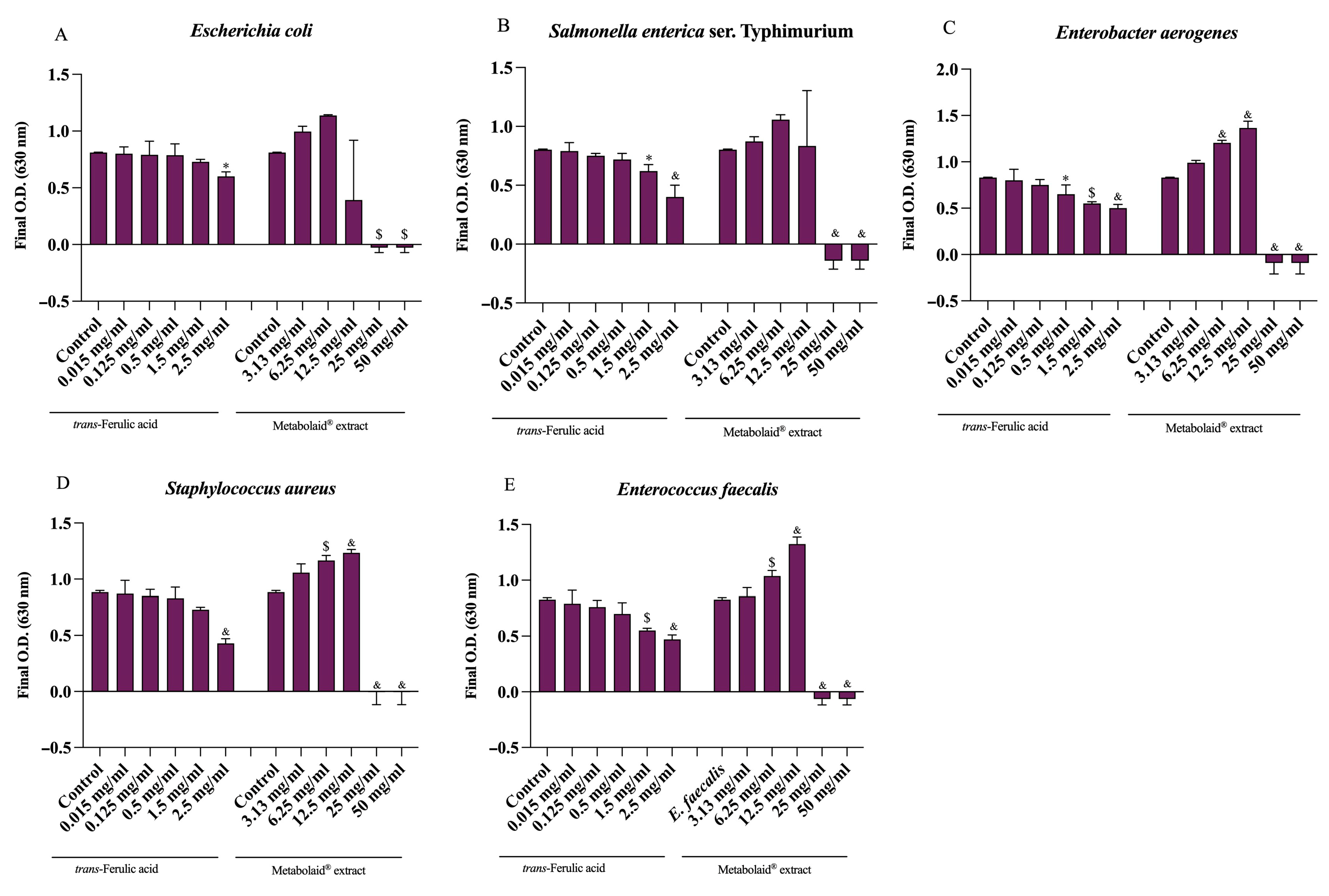
2.5. Antimicrobial Activity

The effect of the sample extract on selected bacteria growth was determined according to [37] with some modifications. The tested bacteria were cultured in MHB at 37 °C for 16 h and diluted to match the turbidity of 0.5 McFarland standard. An amount of 50 microliters of bacterial suspensions (about 1–5 × 10⁵ CFU/mL) was added to 100 µL of MHB and to 100 µL of extract (3.13, 6.25, 12.5, 25, and 50 mg/mL) and trans-ferulic acid as a phenolic standard (0.015–0.125–0.5–1.5 and 2.5 mg/mL) in a 96-well plate. A negative control was included on each microplate. The plates were incubated at 37 °C for 24 h. After incubation, the optical density (O.D.) at 630 nm was spectrophotometrically measured using a microplate reader (Eti-System fast reader, Sorin Biomedica, Modena, Italy). Results are presented as final growth (O.D.), representing the bacterial density at the end of incubation with or without the extracts.

2.6. Statistical Analysis

The data are reported as the mean and standard deviation of triplicates. The statistically significant (p < 0.05) differences between the extract concentrations (3.13, 6.25, 12.5, 25, and 50 mg/mL) on final growth (O.D.) of Gram-negative (Escherichia coli ATCC 25922, S. enterica ser. Typhimurium ATCC 14028, and Enterobacter aerogenes ATCC 13048) and Gram-positive (Staphylococcus aureus ATCC 25923 and Enterococcus faecalis ATCC 29212) bacteria and the control were analyzed by one-way ANOVA followed by Barlett’s test, with p ≤ 0.05. Statistical analyses were performed using Prism, GraphPad Software (San Diego, CA, USA).

3. Results

Different methods were used to measure the antioxidant properties of a Metabolaid® extract. FRAP in our case showed 0.32 mg TE/g f.w. The DPPH• scavenging activity of the extract was 1.32 mg TE/g f.w. The oxygen radical absorbance capacity (ORAC) measures how well antioxidants can absorb and neutralize oxygen radicals. Higher values indicate stronger antioxidant effects. In this extract, there is a 474.13 mg TE/g f.w. There is a 256.10 mg GAE/g f.w. of total phenols, 48.90 mg CE/g f.w. of total flavonoids, and 60.17 mg QE/g f.w of flavonols. Anthocyanins, pigments found in plants (especially in red, blue, and purple fruits) with strong antioxidant effects, were found in a capacity of 3.78 mg C3GE/g f.w. (Table 1).
Table 2 reports the phenolic profile of the Metabolaid® extract by UHPLC-ESI-MS/MS. In total, thirty-five polyphenols were characterized and quantified. The largest part of the phenolic compounds goes to Verbascoside in the Metabolaid® extract with a percentage of 98.93% in the other compounds category. The second largest amount, 75.08%, goes to 3-O-Caffeoylquinic acid (Chlorogenic acid) in the phenolic acid family. Delphinidin 3,5-O-diglucoside (Delphin), with a percentage of 61.47%, is in third place. Then, also from the anthocyanins family, a large part goes to Cyanidin 3-O-glucoside (Kuromanin) 37.75%. With the aim of quantifying specific polyphenols present in the Metabolaid® sample extract, selected reaction monitoring (SRM) transitions and their corresponding phenolic compounds were identified and analyzed, and the results are reported in Supplementary Materials (Table S1).
The antimicrobial activity of the tested compound was evaluated against five bacterial strains using minimum inhibitory concentration (MIC) and minimum bactericidal concentration (MBC) assays. Escherichia coli (ATCC 25922) exhibited an MIC of 12.5 mg/mL, indicating the inhibition of bacterial growth at this concentration, while the MBC was determined to be 25 mg/mL, signifying complete bacterial eradication. Salmonella enterica ser. Typhimurium (ATCC 14028) demonstrated a higher MIC (25 mg/mL) and MBC (50 mg/mL), suggesting that this strain requires a greater concentration of the antimicrobial agent for both growth inhibition and bactericidal activity compared to E. coli. Similarly, Enterobacter aerogenes (ATCC 13048) and Enterococcus faecalis (ATCC 29212) exhibited identical susceptibility profiles, with an MIC of 12.5 mg/mL and an MBC of 25 mg/mL, indicating comparable sensitivity to the antimicrobial compound. In contrast, Staphylococcus aureus (ATCC 25923) presented an MIC of 25 mg/mL and an MBC of 50 mg/mL, demonstrating a lower susceptibility to the tested compound than E. coli and Enterobacter aerogenes, yet a similar response to Salmonella enterica ser. Typhimurium. These findings suggest varying degrees of antimicrobial efficacy across different bacterial species, highlighting the need for targeted dosing strategies (Table 3 and Figure 1).

4. Discussion

The antioxidant capacity of the Metabolaid® extract, as measured by DPPH radical scavenging, demonstrated potent activity, with a concentration of 23.33 µg/mL required to neutralize 50% of DPPH radicals (IC50). This indicates a relatively strong free radical scavenging potential.
In a comparative study, Ficus religiosa extract exhibited 43.415% scavenging activity at an absorbance of 0.550 at 517 nm [38]. Although these results are not expressed as IC50 values, the percentage inhibition at a given concentration allows for a qualitative comparison.
A study by [39] evaluated the antioxidant activity of Limnophila aromatica using different extraction solvents. The FRAP values ranged from 0.96 to 1.85 mg Fe2+/g dry weight, depending on the solvent used. This suggests that the Metabolaid® extract exhibits comparable or higher antioxidant capacity, considering the result based on fresh weight.
The antioxidant activity of total phenols is 256.10 mg GAE/g f.w. The antioxidant activity and total phenolic content varied significantly among the selected plants. According to [40], aqueous extracts of oak (Quercus robur), pine (Pinus maritima), and cinnamon (Cinnamomum zeylanicum) demonstrated the highest antioxidant capacities across most of the methods tested, indicating their potential as rich sources of natural antioxidants. These extracts also contained the highest levels of phenolic compounds, ranging from 300 to 400 mg GAE/g. Additionally, aqueous extracts of mate (Ilex paraguariensis) and clove (Eugenia caryophyllus) exhibited strong antioxidant activity and substantial phenolic content, approximately 200 mg GAE/g. The ORAC value of 1893.77 µmol TE/g f.w. suggests a strong capacity to neutralize peroxyl radicals through hydrogen atom transfer mechanisms. This value is higher than red pepper in [41], which exhibited ORAC values around 175 µmol TE/g. The antimicrobial activity of the tested compound was evaluated using minimum inhibitory concentration (MIC) and minimum bactericidal concentration (MBC) assays, revealing significant inhibitory effects across both Gram-negative and Gram-positive bacterial strains. Escherichia coli, Enterobacter aerogenes, and Enterococcus faecalis were particularly sensitive, with MIC values of 12.5 mg/mL and MBC values of 25 mg/mL. In contrast, Salmonella enterica ser. Typhimurium and Staphylococcus aureus required higher concentrations (MIC: 25 mg/mL, MBC: 50 mg/mL), suggesting greater resistance.
These findings align partially with previous work by [42], who reported that methanolic leaf extracts of Verbascum thapsus exhibited strong antimicrobial activity against several strains, including S. enterica ser. Typhimurium and E. coli, with much lower MIC values (15.625–31.25 µg/mL) and MBC values ranging between 31.25 and 62.5 µg/mL. The discrepancies in MIC/MBC values between their study and the present may be attributed to differences in extract composition, bacterial strain variability, or the presence of synergistic compounds in the V. thapsus methanolic extract.
Nevertheless, both studies support the broad-spectrum antimicrobial potential of phenolic-rich plant extracts. In our case, the presence of high levels of verbascoside and chlorogenic acid likely contributed to the observed effects, as these compounds are well-documented for their ability to disrupt bacterial membranes and interfere with key enzymatic functions [43]. These results reinforce the potential of natural phenolic compounds as viable alternatives or supplements to conventional antibiotics, especially in light of increasing antibiotic resistance.

5. Conclusions

The antioxidant activity of the Metabolaid® extract was evaluated through DPPH radical scavenging, revealing a potent activity and indicating its strong capacity to neutralize free radicals. The total phenolic content of the extract reinforces its overall antioxidant potential. Additionally, the extract demonstrated a high ORAC value, underscoring its ability to neutralize peroxyl radicals and suggesting a robust antioxidant defense mechanism.
In terms of antimicrobial activity, the Metabolaid® extract exhibited significant inhibitory effects against a range of bacterial strains. The minimum inhibitory concentration (MIC) and the minimum bactericidal concentration (MBC) for Escherichia coli, Enterobacter aerogenes, and Enterococcus faecalis indicated strong antimicrobial properties. However, Salmonella enterica ser. Typhimurium and Staphylococcus aureus required higher concentrations, suggesting greater resistance to the extract. The bioactive compounds in Metabolaid®, including verbascoside and chlorogenic acid, are likely key contributors to its antimicrobial and antioxidant effects.
Overall, the findings suggest that Metabolaid® holds promise as a natural alternative or supplement to conventional antibiotics, particularly in the context of increasing antibiotic resistance. The obtained results give valid indications in the process of the evaluation of Metabolaid® as a supplement for food and feed innovative formulation.

Supplementary Materials

The following supporting information can be downloaded at: https://www.mdpi.com/article/10.3390/pr13082405/s1, Table S1: SRM transitions and the corresponding phenolic compounds of Metabolaid® extract by UHPLC-ESI-MS/MS.

Author Contributions

Conceptualization, F.V., V.L. and L.P.; methodology, F.V., L.P. and A.R.; investigation, F.V., A.K., I.S., L.P. and A.R.; data curation, L.P., A.K. and I.S.; writing—original draft preparation, F.V., L.P. and A.K.; writing—review and editing, F.V., L.P. and A.K.; funding acquisition, F.V. and V.L. All authors have read and agreed to the published version of the manuscript.

Funding

The investigation was conducted with the collaboration and contribution of all co-authors. The research was funded by departmental resources of the National Agricultural and Food Centre (Luzianky, Slovakia) and National Research Council (Pisa, Italy).

Data Availability Statement

The original contributions presented in this study are included in the article/Supplementary Materials. Further inquiries can be directed to the corresponding author.

Conflicts of Interest

The authors declare no conflicts of interest.

Abbreviations

The following abbreviations are used in this manuscript:
GAEGallic acid equivalents
DMDry matter
C3GECyanidin-3-glucoside equivalents
FRAPFerric reducing antioxidant power
ORACOxygen radical absorbance capacity
MICMinimum inhibitory concentration
MBCMinimum bactericidal concentration
NF-κBNuclear Factor kappa-light-chain-enhancer of activated B cells
PPARPeroxisome Proliferator-Activated Receptors
AMPKAMP-activated Protein Kinase
DPPH2,2-Diphenyl-1-picrylhydrazyl assay
HCLHydrochloric acid
APPH2,2′-Azobis(2-methylpropionamidine) dihydrochloride

References

  1. Olszowy, M. What is responsible for antioxidant properties of polyphenolic compounds from plants? Plant Physiol. Biochem. 2019, 144, 135–143. [Google Scholar] [CrossRef]
  2. Yahia, Y.; Benabderrahim, M.A.; Tlili, N.; Bagues, M.; Nagaz, K. Bioactive compounds, antioxidant and antimicrobial activities of extracts from different plant parts of two Ziziphus Mill. species. PLoS ONE 2020, 15, e0232599. [Google Scholar] [CrossRef] [PubMed]
  3. Dikpınar, T.; Süzgeç-Selçuk, S. Antimicrobial activities of medicinal plants containing phenolic compounds. J. Nat. Prod. 2020, 10, 514–534. [Google Scholar] [CrossRef]
  4. Singh, B.; Singh, J.P.; Kaur, A.; Singh, N. Antimicrobial potential of pomegranate peel: A review. Int. J. Food Sci. Technol. 2019, 54, 959–965. [Google Scholar] [CrossRef]
  5. Lopez-Corona, A.V.; Valencia-Espinosa, I.; González-Sánchez, F.A.; Sánchez-López, A.L.; Garcia-Amezquita, L.E.; Garcia-Varela, R. Antioxidant, Anti-Inflammatory and Cytotoxic Activity of Phenolic Compound Family Extracted from Raspberries (Rubus idaeus): A General Review. Antioxidants 2022, 11, 1192. [Google Scholar] [CrossRef]
  6. Calderón-Pérez, L.; Llauradó, E.; Companys, J.; Pla-Pagà, L.; Pedret, A.; Rubió, L.; Gosalbes, M.J.; Yuste, S.; Solà, R.; Valls, R.M. Interplay between dietary phenolic compound intake and the human gut microbiome in hypertension: A cross-sectional study. Food Chem. 2021, 344, 128567. [Google Scholar] [CrossRef]
  7. Perveen, S.; Al-Taweel, A. (Eds.) Pharmacognosy—Medicinal Plants; InthechOpen: London, UK, 2019; pp. 59–72. [Google Scholar]
  8. World Health Organization (WHO). Traditional medicine: Report of a WHO Expert Committee. WHO Tech. Rep. Ser. 1976, 622, 1–49. Available online: https://apps.who.int/iris/handle/10665/40995 (accessed on 4 June 2025).
  9. Mickymaray, S. Efficacy and mechanism of traditional medicinal plants and bioactive compounds against clinically important pathogens. Antibiotics 2019, 8, 257. [Google Scholar] [CrossRef]
  10. Sofowora, A.; Ogunbodede, E.; Onayade, A. The role and place of medicinal plants in the strategies for disease prevention. African Journal of Traditional, Complementary and Alternative Medicines 2013, 10, 210–229. [Google Scholar] [CrossRef]
  11. Kazemipoor, M.; Hamzah, S.; Hajifaraji, M.; Radzi, C.W.; Cordell, G.A. Slimming and appetite-suppressing effects of caraway aqueous extract as a natural therapy in physically active women. Phytother. Res. 2016, 30, 981–987. [Google Scholar] [CrossRef]
  12. Duque-Soto, C.; Expósito-Almellón, X.; García, P.; Pando, M.E.; Borrás-Linares, I.; Lozano-Sánchez, J. Extraction, characterization, and bioactivity of phenolic compounds—A case on Hibiscus genera. Foods 2023, 12, 963. [Google Scholar] [CrossRef]
  13. Ojulari, O.V.; Lee, S.G.; Nam, J.O. Beneficial effects of natural bioactive compounds from Hibiscus sabdariffa L. on obesity. Molecules 2019, 24, 210. [Google Scholar] [CrossRef]
  14. Takada, K.; Nakano, S.; Nishio, R.; Muku, D.; Mochizuki, S.; Inui, I.; Okita, K.; Koga, A.; Watanabe, K.; Yoshioka, Y.; et al. Medicinal herbs, especially Hibiscus sabdariffa, inhibit oral pathogenic bacteria. J. Oral Biosci. 2024, 66, 179–187. [Google Scholar] [CrossRef] [PubMed]
  15. Abdallah, E.M. Antibacterial activity of Hibiscus sabdariffa L. calyces against hospital isolates of multidrug resistant Acinetobacter baumannii. J. Acute Dis. 2016, 5, 512–516. [Google Scholar] [CrossRef]
  16. Newall, C.A.; Anderson, L.A.; Phillipson, J.D. Herbal Medicines: A Guide for Healthcare Professionals; Pharmaceutical Press: London, UK, 1996; p. 296. [Google Scholar]
  17. Jamila, F.; Mostafa, E. Ethnobotanical survey of medicinal plants used by people in oriental Morocco to manage various ailments. J. Ethnopharmacol. 2014, 154, 76–87. [Google Scholar] [CrossRef] [PubMed]
  18. Lenoir, L.; Rossary, A.; Joubert-Zakeyh, J.; Vergnaud-Gauduchon, J.; Farges, M.C.; Fraisse, D.; Texier, O.; Lamaison, J.-L.; Vasson, M.P.; Felgines, C. Lemon verbena infusion consumption attenuates oxidative stress in dextran sulfate sodium-induced colitis in the rat. Dig. Dis. Sci. 2011, 56, 3534–3545. [Google Scholar] [CrossRef]
  19. Herranz-López, M.; Barrajón-Catalán, E.; Segura-Carretero, A.; Menéndez, J.A.; Joven, J.; Micol, V. Lemon verbena (Lippia citriodora) polyphenols alleviate obesity-related disturbances in hypertrophic adipocytes through AMPK-dependent mechanisms. Phytomedicine 2015, 22, 605–614. [Google Scholar] [CrossRef]
  20. Alipieva, K.; Korkina, L.; Orhan, I.E.; Georgiev, M. Verbascoside—A review of its occurrence, (bio)synthesis and pharmacological significance. Biotechnol. Adv. 2014, 32, 1065–1076. [Google Scholar] [CrossRef]
  21. Rossi, R.; Mainardi, E.; Vizzarri, F.; Corino, C. Verbascoside-rich plant extracts in animal nutrition. Antioxidants 2024, 13, 39. [Google Scholar] [CrossRef]
  22. Carrera-Quintanar, L.; Funes, L.; Vicente-Salar, N.; Blasco-Lafarga, C.; Pons, A.; Micol, V.; Roche, E. Effect of polyphenol supplements on redox status of blood cells: A randomized controlled exercise training trial. Eur. J. Nutr. 2015, 54, 1081–1093. [Google Scholar] [CrossRef]
  23. Funes, L.; Carrera-Quintanar, L.; Cerdán-Calero, M.; Ferrer, M.D.; Drobnic, F.; Pons, A.; Roche, E.; Micol, V. Effect of lemon verbena supplementation on muscular damage markers, proinflammatory cytokine release and neutrophil oxidative stress in chronic exercise. Eur. J. Appl. Physiol. 2011, 111, 695–705. [Google Scholar] [CrossRef]
  24. Quirantes-Piné, R.; Herranz-López, M.; Funes, L.; Borrás-Linares, I.; Micol, V.; Segura-Carretero, A.; Fernández-Gutiérrez, A. Phenylpropanoids and their metabolites are the major compounds responsible for blood-cell protection against oxidative stress after administration of Lippia citriodora in rats. Phytomedicine 2013, 20, 1112–1118. [Google Scholar] [CrossRef]
  25. Funes, L.; Fernández-Arroyo, S.; Laporta, O.; Pons, A.; Roche, E.; Segura-Carretero, A.; Fernández-Gutiérrez, A.; Micol, V. Correlation between plasma antioxidant capacity and verbascoside levels in rats after oral administration of lemon verbena extract. Food Chem. 2009, 117, 589–598. [Google Scholar] [CrossRef]
  26. Kang, J.S.; Lee, W.K.; Lee, C.W.; Yoon, W.K.; Kim, N.; Park, S.K.; Lee, H.S.; Park, H.K.; Han, S.B.; Yun, J.; et al. Improvement of high-fat diet-induced obesity by a mixture of red grape extract, soy isoflavone and L-carnitine: Implications in cardiovascular and non-alcoholic fatty liver diseases. Food Chem. Toxicol. 2011, 49, 2453–2458. [Google Scholar] [CrossRef] [PubMed]
  27. Walton, R.J.; Whitten, D.L.; Hawrelak, J.A. The efficacy of Hibiscus sabdariffa (rosella) in essential hypertension: A systematic review of clinical trials. Aust. J. Herb. Med. 2016, 28, 48–51. [Google Scholar]
  28. Eddouks, M.; Maghrani, M.; Lemhadri, A.; Ouahidi, M.-L.; Jouad, H. Ethnopharmacological survey of medicinal plants used for the treatment of diabetes mellitus, hypertension and cardiac diseases in the south-east region of Morocco (Tafilalet). J. Ethnopharmacol. 2002, 82, 97–103. [Google Scholar] [CrossRef] [PubMed]
  29. Serban, C.; Sahebkar, A.; Ursoniu, S.; Andrica, F.; Banach, M. Effect of sour tea (Hibiscus sabdariffa L.) on arterial hypertension: A systematic review and meta-analysis of randomized controlled trials. J. Hypertens. 2015, 33, 1119–1127. [Google Scholar] [CrossRef]
  30. Guardiola, S.; Mach, N. Therapeutic potential of Hibiscus sabdariffa: A review of the scientific evidence. Endocrinol. Y Nutr. Órgano De La Soc. Española De Endocrinol. Y Nutr. 2014, 61, 274–295. [Google Scholar] [CrossRef]
  31. Jiang, F.; Zhang, Y.; Dusting, G.J. NADPH oxidase-mediated redox signaling: Roles in cellular stress response, stress tolerance, and tissue repair. Pharmacol. Rev. 2011, 63, 218–242. [Google Scholar] [CrossRef]
  32. Singleton, V.L.; Orthofer, R.; Lamuela-Raventós, R.M. Analysis of total phenols and other oxidation substrates and antioxidants by means of Folin–Ciocalteu reagent. Methods Enzymol. 1999, 299, 152–178. [Google Scholar]
  33. Benzie, I.F.F.; Strain, J.J. Ferric reducing/antioxidant power assay: Direct measure of total antioxidant activity of biological fluids and modified version for simultaneous measurement of total antioxidant power and ascorbic acid concentration. Methods Enzymol. 1999, 299, 15–27. [Google Scholar] [PubMed]
  34. Boudjou, S.; Oomah, B.D.; Zaidi, F.; Hosseinian, F. Phenolics content and antioxidant and anti-inflammatory activities of legume fractions. Food Chem. 2013, 138, 1543–1550. [Google Scholar] [CrossRef] [PubMed]
  35. Gabriele, M.; Parri, E.; Felicioli, A.; Sagona, S.; Pozzo, L.; Biondi, C.; Domenici, V.; Pucci, L. The antioxidant capacity of honey varies according to the origin of the flowers used in its production. Food Chem. 2015, 174, 637–643. [Google Scholar]
  36. Pozzo, L.; Russo, R.; Frassinetti, S.; Vizzarri, F.; Árvay, J.; Vornoli, A.; Casamassima, D.; Palazzo, M.; Croce, C.M.D.; Longo, V. Wild Italian Prunus spinosa L. fruit exerts in vitro antimicrobial activity and protects against in vitro and in vivo oxidative stress. Foods 2020, 9, 5. [Google Scholar] [CrossRef] [PubMed]
  37. Delgado-Adamez, J.; Fernandez-Leon, M.F.; Velardo-Micharet, B.; Gonzalez-Gomez, D. In vitro assays of the antibacterial and antioxidant activity of aqueous leaf extracts from different Prunus salicina Lindl. Cultivars. Food Chem. Toxicol. 2012, 50, 2481–2486. [Google Scholar] [CrossRef]
  38. Baliyan, S.; Mukherjee, R.; Priyadarshini, A.; Vibhuti, A.; Gupta, A.; Pandey, R.P.; Chang, C.M. Determination of antioxidants by DPPH radical scavenging activity and quantitative phytochemical analysis of Ficus religiosa. Molecules 2022, 27, 1326. [Google Scholar] [CrossRef]
  39. Do, Q.D.; Angkawijaya, A.E.; Tran-Nguyen, P.L.; Huynh, L.H.; Soetaredjo, F.E.; Ismadji, S.; Ju, Y.H. Effect of extraction solvent on total phenol content, total flavonoid content, and antioxidant activity of Limnophila aromatica. J. Food Drug Anal. 2014, 22, 296–302. [Google Scholar] [CrossRef]
  40. Dudonné, S.; Vitrac, X.; Coutière, P.; Woillez, M.; Mérillon, J.M. Comparative study of antioxidant properties and total phenolic content of 30 plant extracts of industrial interest using DPPH, ABTS, FRAP, SOD, and ORAC assays. J. Agric. Food Chem. 2009, 57, 1768–1774. [Google Scholar] [CrossRef]
  41. Ou, B.; Huang, D.; Hampsch-Woodill, M.; Flanagan, J.A.; Deemer, E.K. Analysis of Antioxidant Activities of Common Vegetables Employing Oxygen Radical Absorbance Capacity (ORAC) and Ferric Reducing Antioxidant Power (FRAP) Assays: A Comparative Study. J. Agric. Food Chem. 2002, 50, 3122–3128. [Google Scholar] [CrossRef]
  42. Lone, A.S.; Ravindran, K.C.; Jeandet, P. Evaluation of antimicrobial activity and bioactive compound analysis of Verbascum thapsus L.: A folklore medicinal plant. Phytomedicine Plus 2024, 4, 100560. [Google Scholar] [CrossRef]
  43. Puupponen-Pimiä, R.; Nohynek, L.; Meier, C.; Kähkönen, M.; Heinonen, M.; Hopia, A.; Oksman-Caldentey, K.-M. Antimicrobial properties of phenolic compounds from berries. J. Appl. Microbiol. 2001, 90, 494–507. [Google Scholar] [CrossRef]
Figure 1. Effect of trans-ferulic acid as reference standard (0.015, 0.125, 0.5, 1.5, and 2.5 mg/mL) and Metabolaid® extract (3.13, 6.25, 12.5, 25, and 50 mg/mL) on final growth (O.D.) of Gram-negative ((A), Escherichia coli ATCC 25922; (B), Salmonella enterica ser. Typhimurium ATCC 14028; (C), Enterobacter aerogenes ATCC 13048) and Gram-positive ((D), Staphylococcus aureus ATCC 25923; (E), Enterococcus faecalis ATCC 29212) bacteria. Results are expressed as mean ± SD. Significantly different from control by one-way ANOVA with Fisher’s multiple comparison test: & p ≤ 0.001, $ p ≤ 0.01, and * p ≤ 0.05. Results are reported as mean (n = 3) values ± standard deviation.
Processes 13 02405 g001
Table 1. Antioxidant activity of a Metabolaid® extract. Results are reported as mean values ± SD of three replicates, expressed on dry matter.
Value
Antioxidant activityFRAP (mg TE/g)0.32 ± 0.06
DPPH (mg TE/g)1.32 ± 0.05
ORAC (mg TE/g)474.13 ± 7.59
Bioactive compoundsTotal phenols256.10 ± 2.26
Total flavonoids48.90 ± 2.95
Total flavonols60.17 ± 7.68
Anthocyanins3.78 ± 0.17
Table 2. Phenolic compound concentrations of Metabolaid® extract by UHPLC-ESI-MS/MS.
Compound NameConcentration
(µg/100 g)
Concentration (%)
3-O-Caffeoylquinic acid (Chlorogenic acid)27.63 ± 2.3775.08
Gallic acid2.81 ± 0.077.64
Caffeic acid2.81 ± 0.077.64
p-Coumaric acid1.10 ± 0.082.99
Vanillic acid0.89 ± 0.062.42
trans-Ferulic acid0.88 ± 0.062.39
Rosmarinic acid0.68 ± 0.081.85
1,3-Dicaffeoylquinic acid (Cynarin)0.01 ± 0.000.03
∑ Phenolic acids36.80100
Quercetin6.33 ± 0.0539.74
Quercetin 3-O-glucoside0.08 ± 0.030.50
Quercetin 3-O-rutinoside (Rutin)7.15 ± 0.1244.88
Quercetin 3,4-O-diglucoside1.09 ± 0.106.84
Kaempferol 7-O-glucoside0.07 ± 0.010.44
Kaempferol 3-O-glucoside0.78 ± 0.084.90
Kaempferol 3-O-rutinoside0.43 ± 0.042.70
∑ Flavonols15.93100
Cyanidin 3-O-glucoside (Kuromanin)7.26 ± 0.3037.75
Cyanidin 3,5-O-diglucoside (Cyanin)0.02 ± 0.000.10
Delphinidin 3,5-O-diglucoside (Delphin)11.82 ± 0.0961.47
Malvidin 3-O-glucoside (Oenin)0.01 ± 0.010.05
Petunidin 3-O-glucoside0.11 ± 0.030.57
∑ Anthocyanins19.23100
Apigenin0.46 ± 0.0495.83
(+)-Catechin0.01 ± 0.012.08
(−)-Epicatechin0.01 ± 0.002.08
∑ Flavan-3-ols0.48100
Verbascoside233.40 ± 20.3898.93
Luteolin0.83 ± 0.090.35
Hydroxytyrosol0.56 ± 0.060.24
Phloretin0.46 ± 0.040.19
Naringenin0.30 ± 0.080.13
Oleuropein0.24 ± 0.050.10
Ligstroside0.13 ± 0.020.5
Phloridzin0.02 ± 0.010.01
Resveratrol0.02 ± 0.010.01
∑ Others235.92100
Table 3. Minimum inhibitory concentration (MIC) and minimum bactericidal concentration (MBC) of Metabolaid® extract.
Bacterial StrainsMIC * (mg/mL)MBC * (mg/mL)
Escherichia coliATCC 2592212.525
Salmonella enterica ser. TyphimuriumATCC 140282550
Enterobacter aerogenesATCC 1304812.525
Enterococcus faecalisATCC 2921212.525
Staphylococcus aureusATCC 259232550
* Results are reported as mean values ± SD of three replicates.
Disclaimer/Publisher’s Note: The statements, opinions and data contained in all publications are solely those of the individual author(s) and contributor(s) and not of MDPI and/or the editor(s). MDPI and/or the editor(s) disclaim responsibility for any injury to people or property resulting from any ideas, methods, instructions or products referred to in the content.

