You are currently viewing a new version of our website. To view the old version click .
Processes
  • Article
  • Open Access

6 November 2025

Solid Waste Management: Degradation of Commercial and Newly Fabricated Cellulose Acetate Ultrafiltration Membranes

,
,
,
and
1
Water Industry Engineering Department, The Engineering Faculty, Kinneret Academic College on the Sea of Galilee, Zemach 1513200, Israel
2
School of Mechanical Engineering, Faculty of Engineering, Tel Aviv University, Tel Aviv 69978, Israel
3
BioCastle Water Technologies Ltd., Afikim Jordan Valley 1510501, Israel
*
Author to whom correspondence should be addressed.
This article belongs to the Section Environmental and Green Processes

Abstract

Treatment of polymeric solid waste, such as used membranes, is vital for environmental sustainability. Cellulose-based membranes are widely utilized in the water industry due to their resistance to biodegradation. These non-biodegradable membranes can persist in landfills and aquatic environments for extended periods. Our study assessed the biodegradation potential of Trametes versicolor on a newly fabricated cellulose acetate (CA) membrane and a commercially produced membrane under various conditions, including oxidative stress. Additionally, we employed T. versicolor encapsulated in a small bioreactor platform (SBP) for media inoculation and biomass augmentation. Treatment of the commercially produced CA membrane within a timeframe of 30 days was unsuccessful. This was primarily attributed to the structural stability of the membrane over time and the limited ability of the culture to attach to the membrane surface. These results underscore the necessity of exploring alternative biopolymer cellulose-based materials for ultrafiltration (UF) and microfiltration (MF) membrane applications. The custom-made UF membrane, treated by ozonation as a pretreatment, emerged as an effective approach for enhancing biodegradation. Combining these factors, we expect to achieve over 27.75 ± 1.5% weight loss in membrane solids by 30 days of treatment. This study represents the first inquiry into the biodegradation capabilities of T. versicolor on CA-based membranes.

1. Introduction

Membrane technology plays a principal role in water and wastewater treatment due to its numerous advantages, including minimal environmental impact, ease of installation, low cost, compact footprint, and high output with reduced energy and chemical consumption. More than 70% of the world’s desalination capacity is provided by membrane technology; furthermore, membrane bioreactor (MBR) technology is currently taking the forefront in the wastewater treatment sector []. Additionally, membranes for microfiltration (MF), ultrafiltration (UF), nanofiltration (NF), and reverse osmosis (RO) find application across a wide range of sectors, from treating industrial effluent to domestic drinking water. This has led to a massive market for membranes, projected to grow from USD 6.4 billion in 2022 to USD 10.1 billion by 2027, at an annual growth rate of 9.7% []. Commercially available polymeric materials such as polyvinylidene fluoride (PVDF), polyethersulfone (PES), polysulfone, and polyacrylonitrile are derived from petroleum hydrocarbons which are non-biodegradable, environmentally toxic, and can cause secondary pollution in the recycling process []. The environmental concern regarding polymeric membranes primarily revolves around their production, disposal, and contribution to plastic pollution and microplastics worldwide distribution. Non-biodegradable membranes can persist in landfills and aquatic ecosystems for decades. Thus, the absence of efficient downstream recycling technologies presents a significant challenge in the management of membrane elements at the end of their useful life. Reverse osmosis membrane elements constitute the primary solid waste stream in numerous desalination facilities. According to estimates, more than two million end-of-life RO modules may be discarded by 2025 [].
Following their service life, these elements are typically disposed of in landfills or by incineration, both of which have significant drawbacks. Landfill disposal consumes land resources and poses concerns due to the non-biodegradable nature of membranes, resulting in their transformation into microplastics and the release of environmentally harmful gases, including methane. Incineration, an alternative disposal method, produces greenhouse gases and other toxic gases. Hence, there is an urgent need to develop membranes from biodegradable polymers [,].
Among several biodegradable polymers, membranes based on cellulose and cellulose acetate (CA) are widely employed in water treatment technology owing to their abundance, excellent biocompatibility, easy processability, and relatively low cost []. Cellulose acetate is prepared by acetylating cellulose, the most abundant natural polymer. Nevertheless, a significant bottleneck in the life cycle of CA membranes is the fouling propensity of the biopolymer, which shortens their life span. To address these limitations, modifying CA membranes by blending them with other polymers can improve separation performance and increase flux, rendering them more suitable for practical use and applications [,]. However, those supplemental elements increase their resistance to the biodegradation process. Currently, the prevalent method for disposing used CA is through landfill, mainly because recycling these membranes is a complex task. Challenges such as residual contaminants, high energy requirements, limited recycling facilities and markets, as well as the need for regulatory compliance all contribute to widespread landfill practices [].
Biodegradation, a sustainable process that provides an alternative to landfill and incineration, utilizes microorganisms and enzymes to break down complex organic compounds into simpler environmentally friendly substances. It offers an eco-friendly approach for the sustainable disposal of organic waste. Degradation of CA is affected by various conditions and factors, including microorganism type, digestion time (hydraulic retention time (HRT)), and CA membrane composition. Subject to CA membrane composition, the biodegradation rate is governed by the degree of acetyl substitution (DS) and the actual conditions the material is subjected to. The biodegradation rate decreases as the DS increases; therefore, membrane composition must be considered to determine biodegradation conditions and operation in designated membrane treatment facilities. Despite this, microorganism culture type seems to have a major effect, perhaps more than the DS, as indicated by Sakai and his coworkers [], who achieved effective CA (2.3 DS) membrane biodegradation in 20 days, using Neisseria sicca SC bacterial culture (Table 3).
Another aspect of membrane waste treatment approach is the design of membranes that offer lower resistance to the biodegradation process. In a previous study, we fabricated a UF membrane made of CA blended with polycaprolactone (PCL) using a non-solvent-induced phase-separation technique, with added titanium dioxide (TiO2) nanoparticles for degrading organic contaminants under 365 nm UVA irradiation []. The present study will also demonstrate the degradation of these novel UF membranes using sequential physical and biological treatments. The biological treatment will include suspended and encapsulated white rot fungus Trametes versicolor; the latter to allow recycling and reuse of the fungus. Trametes versicolor is characterized by its non-specific and non-selective enzymatic machinery, which is capable of degrading and mineralizing polysaccharides, such as cellulose, hemicellulose, starch, and pectin, as well as non-polysaccharide polymers, such as lignin [,]. This ability makes T. versicolor an attractive option for degrading cellulose waste materials. The encapsulation method known as small bioreactor platform (SBP) capsules is based on macro-encapsulation in confined environments protected by structural microfiltration membranes made of CA. The structure, composition, and validation of the SBP capsule technology has been patented under article number PCT/IL2010/000256 []. The 3D SBP capsule (2.5 cm length and 0.8 cm diameter) creates an appropriate growth environment by providing a constant supply of nutrients and physical protection from environmental stress and prevents competition with other microorganisms [,]. Once the fungus reaches a substantial biomass concentration inside the capsule, it can breach the capsule CA membrane and exit into the external environment, where it continues to grow vigorously. Therefore, in this study, SBP technology is being used for the first time as a method for bioreactor inoculation and biomass maintained.
The aim of this study is to evaluate a relatively short treatment process for the degradation of both a self-fabricated innovative UF membrane and a commercial membrane in order to compare treatment efficacy and gain a deeper understanding of the underlying biodegradation mechanisms. This study is particularly relevant due to the increasing amounts of cellulosic derivative membrane waste. Moreover, our goal is to propose a commercial-scale design framework for the treatment of solid wastes, such as polymers. To achieve this goal, we used biodegradable polymer-based membranes with anti-fouling properties that were developed and characterized in our previous study []. The current study aimed to establish a model system that can be upscaled to the industrial scale for CA derivate UF membrane degradation, which includes both physical and biological treatment methods. The physical process employed ultraviolet (UV) irradiation at 365 nm, and membrane-integrated TiO2, suspended TiO2, ozone, and H2O2, followed by biological treatment with suspended and immobilized white rot fungus, T. versicolor. The custom-made UF-CA membranes were assessed and compared for their physical and biological biodegradability and compared with a commercialized CA membrane. To evaluate membrane loss due to the degradation treatment, net dry weight loss was measured after residual organic matter cleaning.

2. Materials and Methods

2.1. Solutions, Materials, and Cultures

Polycaprolactone (PCL) was obtained from Angene Chemical, Secunderabad, India. Titanium dioxide (TiO2) and cellulose acetate (CA) with an average molecular weight of ≈50,000 mol/g were obtained from Sigma Aldrich chemicals (Saint Louis, MO, USA). Dichloromethane (DCM) was obtained from Acros Organics (Milano, Italy). Polyethylene glycol 400 (PEG 400) was obtained from Alfa Aesar chemicals (Petach Tikva, Israel). N,N-dimethylformamide (DMF) was obtained from Bio-Lab Ltd. (Jerusalem, Israel). All chemicals were of analytical grade. Millipore water was used to prepare all the standard solutions throughout the experiments. The composition of the M9 media used for culture enrichment was mineral salts media (MSM) composed of 1 g/L NH4Cl, 3 g/L KH2PO4, 6 g/L Na2HPO4, 5 g/L NaCl, 1 mmol/L MgSO4, and 0.1 mmol/L CaCl2. Prior to sterilization of the prepared MSM, the pH of the media was adjusted to 7.0 with NaOH. In this study, we selected a fungal culture of Trametes versicolor, which is known to secrete carbohydrate esterases capable of hydrolyzing acetyl groups, thereby initiating the biodegradation process of cellulose []. Trametes versicolor was provided by the laboratory of Eyal Kurzbaum from Shamir Research Institute, Israel. The SBP capsules, containing T. versicolor in a dry state, were purchased from BioCastle Water Technologies Ltd., Affikim, Israel.

2.2. Fabrication of Membranes

The optimized polycaprolactone (PCL)-CA blend membrane was prepared according to our previous study []. We used DMF to dissolve CA, and DCM to dissolve PCL; the solvent mixture of DMF and DCM in % v/v was maintained according to the mass ratio of CA and PCL. The PCL-CA blend membranes were prepared by taking CA and PCL at ratios of 70:30 (w%). In a typical experiment, the doped solution for UFM-70 was prepared by adding 2.625 g CA and 1.125 g PCL to a conical flask containing 15.75 mL DMF, 6.75 mL DCM, and 2.5 mL PEG 400 (15% solids). The mixture was stirred constantly at 200 rpm using a shaking incubator for 12 h at 35 °C. The solutions were cooled to room temperature before being cast on a steel plate with a MemcastTM (Nazareth-De Pinte, Belgium) device. The casting time was usually between 7 and 10 s, including the time required to transfer the steel plate to the deionized (DI) water bath at 25 °C. After 24 h of thorough solvent exchange, the membrane was detached from the steel plate and stored in a plastic bag with DI water to keep it moist. The composition of all the membranes is listed in Table 1.
Table 1. Composition of all the ultrafiltration membranes.
S/NMembraneCA (% w/v)PCL (% w/v)DMF (% v/v)DCM (% v/v)PEG (% v/v)DS
01UFM70306327101.8
02UFM-T *70306327101.8
* T—2% w/v H-TiO2 catalyst was added as a photocatalyst. S/N—serial number, CA—cellulose acetate, PCL—polycaprolactone, DMF—dimethylformamide, DCM—dichloromethane, PEG—polyethylene glycol, DS—degree of acetyl substitution.

2.3. Fabrication of Photo-Assisted Self-Cleaning Membranes

First, commercially available TiO2 was calcined at 550 °C for 2 h at room temperature to obtain TiO2 in the pure anatase phase (H-TiO2) []. A self-cleaning membrane was fabricated by dispersing 1%, 1.5%, and 2% (w/v) H-TiO2 as nanoparticles into the optimized composition (UFM). Typically, 500 mg of the nanomaterial was dispersed in 25 mL of the solvent mixture in a conical flask, followed by sonication for 5 min using an ultrasonicator to obtain 2% (w/v) TiO2-loaded dope solution. The optimized weight of the polymer was then added and dissolved by continuous stirring at 200 rpm, and the doped solution was cast on the metal sheet using a MemcastTM unit following the procedure outlined in Section 2.1 to produce the UFM-T membrane. The UFM and UFM-T membranes were presented and characterized in our previous study [].

2.4. Ozonation

Ozonation was applied as a standalone pretreatment using a constant ozone dose of 40 g Nm−3 delivered at 0.44 L min−1 for 30 min. Ozone was generated by a corona-discharge generator and introduced through a fine-pore diffuser into a 500 mL membrane suspension held at 20 ± 2 °C with magnetic stirring at 300 rpm to ensure uniform bubble dispersion and maximize mass transfer. After treatment, membranes were separated by vacuum filtration, air-dried at room temperature, then transferred to biodegradation reactors, ensuring no oxidative by-products entered the fungal system. Off-gas was vented through laboratory fume extraction (industrial scale would employ catalytic ozone destruct units). Trametes versicolor exhibited no inhibition due to ozonation-induced surface modifications enhancing fungal attachment.

2.5. Study System Setup

The goal of this study was to create a membrane degradation system that could be readily scaled up for industrial applications (Figure 1). Hydraulic retention time (HRT) affects both the bioreactor volume and overall infrastructure. In this study, we aimed to shorten the HRT to a duration similar to that commonly used in industrial wastewater treatment. For instance, refinery wastewater treatment typically requires an HRT of 1–15 days [], whey wastewater in a sequencing batch reactor requires around 4 days [], and municipal leachate treatment requires approximately 4.5 days []. Accordingly, the following few key parameters were established for the system design:
(1)
Pretreatment stage: This chamber serves to achieve an adequate HRT, which in turn decreases membrane structural stability while promoting membrane biocompatibility for the attachment of fungal biomass. Additionally, it offers flexibility in combining various treatment methods.
(2)
Bioreactor: Designed to ensure an adequate HRT for biodegradation, but not excessively long, as prolonged HRT can reduce the efficiency of the process while restricting its potential for commercial scalability. The bioreactor was operated under presumed aerobic conditions (although dissolved oxygen (D.O.) was not measured), using two air diffusers for aeration. The pH of the bioreactor medium ranged between 6.5 and 7.5, with a temperature of 30 °C. Within this treatment setup, two distinct retention times were identified as follows: (1) the solids retention time (SRT) of the membrane fragments, encompassing both the pretreatment stage and the bioreactor, and (2) the hydraulic retention time (HRT) specific to the bioreactor. The separate HRT for pretreatment and bioreactor was set at 15 days, while solids (membrane particles) retention time within the treatment system was 30 days. Both are considered relatively short. In contrast, CA degradation studies typically involve much longer HRT, ranging up to 365 days (Table 3).
(3)
The system is designed in a continuous flow configuration that allows for the accumulation of membrane solids, including biomass and membrane particles, over time. Therefore, the approach involves the use of SBP technology to control the accumulated immobilized biomass. The bioreactor was inoculated with 50 SBP capsules.
(4)
Phase separation: In this phase, there is an effluent separation chamber that facilitates the refreshment of the growth medium and solids (membrane fragments) circulated back to the bioreactor.
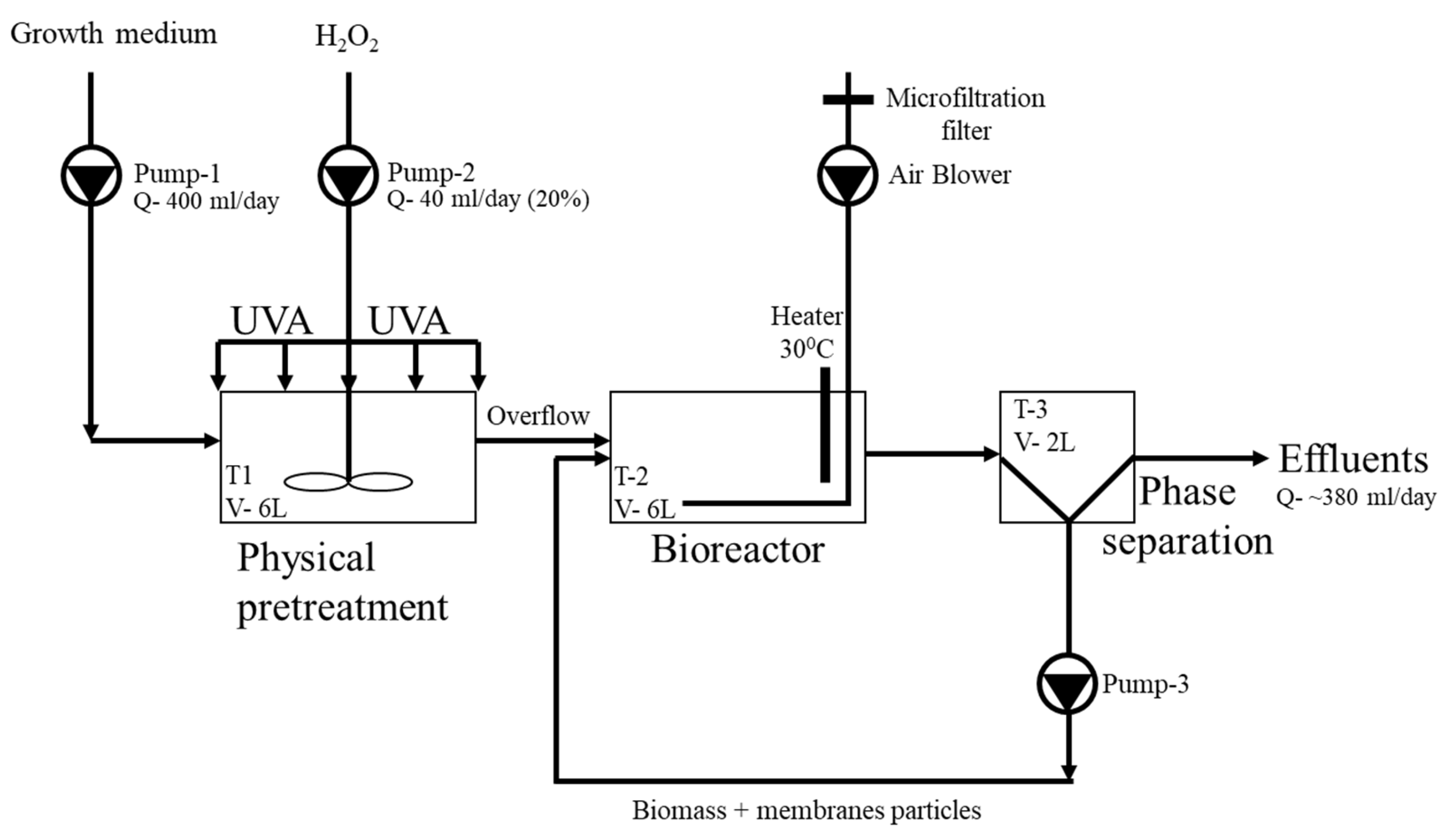
Figure 1. Process flow diagram (PFD) of the treatment system. The system operated in a continuous flow configuration. Solid materials (membrane fragments) were loaded into the pretreatment chamber (T-1) for a retention period of 15 days, followed by a biological treatment phase of an additional 15 days in the bioreactor (T-2). T-3 demonstrates phase separation. The total treatment duration was 30 days.
The treatment system model described above was subject to more than 15 cycles of testing over a span of two years, with the results from 13 of these cycles being presented in this article. We tested degradation of the custom-made CA membranes (UFM and UFM-T) in a 30-day continuous configuration, with pretreatments in different combinations, compared with a standard industrial CA membrane with a DS of 2.6 without pretreatment as a control (membrane filter 0.45 µm, 90 mm, Sartorius Stedim Biotech, Aubagne, France) (Figure 1). All tested membranes were exposed to light at 365 nm (15 W, Nuochong), at an intensity of 10 mW/cm2 for 15 min per hour for the entire test period, with a constant flow feed of H2O2 (40 mL 20% H2O2 per day for the entire test period). In some experiments we also included suspended TiO2 nanoparticles (0.5 g TiO2 in a 6 L solution of pretreatment chamber medium, 83 mg/L). Table 2 presents the membrane degradation results. Studies 1–3 present the use of suspended T. versicolor culture, while studies 4 and 5 represent the use of SBP-encapsulated T. versicolor as a tool for culture propagation. Studies 1 and 4 were used as a control for both culture propagation methods, demonstrating the treatment efficacy on the industrial membrane. Test cycles 2, 3, and 5 examined the new custom-made membranes, incorporated with TiO2 nanoparticles (UFM-T) or without TiO2 nanoparticles (UFM).
We used a culture of T. versicolor in suspended form or within SBP capsules (50 capsules per 6 L bioreactor) in aerobic conditions at a constant temperature (30 °C). The growth medium was M9 MSM minimal nutrient solution (without glucose) containing 0.006% lysogeny broth (LB) (40 mL per 6 L, once a week) and chloramphenicol (0.03 mg/mL) to inhibit bacterial growth. Each experiment was run for 30 days.
The treatment system (Figure 1) comprised four chambers with peristaltic pumps to control the system flow (nutrients, H2O2, and effluent). The first container (2 L, not shown in Figure 1) was used for inflow nutrient feed (mineral salts medium M9 MSM with 1% LB and 0.03 mg/mL chloramphenicol) with a flow rate of 400 mL/day, followed by a pretreatment tank (6 L) and a bioreactor (6 L) containing the fungal culture with an HRT of 15 days. Solids (membrane particles—9 mm2) retention time within the treatment system module was 30 days. The second chamber (pretreatment reactor) was used for physical pretreatment and contained an upper vortex (ca. 250 rpm) for medium agitation and homogenizing, UVA radiation (low pressure (LP) UV radiation 15 W power with 15 min/hour intervals) and, in some cases, constant flow feeds of H2O2 (40 mL 20% H2O2 per day for the entire experimental period). Studies 2, 3, and 5 included an additional pretreatment using ozone (40 g/Nm3 ozone for 30 min, 0.44 L/min). To the pretreatment chamber we introduced 3 mm × 3 mm CA membrane strips. The bioreactor chamber (6 L) contained T. versicolor in suspended or encapsulated form. The chamber also contained an air stone to introduce atmospheric oxygen into the solution and agitate the medium (connected to a microfiltration membrane), and a thermostat to maintain a constant temperature of 30 °C. The last chamber was used for phase separation by gravitation and overflow from the reactor, where the solids (biomass and membrane particles) circulated manually back into the reactor and effluents were collected.

2.6. Membrane Degradation Analysis

Before starting the experiment, membranes strips were dried in a vacuum oven for 24–8 h at 60 °C and weighed (W0). It is important to note that the post-treatment membrane fragments contained organic matter, including fungal hyphae that penetrated the membrane structure. Fungal hyphae suggest extensive penetration deep into the polymer, Schirp and Wolcott []. Therefore, a washing protocol was implemented to significantly reduce the presence of this organic material. Shortly after the experiment ended, membrane fragments were separated from the suspended biomass via vacuum filtration using an aluminum foil with 250 nm pores to create a barrier that helped prevent membranes from escaping into the filtrated solution. Once the entire solution was filtered, the residue was washed with double-distilled water (DW) and 35% ethanol and filtered again to remove the attached biomass from the membrane particles. The washing procedure was repeated until the filtrated solution was clear, and microscope imaging revealed no presence of T. versicolor fragments on the membrane surface. The clean membrane residuals were dried and weighed (Wt). The filtered medium containing the fungal particles was concentrated and transferred into the next batch study. The above procedures were performed in triplicate to determine %WL, the relative membrane weight loss over the course of the experiment, using Equation (1).
% WL = W 0 W t W 0 × 100

2.7. Statistics

A comparison between the commercial CA membrane and the UFM-T and UFM membranes was conducted for both suspended and SBP-encapsulated culture operations using a Student’s t-test, assuming equal variances. The Student’s t-test was chosen over Welch’s test, since the F-test confirmed homogeneity of variances (p > 0.082 in both cases).

3. Results and Discussion

3.1. Membrane Degradation by Pretreatment and Suspended Culture

Our aims in this study are to assess and compare the physical and biological degradability of the custom-made CA membranes, with and without incorporated TiO2, in comparison with an industrial CA membrane. Although CA can be produced with a range of DS, the most common is a DS of 2.5, due to its good solubility in common solvents, molecular weights, and melt properties. In their study, Komarek and coworkers [] compared CA with a DS of 1.85, 2.07, and 2.57, and found that biodegradation rates increased with decreasing DS, but were not inhibited by the higher levels of acetyl. Cellulose is readily biodegraded by organisms that utilize cellulase enzymes, but, due to the additional acetyl groups, CA requires the presence of esterases for the first stage of biodegradation. Esterases are hydrolase enzymes that split esters into alcohols and acids by the addition of water molecules. They are capable of catalyzing the cleavage and formation of ester bonds particularly in branching groups such as acetyl groups. Acetyl esterase is suggested to be distributed widely in wood-rotting fungi for the degradation of native acetylated hemicelluloses. Once partial deacetylation has been accomplished, either by enzymes or by partial chemical hydrolysis, the polymer’s cellulose backbone is readily biodegraded []. Due to their diversity of digestive enzymes, fungal cultures offer a promising contribution to CA remediation. Trametes versicolor secretes carbohydrate esterases that can hydrolyze the acetyl group and thus initiate the cellulose biodegradation process []. To the best of our knowledge, this is the first time T. versicolor has been used to biodegrade CA membranes.
Table 2. Experimental setup and results. Continuous configuration treatment simulation. Each test cycle was performed in triplicate, and the average values along with the standard deviation (±SD) were calculated.
Pretreatment
Study #Membrane TypeTrametes versicolor Culture TypeAdditional Carbon * NanoparticlesUVAH2O2OzoneInitial WeightWeight Change% Weight Loss
1ACommercial
CA membrane
Suspended-+++-5.080.30486
1B-++--4.81870.38558
1C+----5.0144.75234.94
Average (±SD)6.31 (±1.55)
2AUFM-TSuspended-+++-4.19841.049625
2B-+++-3.75450.288913
2C-++++2.45530.655326.7
Average (±SD)21.87 (±7.79)
3AUFM Suspended--++-3.70350.618516.7
3B--++-4.22060.637915.1
3C--+++1.28330.369128.8
Average (±SD)20.2 (±7.49)
4ACommercial
CA membrane
SBP-encapsulated +----5.0330.624412.4
4B-----4.88890.25675.25
4C-----5.06960.24864.9
Average (±SD)7.52 (±4.23)
5AUFMSBP-encapsulated+----5.08080.40598
5B+----5.08080.48669.58
5C+---+5.04570.656113
Average (±SD)10.19 (±2.56)
*An additional carbon source added to the medium during the study period.
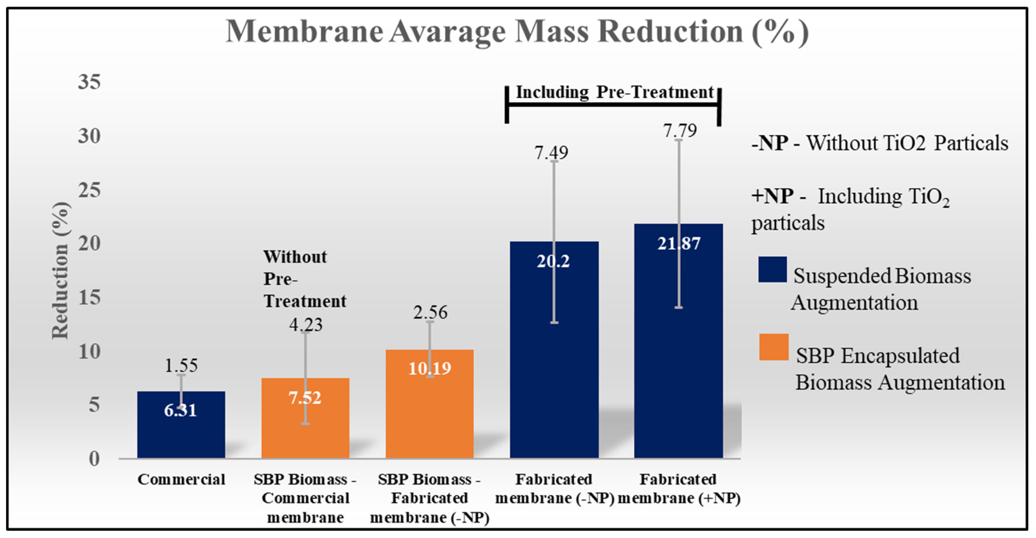
Photo degradation with TiO2 causes surface roughness, thus increasing a material’s surface area to enhance biodegradation. The combination of both photolysis by UV at 280 nm and biodegradation generates a synergy that enhances the overall degradation rate []. In this study, we did not observe surface roughness or any effect induced by the oxidative process following commercial CA membrane degradation, with an average of 6.3% (6%, 8%, and 4.9%) degradation (Table 2, studies 1A–C). Those were obtained with exposure to the oxidative process in the presence of TiO2 and induction by UVA radiation to form radicals that are expected to reduce the membrane stability structure. The use of H2O2 aimed to amplify the effect of the oxidative process by creating hydroxyl radicals under exposure to UV radiation. Briefly, inducing a pretreatment process (photolysis and radicals) did not have a significant effect on industrial membrane degradation (Figure 2).
Figure 2. Summary of membrane weight reduction (%) across five treatment and membrane categories. Each treatment was performed in triplicate, and the bars represent standard deviation values.
For the biological treatment, we expected the fungal culture to produce hyphae that could penetrate the membrane sheet, resulting in decreased membrane stability. Fungal hyphae were observed to attach to and penetrate certain areas of the membrane (Figure 3), with a pronounced effect at the membrane edges. These edges are created by the dissection of the membrane sheet, exposing its internal structure. Given that the exact composition of the commercial membrane is unknown, we hypothesize that its surface may be integrated or coated to limit microbial attachment, potentially preventing biofilm formation that could obstruct membrane porosity. Abu-Zurayk and colleagues [] described various preventative strategies for biofilm formation and microorganisms’ attachment, including surface coating and modification. This could help explain the challenges in degrading the commercial membrane and the limited degradation observed after 30 days of combined treatment.
Figure 3. Light microscope image (magnification ×100) representation of the overall degradation process for each of the tested membranes.
Due to the complexity involved in separating the biomass from the membranes, a restricted washing procedure was developed. Nevertheless, we acknowledge that some residual organic matter remained adhered to the membrane, suggesting that the actual weight loss may have been greater than what was recorded.
Study 1A-C presents the effect of the degradation process on commercial CA membranes. These membranes, used as a control, were the most resistant to pretreatment and biodegradation (average of 6.3% weight loss).
Figure 3 illustrates the interactions between fungal hyphae and various membrane types within the pretreatment chamber after 15 days of incubation, as well as in the bioreactor after 5 and 15 days, at the conclusion of this study. After 15 days of incubation in the bioreactor (Figure 3(1D–3D)), a high concentration of hyphae was detected in the medium, indicating the effectiveness of the augmentation method. It can be observed that the self-fabricated membranes (Figure 3(2A–2D,3A–3D)) exhibited a different surface structure, potentially indicating a larger surface area or a rougher texture compared with the commercial membrane (Figure 3(1A–1D)). During this study, the control group, which did not include pretreatment or biological treatment (Figure 3(1A–3A)), showed no structural changes. Following pretreatment (Figure 3(1B–3B)), no structural alterations were observed on the membrane surface or within its structure. Subject to the biological treatment, light microscopic analysis showed that the fungal hyphae could not penetrate the membrane surface and the only attachments were on the membrane edges; consequently, the biodegradation process was restricted to the membrane edges (Figure 3(1A–1D)), thus explaining the low membrane biodegradation results. Pretreatment with suspended nanoparticles (TiO2) in the presence of UVA light, with or without H2O2 (to enhance hydroxyl radical generation), followed by suspended T. versicolor, did not have a significant effect on commercial (control) membrane degradation (Figure 3(1B)). This indicates that the commercial membrane possesses a robust stable structure; thus, fungi encounter significant difficulties in attaching to the membrane surface and subsequently penetrating it as necessary for the digestion process. All biodegradation activity was reduced and limited to the membrane edges where the fungal hyphae could penetrate the membrane and subsequently attack it. Thus, we can conclude that only a small surface area of the membrane is accessible for the biodegradation process.
Using custom-made CA membranes (UFM and UFM-T) followed by pretreatment and biodegradation by T. versicolor culture demonstrated better degradation results in comparison with the commercial membrane. However, the incorporation of nanoparticles (UFM-T) into the custom-made membrane only slightly improved CA membrane degradation compared with the membrane without nanoparticles (UFM) (Table 2, test cycles 2 and 3). The new membrane structure combined with oxidative pretreatment (UVA and H2O2) affected membrane weight loss after 30 days of incubation, where slightly better mass loss was achieved with the UFM-T membrane (21.9%) compared with the UFM membrane (20.2%) (Figure 3(2D,3D), Table 2, test cycles 2 and 3). It seems that the composition of the nanoparticles made a small contribution to the oxidative inducted reaction on the membrane as a pretreatment and also increased the membrane surface area for fungal attachment and the biodegradation process. Schirp and Wolcott [] reported the presence of fungal hyphae within the wood–plastic interface, demonstrating the fungi’s ability to deeply penetrate cellulose-based materials such as CA. This may represent a critical stage in the mechanism of polymer degradation, involving both physical disruption of the polymer by fungal hyphae and enzymatic digestion through secreted enzymes. Because the contribution of nanoparticle degradation is minimal and possibly negligible, we did not investigate the effect of the nanoparticle composition on membrane characterization. We also acknowledge that the reduced DS in the custom-made UF membrane probably has a positive effect on membrane degradation, as already mentioned by Komarek and co-workers []. However, we propose that the DS level is not the primary factor affecting the degradation process; instead, the membrane’s resistance to fungi hyphae attachment may be the main reason for the low efficacy of biodegradation. This hypothesis is supported by microscopic observations of membrane–fungi interactions (Figure 2), which reveal notable surface differences between the commercial and fabricated membranes. The fabricated membrane exhibits a less smooth surface, potentially enhancing hyphal attachment and activity. Correspondingly, biodegradation efficiency, as indicated by membrane weight loss, is substantially higher in the self-fabricated membranes compared with the commercial ones.
Ozone pretreatment resulted in the highest mass loss of 26.7% and 28.8% for the UFM-T and UFM membranes, respectively (Table 2, test cycles 2 and 3). Ozone is a strong oxidant applied in water treatment for disinfection and for the removal of organic and inorganic pollutants. Using ozone as an oxidative reagent significantly improved the UF membrane degradation results, as presented in Figure 3(2C,3C,2D,3D), backup with quantitative results in Table 3, suggesting that ozone markedly accelerates the aging of polymeric materials and therefore might damage the membrane surface. This process in saturated polymers is accompanied by the intensive formation of oxygen-containing compounds, a change in molecular weight, and impairment of the mechanical and electrical properties of the specimens [].

3.2. Membrane Degradation by SBP-Encapsulated Culture

In these studies (Table 2, test cycles 4–5) we attempted to increase the concentration and persistence of T. versicolor within the bioreactor, using SBP capsules. The capsules were designed to provide the fungi culture with optimal growth conditions in a confined environment, protecting it from shear forces within the bioreactor, as well as facilitating the production and extraction of extracellular enzymes. Nonetheless, we do not anticipate that this technology will enhance the attachment of fungal hyphae to the membrane. From our previous experience we know that T. versicolor demonstrates rapid growth, resulting in a notable accumulation of hyphae within the SBP capsule. These hyphae fragments gradually permeate the CA membrane of the SBP capsule, dispersing into the host medium. Within the capsules, the culture achieves a high hyphae fragment density, consistently serving as a dependable inoculant and fostering additional biomass colonies within the host medium. In essence, the use of SBP capsules significantly enhances our ability to accumulate fungal biomass for biochemical processes. One key advantage of SBP capsules, when compared with the suspended form, lies in their ability to offer protection against bioreactor shear forces, thereby creating more favorable conditions for fungal culture growth. In summary, SBP technology was used as an effective means to proliferate fungal cultures within the bioreactor. Notably, the immobilized culture reduced the need for recirculation into the host bioreactor, ensuring a sustained biomass within the bioreactor operating in a continuous configuration. This is the first publication of SBP-encapsulated T. versicolor.
The commercial membrane degradation findings indicate that the use of SBP-encapsulating technology with T. versicolor results in an average weight loss of 7.52%, similar to that of suspended T. versicolor culture (up to 6.3%). Carbon supplementation to SBP-encapsulated T. versicolor increased the degradation component from 4.9–5.25% (Table 2, test cycles 4B and 4C) to 12.4% (Table 2, experiment 4A), thus the additional of a carbon source into the medium may increase the amount of suspended biomass and, in turn, the metabolic activity and the biodegradation rate. This result highlights the added value of utilizing this method for bioreactor medium inoculation and biomass accumulation. Without the use of an additional carbon source, weight loss remains as recorded in the suspension culture subject to industrial membrane biodegradation. Utilizing SBP-encapsulated T. versicolor supplemented with an additional carbon source appears to have significantly boosted biomass production and commercial membrane (control) weight loss, where weight loss increased by over 130% from the initial 5.25%, indicating that biomass type and growth conditions are critical factors for CA membrane degradation. This discovery underscores the critical importance of an exogenous carbon source for membrane biodegradation. It also highlights the challenges faced by the fungal culture in sustaining the membrane as the sole carbon source for assimilation and energy generation.
The degradation of commercial membranes, whether employing encapsulated or suspended T. versicolor, has been proven inadequate within the given HRT on an industrial scale. However, with respect to the custom-made membranes, encapsulated T. versicolor achieved lower biodegradation, with an average weight loss of 10.19% (SD ± 2.56) (Table 2, test cycle 5) compared with suspended T. versicolor, which showed average weight losses of 20.2% (SD ± 7.49) and 21.87% (SD ± 7.79) for UFM (Table 2, test cycle 4) and UFM-T (Table 2, test cycle 2), respectively. Although encapsulation increased T. versicolor biomass within the bioreactor in comparison with suspended T. versicolor, for the UFM membrane (Table 2, test cycles 5 vs. 3), the biodegradation results showed lower membrane weight loss with encapsulated T. versicolor compared with suspended T. versicolor. Since the SBP capsule membrane comprises CA, it was hypothesized that the capsule membranes could also serve as an additional carbon source and may be partially degraded. Consequently, a decrease in the degradation of suspended membrane particles was anticipated to explain the lower biodegradation results observed with encapsulated T. versicolor compared with suspended T. versicolor. This study aberration is anticipated to occur in small volume tests, particularly when the ratio between membrane particles and SBP capsules (membrane) is high (1.2–5 gr vs. 2.5 gr, respectively). The t-test indicated a statistically significant difference between the commercial membrane and both UFM-T and UFM (p = 0.026 and p = 0.034, respectively) when operated with suspended culture. In contrast, the comparison between the commercial CA membrane and the UFM membrane operated with SBP-encapsulated culture showed no significant difference (p = 0.4).
Ozonation as a pretreatment seems to have had the greatest effect on membrane biodegradation as seen in test cycles 2C and 3C (Table 2), when using suspended culture and our custom-made UF membrane, achieving weight losses of 26.7 and 28.8%, respectively, with only a slight effect of the presence of nanoparticles incorporated into the membrane. Ozonation as a pretreatment also contributed to the degradation process by SBP-encapsulated T. versicolor, as observed in experiment 5C (Table 2). These results also emphasize the contribution of ozone pretreatment to increasing membrane degradation. In our previous study, which integrated ozonation as a pretreatment with SBP-based bioremediation, we observed synergistic degradation of phenols. This underscores the advantages of employing a combined treatment approach []. Therefore, ozonation as a pretreatment for the waste membrane degradation process can be a good candidate for upscaling the treatment system, particularly since it allows us to reduce the use of chemicals, such as H2O2 and nanoparticles (TiO2).
Table 3. Literature review of biodegradation of cellulose-acetate (CA)-based membranes in comparison with study results.
CultureTypeDS **CA Weight Loss (%)Incubation Time (Days)References
Rhizobium melilotiBacteria0.4–0.834150[]
Neisseria sicc SBBacteria1.8151 & 40 *20[]
Neisseria sicc SCBacteria2.360 & 45 *20[]
Alcaligens xylosoxidansBacteria0.4–0.823150[]
Various biota from natural water sources2.210365[]
Trametes versicolorFungi2.6Up to 28.830Present study
* The different values refer to experiments conducted under different environmental conditions. ** DS—degree of acetyl substitution.
In summary, for CA membranes, the biodegradation rate is influenced by the DS content and the specific conditions to which the material is exposed. The rate of biodegradation decreases as the DS content increases. Thus, deacetylation is the rate determining step during biodegradation of CA. In addition to the abovementioned structural features, many environmental factors further influence the degradation rate at any given location. These include temperature, humidity, pH, sunlight, and the availability of oxygen, nutrients, and microorganisms []. We acknowledge that in all test cycles, the custom-made membranes, whether containing nanoparticles or not, were biodegraded more efficiently than the commercial membranes, suggesting structural modifications that enhance the culture’s ability to attach to the membrane more effectively. The microorganism culture type is critical for increasing process efficacy and reducing the required HRT, both of which affect the required bioreactor volume. Similar to our results, a previous study also found relatively slow biodegradation rates when using fungi []; however, our study achieved these results in only one-fifth of the time (HRT), probably due to the contributions of pretreatment and the custom-made UF membranes. We estimate that pretreatment processes decrease polymer structural stability while increasing the surface area available for microorganisms’ attachment and activity close to the membrane. This, in turn, provides more favorable physical conditions for the biological degradation of CA membranes and, consequently, reduces the required HRT for treatment.

4. Conclusions

The literature on the degradation of CA-based membranes in different environments and biomass types is surprisingly sparse and contradictory. This study presents an innovative CA treatment involving a continuous flow configuration that combines a pretreatment stage with a subsequent biodegradation phase using suspended or encapsulated fungal biomass inoculation. The custom-made UF membranes (with or without incorporated TiO2 nanoparticles), proved to be more biodegradable than the commercial CA membrane. This is likely due to structural modifications that may enhance the interactions between fungal hyphae and the membrane surface.
Ozonation as a pretreatment seems to be an effective approach for the custom-made UF membrane, with an expected weight loss exceeding 28% over a membrane SRT of 30 days, which is considered a relatively short treatment period. The innovative utilization of an SBP-encapsulated T. versicolor culture proved to be an efficient strategy for fostering fungal cultures within the bioreactor and ensuring sustained biomass within the continuously operating bioreactor.
In this study, some variables remain unaccounted for and may influence the reported membrane degradation results: (1) trapped organic matter, likely due to fungal hyphae penetrating the polymer structure, and (2) the portion of CA+ in the capsule membrane that has been biodegraded. These factors limit our ability to perform a complete mass balance. For future studies, we recommend monitoring additional time points of CA degradation to evaluate its kinetic rate, as well as exploring combinations of different fungal cultures to take advantage of co-metabolic processes.

Author Contributions

Conceptualization, H.M. and O.A.M.; Methodology, H.M., M.H.M. and O.A.M.; Validation, M.H.M., B.H. and O.A.M.; Formal analysis, C.S., B.H. and O.A.M.; Investigation, C.S. and H.M.; Writing—original draft, O.A.M.; Supervision, O.A.M. All authors have read and agreed to the published version of the manuscript.

Funding

This study was carried out with funding from the Ministry of Innovation, Science, and Technology of the State of Israel (Grant number 3-17393), as well as the research fund of Kinneret Academic College.

Data Availability Statement

The original contributions presented in this study are included in the article. Further inquiries can be directed to the corresponding author.

Acknowledgments

This study is dedicated to the memory of Nevo Arad (1998–2023), a fourth year student in the Water Industry Engineering Department, Kinneret Academic College on the Sea of Galilee, Israel.

Conflicts of Interest

Author Ofir A. Menashe was employed by the company BioCastle Water Technologies Ltd. The remaining authors declare that the research was conducted in the absence of any commercial or financial relationships that could be construed as a potential conflict of interest. The company had no role in the design of the study; in the collection, analyses, or interpretation of data; in the writing of the manuscript, or in the decision to publish the results.

References

  1. Cao, L.; Zhang, Y.; Nei, L.; Feng, X. A novel loosely structured nanofiltration membrane bioreactor for wastewater treatment: Process performance and membrane fouling. J. Membr. Sci. 2022, 644, 120128. [Google Scholar] [CrossRef]
  2. Markets and Markets, Membranes Market by Material (Polymeric, Ceramic), Technology (RO, MF, UF, NF), Application (Water & Wastewater Treatment, Industrial Processing), & Region (North America, EU, APAC, Middle East & Africa, South America)—Global forecast 2027. 2022. Available online: https://www.marketsandmarkets.com/Market-Reports/membranes-market-1176.html (accessed on 3 December 2024).
  3. Maalige, N.R.; Mruthunjayappa, M.H.; Nataraj, S.K. Sustainable polymer-based materials for energy and environmental applications. In Polymer-Based Advanced Functional Materials for Energy and Environmental Applications; Subramani, N.K., Nataraj, S.K., Patel, C., Shivanna, S., Eds.; Springer: Singapore, 2022; pp. 9–30. [Google Scholar] [CrossRef]
  4. Senán-Salinas, J.; Blanco, A.; Garcia-Pacheco, R.; Landaburu-Aguirre, J.; Garcia-Calvo, E. Prospective Life Cycle Assessment and economic analysis of direct recycling of end-of-life reverse osmosis membranes based on Geographic Information Systems. J. Clean. Prod. 2021, 282, 124400. [Google Scholar] [CrossRef]
  5. Bandehali, S.; Sanaeepur, H.; Ebadi Amooghin, A.; Shirazian, S.; Ramakrishna, S. Biodegradable polymers for membrane separation. Sep. Purif. Technol. 2021, 269, 118731. [Google Scholar] [CrossRef]
  6. Zhang, R.; Liu, Y.; He, M.; Su, Y.; Zhao, X.; Elimelech, M.; Jiang, Z. Antifouling membranes for sustainable water purification: Strategies and mechanisms. Chem. Soc. Rev. 2016, 45, 5888–5924. [Google Scholar] [CrossRef] [PubMed]
  7. Li, S.; Wang, X.; Guo, Y.; Hu, J.; Lin, S.; Tu, Y.; Chen, L.; Ni, Y.; Huang, L. Recent advances on cellulose-based nanofiltration membranes and their applications in drinking water purification: A review. J. Clean. Prod. 2022, 333, 130171. [Google Scholar] [CrossRef]
  8. Arockiasamy, D.L.; Nagendran, A.; Shobana, K.H.; Mohan, D. Preparation and characterization of cellulose acetate/aminated polysulfone blend ultrafiltration membranes and their application studies. Sep. Sci. Technol. 2009, 44, 398–421. [Google Scholar] [CrossRef]
  9. Han, B.; Zhang, D.; Shao, Z.; Kong, L.; Lv, S. Preparation and characterization of cellulose acetate/carboxymethyl cellulose acetate blend ultrafiltration membranes. Desalination 2013, 311, 80–89. [Google Scholar] [CrossRef]
  10. El Seoud, O.A.; Kostag, M.; Jedvert, K.; Malek, N.I. Cellulose regeneration and chemical recycling: Closing the “cellulose gap” using environmentally benign solvents. Macromol. Mater. Eng. 2020, 305, 1900832. [Google Scholar] [CrossRef]
  11. Sakai, K.; Yamauchi, T.; Nakasu, F.; Ohe, T. Biodegradation of cellulose acetate by Neisseria sicca. Biosci. Biotech. Biochem. 1996, 60, 1617–1622. [Google Scholar] [CrossRef]
  12. Mruthunjayappa, M.H.; Shachar, C.; Imbar, A.; Menashe, O.A.; Mamane, H. Cellulose acetate and polycaprolactone based photoactive ultrafiltration membrane: A novel approach with UV-switchable photocatalytic activity. Sep. Purif. Technol. 2023, 329, 125102. [Google Scholar] [CrossRef]
  13. Andlar, M.; Rezić, T.; Marđetko, N.; Kracher, D.; Ludwig, R.; Šantek, B. Lignocellulose degradation: An overview of fungi and fungal enzymes involved in lignocellulose degradation. Eng. Life Sci. 2018, 18, 768–778. [Google Scholar] [CrossRef] [PubMed]
  14. Qi, J.; Li, F.; Jia, L.; Zhang, X.; Deng, S.; Luo, B.; Zhou, Y.; Fan, M.; Xia, Y. Fungal selectivity and biodegradation effects by white and brown rot fungi for wood biomass pretreatment. Polymers 2023, 15, 1957. [Google Scholar] [CrossRef] [PubMed]
  15. Ishak, S.; Malakahmad, A.; Isa, M.H. Refinery wastewater biological treatment: A short review. J. Sci. Ind. Res. 2012, 71, 251–256. [Google Scholar]
  16. Menashe, O.; Kurzbaum, E. Small-bioreactor platform technology as a municipal wastewater additive treatment. Water Sci. Technol. 2014, 69, 504–510. [Google Scholar] [CrossRef] [PubMed]
  17. Menashe, O.; Rosen-Kligvasser, J.; Kurzbaum, E.; Suckeveriene, R.Y. Structural properties of a biotechnological capsule confined by a 3D-cellulose acetate membrane. Polym. Adv. Technol. 2020, 32, 681–689. [Google Scholar] [CrossRef]
  18. Machado, A.S.; Valadares, F.; Silva, T.F.; Milagres, A.M.F.; Segato, F.; Ferraz, A. The secretome of Phanerochaete chrysosporium and Trametes versicolor grown in microcrystalline cellulose and use of the enzymes for hydrolysis of lignocellulosic materials. Front. Bioeng. Biotechnol. 2020, 8, 826. [Google Scholar] [CrossRef]
  19. Qiu, Y.; Ouyang, F. Fabrication of TiO2 hierarchical architecture assembled by nanowires with anastase/TiO2(B) phase-junctions for efficient photocatalytic hydrogen production. Appl. Surf. Sci. 2017, 403, 691–698. [Google Scholar] [CrossRef]
  20. Zhao1, K.; Wu1, Y.W.; Young, S.; Chen, X.J. Biological Treatment of Dairy Wastewater: A Mini Review. J. Environ. Inform. Lett. 2020, 4, 22–31. [Google Scholar] [CrossRef]
  21. Im, J.H.; Woo, H.J.; Choi, M.W.; Han, K.B.; Kim, C.W. Simultaneous organic and nitrogen removal from municipal landfill leachate using an anaerobic–aerobic system. Water Res. 2001, 35, 2403–2410. [Google Scholar] [CrossRef]
  22. Schirp, A.; Wolcott, M.P. Fungal degradation of wood-plastic composites and evaluation using dynamic mechanical analysis. J. Appl. Polym. Sci. 2006, 99, 3138–3146. [Google Scholar] [CrossRef]
  23. Komarek, R.J.; Gardner, R.M.; Buchanan, C.M.; Gedon, S. Biodegradation of radiolabeled cellulose acetate and cellulose propionate. J. Appl. Polym. Sci. 1993, 50, 1739–1746. [Google Scholar] [CrossRef]
  24. Puls, J.; Wilson, S.A.; Hölter, D. Degradation of cellulose acetate–based materials: A review. J. Polym. Environ. 2011, 19, 152–165. [Google Scholar] [CrossRef]
  25. Abu-Zurayk, R.; Alnairat, N.; Khalaf, A.; Alqader Ibrahim, A.; Halaweh, G. Cellulose Acetate Membranes: Fouling Types and Antifouling Strategies: A Brief Review. Processes 2023, 11, 489. [Google Scholar] [CrossRef]
  26. Andrady, A.L.; Harnid, S.H.; Hu, X.; Torikai, A. Effects of increased solar ultraviolet radiation on materials. J. Photochem. Photobiol. B Biol. 1998, 46, 96–103. [Google Scholar] [CrossRef] [PubMed]
  27. Bar Oz, Y.; Mamane, H.; Menashe, O.; Cohen-Yaniv, V.; Kumar, R.; Iasur Kruh, L.; Kurzbaum, E. Treatment of olive mill wastewater using ozonation followed by an encapsulated acclimated biomass. J. Environ. Chem. Eng. 2018, 6, 5014–5023. [Google Scholar] [CrossRef]
  28. Gu, J.-D.; Eberiel, D.; McCarthy, S.P.; Gross, R.A. Degradation and mineralization of cellulose acetate in simulated thermophilic compost environments. J. Environ. Polym. Degrad. 1993, 1, 281–291. [Google Scholar] [CrossRef]
  29. Yadav, N.; Hakkarainen, M. Degradation of cellulose acetate in simulated aqueous environments: One-year study. Mater. Eng. 2002, 307, 2100951. [Google Scholar] [CrossRef]
  30. Laycock, B.; Nikolić, M.; Colwell, J.M.; Gauthier, E.; Halley, P.; Bottle, S.; George, G. Lifetime prediction of biodegradable polymers. Prog. Polym. Sci. 2017, 71, 144–189. [Google Scholar] [CrossRef]
Disclaimer/Publisher’s Note: The statements, opinions and data contained in all publications are solely those of the individual author(s) and contributor(s) and not of MDPI and/or the editor(s). MDPI and/or the editor(s) disclaim responsibility for any injury to people or property resulting from any ideas, methods, instructions or products referred to in the content.

Article Metrics

Citations

Article Access Statistics

Article metric data becomes available approximately 24 hours after publication online.