

Neurodegenerative Disease-Specific Relations Between Temporal and Kinetic Gait Features Identified Using InterCriteria Analysis


Abstract
1. Introduction
2. Materials and Methods
2.1. Database
2.2. Gait Features
2.2.1. Temporal Gait Features
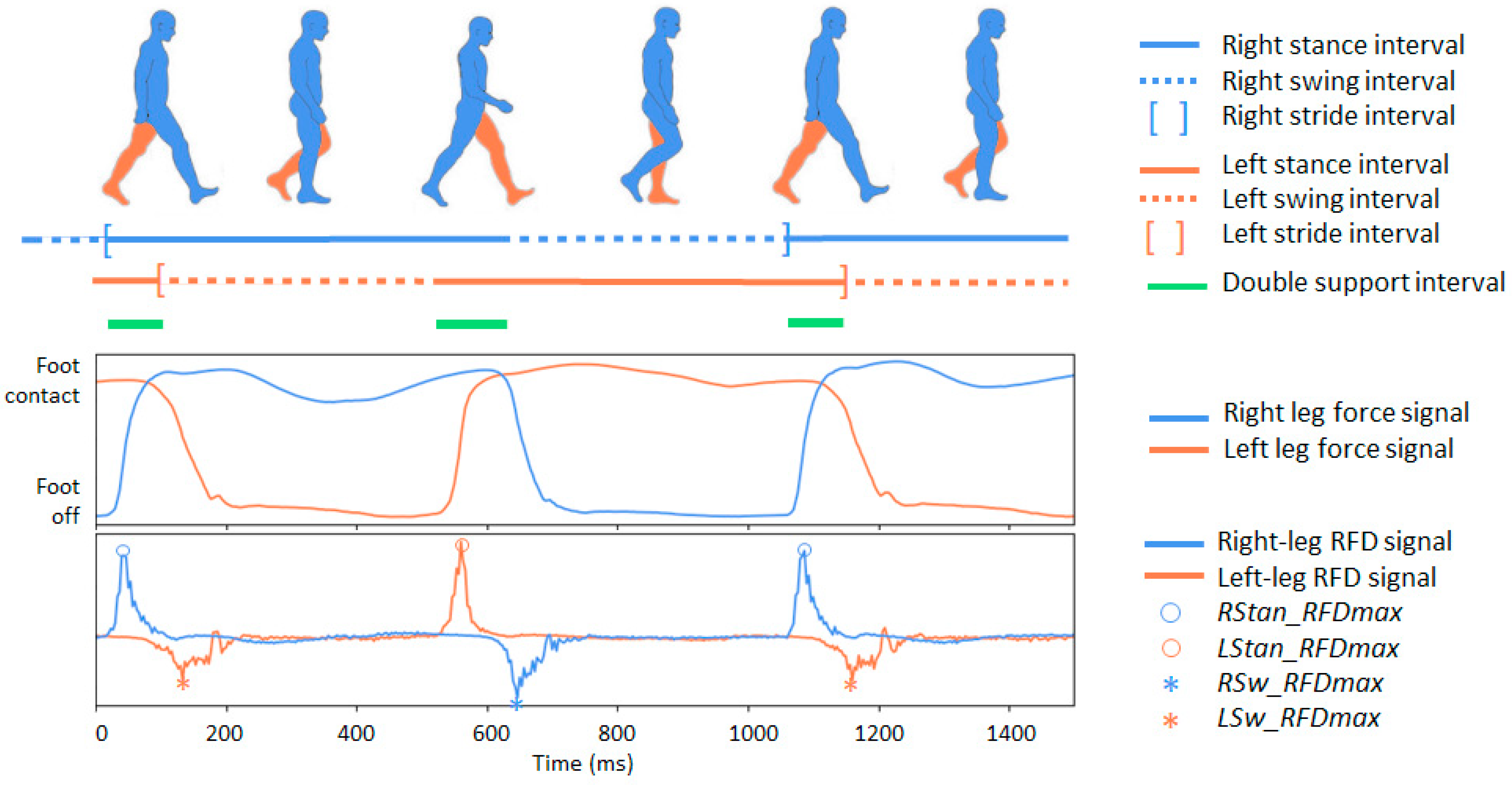
- C01–C12: Twelve primary stride-to-stride temporal gait measures read directly from the database time-series (.ts) files. They characterize the stride interval, swing interval, stance interval, and double-support interval, provided both in the absolute time (seconds) and as a percentage of the stride for the left and right legs. Figure 1 (top) illustrates the sequence of intervals for the two legs in a normal gait. These fundamental temporal parameters follow established gait-analysis conventions and definitions [50,51,52] and are widely used to evaluate walking patterns, postural stability, and deviations from normative gait behavior [53,54,55].
- C13–C16: Four secondary temporal features capturing broader aspects of gait dynamics, including the cadence (walking rate), swing asymmetry (left–right imbalance in swing duration), swing variability (stride-to-stride fluctuations in swing timing), and gait variability (stride-interval instability reflecting reduced rhythmicity). All four measures are analytically derived from the primary temporal parameters (C01-C12) using the definitions summarized in Table 2. These descriptors are established markers of gait stability and motor impairment in NDD [51,56,57].

| ID | Abbreviation | Description |
|---|---|---|
| Primary temporal features of the gait cycle | ||
| C01 | LStrInt (s) | Left stride interval: Time between two consecutive heel strikes of the left foot |
| C02 | RStrInt (s) | Right stride interval: Time between two consecutive heel strikes of the right foot |
| C03 | LSwInt (s) | Left swing interval: Duration when the left foot is not in contact with the ground during a stride |
| C04 | RSwInt (s) | Right swing interval: Duration when the right foot is not in contact with the ground during a stride |
| C05 | LSwInt (% stride) | Left swing interval expressed as a percentage of the left stride duration |
| C06 | RSwInt (% stride) | Right swing interval expressed as a percentage of the right stride duration |
| C07 | LStanInt (s) | Left stance interval: Time during which the left foot is in contact with the ground within a stride |
| C08 | RStanInt (s) | Right stance interval: Time during which the right foot is in contact with the ground within a stride |
| C09 | LStanInt (% stride) | Left stance interval expressed as a percentage of the left stride duration |
| C10 | RStanInt (% stride) | Right stance interval expressed as a percentage of the right stride duration |
| C11 | DSupInt (s) | Double-support interval: Total time within a stride when both feet are simultaneously in contact with the ground |
| C12 | DSupInt (% stride) | Double-support interval expressed as a percentage of stride duration |
| Secondary temporal features of the gait dynamics | ||
| C13 | Cadance (steps/minute) | Number of steps taken per minute (walking rate). |
| C14 | SwAsymetry (% stride) | Swing asymmetry: Difference between the left and right swing intervals, expressed as a percentage of the stride: |
| C15 | SwVariability (%) | Swing Variability: Stride-to-stride variability of the swing interval, quantifying how much the swing duration changes from one stride to the next. The formula is shown for the left leg, where n and n−1 denote the current and previous strides, respectively: |
| C16 | GaitVariability (%) | Gait variability: Stride-to-stride variability of the stride interval (in seconds), normalized by the duration of the current stride. The formula is expressed for the left leg, where n and n−1 denote the current and previous strides, respectively: |
2.2.2. Kinetic Gait Features
- C26–C29: Inter-limb correlation features capturing bilateral symmetry in the left- and right-leg GRF and RFD waveforms over a step. Classical zero-lag and lag-optimized cross-correlation measures are employed to quantify bilateral waveform synchrony, as commonly applied in gait coordination and signal-processing analyses [52,64,65]. Zero-lag correlations (C26–C27) reflect both the waveform shape similarity and temporal alignment at identical time instants. For lag-optimized correlations (C28–C29), only the maximum correlation magnitude observed within the step interval is retained. These measures therefore quantify waveform-shape synchrony independent of explicit timing offset as the associated lag values are not further analyzed.
- C30–C33: Inter-limb RFD asymmetry features describing differences between the left and right legs in maximum and average RFD amplitudes during stance and swing.
- C34–C37: Intra-limb stance–swing RFD asymmetry features reflecting differences between stance and swing phases in maximum and average RFD amplitudes within each leg.
- C38–C41: Global stance–swing RFD asymmetry features capturing the following: (i) differences between stance and swing in maximum and average RFD amplitudes averaged across both legs; and (ii) ratios of stance- and swing-phase RFD amplitudes relative to double-support.
| ID | Abbreviation | Description |
|---|---|---|
| Primary RFD Features (Force-Derivative Based) | ||
| C17 | LStan_RFDmax | Maximum positive deflection of the left-leg RFD signal during the stance interval—illustrated in Figure 1 (bottom). Represents the peak rate at which force is generated by the left leg while in ground contact. This value reflects the leg’s ability to rapidly produce force during weight acceptance and propulsion—critical components of a stable gait. Reduced or delayed stance-phase RFDmax is frequently observed in individuals with impaired neuromuscular control, such as those with NDDs. |
| C18 | LSw_RFDmax | Maximum negative deflection of the left-leg RFD signal during the swing interval (absolute value used)—illustrated in Figure 1 (bottom). Captures the largest rate of force fluctuation while the left foot is off the ground. Although overall forces are lower during swing, RFDmax reflects rapid limb deceleration/acceleration needed for foot clearance and step preparation. Abnormalities may indicate impaired motor coordination or compensatory movement strategies. |
| C19 | RStan_RFDmax | Maximum positive deflection of the right-leg RFD signal during the stance interval—illustrated in Figure 1 (bottom). Right-leg analogue of C17. Indicates the capacity of the right leg to rapidly generate force during ground contact. Asymmetry between C17 and C19 may highlight unilateral weakness, a gait imbalance, or NDD-specific lateralization. |
| C20 | RSw_RFDmax | Maximum negative deflection of the right-leg RFD signal during the swing interval (absolute value used)—illustrated in Figure 1 (bottom). Right-leg analogue of C18. Reflects rapid force changes during the right leg’s swing phase. Elevated variability or reduced peaks can indicate difficulties in limb control or instability during segmental transitions. |
| C21 | LStan_RFDavg | Average of the absolute left-leg RFD signal during the stance interval. Represents the overall rate of force development sustained throughout the stance phase on the left side. Unlike C17 (which captures the peak), this feature describes the general force-generation profile. Lower average RFD may correspond to reduced push-off efficiency or diminished neuromuscular responsiveness. |
| C22 | LSw_RFDavg | Average of the absolute left-leg RFD signal during the swing interval. Reflects the average rate at which force changes occur while the left foot is in the air. This measure indicates the smoothness of limb motion and the consistency of force regulation during swing, both of which may be impaired in NDD gait. |
| C23 | RStan_RFDavg | Average of the absolute right-leg RFD signal during the stance interval. The right-leg analogue to C21, capturing the mean force-development rate while the leg supports body weight. Comparing C21 and C23 provides insight into stance-phase symmetry and inter-limb coordination. |
| C24 | RSw_RFDavg | Average of the absolute right-leg RFD signal during the swing interval. Complementing C22, this metric characterizes the force-change dynamics of the right leg during swing. Altered values may indicate difficulties in executing controlled, rhythmic limb advancement. |
| C25 | DSup_RFDavg | Average of the absolute RFD signal during double support interval. Separate values are measured from the RFD for the left and right legs and averaged. This measure reflects how quickly force is generated jointly by both legs during one of the most stability-critical phases of gait. Double support requires smooth and coordinated load transfer from one limb to the other; thus, abnormalities in DSup_RFDavg may indicate compromised balance control or impaired bilateral coordination—frequent findings in NDD. |
| Inter-limb Synchrony Features (Force and RFD Correlation) | ||
| C26 | CorLR_F(lag0) | Zero-lag correlation between left and right raw force signals within a stride. This feature quantifies how synchronized both legs are in producing ground reaction forces at the same moment. High values indicate smooth bilateral coordination, whereas reduced synchrony may reflect impaired timing, asymmetry, or compensatory loading strategies commonly observed in NDD gait. |
| C27 | CorLR_RFD(lag0) | Zero-lag correlation between left and right RFD signals within a stride. Captures how closely the instantaneous force-generation dynamics of both legs align in time. Because RFD reflects neuromuscular responsiveness, this metric is sensitive to disruptions in bilateral motor control and can highlight subtle coordination deficits. |
| C28 | CorLR_F(maxlag) | Maximum cross-correlation between left- and right-leg force signals computed across the duration of a single step. This feature identifies the highest possible bilateral synchrony in the force waveform shape after allowing for any temporal shift between limbs within the step interval. A lower value indicates reduced coordination quality, even when optimally time-aligned. |
| C29 | CorLR_RFD(maxlag) | Maximum cross-correlation between left- and right-leg RFD signals computed across the duration of a single step. This feature captures the highest similarity in neuromuscular force-generation dynamics between limbs when time-alignment is optimized within the step. Only the correlation magnitude is analyzed here; the lag at which it occurs is not interpreted further. |
| Left–Right Leg Asymmetry Features (%) | ||
| C30 | Stan_RFDmax(L-R)% | Percent asymmetry between legs in terms of maximum RFD deflection during the stance: Reflects unilateral differences in how force is rapidly produced during weight-bearing. Elevated asymmetry is often associated with unilateral weakness, impaired balance, or lateralized disease progression. |
| C31 | Sw_RFDmax(L-R)% | Percent asymmetry between legs in terms of maximum RFD deflection during swing: Measures left–right differences in limb acceleration and control during swing. Larger values may indicate impaired motor coordination, reduced foot-clearance control, or abnormal limb sequencing. |
| C32 | Stan_RFDavg(L-R)% | Percent asymmetry between legs in terms of average RFD during stance: Evaluates persistent loading imbalance rather than peak differences. Elevated asymmetry may signal chronic compensatory gait patterns. |
| C33 | Sw_RFDavg(L-R)% | Percent asymmetry between legs in terms of average RFD during swing: Reflects sustained differences in inter-limb force-change dynamics during the non-contact portion of gait, often heightened in conditions with motor rhythm disruption. |
| Intra-Limb Stance–Swing RFD Asymmetry (%) | ||
| C34 | L_RFDmax(stance-swing)% | Percent difference between left stance and left swing maximum RFD: Highlights how effectively the left leg adapts between load-bearing (stance) and limb-advancement (swing). Abnormal values may indicate poor phase modulation. |
| C35 | R_RFDmax(stance-swing)% | Percent difference between right stance and right swing maximum RFD: Assesses intra-limb force-generation adaptability for the right leg. Phase-dependent modulation deficits are common in NDD gait patterns. |
| C36 | L_RFDavg(stance-swing)% | Percent difference between left stance and left swing average RFD: Indicates whether the left leg maintains consistent force-change dynamics across phases. Reduced differentiation may reflect impaired gait rhythm or stiffened motor patterns. |
| C37 | R_RFDavg(stance-swing)% | Percent difference between right stance and right swing average RFD: Provides the right-side analogue to C36, showing how the leg transitions between functional roles. Deviations often appear in asymmetric or progression-based gait impairments. |
| Global Stance–Swing RFD Asymmetry (%) | ||
| C38 | RFDmax(stance-swing)% | Percent difference between stance-phase and swing-phase maximum RFD, averaged across both legs: Reflects global phase-dependent differences in rapid force-generation capability. A healthy gait typically shows clear differentiation; reduced contrast may indicate impaired phase modulation or diminished neuromuscular flexibility. |
| C39 | RFDavg(stance-swing)% | Percent difference between stance-phase and swing-phase average RFD, averaged across both legs: Quantifies the overall modulation of force-change dynamics between weight-bearing and limb-advancement phases. Altered values may signal global gait dysregulation, reduced adaptability, or impaired neuromechanical control. |
| C40 | DSup_to_Stan_RFD% | Ratio between double-support RFD and stance-phase maximal RFD. This feature quantifies how the average bilateral rate of force development during the double-support interval compares with the maximum RFD observed during stance, averaged across both legs. It expresses how force-generation dynamics during double support relate to the peak force-generation capacity during single-leg loading: |
| C41 | DSup_to_Sw_RFD% | Ratio between double-support RFD and swing-phase maximal RFD. This feature quantifies how the average bilateral rate of force development during the double-support interval compares with the maximum RFD measured during the swing phase, averaged across both legs. It reflects how force-generation capability during double support relates to the peak force-production capacity when each leg is unloaded and preparing for the next stance phase: |
2.3. InterCriteria Analysis
| C1 | … | Ck | … | Cn | |
| O1 | … | … | |||
| … | … | … | … | … | … |
| Oi | … | … | |||
| … | … | … | … | … | … |
| Om | … | … |
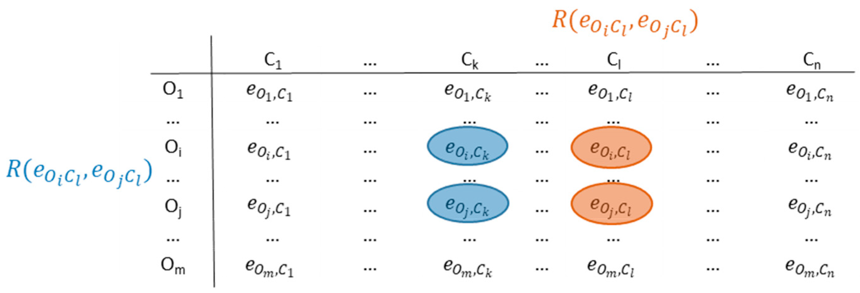
- counts the number of cases in which the relations and (or their duals and ) are simultaneously satisfied.
- counts the number of cases in which and (or and ) are simultaneously satisfied.
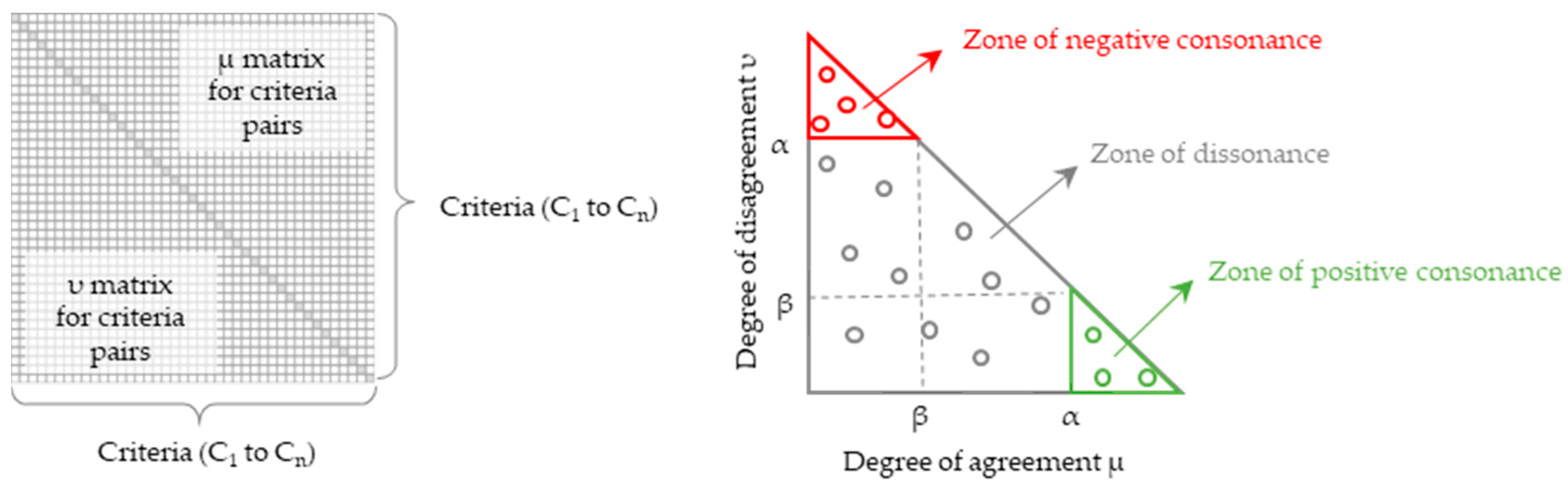
- which, in terms of ICrA, is known as the degree of agreement or positive consonance, and
- , known as degree of disagreement or negative consonance.
| C1 | … | Ck | … | Cn | |
| C1 | … | … | |||
| … | … | … | … | … | … |
| Ck | … | … | |||
| … | … | … | … | … | … |
| Cn | … | … |
- positive consonance, if > α and < β;
- negative consonance, if < β and > α;
- dissonance, otherwise.
3. Results
3.1. Test Setting
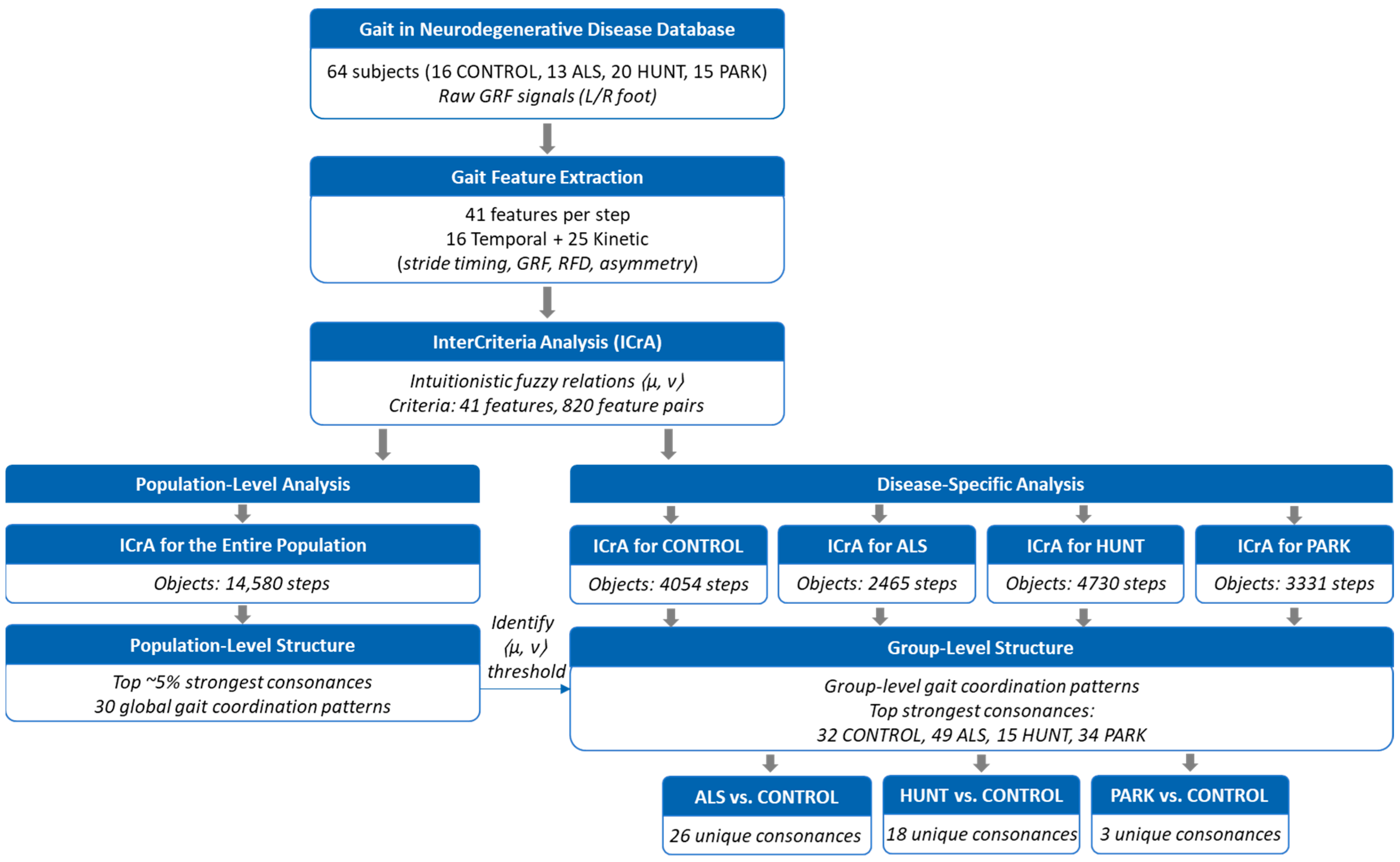
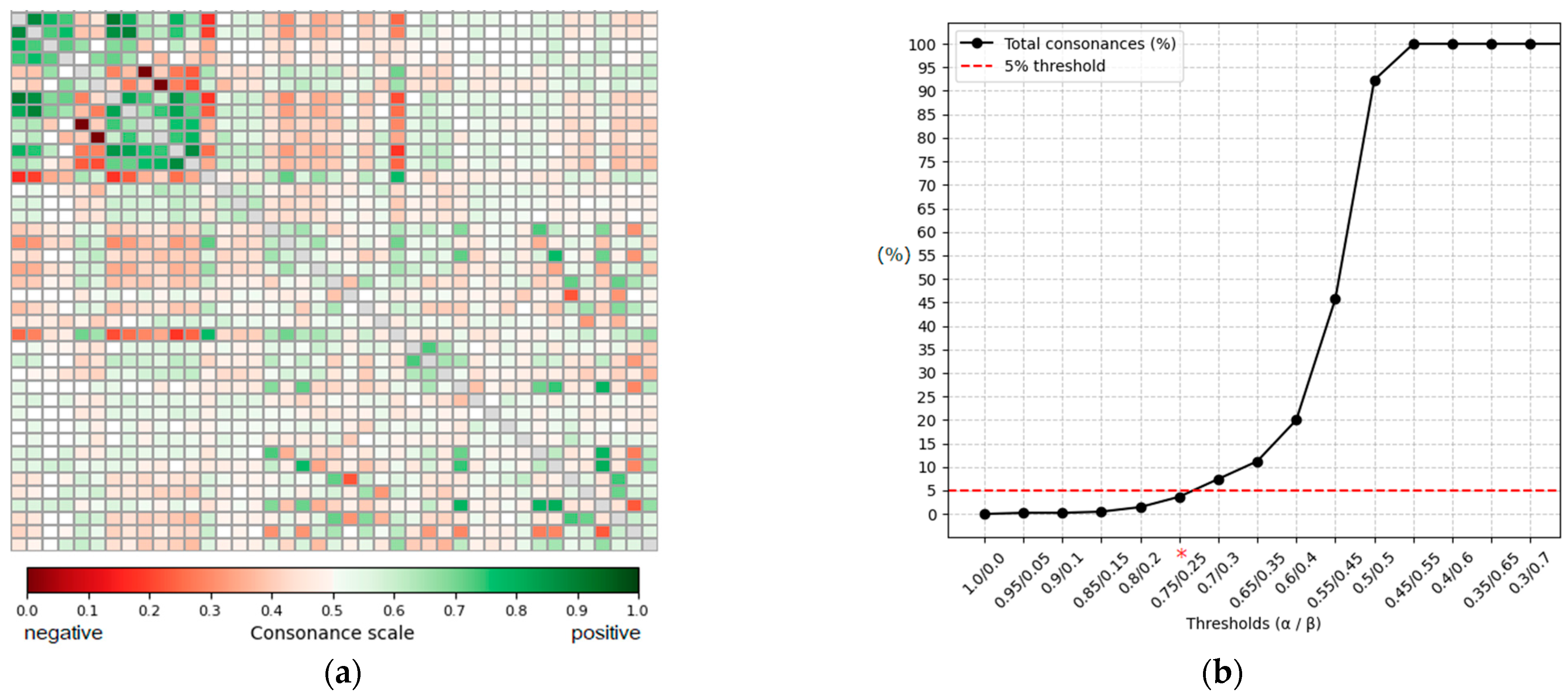
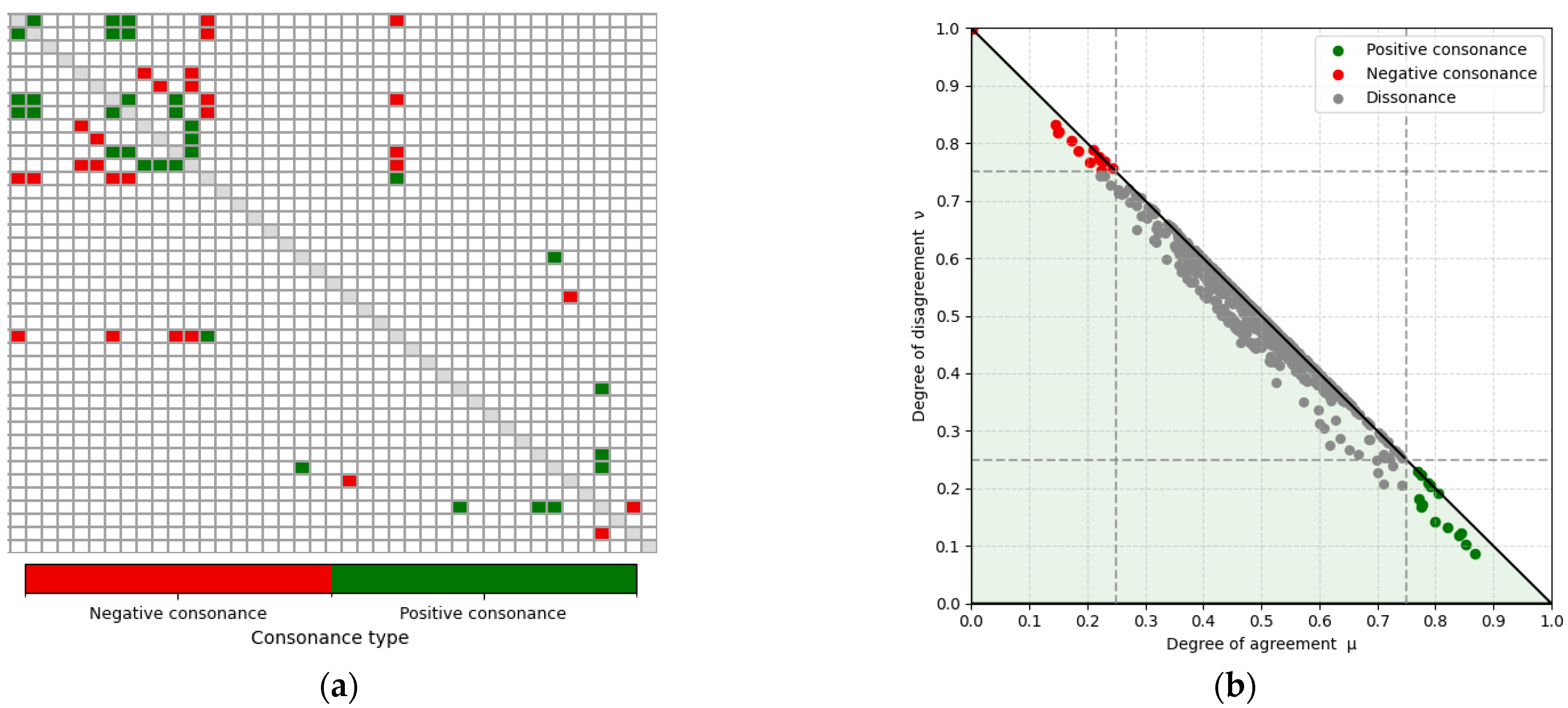
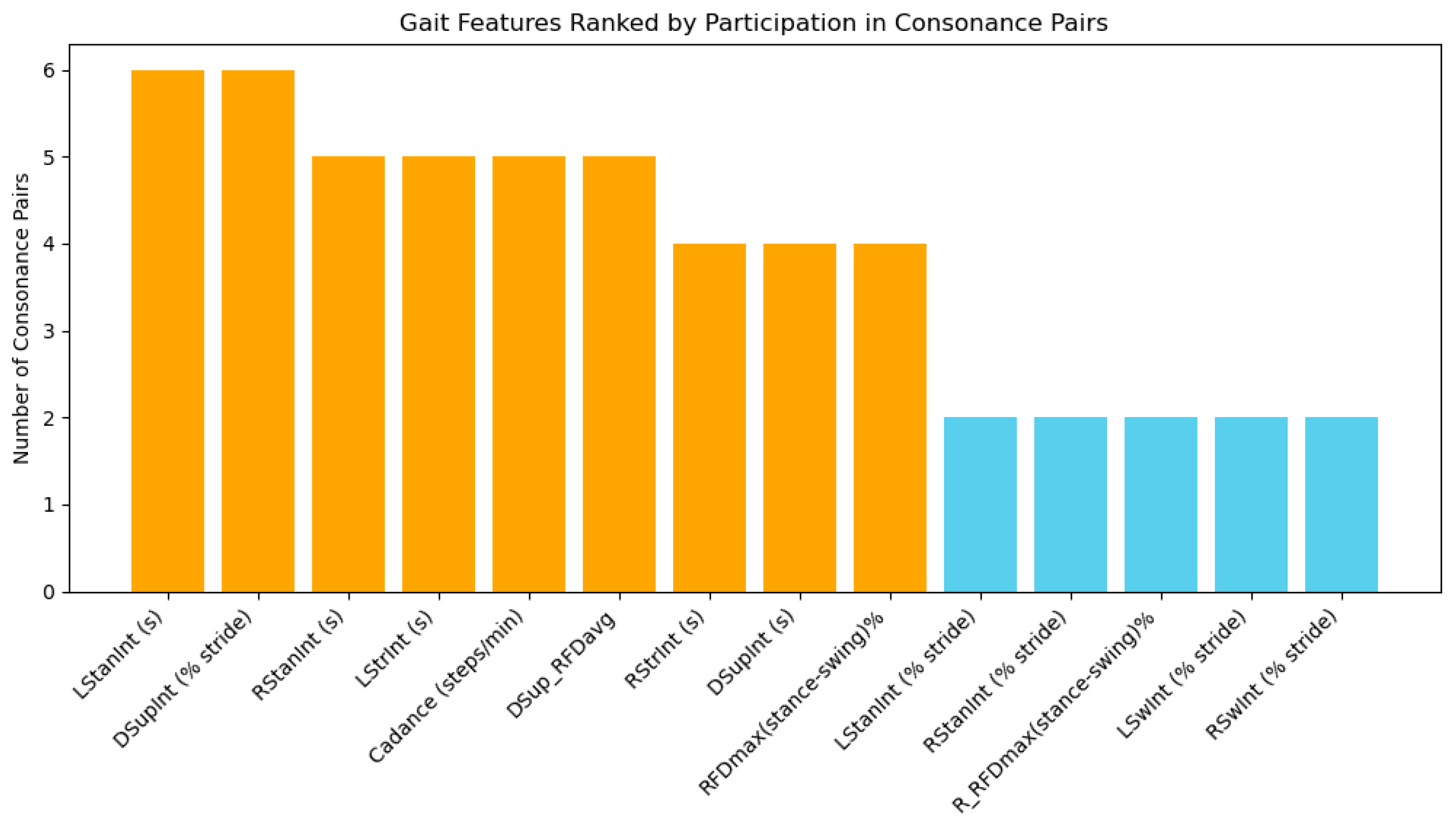
- Population-level intercriteria relations: Feature relations were analyzed across the entire dataset to identify the most prominent (up to 5%) gait features exhibiting strong consonance (i.e., statistical agreement or dependence).
- Group-specific intercriteria relations: The analysis was performed separately within each subject group (CONTROL, ALS, HUNT, and PARK) followed by pairwise comparisons between the CONTROL and each NDD group (CONTROL vs. ALS; CONTROL vs. HUNT; CONTROL vs. PARK). Our focus was to identify feature relations presenting strong positive or negative consonance that are unique either to the CONTROL group or to a specific neurodegenerative disorder.
3.2. Population-Level Intercriteria Relations
3.3. Group-Specific Intercriteria Relations
- CONTROL group: 3.9% (32 of 820 pairs), 20 positive and 12 negative consonances;
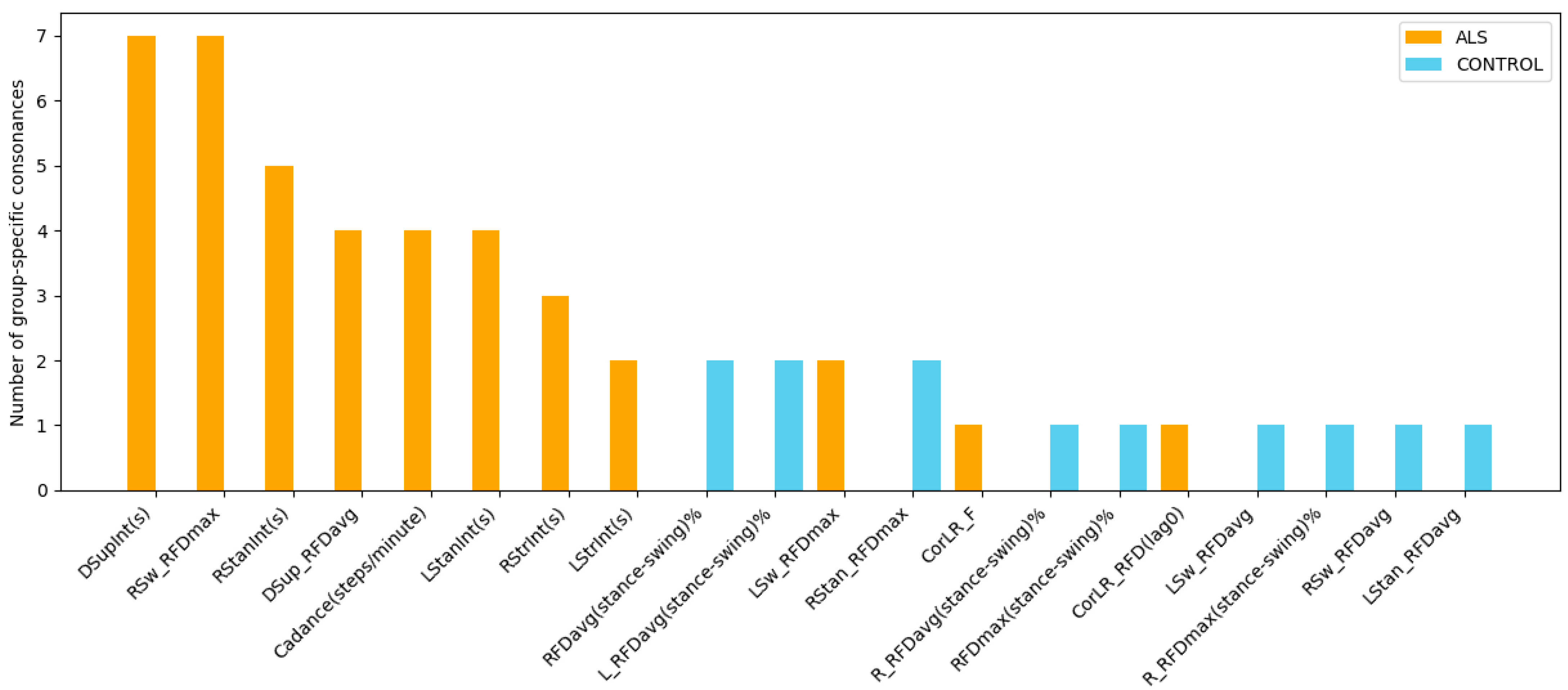
- ALS group: 5.98% (49 of 820 pairs), 25 positive and 24 negative consonances;
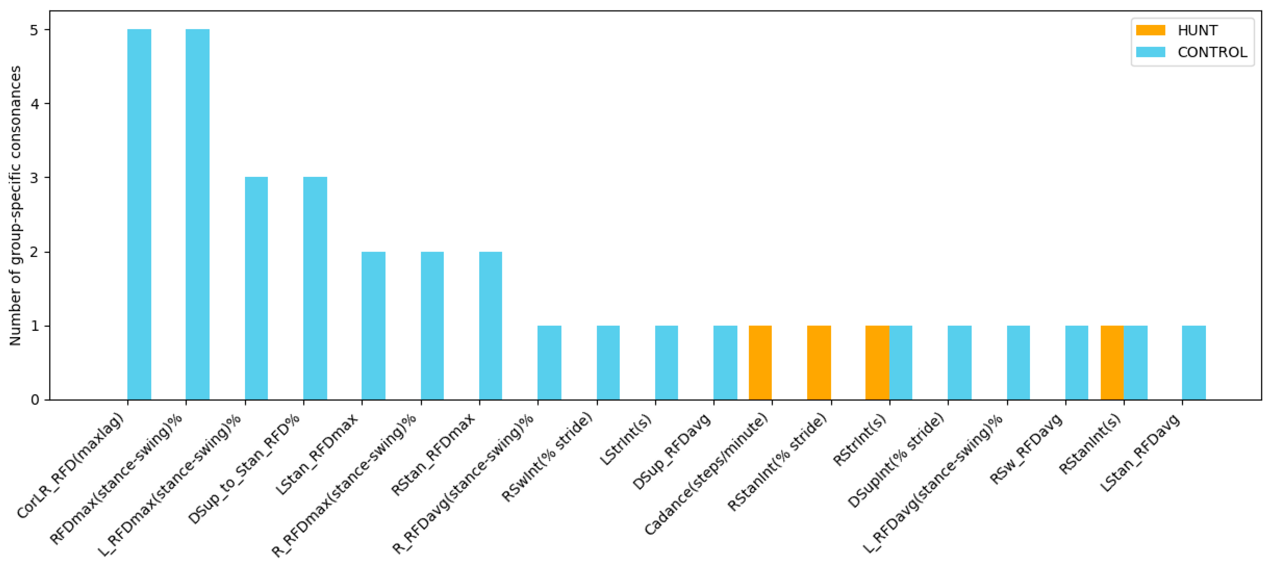
- HUNT group: 1.83% (15 of 820 pairs), 6 positive and 9 negative consonances;
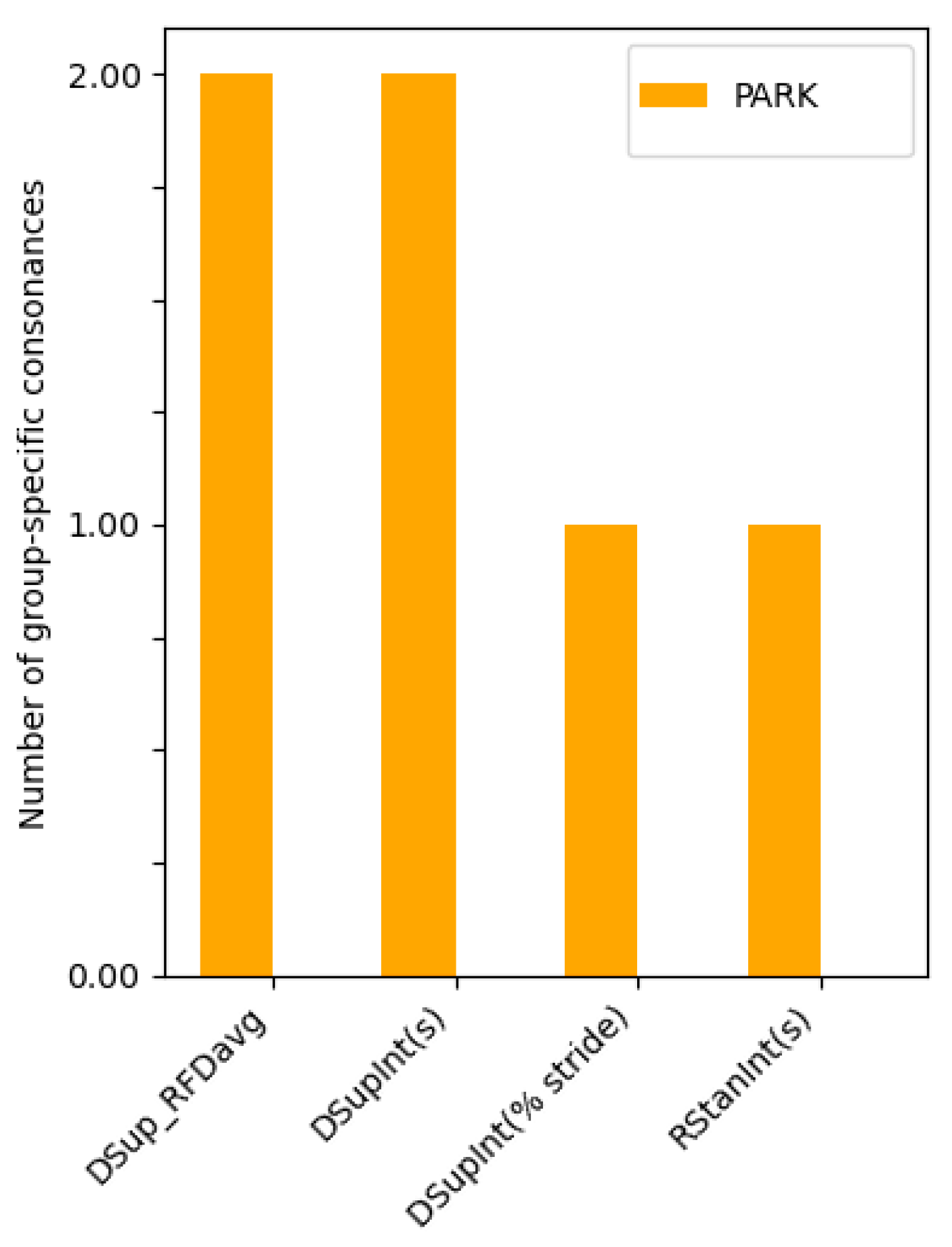
- PARK group: 4.15% (34 of 820 pairs), 20 positive and 14 negative consonances;
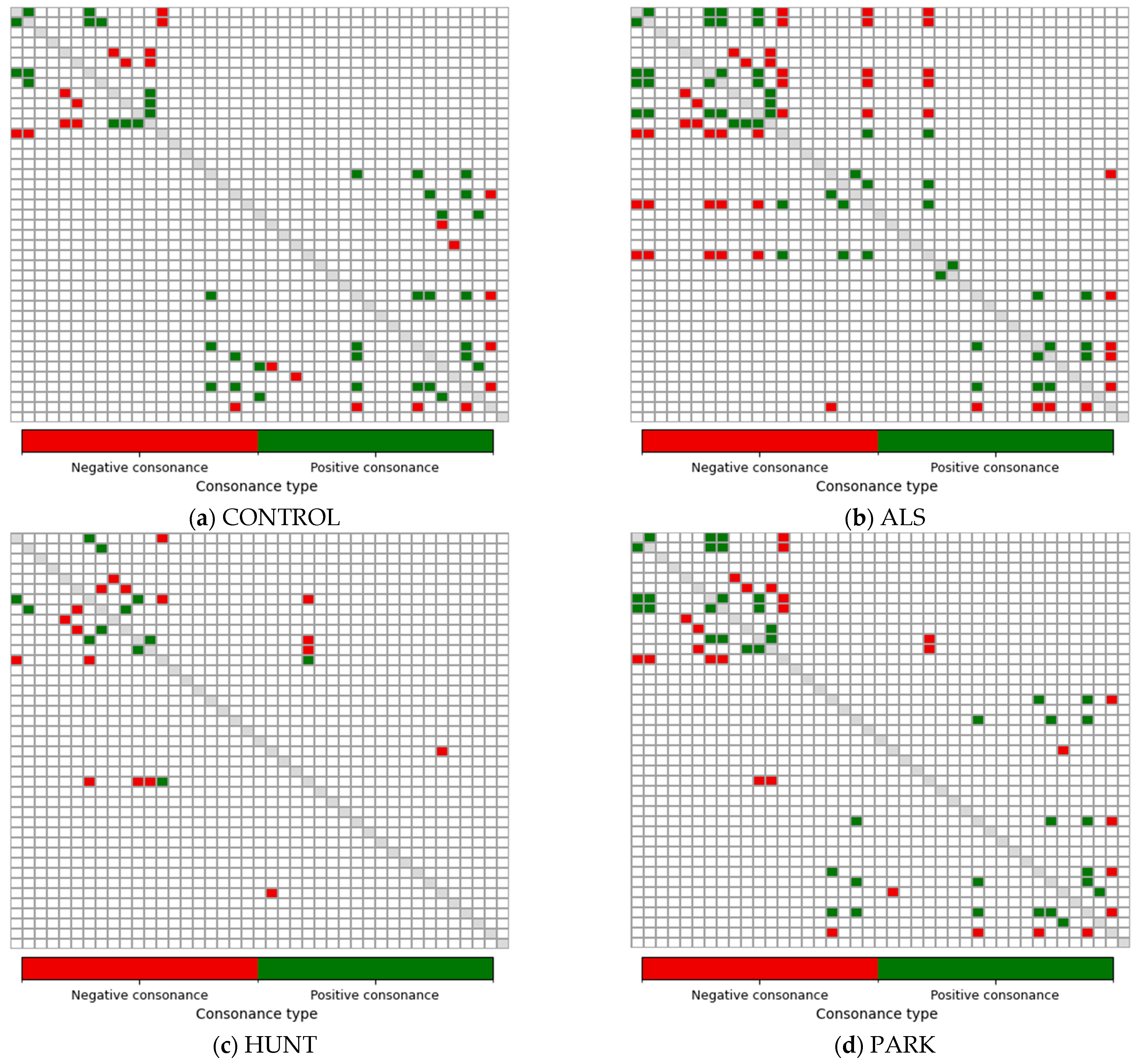
- Positive consonances present only in one group (µ ≥ 0.75 and υ ≤ 0.25);
- Negative consonances present only in one group (µ ≤ 0.25 and υ ≥ 0.75),
- Pairs that satisfy either of the above two conditions and additionally show a substantial between-group difference, defined as follows: |µ_CONTROL − µ_NDD| ≥ 0.1 and |υ_CONTROL − υ_NDD| ≥ 0.1.
3.4. Benchmarking Population-Level Intercriteria Relations Against Reference Correlation Method
4. Discussion
4.1. Population-Level Intercriteria Relations
4.2. Group-Specific Intercriteria Relations
4.2.1. ALS Patients vs. Healthy Controls
- CONTROL-specific consonances between temporal features:
- Positive consonance between LStrInt(s) and RStrInt(s): This bilateral dependency reflects stable, symmetric regulation of left- and right-leg stride intervals typical of healthy gait. Its absence in the ALS group corresponds to the well-documented increase in temporal irregularity, impaired step-to-step consistency, and reduced ability to maintain stable rhythm in ALS [15,16].
- Negative consonance between RStrInt(s) and Cadence(steps/minute): This inverse relation—longer stride intervals paired with lower cadence—is present in a normal gait. Its absence in ALS patients suggests a pathological decoupling between the stride duration and step rhythm, consistent with reduced walking speed and timing variability associated with neuromuscular weakness [5,7,16].
- ALS-specific consonances between temporal features:
- Positive consonances of LStrInt(s), RStrInt(s), LStanInt(s), RStanInt(s) vs. DSupInt(s): These multiple interdependencies indicate that ALS patients exhibit globally coupled timing patterns across stride, stance, and double-support intervals. Such coupling reflects the characteristic prolongation of stance and overall gait cycle duration due to muscle weakness, instability, and reduced motor control efficiency [16].
- Negative consonances between stance-related intervals (LStanInt(s), RStanInt(s), DSupInt(s)) and Cadence(steps/minute): In ALS, increases in stance or double-support duration directly correspond to reduced cadence, highlighting a slowed gait driven by a prolonged weight-bearing time [15,16,17]. This relation is not present in controls and aligns with compensatory strategies used to maintain stability when limb force generation is compromised.
- CONTROL-specific consonances between kinetic RFD-based features:
- Positive consonances of RFDmax(stance–swing)%, L_RFDmax(stance–swing)%, R_RFDmax(stance–swing)%, LStan_RFDmax vs. RStan_RFDmax: Reflect coordinated bilateral modulation of force production during stance–swing transitions—indicative of efficient neuromuscular control and symmetrical loading in a healthy gait.
- Positive consonances linking CorLR_RFD(maxlag) to L_RFDmax(stance–swing)%, R_RFDmax(stance–swing)%, RFDmax(stance–swing)%, and LStan_RFDmax: Demonstrate robust left–right synchronization of force-generation dynamics, reflecting preserved inter-limb coordination in healthy gait. This synchrony is markedly reduced in ALS, where neuromuscular degeneration disrupts the temporal alignment of limb kinetics and leads to desynchronized force-production patterns [80].
- Positive consonance between LStan_RFDavg and L_RFDavg(stance–swing)%: Indicates that higher stance-phase RFD is associated with greater overall force-development capacity across the full stance–swing cycle, typical of healthy, well-regulated loading patterns.
- Negative consonances between corresponding kinetic features of the left and right legs, specifically LSw_RFDavg vs. L_RFDavg(stance–swing)% and RSw_RFDavg vs. R_RFDavg(stance–swing)%: Indicate that force development is naturally lower during swing than during stance, consistent with normal biomechanical demands. The absence of these relations in ALS suggests impaired modulation of force across gait phases, reflecting a reduced ability to dynamically regulate limb loading [81].
- ALS-specific consonances between kinetic features:
- Positive consonances between LSw_RFDmax, RSw_RFDmax, and DSup_RFDavg: These relations suggest altered coordination of force generation during swing and double-support phases. They reflect compensatory adaptations linked to muscle weakness, reduced hip control, and increased instability characteristic of an ALS gait [15,16,17].
- Positive consonance between CorLR_F(lag0) and CorLR_RFD(lag0): Indicates that reductions in bilateral force coordination are closely coupled with the impaired synchronization of RFD. Such coupling is consistent with the ALS-related degeneration of motor-unit recruitment and inter-limb communication [82].
- Negative consonances of RSw_RFDmax and DSup_RFDavg vs. temporal intervals (RStrInt(s), LStanInt(s), RStanInt(s), and DSupInt(s)): Show that prolonged stance or double-support phases constrain dynamic force production during swing. These interactions, absent in healthy controls, reflect the tendency of ALS patients to adopt longer support phases [17], while reducing the capacity for rapid force generation in swing.
4.2.2. HUNT Patients vs. Healthy Controls
- CONTROL-specific consonances between temporal features:
- Positive consonance between LStrInt(s) and RStrInt(s): This bilateral dependency reflects stable, symmetric regulation of left- and right-leg stride intervals typical of a healthy gait. Its absence in the HUNT group corresponds to well-known Huntington’s disease features such as elevated temporal variability, irregular step-to-step transitions, and difficulty in sustaining consistent forward progression [7,13,14].
- Negative consonance between RStrInt(s) and Cadance(steps/minute): This inverse relation—longer stride intervals paired with lower cadence—is typical for a normal gait. Its absence in HUNT patients suggests a pathological decoupling between stride duration and stepping rhythm, consistent with irregular pacing and increased stride-timing variability.
- HUNT-specific consonances between temporal features:
- Positive consonance between RStanInt(s) and RStanInt(% stride): This indicates that in HUNT patients, the absolute and relative durations of the right stance phase increase proportionally—a dependency consistent with the compensatory lengthening of stance time often reported in individuals with instability, chorea, or fear of falling [83]. This feature is not present in controls, underscoring a disease-specific alteration in stance-phase organization.
- Negative consonance between temporal features RStanInt(s) and RSwInt(% stride): Increased stance time corresponds to proportional reductions in swing time. This asymmetrical temporal adjustment is not seen in controls and supports the interpretation that HUNT patients adopt prolonged stance [84] as a compensatory mechanism to maintain stability.
- CONTROL-specific consonances between kinetic RFD variables:
- Positive consonances among RFDmax(stance–swing)%, L_RFDmax(stance–swing)%, R_RFDmax(stance–swing)%, LStan_RFDmax, and RStan_RFDmax: Reveal coordinated bilateral modulation of force production during stance–swing transitions—an indicator of intact motor control in healthy gait.
- Positive consonance linking CorLR_RFD(maxlag) to L_RFDmax(stance–swing)%, R_RFDmax(stance–swing)%, RFDmax(stance–swing)%, and LStan_RFDmax: Reflects strong left–right synchronization of RFD, further underscoring coordinated bilateral force generation.
- Positive consonance between LStan_RFDavg and L_RFDavg(stance–swing)%: Suggests that higher stance-phase RFD corresponds to higher overall force-development capacity across the stance–swing cycle.
- Negative consonance between DSup_to_Stan_RFD% to RStan_RFDmax, CorLR_RFD(maxlag), and L_RFDmax(stance–swing)%: Indicate efficient redistribution of force from double-support to stance and coordinated inter-limb regulation of loading.
- Negative consonance between RSw_RFDavg and R_RFDavg(stance–swing)%: Shows that force development is naturally lower during swing than stance, consistent with expected biomechanical demands of a healthy gait. The absence of these negative kinetic consonances in HUNT suggests disruption in normal load-transfer strategies and impaired modulation of force across gait phases.
- HUNT-specific consonances between temporal and kinetic RFD features:
4.2.3. PARK Patients vs. Healthy Controls
- CONTROL-specific consonances between gait features are not identified, indicating that the distinguishing relations arise exclusively from PARK-specific alterations.
- PARK-specific consonances between temporal and kinetic features:
- Positive consonance between RStanInt(s) and DSupInt(s): This dependency indicates that in PARK patients, prolongation of the right stance phase is directly associated with increased double-support time. This pattern aligns with characteristic shuffling gait in Parkinson’s disease, where feet are insufficiently lifted and weight transfer becomes slower and more prolonged [85]. The absence of this relation in healthy controls suggests that this coupling reflects disease-specific compensation for postural instability and bradykinetic movement.
- Negative consonance between DSupInt(s) and DSup_RFDavg: Longer double-support duration corresponds to reduced force development during double support. This relationship reflects the slow, hesitant, and low-amplitude movements typical of a Parkinsonian gait [8,9], where decreased push-off dynamics and stiffness reduce the force generated during weight transfer.
- Negative consonance between DSupInt(% stride) and DSup_RFDavg: A proportional increase in the relative duration of double support is accompanied by reduced RFD during the same phase. This reinforces the interpretation that prolonged stabilization phases in Parkinson’s disease come at the cost of diminished dynamic force production, consistent with bradykinesia and decreased lower-limb motor drive.
4.3. Related Work and Comparisons
5. Study Limitations
6. Conclusions
- ALS vs. CONTROL: ICrA identifies numerous temporal and kinetic consonances unique to ALS, indicating widespread alterations in the stance duration, stride timing, force-generation dynamics, and RFD modulation. The disrupted relationships observed among stance–swing timing, inter-limb synchronization, and intra-limb RFD asymmetries point to impaired neuromuscular control and compensatory strategies characteristic of an ALS gait. In contrast, controls show coherent bilateral regulation of RFD and consistent coupling between temporal and kinetic features, suggesting preserved coordination mechanisms absent in ALS.
- HUNT vs. CONTROL: HUNT patients show the most pronounced breakdown of relational gait structure. The loss of nearly all control-specific consonances, coupled with the emergence of only a few weak disease-specific dependencies, indicates severe disruption in the timing relationships that normally link stride intervals, stance–swing organization, and force-production patterns. These findings align with the well-known high temporal variability, instability, and irregular step-to-step transitions characteristic of Huntington’s disease.
- PARK vs. CONTROL: Compared with ALS and HUNT, Parkinson’s disease exhibits only a small number of group-specific consonances. The differences are subtle and largely confined to relationships involving stance duration, double-support timing, and reduced RFD during double support. These limited alterations suggest that inter-feature coordination in a Parkinsonian gait remains mostly preserved, with detectable disruptions reflecting hallmark features such as shuffling, slowed step initiation, and reduced propulsive force.
Supplementary Materials
Author Contributions
Funding
Institutional Review Board Statement
Informed Consent Statement
Data Availability Statement
Conflicts of Interest
References
- Ciurea, A.V.; Mohan, A.G.; Covache-Busuioc, R.-A.; Costin, H.-P.; Glavan, L.-A.; Corlatescu, A.-D.; Saceleanu, V.M. Unraveling Molecular and Genetic Insights into Neurodegenerative Diseases: Advances in Understanding Alzheimer’s, Parkinson’s, and Huntington’s Diseases and Amyotrophic Lateral Sclerosis. Int. J. Mol. Sci. 2023, 24, 10809. [Google Scholar] [CrossRef] [Scilit]
- Beyrami, S.M.G.; Ghaderyan, P. A robust, cost-effective and non-invasive computer-aided method for diagnosis three types of neurodegenerative diseases with gait signal analysis. Measurement 2020, 156, 107579. [Google Scholar] [CrossRef] [Scilit]
- Dorsey, E.R.; Elbaz, A.; Nichols, E.; Abd-Allah, F.; Abdelalim, A.; Adsuar, J.C.; Ansha, M.G.; Brayne, C.; Choi, J.-Y.J.; Collado-Mateo, D. Global, Regional, and National Burden of Parkinson’s Disease, 1990–2016: A Systematic Analysis for the Global Burden of Disease Study 2016. Lancet Neurol. 2018, 17, 939–953. [Google Scholar] [CrossRef] [Scilit]
- Mathis, S.; Goizet, C.; Soulages, A.; Vallat, J.-M. Genetics of amyotrophic lateral sclerosis: A review. J. Neurol. Sci. 2019, 15, 217–226. [Google Scholar] [CrossRef] [Scilit]
- Gadhave, D.G.; Sugandhi, V.V.; Jha, S.K.; Nangare, S.N.; Gupta, G.; Singh, S.K.; Dua, K.; Cho, H.; Hansbro, P.M.; Paudel, K.R. Neurodegenerative disorders: Mechanisms of degeneration and therapeutic approaches with their clinical relevance. Ageing Res. Rev. 2024, 99, 102357. [Google Scholar] [CrossRef] [Scilit]
- Lamptey, R.N.L.; Chaulagain, B.; Trivedi, R.; Gothwal, A.; Layek, B.; Singh, J. A Review of the Common Neurodegenerative Disorders: Current Therapeutic Approaches and the Potential Role of Nanotherapeutics. Int. J. Mol. Sci. 2022, 23, 1851. [Google Scholar] [CrossRef] [Scilit]
- Harmanşa, Y.K.; Erbaş, O. Diagnostic and Therapeutic Biomarkers for Neurodegeneration. J. Exp. Basic Med. Sci. 2022, 3, 47–53. [Google Scholar] [CrossRef]
- Politis, M.; Wu, K.; Molloy, S.; Bain, P.G.; Chaudhuri, K.R.; Piccini, P. Parkinson’s Disease Symptoms: The Patient’s Perspective. Mov. Disord. 2010, 25, 1646–1651. [Google Scholar] [CrossRef] [Scilit] [PubMed]
- Balestrino, R.; Schapira, A.H.V. Parkinson Disease. Eur. J. Neurol. 2020, 27, 27–42. [Google Scholar] [CrossRef] [Scilit] [PubMed]
- Bhat, S.; Acharya, U.R.; Hagiwara, Y.; Dadmehr, N.; Adeli, H. Parkinson’s Disease: Cause Factors, Measurable Indicators, and Early Diagnosis. Comput. Biol. Med. 2018, 102, 234–241. [Google Scholar] [CrossRef] [Scilit]
- Carvalho, L.P.; Mate, K.K.V.; Cinar, E.; Abou-Sharkh, A.; Lafontaine, A.-L.; Mayo, N.E. A new approach toward gait training in patients with Parkinson’s Disease. Gait Posture 2020, 81, 14–20. [Google Scholar] [CrossRef] [Scilit]
- Moreau, C.; Cantiniaux, S.; Delval, A.; Defebvre, L.; Azulay, J.-P. Gait disorders in Parkinson’s disease: And pathophysiological approaches. Rev. Neurol. 2010, 166, 158–167. [Google Scholar] [CrossRef] [Scilit]
- Koller, W.C.; Trimble, J. The gait abnormality of Huntington’s disease. Neurology 1985, 35, 1450–1454. [Google Scholar] [CrossRef] [Scilit] [PubMed]
- Delval, A.; Krystkowiak, P.; Blatt, J.L.; Labyt, E.; Dujardin, K.; Destée, A.; Derambure, P.; Defebvre, L. Role of hypokinesia and bradykinesia in gait disturbances in Huntington’s disease: A biomechanical study. J. Neurol. 2006, 253, 73–80. [Google Scholar] [CrossRef] [Scilit]
- Dubbioso, R.; Spisto, M.; Hausdorff, J.M.; Aceto, G.; Iuzzolino, V.V.; Senerchia, G.; De Marco, S.; Marcuccio, L.; Femiano, C.; Iodice, R.; et al. Cognitive impairment is associated with gait variability and fall risk in amyotrophic lateral sclerosis. Eur. J. Neurol. 2023, 30, 3056–3067. [Google Scholar] [CrossRef] [Scilit] [PubMed]
- Hausdorff, J.M.; Lertratanakul, A.; Cudkowicz, M.E.; Peterson, A.L.; Kaliton, D.; Goldberger, A.L. Dynamic markers of altered gait rhythm in amyotrophic lateral sclerosis. J. Appl. Physiol. 2000, 88, 2045–2053. [Google Scholar] [CrossRef] [Scilit] [PubMed]
- Goldfarb, B.J.; Simon, S.R. Gait patterns in patients with amyotrophic lateral sclerosis. Arch. Phys. Med. Rehabil. 1984, 65, 61–65. [Google Scholar]
- Berry, J.D.; Blanchard, M.; Bonar, K.; Drane, E.; Murton, M.; Ploug, U.; Ricchetti-Masterson, K.; Savic, N.; Worthington, E.; Heiman-Patterson, T. Epidemiology and economic burden of amyotrophic lateral sclerosis in the United States: A literature review. Amyotroph. Lateral Scler. Front. Degener. 2023, 24, 436–448. [Google Scholar] [CrossRef] [Scilit] [PubMed]
- Kruse, C.; Lipinski, A.; Verheyen, M.; Balzer-Geldsetzer, M.; Wittenberg, M.; Lorenzl, S.; Richinger, C.; Schmotz, C.; Tönges, L.; Woitalla, D.; et al. Care of Late-Stage Parkinsonism: Resource Utilization of the Disease in Five European Countries. Mov. Disord. 2024, 39, 571–584. [Google Scholar] [CrossRef] [Scilit] [PubMed]
- Rodríguez-Santana, I.; Mestre, T.; Squitieri, F.; Willock, R.; Arnesen, A.; Clarke, A.; D’Alessio, B.; Fisher, A.; Fuller, R.; Hamilton, J.L.; et al. Economic burden of Huntington disease in Europe and the USA: Results from the Huntington’s Disease Burden of Illness study. Eur. J. Neurol. 2023, 30, 1109–1117. [Google Scholar] [CrossRef] [Scilit]
- Achtert, K.; Kerkemeyer, L. The economic burden of amyotrophic lateral sclerosis: A systematic review. Eur. J. Health Econ. 2021, 22, 1151–1166. [Google Scholar] [CrossRef] [Scilit]
- Beck, M.; Giess, R.; Würffel, W.; Magnus, T.; Ochs, G.; Toyka, K.V. Comparison of maximal voluntary isometric contraction and Drachman’s hand-held dynamometry in evaluating patients with amyotrophic lateral sclerosis. Muscle Nerve 1999, 22, 1265–1270. [Google Scholar] [CrossRef] [Scilit]
- Bowser, R.; Cudkowicz, M.; Kaddurah-Daouk, R. Biomarkers for amyotrophic lateral sclerosis. Expert Rev. Mol. Diagn. 2006, 6, 387–398. [Google Scholar] [CrossRef] [Scilit]
- Erdaş, Ç.B.; Sümer, E.; Kibaroğlu, S. Neurodegenerative diseases detection and grading using gait dynamics. Multimed. Tools Appl. 2023, 82, 22925–22942. [Google Scholar] [CrossRef] [Scilit]
- Khera, P.; Kumar, A.; Kapila, R. Decision tree for severity assessment of neurodegenerative diseases using possibility approach and gait dynamics. Sci. Rep. 2025, 15, 26247. [Google Scholar] [CrossRef] [Scilit] [PubMed]
- Ru, Y.; Zhao, X.; Baghersad, Y.; Andani, H.T.; Hekmatifar, M.; Bouallegue, B. Gait analysis and Machine learning algorithms to distinguish between Parkinson’s disease, amyotrophic lateral sclerosis, and Huntington’s disease. Biomed. Signal Process. Control 2025, 110, 108216. [Google Scholar] [CrossRef] [Scilit]
- Cicirelli, G.; Impedovo, D.; Dentamaro, V.; Marani, R.; Pirlo, G.; D’Orazio, T.R. Human Gait Analysis in Neurodegenerative Diseases: A Review. IEEE J. Biomed. Health Inform. 2022, 26, 229–242. [Google Scholar] [CrossRef] [Scilit]
- Atanassov, K.; Mavrov, D.; Atanassova, V. Intercriteria Decision making: A new approach for multicriteria decision making, based on index matrices and intuitionistic fuzzy sets. Issues IFSs GNs 2014, 11, 1–8. [Google Scholar]
- Atanassov, K.; Atanassova, V.; Gluhchev, G. Intercriteria analysis: Ideas and problems. Notes Intuitionistic Fuzzy Sets 2015, 21, 81–88. [Google Scholar]
- Atanassova, V. Interpretation in the intuitionistic fuzzy triangle of the results, obtained by the intercriteria analysis. In Proceedings of the 2015 Conference of the International Fuzzy Systems Association and the European Society for Fuzzy Logic and Technology (IFSA-EUSFLAT-15), Gijón, Spain, 30 June 2015; Atlantis Press: Paris, France, 2015; pp. 1369–1374. [Google Scholar]
- Doukovska, L.; Atanassova, V.; Sotirova, E.; Vardeva, I.; Radeva, I. Defining consonance thresholds in intercriteria analysis: An overview. Stud. Comput. Intell. 2019, 757, 161–179. [Google Scholar]
- Pencheva, T.; Roeva, O.; Angelova, M. Investigation of genetic algorithm performance based on different algorithms for intercriteria relations calculation. Lect. Notes Comput. Sci. 2018, 10665, 390–398. [Google Scholar]
- Vassilev, P.; Ribagin, S.; Todorova, L. On an aggregation of expert value assignments using index matrices. Notes Intuitionistic Fuzzy Sets 2017, 23, 75–78. [Google Scholar]
- Roeva, O.; Pencheva, T.; Angelova, M.; Vassilev, P. Intercriteria analysis by pairs and triples of genetic algorithms application for models identification. Stud. Comput. Intell. 2016, 655, 193–218. [Google Scholar]
- Traneva, V.; Tranev, S.; Kahraman, C. A multidimensional intuitionistic fuzzy InterCriteria analysis in the restaurant. J. Intell. Fuzzy Syst. Appl. Eng. Technol. 2020, 39, 6059–6071. [Google Scholar] [CrossRef] [Scilit]
- Tsakovska, I.; Alov, P.; Ikonomov, N.; Atanassova, V.; Vassilev, P.; Roeva, O.; Jereva, D.; Atanassov, K.; Pajeva, I.; Pencheva, T. Intercriteria analysis implementation for exploration of the performance of various docking scoring functions. Stud. Comput. Intell. 2021, 902, 88–98. [Google Scholar]
- Vankova, D.; Sotirova, E.; Bureva, V. An application of the intercriteria analysis approach to health-related quality of life. Notes Intuitionistic Fuzzy Sets 2015, 21, 40–48. [Google Scholar]
- Todinova, S.; Mavrov, D.; Krumova, S.; Marinov, P.; Atanassova, V.; Atanassov, K.; Taneva, S.G. Blood plasma thermograms dataset analysis by means of intercriteria and correlation analyses for the case of colorectal cancer. Int. J. Bioautomat. 2016, 20, 115–124. [Google Scholar]
- Krumova, S.; Todinova, S.; Mavrov, D.; Marinov, P.; Atanassova, V.; Atanassov, K.; Taneva, S. Intercriteria analysis of calorimetric data of blood serum proteome. Biochim. Biophys. Acta Gen. Subj. 2017, 1861, 409–417. [Google Scholar] [CrossRef] [Scilit]
- Sotirov, S.; Petrova, Y.; Bozov, H.; Sotirova, E. A Hybrid Algorithm for Multilayer Perceptron Design with Intuitionistic Fuzzy Logic Using Malignant Melanoma Disease Data. In Proceedings of the International Conference on Intelligent and Fuzzy Systems (INFUS 2022), Izmir, Turkey, 19–21 July 2022; Kahraman, C., Tolga, A.C., Cevik Onar, S., Cebi, S., Oztaysi, B., Sari, I.U., Eds.; Springer: Berlin/Heidelberg, Germany, 2022; Volume 504. [Google Scholar] [CrossRef] [Scilit]
- Zaharieva, B.; Doukovska, L.; Ribagin, S.; Radeva, I. Intercriteria analysis of data obtained from patients with Behterev’s disease. Int. J. Bioautomat. 2020, 24, 5–14. [Google Scholar] [CrossRef] [Scilit]
- Jekova, I.; Vassilev, P.; Stoyanov, T.; Pencheva, T. InterCriteria Analysis: Application for ECG Data Analysis. Mathematics 2021, 9, 854. [Google Scholar] [CrossRef] [Scilit]
- Dobrev, P.; Sotirova, E. An Intelligent Data Analysis Approach for Women with Menopausal Genitourinary Syndrome with Intuitionistic Fuzzy Logic. In Proceedings of the International Conference on Intelligent and Fuzzy Systems 2023 (INFUS 2023), Istanbul, Turkey, 22–24 August 2023; Kahraman, C., Sari, I.U., Oztaysi, B., Cebi, S., Cevik Onar, S., Tolga, A.Ç., Eds.; Springer: Berlin/Heidelberg, Germany, 2023; Volume 758. [Google Scholar] [CrossRef] [Scilit]
- Angelova, S.; Angelova, M.; Raikova, R. Estimating Surface EMG Activity of Human Upper Arm Muscles Using InterCriteria Analysis. Math. Comput. Appl. 2024, 29, 8. [Google Scholar] [CrossRef] [Scilit]
- Angelova, M.; Angelova, S.; Raikova, R. How to Optimize the Experimental Protocol for Surface EMG Signal Measurements Using the InterCriteria Decision-Making Approach. Appl. Sci. 2024, 14, 5436. [Google Scholar] [CrossRef] [Scilit]
- Mancheva, K.; Angelova, M.; Kossev, A.; Angelova, S. Assessing the Effects of TMS Intensities and Muscle Conditions on the Evoked Responses of the First Dorsal Interosseous Muscle Using Statistical Methods and InterCriteria Analysis. Appl. Sci. 2025, 15, 5236. [Google Scholar] [CrossRef] [Scilit]
- Angelova, S.; Raikova, R.; Angelova, M. Intercriteria Decision-Making Method for Speed and Load Effects Evaluation on Upper Arm Muscles in the Horizontal Plane. Appl. Sci. 2025, 15, 11213. [Google Scholar] [CrossRef] [Scilit]
- Goldberger, A.; Amaral, L.; Glass, L.; Hausdorff, J.; Ivanov, P.C.; Mark, R.; Mietus, J.E.; Moody, G.B.; Peng, C.-K.; Stanley, H.E. PhysioBank, PhysioToolkit, and PhysioNet: Components of a new research resource for complex physiologic signals. Circulation 2000, 101, e215–e220. [Google Scholar] [CrossRef] [Scilit] [PubMed]
- Hausdorff, J. Gait in Neurodegenerative Disease Database; Version: 1.0.0; PhysioNet: Cambridge, MA, USA, 2000; Available online: https://www.physionet.org/content/gaitndd/1.0.0/ (accessed on 4 November 2025).
- Murray, M.P.; Drought, A.B.; Kory, R.C. Walking patterns of normal men. J. Bone Jt. Surg. Am. 1974, 56, 777–790. [Google Scholar] [CrossRef] [Scilit]
- Perry, J.; Burnfield, J.M. Gait Analysis: Normal and Pathological Function, 2nd ed.; SLACK Incorporated: Thorofare, NJ, USA, 2010. [Google Scholar]
- Whittle, M.W. Gait Analysis: An Introduction, 5th ed.; Butterworth-Heinemann: Oxford, UK, 2014. [Google Scholar]
- Erdaş, Ç.B.; Sümer, E. CNN-Based Neurodegenerative Disease Classification Using QR-Represented Gait Data. Brain Behav. 2024, 14, e70100. [Google Scholar] [CrossRef] [Scilit]
- Yang, M.; Zheng, H.; Wang, H.; McClean, S. Feature Selection and Construction for the Discrimination of Neurodegenerative Diseases Based on Gait Analysis. In Proceedings of the 3rd International Conference on Pervasive Computing Technologies for Healthcare, London, UK, 1–3 April 2009; pp. 1–7. [Google Scholar] [CrossRef] [Scilit]
- Xia, Y.; Gao, Q.; Ye, Q. Classification of Gait Rhythm Signals Between Patients with Neuro-degenerative Diseases and Normal Subjects: Experiments with Statistical Features and Different Classification Models. Biomed. Signal Process. Control 2015, 18, 254–262. [Google Scholar] [CrossRef] [Scilit]
- Hausdorff, J.M. Gait variability: Methods, modeling and meaning. J. NeuroEng. Rehabil. 2005, 2, 19. [Google Scholar] [CrossRef] [Scilit] [PubMed]
- Galna, B.; Lord, S.R.; Rochester, L. Is gait variability reliable in older adults and Parkinson’s disease? Towards an optimal testing protocol. Gait Posture 2013, 37, 580–585. [Google Scholar] [CrossRef] [Scilit] [PubMed]
- Lin, D.C.; McGowan, C.P.; Blum, K.P.; Ting, L.H. Yank: The Time Derivative of Force is an Important Biomechanical Variable in Sensorimotor Systems. J. Exp. Biol. 2019, 222, jeb180414. [Google Scholar] [CrossRef] [Scilit]
- Andersen, L.L.; Aagaard, P. Influence of maximal muscle strength and intrinsic muscle contractile properties on contractile rate of force development. Eur. J. Appl. Physiol. 2006, 96, 46–52. [Google Scholar] [CrossRef] [Scilit]
- Maffiuletti, N.A.; Aagaard, P.; Blazevich, A.J.; Folland, J.; Tillin, N.; Duchateau, J. Rate of force development: Physiological and methodological considerations. Eur. J. Appl. Physiol. 2016, 116, 1091–1116. [Google Scholar] [CrossRef] [Scilit] [PubMed]
- Bohannon, R.W. Considerations and Practical Options for Measuring Muscle Strength: A Narrative Review. BioMed Res. Int. 2019, 2019, 8194537. [Google Scholar] [CrossRef] [Scilit]
- Winter, D.A. The Biomechanics and Motor Control of Human Gait: Normal, Elderly and Pathological, 2nd ed.; University of Waterloo Press: Waterloo, ON, Canada, 1991. [Google Scholar]
- Aagaard, P.; Simonsen, E.B.; Andersen, J.L.; Magnusson, P.; Dyhre-Poulsen, P. Increased rate of force development and neural drive of human skeletal muscle following resistance training. J. Appl. Physiol. 2002, 93, 1318–1326. [Google Scholar] [CrossRef] [Scilit] [PubMed]
- Winter, D.A. Biomechanics and Motor Control of Human Movement, 4th ed.; John Wiley & Sons: Hoboken, NJ, USA, 2009. [Google Scholar]
- Bendat, J.S.; Piersol, A.G. Random Data: Analysis and Measurement Procedures, 4th ed.; John Wiley & Sons: Hoboken, NJ, USA, 2010. [Google Scholar]
- Robinson, R.O.; Herzog, W.; Nigg, B.M. Use of force platform variables to quantify the effects of chiropractic manipulation on gait symmetry. J. Manip. Physiol. Ther. 1987, 10, 172–176. [Google Scholar]
- Herzog, W.; Nigg, B.M.; Read, L.J.; Olsson, E. Asymmetries in ground reaction force patterns in normal human gait. Med. Sci. Sports Exerc. 1989, 21, 110–114. [Google Scholar] [CrossRef] [Scilit]
- Patterson, K.K.; Gage, W.H.; Brooks, D.; Black, S.E.; McIlroy, W.E. Evaluation of gait symmetry after stroke: A comparison of current methods and recommendations for standardization. Gait Posture 2010, 31, 241–246. [Google Scholar] [CrossRef] [Scilit]
- Atanassov, K. Generalized index matrices. Compt. Rend. Acad. Bulg. Sci. 1987, 40, 15–18. [Google Scholar]
- Atanassov, K. Index Matrices: Towards an Augmented Matrix Calculus; Springer: Cham, Switzerland, 2014. [Google Scholar]
- Atanassov, K. Intuitionistic fuzzy sets. Int. J. Bioautom. 2016, 20, S1–S6. [Google Scholar]
- Atanassov, K. On Intuitionistic Fuzzy Sets Theory; Springer: Cham, Switzerland, 2012. [Google Scholar]
- Atanassov, K. Intuitionistic Fuzzy Logics; Springer: Cham, Switzerland, 2017. [Google Scholar]
- Hošková-Mayerová, S.; Maturo, A. Decision-making process using hyperstructures and fuzzy structures in social sciences. Stud. Fuzziness Soft Comput. 2018, 357, 103–111. [Google Scholar] [CrossRef] [Scilit]
- Szmidt, E.; Kacprzyk, J. An application of intuitionistic fuzzy set similarity measures to a multi-criteria decision making problem. Lect. Notes Comput. Sci. 2006, 4029, 314–323. [Google Scholar] [CrossRef] [Scilit]
- Singh, A.P.; Athisayamani, S. Segmentation of Brain Tumors with Multi-kernel Fuzzy C-means Clustering in MRI. In Data Engineering and Intelligent Computing; Bhateja, V., Khin Wee, L., Lin, J.C.W., Satapathy, S.C., Rajesh, T.M., Eds.; Lecture Notes in Networks and Systems; Springer: Singapore, 2022; Volume 446. [Google Scholar] [CrossRef] [Scilit]
- Atanassova, V.; Mavrov, D.; Doukovska, L.; Atanassov, K. Discussion on the Threshold Values in the InterCriteria Decision Making Approach. Int. J. Notes Intuitionistic Fuzzy Sets 2014, 20, 94–99. [Google Scholar]
- Xie, C.; McCullum, L.; Johnson, A.; Pollard, T.; Gow, B.; Moody, B. Waveform Database Software Package (WFDB) for Python (version 4.1.0). In PhysioNet; RRID:SCR_007345; MIT Laboratory for Computational Physiology: Cambridge, MA, USA, 2023. [Google Scholar] [CrossRef]
- Ikonomov, N.; Vassilev, P.; Roeva, O. ICrAData—Software for intercriteria analysis. Int. J. Bioautom. 2018, 22, 1–10. [Google Scholar] [CrossRef] [Scilit]
- Keon, M.; Musrie, B.; Dinger, M.; Brennan, S.E.; Santos, J.; Saksena, N.K. Destination Amyotrophic Lateral Sclerosis. Front. Neurol. 2021, 12, 596006. [Google Scholar] [CrossRef] [Scilit]
- Rushton, D.J.; Andres, P.L.; Allred, P.; Baloh, R.H.; Svendsen, C.N. Patients with ALS show highly correlated progression rates in left and right limb muscles. Neurology 2017, 89, 196–206. [Google Scholar] [CrossRef] [Scilit] [PubMed]
- Nishikawa, Y.; Holobar, A.; Watanabe, K.; Takahashi, T.; Ueno, H.; Maeda, N.; Maruyama, H.; Tanaka, S.; Hyngstrom, A.S. Detecting Motor Unit Abnormalities in Amyotrophic Lateral Sclerosis Using High-density Surface EMG. Clin. Neurophysiol. 2022, 142, 262–272. [Google Scholar] [CrossRef] [Scilit]
- Grimbergen, Y.A.M.; Knol, M.J.; Bloem, B.R.; Kremer, B.P.H.; Roos, R.A.C.; Munneke, M. Falls and gait disturbances in Huntington’s disease. Mov. Disord. 2008, 23, 970–976. [Google Scholar] [CrossRef] [Scilit]
- Talman, L.S.; Hiller, A.L. Approach to Posture and Gait in Huntington’s Disease. Front. Bioeng. Biotechnol. 2021, 9, 668699. [Google Scholar] [CrossRef] [Scilit]
- Morris, M.E.; Iansek, R.; Matyas, T.A.; Summers, J.J. Stride length regulation in Parkinson’s disease. Normalization strategies and underlying mechanisms. Brain 1996, 119, 551–568. [Google Scholar] [CrossRef] [Scilit]
- Giri, D.; Panigrahi, R.; Bhandari, S.S.; Pramanik, M.; de Albuquerque, V. Gait-Based Neurodegenerative Disease Detection via Recurrent Neural Networks. In Proceedings of the Innovations in Data Analytics 2024 (ICIDA 2024), Kolkata, India, 18–19 December 2024; Bhattacharya, A., Dutta, S., Yang, X.S., Goon, S., Eds.; Lecture Notes in Networks and Systems; Springer: Singapore, 2025; Volume 1408. [Google Scholar] [CrossRef] [Scilit]
- Lin, C.-W.; Wen, T.-C.; Setiawan, F. Evaluation of Vertical Ground Reaction Forces Pattern Visualization in Neurodegenerative Diseases Identification Using Deep Learning and Recurrence Plot Image Feature Extraction. Sensors 2020, 20, 3857. [Google Scholar] [CrossRef] [Scilit]
- Torghabeh, F.A.; Moghadam, E.A.; Hosseini, S.A. Simultaneous time-frequency analysis of gait signals of both legs in classifying neurodegenerative diseases. Gait Posture 2024, 113, 443–451. [Google Scholar] [CrossRef] [Scilit]
- Rahman, K.A.; Shair, E.F.; Abdullah, A.R.; Lee, T.H.; Nazmi, N.; Zoha, M.S. Classifying Gait Disorder in Neurodegenerative Disorders Among Older Adults Using Deep Learning. In Proceedings of the 2024 IEEE-EMBS Conference on Biomedical Engineering and Sciences (IECBES), Penang, Malaysia, 11–13 December 2024; pp. 209–214. [Google Scholar] [CrossRef] [Scilit]
- Setiawan, F.; Liu, A.-B.; Lin, C.-W. Development of Neuro-Degenerative Diseases’ Gait Classification Algorithm Using Convolutional Neural Network and Wavelet Coherence Spectrogram of Gait Synchronization. IEEE Access 2022, 10, 38137–38153. [Google Scholar] [CrossRef] [Scilit]
- Mengarelli, A.; Tigrini, A.; Fioretti, S.; Verdini, F. Identification of Neurodegenerative Diseases from Gait Rhythm Through Time Domain and Time-Dependent Spectral Descriptors. IEEE J. Biomed. Health Inform. 2022, 26, 5974–5982. [Google Scholar] [CrossRef] [Scilit] [PubMed]
- Prabhu, P.; Karunakar, A.K.; Anitha, H.; Pradhan, N. Classification of gait signals into different neurodegenerative diseases using statistical analysis and recurrence quantification analysis. Pattern Recognit. Lett. 2020, 139, 10–16. [Google Scholar] [CrossRef] [Scilit]
- Chatterjee, S. Analysis of the Human Gait Rhythm in Neurodegenerative Disease: A Multifractal Approach Using Multifractal Detrended Cross Correlation Analysis. Phys. A Stat. Mech. Its Appl. 2020, 540, 123154. [Google Scholar] [CrossRef] [Scilit]
- Nguyen, Q.D.N.; Liu, A.-B.; Lin, C.-W. Development of a Neurodegenerative Disease Gait Classification Algorithm Using Multiscale Sample Entropy and Machine Learning Classifiers. Entropy 2020, 22, 1340. [Google Scholar] [CrossRef] [Scilit] [PubMed]

| Disease | Nb Subjects | Age (Years) | Height (m) | Weight (kg) | Gender | Steps |
|---|---|---|---|---|---|---|
| CONTROL | 16 | 39 ± 19 | 1.83 ± 0.08 | 67 ± 11 | 12.5% male | 255 ± 18 |
| ALS | 13 | 56 ± 13 | 1.74 ± 0.10 | 77 ± 21 | 76.9% male | 196 ± 38 |
| HUNT | 20 | 47 ± 13 | 1.83 ± 0.11 | 72 ± 17 | 30% male | 242 ± 30 |
| PARK | 15 | 67 ± 11 | 1.87 ± 0.15 | 75 ± 17 | 66.7% male | 246 ± 24 |
| ICrA Result | ICrA Criteria Pairs Formed by Two Gait Features | ICrA Ordered Pair | ||
|---|---|---|---|---|
| Relation | Gait Feature 1 | Gait Feature 2 | µ | υ |
| Positive Consonance | C01: LStrInt (s) | C02: RStrInt (s) | 0.84 | 0.12 |
| C01: LStrInt (s) | C07: LStanInt (s) | 0.87 | 0.09 | |
| C01: LStrInt (s) | C08: RStanInt (s) | 0.77 | 0.18 | |
| C02: RStrInt (s) | C07: LStanInt (s) | 0.82 | 0.13 | |
| C02: RStrInt (s) | C08: RStanInt (s) | 0.85 | 0.10 | |
| C07: LStanInt (s) | C08: RStanInt (s) | 0.78 | 0.17 | |
| C07: LStanInt (s) | C11: DSupInt (s) | 0.80 | 0.14 | |
| C08: RStanInt (s) | C11: DSupInt (s) | 0.78 | 0.17 | |
| C09: LStanInt (% stride) | C12: DSupInt (% stride) | 0.77 | 0.23 | |
| C10: RStanInt (% stride) | C12: DSupInt (% stride) | 0.79 | 0.21 | |
| C11: DSupInt (s) | C12: DSupInt (% stride) | 0.84 | 0.12 | |
| C13: Cadance (steps/minute) | C25: DSup_RFDavg | 0.77 | 0.22 | |
| C19: RStan_RFDmax | C35: R_RFDmax(stance-swing)% | 0.77 | 0.22 | |
| C29: CorLR_RFD(maxlag) | C38: RFDmax(stance-swing)% | 0.79 | 0.20 | |
| C34: L_RFDmax(stance-swing)% | C38: RFDmax(stance-swing)% | 0.79 | 0.21 | |
| C35: R_RFDmax(stance-swing)% | C38: RFDmax(stance-swing)% | 0.81 | 0.19 | |
| Negative Consonance | C01: LStrInt (s) | C13: Cadance (steps/minute) | 0.14 | 0.83 |
| C01: LStrInt (s) | C25: DSup_RFDavg | 0.22 | 0.75 | |
| C02: RStrInt (s) | C13: Cadance (steps/minute) | 0.17 | 0.80 | |
| C05: LSwInt (% stride) | C09: LStanInt (% stride) | 0.00 | 1.00 | |
| C05: LSwInt (% stride) | C12: DSupInt (% stride) | 0.23 | 0.77 | |
| C06: RSwInt (% stride) | C10: RStanInt (% stride) | 0.00 | 1.00 | |
| C06: RSwInt (% stride) | C12: DSupInt (% stride) | 0.21 | 0.79 | |
| C07: LStanInt (s) | C13: Cadance (steps/minute) | 0.15 | 0.82 | |
| C07: LStanInt (s) | C25: DSup_RFDavg | 0.18 | 0.79 | |
| C08: RStanInt (s) | C13: Cadance (steps/minute) | 0.21 | 0.77 | |
| C11: DSupInt (s) | C25: DSup_RFDavg | 0.15 | 0.82 | |
| C12: DSupInt (% stride) | C25: DSup_RFDavg | 0.24 | 0.76 | |
| C22: LSw_RFDavg | C36: L_RFDavg(stance-swing)% | 0.22 | 0.78 | |
| C38: RFDmax(stance-swing)% | C40: DSup_to_Stan_RFD% | 0.22 | 0.77 | |
| ICrA Criteria Pairs Formed by Two Gait Features | CONTROL | ALS | |||||
|---|---|---|---|---|---|---|---|
| Gait Feature 1 | Gait Feature 2 | µ | υ | +Cons | µ | υ | +Cons |
| C01: LStrInt (s) | C08: RStanInt (s) | 0.74 | 0.16 * | N | 0.85 * | 0.11 * | Y |
| C01: LStrInt (s) | C11: DSupInt (s) | 0.59 | 0.30 | N | 0.78 * | 0.16 * | Y |
| C02: RStrInt (s) | C11: DSupInt (s) | 0.57 | 0.32 | N | 0.76 * | 0.18 * | Y |
| C07: LStanInt (s) | C08: RStanInt (s) | 0.72 | 0.17 * | N | 0.84 * | 0.11 * | Y |
| C07: LStanInt (s) | C11: DSupInt (s) | 0.69 | 0.19 * | N | 0.82 * | 0.12 * | Y |
| C08: RStanInt (s) | C11: DSupInt (s) | 0.68 | 0.20 * | N | 0.80 * | 0.14 * | Y |
| C13: Cadance (steps/minute) | C20: RSw_RFDmax | 0.58 | 0.42 | N | 0.77 * | 0.23 * | Y |
| C18: LSw_RFDmax | C20: RSw_RFDmax | 0.57 | 0.43 | N | 0.79 * | 0.21 * | Y |
| C18: LSw_RFDmax | C25: DSup_RFDavg | 0.64 | 0.36 | N | 0.75 * | 0.25 * | Y |
| C20: RSw_RFDmax | C25: DSup_RFDavg | 0.51 | 0.49 | N | 0.79 * | 0.21 * | Y |
| C26: CorLR_F(lag0) | C27: CorLR_RFD(lag0) | 0.68 | 0.31 | N | 0.84 * | 0.16 * | Y |
| C19: RStan_RFDmax | C35: R_RFDmax(stance-swing)% | 0.83 * | 0.17 * | Y | 0.70 | 0.30 | N |
| C19: RStan_RFDmax | C38: RFDmax(stance-swing)% | 0.80 * | 0.20 * | Y | 0.65 | 0.34 | N |
| C21: LStan_RFDavg | C39: RFDavg(stance-swing)% | 0.76 * | 0.24 * | Y | 0.62 | 0.38 | N |
| C36: L_RFDavg(stance-swing)% | C39: RFDavg(stance-swing)% | 0.75 * | 0.25 * | Y | 0.65 | 0.34 | N |
| ICrA Criteria Pairs Formed by Two Gait Features | CONTROL | ALS | |||||
|---|---|---|---|---|---|---|---|
| Gait Feature 1 | Gait Feature 2 | µ | υ | –Cons | µ | υ | –Cons |
| C02: RStrInt (s) | C20: RSw_RFDmax | 0.38 | 0.57 | N | 0.22 * | 0.76 * | Y |
| C02: RStrInt (s) | C25: DSup_RFDavg | 0.27 | 0.68 | N | 0.20 * | 0.78 * | Y |
| C07: LStanInt (s) | C13: Cadance (steps/minute) | 0.21 * | 0.72 | N | 0.15 * | 0.81 * | Y |
| C07: LStanInt (s) | C20: RSw_RFDmax | 0.42 | 0.51 | N | 0.21 * | 0.76 * | Y |
| C08: RStanInt (s) | C13: Cadance (steps/minute) | 0.22 * | 0.71 | N | 0.14 * | 0.83 * | Y |
| C08: RStanInt (s) | C20: RSw_RFDmax | 0.36 | 0.57 | N | 0.19 * | 0.78 * | Y |
| C11: DSupInt (s) | C13: Cadance (steps/minute) | 0.38 | 0.54 | N | 0.20 * | 0.76 * | Y |
| C11: DSupInt (s) | C20: RSw_RFDmax | 0.42 | 0.51 | N | 0.19 * | 0.77 * | Y |
| C11: DSupInt (s) | C25: DSup_RFDavg | 0.21 * | 0.71 | N | 0.15 * | 0.81 * | Y |
| C22: LSw_RFDavg | C36: L_RFDavg(stance-swing)% | 0.22 * | 0.78 * | Y | 0.31 | 0.68 | N |
| C24: RSw_RFDavg | C37: R_RFDavg(stance-swing)% | 0.21 * | 0.78 * | Y | 0.35 | 0.65 | N |
| ICrA Criteria Pairs Formed by Two Gait Features | CONTROL | HUNT | |||||
|---|---|---|---|---|---|---|---|
| Gait Feature 1 | Gait Feature 2 | µ | υ | +Cons | µ | υ | +Cons |
| C08: RStanInt (s) | C10: RStanInt (% stride) | 0.59 | 0.34 | N | 0.75 * | 0.22 * | Y |
| C01: LStrInt (s) | C02: RStrInt (s) | 0.85 * | 0.07 * | Y | 0.74 | 0.22 * | N |
| C29: CorLR_RFD (maxlag) | C17: LStan_RFDmax | 0.79 * | 0.21 * | Y | 0.57 | 0.43 | N |
| C38: RFDmax(stance-swing)% | C17: LStan_RFDmax | 0.76 * | 0.24 * | Y | 0.60 | 0.40 | N |
| C38: RFDmax(stance-swing)% | C19: RStan_RFDmax | 0.80 * | 0.20 * | Y | 0.66 | 0.34 | N |
| C21: LStan_RFDavg | C36: L_RFDavg(stance-swing)% | 0.76 * | 0.24 * | Y | 0.66 | 0.34 | N |
| C29: CorLR_RFD(maxlag) | C34: L_RFDmax(stance-swing)% | 0.80 * | 0.19 * | Y | 0.60 | 0.40 | N |
| C29: CorLR_RFD(maxlag) | C34: R_RFDmax(stance-swing)% | 0.77 * | 0.22 * | Y | 0.66 | 0.34 | N |
| C38: RFDmax(stance-swing)% | C29: CorLR_RFD(maxlag) | 0.84 * | 0.16 * | Y | 0.73 | 0.26 | N |
| C38: RFDmax(stance-swing)% | C34: L_RFDmax(stance-swing)% | 0.84 * | 0.16 * | Y | 0.69 | 0.30 | N |
| C38: RFDmax(stance-swing)% | C35: R_RFDmax(stance-swing)% | 0.85 * | 0.15 * | Y | 0.74 | 0.26 | N |
| ICrA Criteria Pairs Formed by Two Gait Features | CONTROL | HUNT | |||||
|---|---|---|---|---|---|---|---|
| Gait Feature 1 | Gait Feature 2 | µ | υ | –Cons | µ | υ | –Cons |
| C02: RStrInt (s) | C13: Cadance (steps/minute) | 0.14 * | 0.82 * | Y | 0.28 | 0.70 | N |
| C06: RSwInt (% stride) | C08: RStanInt (s) | 0.34 | 0.59 | N | 0.22 * | 0.75 * | Y |
| C12: DSupInt (% stride) | C25: DSup_RFDavg | 0.34 | 0.65 | N | 0.24 * | 0.76 * | Y |
| C19: RStan_RFDmax | C40: DSup_to_Stan_RFD% | 0.22 * | 0.77 * | Y | 0.34 | 0.65 | N |
| C22: RSw_RFDavg | C37: R_RFDavg(stance-swing)% | 0.21 * | 0.78 * | Y | 0.39 | 0.61 | N |
| C29: CorLR_RFD (maxlag) | C40: DSup_to_Stan_RFD% | 0.24 * | 0.75 * | Y | 0.38 | 0.61 | N |
| C34: L_RFDmax(stance-swing)% | C40: DSup_to_Stan_RFD% | 0.22 * | 0.77 * | Y | 0.40 | 0.59 | N |
| ICrA Criteria Pairs Formed by Two Gait Features | CONTROL | PARK | |||||
|---|---|---|---|---|---|---|---|
| Gait Feature 1 | Gait Feature 2 | µ | υ | +Cons | µ | υ | +Cons |
| C08: RStanInt (s) | C11: DSupInt (s) | 0.68 | 0.20 * | N | 0.78 * | 0.16 * | Y |
| ICrA Criteria Pairs Formed by Two Gait Features | CONTROL | PARK | |||||
|---|---|---|---|---|---|---|---|
| Gait Feature 1 | Gait Feature 2 | µ | υ | –Cons | µ | υ | –Cons |
| C11: DSupInt (s) | C25: DSup_RFDavg | 0.21 * | 0.71 | N | 0.15 * | 0.82 * | Y |
| C12: DSupInt (% stride) | C25: DSup_RFDavg | 0.34 | 0.65 | N | 0.25 * | 0.75 * | Y |
Disclaimer/Publisher’s Note: The statements, opinions and data contained in all publications are solely those of the individual author(s) and contributor(s) and not of MDPI and/or the editor(s). MDPI and/or the editor(s) disclaim responsibility for any injury to people or property resulting from any ideas, methods, instructions or products referred to in the content. |
© 2026 by the authors. Licensee MDPI, Basel, Switzerland. This article is an open access article distributed under the terms and conditions of the Creative Commons Attribution (CC BY) license.
Share and Cite
Jekova, I.; Krasteva, V.; Stoyanov, T. Neurodegenerative Disease-Specific Relations Between Temporal and Kinetic Gait Features Identified Using InterCriteria Analysis. Mathematics 2026, 14, 340. https://doi.org/10.3390/math14020340
Jekova I, Krasteva V, Stoyanov T. Neurodegenerative Disease-Specific Relations Between Temporal and Kinetic Gait Features Identified Using InterCriteria Analysis. Mathematics. 2026; 14(2):340. https://doi.org/10.3390/math14020340
Chicago/Turabian StyleJekova, Irena, Vessela Krasteva, and Todor Stoyanov. 2026. "Neurodegenerative Disease-Specific Relations Between Temporal and Kinetic Gait Features Identified Using InterCriteria Analysis" Mathematics 14, no. 2: 340. https://doi.org/10.3390/math14020340
APA StyleJekova, I., Krasteva, V., & Stoyanov, T. (2026). Neurodegenerative Disease-Specific Relations Between Temporal and Kinetic Gait Features Identified Using InterCriteria Analysis. Mathematics, 14(2), 340. https://doi.org/10.3390/math14020340