Correction: Miranda et al. Antioxidant and Anti-Inflammatory Potential of Brassica oleracea Accelerates Third-Degree Burn Healing in Rats. Cosmetics 2024, 11, 27

 ,
,  ,
,  ,
,  and
and 
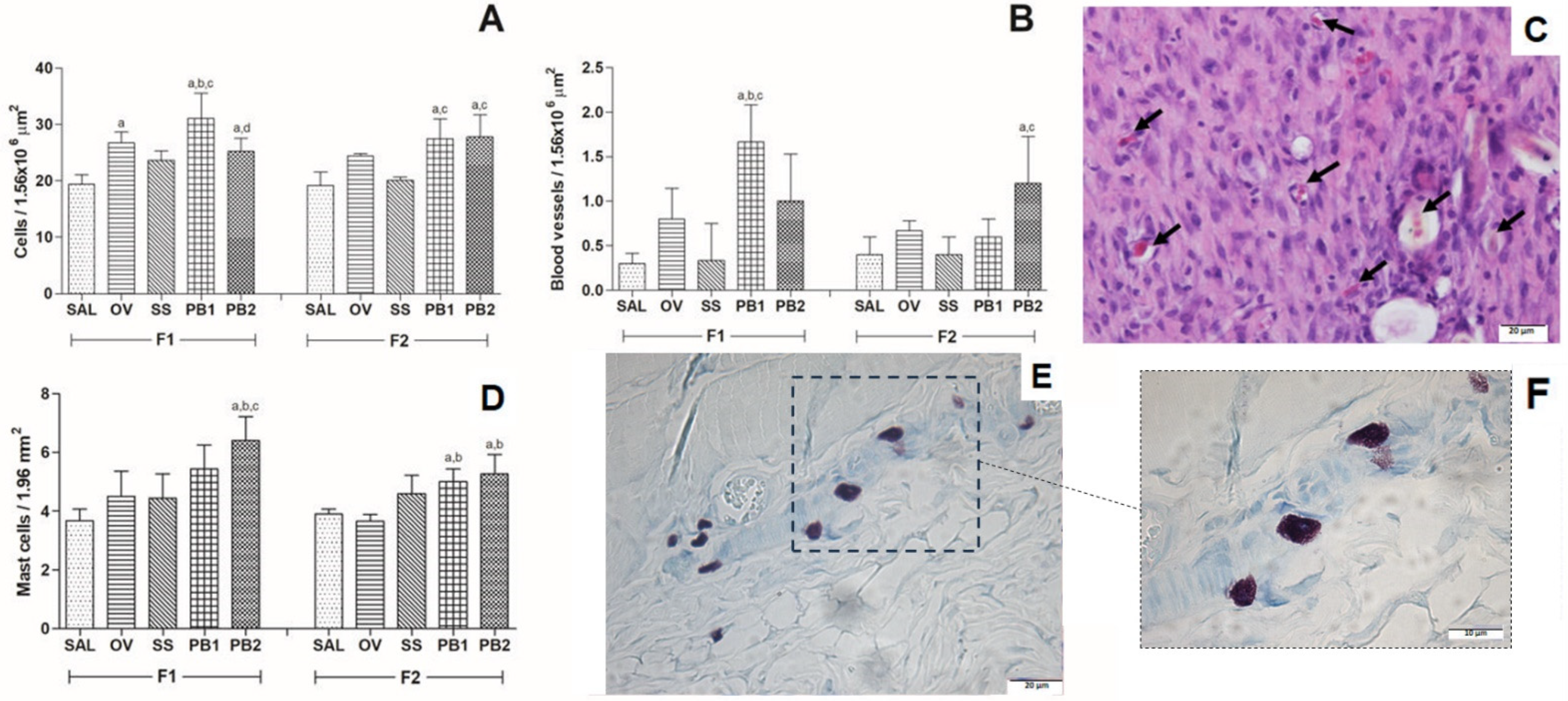
Error in Figure
Reference
- Miranda, L.L.; Sarandy, M.M.; Altoé, L.S.; Bastos, D.S.S.; Melo, F.C.S.A.; Novaes, R.D.; Esposito, D.A.; Gonçalves, R.V. Antioxidant and Anti-Inflammatory Potential of Brassica oleracea Accelerates Third-Degree Burn Healing in Rats. Cosmetics 2024, 11, 27. [Google Scholar] [CrossRef]
Disclaimer/Publisher’s Note: The statements, opinions and data contained in all publications are solely those of the individual author(s) and contributor(s) and not of MDPI and/or the editor(s). MDPI and/or the editor(s) disclaim responsibility for any injury to people or property resulting from any ideas, methods, instructions or products referred to in the content. |
© 2025 by the authors. Licensee MDPI, Basel, Switzerland. This article is an open access article distributed under the terms and conditions of the Creative Commons Attribution (CC BY) license (https://creativecommons.org/licenses/by/4.0/).
Share and Cite
Miranda, L.L.; Sarandy, M.M.; Altoé, L.S.; Bastos, D.S.S.; Melo, F.C.S.A.; Novaes, R.D.; Esposito, D.A.; Gonçalves, R.V. Correction: Miranda et al. Antioxidant and Anti-Inflammatory Potential of Brassica oleracea Accelerates Third-Degree Burn Healing in Rats. Cosmetics 2024, 11, 27. Cosmetics 2025, 12, 110. https://doi.org/10.3390/cosmetics12030110
Miranda LL, Sarandy MM, Altoé LS, Bastos DSS, Melo FCSA, Novaes RD, Esposito DA, Gonçalves RV. Correction: Miranda et al. Antioxidant and Anti-Inflammatory Potential of Brassica oleracea Accelerates Third-Degree Burn Healing in Rats. Cosmetics 2024, 11, 27. Cosmetics. 2025; 12(3):110. https://doi.org/10.3390/cosmetics12030110
Chicago/Turabian StyleMiranda, Lyvia Lopes, Mariáurea Matias Sarandy, Luciana Schulthais Altoé, Daniel Silva Sena Bastos, Fabiana Cristina Silveira Alves Melo, Rômulo Dias Novaes, Debora Araújo Esposito, and Reggiani Vilela Gonçalves. 2025. "Correction: Miranda et al. Antioxidant and Anti-Inflammatory Potential of Brassica oleracea Accelerates Third-Degree Burn Healing in Rats. Cosmetics 2024, 11, 27" Cosmetics 12, no. 3: 110. https://doi.org/10.3390/cosmetics12030110
APA StyleMiranda, L. L., Sarandy, M. M., Altoé, L. S., Bastos, D. S. S., Melo, F. C. S. A., Novaes, R. D., Esposito, D. A., & Gonçalves, R. V. (2025). Correction: Miranda et al. Antioxidant and Anti-Inflammatory Potential of Brassica oleracea Accelerates Third-Degree Burn Healing in Rats. Cosmetics 2024, 11, 27. Cosmetics, 12(3), 110. https://doi.org/10.3390/cosmetics12030110