Abstract
Electrospinning of poly(lactic acid) (PLA) commonly relies on toxic organic solvents, which limit its sustainability and biomedical applicability. In this work, a green electrospinning process was developed using dimethyl carbonate (DMC), a biodegradable and low-toxicity solvent, combined with acetone as a volatile co-solvent to promote efficient jet solidification. Three commercial PLA grades were evaluated for solubility and spinnability, and PLA 4043D was identified as the most suitable for DMC and acetone systems. The electrospinning parameters, including solvent ratio, flow rate, and applied voltage, were systematically optimized to achieve stable jet formation and uniform fiber morphology. Under optimized conditions, the process produced continuous, bead-free nanofibers with a mean diameter of ~1 µm and uniform nanoscale surface porosity resulting from differential solvent evaporation. The resulting fibers were characterized in terms of morphology, structure, thermal behavior, and mechanical performance, confirming increased amorphous content, high porosity (about 78%), and tensile strength of ~3 MPa for the selected electrospinning condition. This study demonstrates that DMC-based solvent systems enable a sustainable and potentially biocompatible route, considering the lower toxicity of the solvents employed, offering a green alternative to conventional toxic processes for the fabrication of medical scaffolds.
1. Introduction
Poly(lactic acid) (PLA) is a widely adopted degradable polyester in biomedical engineering due to its biocompatibility, controlled degradability, and its extensive use in FDA-approved temporary implantable devices such as sutures, scaffolds, and drug-delivery carriers. Its versatility in formulation and processing enables the fabrication of materials with tailored mechanical and degradation profiles, which is essential for regenerative medicine and tissue engineering applications [1,2,3]. PLA can be processed through a wide range of manufacturing techniques, each enabling the production of structures with different architectures and functionalities. A possible drawback in the adoption of PLA is its inherent brittleness, which has led researchers to investigate methods to improve the mechanical performance and unlock functional properties of this polymer [4,5,6,7]. Conventional methods such as solvent casting, melt extrusion, and compression or injection molding are commonly used to obtain films, bulk components, or three-dimensional materials with controlled geometry [8,9]. In the biomedical field, additive manufacturing (AM) methods, including fused filament fabrication (FFF), stereolithography (SLA), and selective laser sintering (SLS), facilitate the design of patient-specific scaffolds with defined macroporosity and tunable mechanical properties [10]. For instance, recent AM studies have demonstrated that FFF-printed PLA scaffolds can accurately reproduce the morphology of bone defects while maintaining an interconnected pore network suitable for osteointegration, highlighting the importance of processability in clinical translation [11]. Although AM techniques provide excellent control over macroscopic geometry, they are generally unable to reproduce the nanoscale fibrous architecture characteristic of the extracellular matrix (ECM), which is crucial for regulating cell adhesion, migration, and nutrient transport. For this reason, solution-based techniques such as phase separation, particulate leaching, and thermally induced phase separation (TIPS) are established approaches for producing porous PLA-based matrices, although their use in soft-tissue engineering remains limited by the intrinsic stiffness of PLA and the mechanical properties of the resulting scaffolds [12].
Among the available processing techniques, electrospinning stands out for its ability to produce PLA-based micro- and nanofibrous mats with controllable morphology, including fiber diameter, alignment, and pore structure [13]. These features are particularly relevant in biomedical contexts, where the high surface area and interconnected porosity enhance cell adhesion, nutrient diffusion, and drug release kinetics [14]. Moreover, electrospinning is compatible with continuous operation and potentially scalable equipment, making it a promising technology for large-scale fabrication of fibrous scaffolds under controlled conditions. Despite these advantages, the electrospinning of PLA remains heavily reliant on toxic and non-sustainable organic solvents such as chloroform and dichloromethane (DCM) [15,16,17]. While these solvents exhibit strong solvation power and fast evaporation rates that facilitate fiber formation, their toxicity, volatility, and potential cytotoxic residues raise concerns for both environmental sustainability and biomedical use. In particular, the presence of toxic solvent residues in fibers intended for tissue-contacting applications represents a significant barrier to clinical translation. To address these limitations, greener solvent systems have attracted increasing interest. Dimethyl carbonate (DMC) is a low-toxicity, biodegradable, and environmentally benign solvent with a favorable safety and regulatory profile, making it an appealing alternative for polymer processing [18,19,20,21,22,23]. However, its relatively low volatility and a boiling point that is higher than that of common chlorinated electrospinning solvents (e.g., DCM and chloroform) hinder rapid solvent evaporation during electrospinning, often preventing complete jet solidification and leading to bead formation or other morphological defects. To overcome this issue, acetone, a relatively low-toxicity solvent [24], compared to chlorinated ones, with a high evaporation rate, can be introduced as a co-solvent, improving the evaporation dynamics and stabilizing the electrospinning process under ambient conditions [25].
In this work, we investigate the feasibility of electrospinning PLA using a fully green binary solvent system based on DMC and acetone, with the objective of providing a sustainable and biocompatible alternative to conventional toxic solvents. Three commercial PLA grades with different molecular weights and D-lactide contents were systematically examined to assess their solubility, phase stability, and electrospinnability in DMC/acetone mixtures. By identifying the PLA grade that exhibited the most favorable balance between solubility and processability, we optimized the electrospinning parameters to obtain continuous, bead-free nanofibers under ambient conditions. Particular attention was devoted to the formation of nanoscale surface porosity, which arises from the differential evaporation rates of the two solvents and may play a role in applications requiring enhanced surface area or controlled release. The resulting fibers were characterized through morphological (SEM), structural (XRD), thermal (DSC), and mechanical testing to determine the suitability of this green process for biomedical scaffold fabrication.
Overall, this study demonstrates that DMC-based binary solvents can support PLA electrospinning under appropriately selected conditions, offering a sustainable and biocompatible alternative to conventional toxic solvents while maintaining control over fiber morphology and structural properties.
2. Materials and Methods
2.1. Materials
PLA IngeoTM 3251D and PLA IngeoTM 4043D were provided by NatureWorks LLC (Plymouth, MN, USA), while PLA Luminy® LX175 was provided by TotalEnergies Corbion (Gorinchem, The Netherlands). From now on, such grades of PLA are referred simply as 3251D, 4043D and LX175, respectively. For all grades, the materials were supplied in the form of pellets. The main properties of the three grades of material, obtained from literature and manufacturers’ data [26,27,28,29,30,31,32], are reported in Table 1. Dimethyl Carbonate (DMC), purity > 99%, was purchased from Sigma-Aldrich (Burlington, MA, USA). Acetone (AC), purity > 99%, was purchased from Supelco (Bellefonte, PA, USA). All the materials in the present study were used as received.

Table 1.
Properties of PLA grades.
2.2. X-Ray Diffraction Analysis of PLA Grades
The micro-structure of the three PLA grades was investigated by means of X-ray diffraction (XRD), using a Philips X’Pert diffractometer, operating at 40 KV and 40 mA with CuKα1 radiation. The diffraction pattern was recorded between 2 values of 5 and 70°, with an angle increment step of 0.02° and an acquisition time of 2 s. Prior to diffraction analysis, the polymers were ground down to 1 mm with a Retsch ZM300 milling machine.
2.3. Solution Preparation and Stability
Different ternary solutions of PLA/DMC/AC were prepared using the three PLA grades, varying the polymer concentration (≈5, 10, 15, 20 wt%) and the solvent weight ratio (DMC/AC = 100/0, 80/20, 67/33, 50/50 wt%). In each case, a few pellets were placed in small vials with the appropriate amount of mixed solvents to obtain approximately 5 mL of solution. The actual weight fractions of the three components were recorded. The vials were then sealed and placed in a water bath at 60 °C. The solutions were magnetically stirred for 1 h, after which the dissolution of the pellets was complete for all the conditions tested. The weight of each vial was recorded to ensure that solvent evaporation did not occur; the vials were weighed immediately after stirring and again after 1 and 2 days. Solutions showing evidence of solvent loss were discarded and prepared again.
2.4. Viscosity Measurements
The viscosity of the PLA/DMC/AC solutions was measured using a rotational viscometer (Visco Basic Lab, Fungilab, Sant Feliu de Llobregat, Spain) equipped with an L1 spindle. Measurements were carried out at room temperature (25 ± 1 °C). For each solution, the viscosity value was recorded once a steady-state reading was reached. All measurements were performed in triplicate, and the mean value was used for subsequent analysis. The spindle was operated at the rotation speed that ensured stable and reliable steady-state reading.
2.5. Electrospinning Process
Electrospinning experiments were performed using a home-made electrospinning setup consisting of a high-precision syringe pump (NE-300, New Era Pump Systems Inc., NY, USA) to deliver the polymer solutions at controlled flow rates [33,34]. The solution was extruded through a stainless steel 4-way G21 needle (inner diameter: 0.51 mm) connected to a high-voltage power supply (CM-5, SIMCO ION, Lochem, The Netherlands) capable of generating up to 30 kV.
The entire electrospinning setup was placed inside an enclosure to ensure stable ambient conditions during processing. The enclosure maintained a constant temperature of approximately 25 °C and a relative humidity below 50%, as both parameters are known to influence jet solidification and fiber morphology during electrospinning [35]. Controlling these environmental conditions ensures stable processing and enhances the reproducibility of the resulting fiber structures. A fixed collecting distance of 10 cm was used for all experiments. The applied voltage (10–20 kV) and the flow rate (0.25–1.0 mL h−1) were systematically varied in order to identify stable spinning conditions that yielded continuous fibers.
Fibers were collected on a grounded flat aluminum foil attached to the collector. After deposition, the electrospun mats were dried in the oven at 40 °C for 40 h to ensure complete solvent removal prior to further characterization.
2.6. Morphological Characterization (SEM)
The morphology and microstructure of the nanofibers were evaluated by scanning electron microscopy (SEM) using a Tescan Mira 3 instrument (Tescan, Brno, Czech Republic). The nanofibers were imaged at an accelerating voltage of 10 kV and different magnifications (5000×, 10,000×, 15,000×, 20,000×). Before imaging, an Edwards Sputter Coater S150B (Burgess Hill, UK) was used for the deposition of a gold coating on the samples to enhance electrical conductivity. Fiber diameter measurements were carried out using a modified version of the SIMPoly algorithm, introduced by Murphy et al. [36], implemented in MATLAB R2024b, allowing automated extraction of diameter distributions from SEM images. At least 8 images obtained in different regions of the electrospun mat samples were analyzed for each testing condition, with a number of fibers per image ranging from hundreds to tens of thousands, depending on the magnification.
The thickness of the electrospun mats was measured with a micrometer, carefully avoiding deforming the material, in several different locations to account for differential accumulations of nanofibers. Once the average thickness was measured, it was possible to evaluate the porosity as follows: rectangular specimens of known area were cut and weighed and, by using the thickness to estimate the volume, the apparent density of the electrospun mats was evaluated. The porosity was then determined as:
where the bulk density used was the value provided by the supplier of the material.
2.7. Thermal Properties by DSC
The thermal properties of the PLA pellets and electrospun nanofibers were analyzed via differential scanning calorimetry (DSC) using a DSC 8500 instrument (PerkinElmer, Waltham, MA, USA). Approximately 10 mg of each sample was sealed in standard aluminum pans with lids and analyzed under a nitrogen atmosphere at a flow rate of 40 mL min−1. The thermal program consisted of five steps: (i) heating from 25 °C to 250 °C, (ii) isothermal holding at 250 °C for 1 min, (iii) cooling to 25 °C, (iv) isothermal holding at 25 °C for 1 min, and (v) a second heating to 250 °C. All heating and cooling segments were performed at a rate of 10 °C min−1. The glass transition temperature (Tg) was determined from the second heating run using the inflection point method, in accordance with the ASTM E1356-08 standard [37]. The cold crystallization temperature (Tcc), melting temperature (Tm), and corresponding enthalpy changes (ΔHc and ΔHm) were obtained from the first heating run. The degree of crystallinity (Xc) was calculated according to Equation (2) [38]:
where ΔH100 = 93.6 J g−1 is the melting enthalpy of 100% crystalline PLA [39].
2.8. Tensile Testing
Tensile tests on electrospun mats were performed as follows: strips of 50 × 5 mm2 were cut with a scalpel. The specimens were then mounted on windowed paper frames with adhesive tape, to handle the specimens before testing. A Z2.5 universal testing machine (Zwick/Roell, Ulm, Germany), equipped with a 100 N load cell, was used to perform the tensile tests at a displacement rate of 1 mm/min and at room temperature. The elongation of the specimens was evaluated from the crosshead displacement of the tensile machine. Once the specimens were mounted on the tensile grips, the paper frames were cut, allowing the load to be carried solely by the PLA electrospun mats. A total of 5 specimens were tested.
3. Results and Discussion
3.1. Crystalline Structure of PLA Grades by XRD
Diffractograms of the three grades of PLA are shown in Figure 1. The experimental diffraction patterns, after background baseline removal, were analyzed with a custom script in MATLAB. The eventual presence of instrumental peak broadening was not corrected with an additional dedicated procedure. Deconvolution of the peaks was carried out by fitting a series of Gaussian distributions, with center values and FWHM (Full Width at Half Maximum) values obtained from literature [40,41,42,43,44,45] as initial guess values.
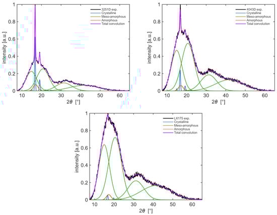
Figure 1.
XRD patterns for the three grades of PLA: (top, left) 3251D, (top, right) 4043D and (bottom) LX175. Black lines are experimental data, blue, orange and green lines indicate crystalline peaks, meso-amorphous halos and amorphous halos, respectively. Magenta lines indicate the cumulative fitting curve.
The peaks found with curve fitting were assigned to the different crystalline reflection planes or to meso-amorphous or amorphous halos. Peaks relevant to the (010), (110/200), (203), (210), (213) and (116) crystallographic planes were found at values of 2 in the immediate vicinity of 14.8°, 16.5°, 19°, 22°, 23.5° and 24.5°, respectively. These peaks belong to well-ordered structures, as suggested by their low values of FWHM (~0.5°), in agreement with [43]. Broader peaks with FWHM close to 8°, relevant to the amorphous phase, were found at angles close to 15°, 21°, 31° and 42° [42,43]. Finally, a peak centered around 2 = 16.5° with an intermediate FWHM (~3°) suggests the presence of an intermediate phase, referred to as meso-amorphous [41,42]. Once the peaks were isolated and properly assigned to the different phases, it was possible to determine their relative intensity as the area underneath the curves (Figure 2 (left)) and then the amount of the different phases inside the polymers was evaluated. Figure 2 (right) shows the amount of crystalline and meso-amorphous phases (amorphous phase was left out for scale purposes) as a function of the d-lactide content. Although it is difficult to establish a trend, since 4043D and LX175 show very similar values, it is possible to infer that a higher content of d-lactide determines an almost completely amorphous micro-structure.
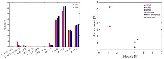
Figure 2.
Relative intensities of deconvoluted peaks of the three grades of PLA (left). Phase contents as a function of the nominal d-lactide content of the three grades of PLA (right).
3.2. Solution Stability and Solubility Behavior
The stability of the solutions was evaluated following the approach proposed by [46]. The vials were stored for three days after preparation and, once the lack of solvent evaporation was confirmed, the solution was checked for phase separation, flocculation or deposition. The solutions that appeared clear and did not show any separated phase were flagged as stable.
Figure 3 shows the ternary plots for the different solutions prepared with the three grades of PLA under investigation. It can be observed that with PLA 3251D, it was never possible to achieve stability of the solutions, apart from those with a low concentration of polymer and low amounts of acetone. On the contrary, PLA 4043D and LX175 showed a wide region of solubility and only in a few cases, at higher PLA concentrations and acetone fractions, phase separation occurred. This result, in combination with data from Figure 2 (right) suggests a direct correlation between the solubility of PLA and the amount of amorphous domains and/or d-lactide content found by XRD analysis (Section 3.1). This result is in agreement with the findings of [47].
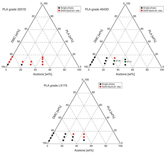
Figure 3.
Ternary plots for PLA/DMC/AC solutions, (top, left) 3251D, (top, right) 4043D and (bottom) LX175. The solutions selected for the subsequent phase of electrospinning are highlighted with green border and relevant labels.
Based on the solubility studies previously carried out and the larger amount of amorphous domains which results in an ease of processing, PLA grade 4043D was chosen for electrospinning. In particular, two solutions were selected: ID 14 (9/45.5/45.5 PLA/DMC/AC wt%) and ID 28 (10.9/29.4/59.7 PLA/DMC/AC wt%). They are highlighted in Figure 3 (top, right). In the electrospinning process, several combinations of flow rate, applied voltage and type of solution were used, as explained in Section 2. After a spinning time of 30 min to 1 h, the PLA fiber mats were removed from the target without detaching them from the aluminum foil. The mats were then observed with SEM.
3.3. Morphology of Electrospun Fibers
Based on the solubility and stability results, PLA 4043D was selected for electrospinning, as it was the only grade able to form homogeneous and stable solutions in DMC/acetone mixtures across a wide composition range. Preliminary spinning tests showed that continuous fibers with generally uniform morphology could be obtained; however, in several conditions, beads or locally thickened segments were also observed. Since incomplete solvent evaporation during jet flight is a known cause of such defects, the potential presence of residual solvent trapped within the fibers was investigated. Selected electrospun mats were subjected to a post-spinning thermal treatment in the oven at 40 °C for 40 h to promote further solvent removal. After drying, the samples were analyzed via SEM and compared with the corresponding as-spun fibers. No morphological differences were detected, and defects such as beads or local diameter variations were still present in the same regions. This indicates that the irregularities do not originate from solvent retention but rather from unstable electrospinning conditions. Representative SEM images before and after thermal treatment are shown in Figure 4.
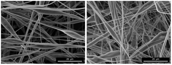
Figure 4.
SEM micrographs of defective PLA fibers spun at 1 mL/h and 10 kV from a solution with 10.9/29.4/59.7 PLA/DMC/AC wt% (Test ID 11) as spinned (left) and after drying (right).
The spinning conditions that led to irregular morphologies, such as beads or local diameter thickening, were discarded and not considered in subsequent analysis. Only the combinations of voltage, flow rate, and solution composition that resulted in stable jet formation and reproducible fiber morphologies were retained, and these are reported in Table 2 (the inconsistency of test ID numbering is related to the fact that some conditions were discarded, as mentioned above, therefore relevant numbers were skipped). For consistency, all collected fibers were dried under identical conditions (40 h at 40 °C) to ensure complete solvent removal before characterization.
Table 2.
Electrospinning conditions selected for the processing of PLA 4043D in DMC/acetone solutions.
An example of the fibers obtained without defects after spinning is shown in Figure 5. By observing the fibers at high magnification, it is possible to observe that the surface is not smooth; rather, it reveals the presence of nanometric pores distributed along the surface of the fibers. This nanoporosity is observed consistently across multiple samples and does not depend on the specific spinning parameters, indicating that it is likely governed by solvent dynamics rather than flow instability. The rapid evaporation of acetone during jet elongation, in contrast with the slower evaporation rate of dimethyl carbonate, can create a concentration gradient at the fiber surface. This condition may promote localized phase separation or incomplete solvent removal, resulting in the formation of nanopores upon solidification. Similar surface features have been reported in electrospun systems where phase separation is driven by differential solvent volatility [48]. It has been reported that polymer stereochemistry and the associated amorphous content play a key role in fiber formation during electrospinning, as variations in DL-lactide content can influence jet stability, fiber diameter, surface porosity, and defect formation in poly(lactide) copolymers [49]. From an application standpoint, this nanoscale surface porosity represents a beneficial feature for biomedical scaffolds, as it increases the specific surface area available for cell–material interactions and protein adsorption, while facilitating permeability and mass transport across the fibrous mat, which are critical aspects in tissue engineering [50,51]. Notably, this nanoporosity is intrinsically generated by the selected DMC/acetone solvent system, without requiring additional post-processing steps.
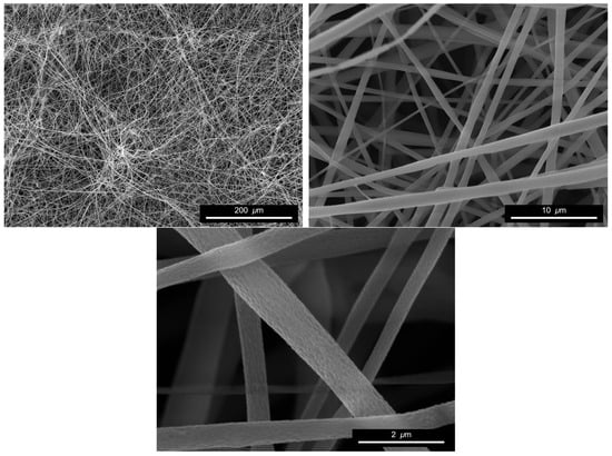
Figure 5.
SEM micrographs of PLA fibers spun at 0.5 mL/h and 20 kV from a solution with 9/45.5/45.5 PLA/DMC/AC wt% (Test ID 2).
The diameter distribution of the spun fibers was measured with a modified version of the SIMPoly MATLAB tool introduced by Murphy et al. [36]. To investigate the sensitivity of the automated analysis to the magnification of SEM micrographs fed to the tool, results from subsets of images taken at 5k×, 10k×, 15k× and 20k× were registered for selected tests. An example of diameter distributions is given in Figure 6. The histograms clearly indicate that the effect of the image magnification on the measurement is negligible: the analysis tool proved to be robust in the chosen range, indicating that the number of fibers and the resolution were large enough to provide valid statistics. The diameter distributions show a positive skewness; therefore, a Log-normal distribution function was used to fit the data, thus producing the solid lines of Figure 6.
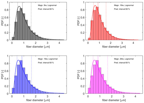
Figure 6.
Diameter distributions obtained from subsets of images obtained at different magnifications (black = 5k×, red = 10k×, blue = 15k× and magenta = 20k×) from fibers spun at 0.5 mL/h and 20 kV from a solution with 9/45.5/45.5 PLA/DMC/AC wt% (Test ID 2). Solid lines represent a fit with Log-normal distribution and vertical solid and dashed lines indicate median values and the prediction intervals containing 68% of the observations, respectively.
The statistics used to describe the distributions were the median values and the width of the prediction intervals. Once it was verified that the magnification of SEM images did not influence the outcome of the analysis, results obtained from images taken at a magnification of 10k× were selected to investigate the dependence of fiber morphology on the parameters used during electrospinning. The median diameters and the width of the prediction intervals containing 68% of the observations for the different tests performed are shown in Figure 7 (left) and (right), respectively.
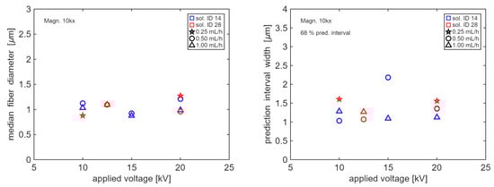
Figure 7.
Median fiber diameters (left) and widths of prediction intervals containing 68% of the observations (right) as a function of applied voltage. Blue and red markers represent solutions 14 and 28, respectively (refer to Table 2 for details on composition). Stars, circles and triangles represent tests performed with a flow rate of 0.25, 0.5 and 1 mL/h, respectively.
It can be observed that the morphology of the fibers seems to be unaffected by the spinning conditions or the solution composition. The median diameter oscillates around an average value of 1 and the width of the prediction interval is between 1 and 1.5 ; although slightly different values are observed for the different tests performed, there is no evidence of a clear trend or dependence of fiber morphology on a specific spinning parameter. If any, it appears that solution 14 (9/45.5/45.5.5 PLA/DMC/AC wt%) presents smaller values of the prediction interval width with respect to solution 28 (10.9/29.4/59.7 PLA/DMC/AC wt%).
Since the spinning parameters appear to have a limited influence on the resulting fibers, further characterization tests were performed for a selected testing condition only. The sample spun at 0.5 mL/h and 15 kV from a solution with 9/45.5/45.5 PLA/DMC/AC wt% (Test ID 1) was picked to preliminarily assess whether the material would be suitable for the intended application. The choice was based on the lower mean diameter value and the general appearance of the fibers, which showed the most homogeneous morphology.
3.4. Thermal Behavior by DSC
The thermal properties of PLA pellets and electrospun nanofibers were investigated by DSC to evaluate the influence of processing on the polymer’s structure and crystallization behavior. Figure 8 shows the thermograms of the as-received pellets and electrospun nanofibers recorded during the first and second heating runs. Results of DSC measurements are summarized in Table 3.
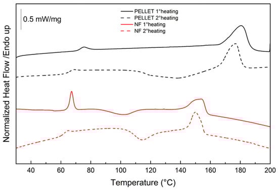
Figure 8.
DSC thermograms of PLA pellets and PLA nanofibers (NF) during the first and second heating.
Table 3.
Values obtained by DSC of PLA pellet and PLA nanofibers (NF): Tg, ΔCp, Tcc, ΔHc, Tm, ΔHm (mean ± SD, n = 3).
The glass transition temperature (Tg), determined from the second heating run in accordance with ASTM E1356-08, was slightly lower for the nanofibers than for the pellets, reflecting their higher amorphous content and reduced chain packing. This shift can be attributed to the rapid solvent evaporation and strong elongational forces occurring during electrospinning, which kinetically freeze the polymer chains in a disordered state [52]. The slightly higher ΔCp of the nanofibers (0.559 vs. 0.50 J/g·°C) also supports their increased amorphous fraction.
During the first heating, the pellets did not exhibit any cold-crystallization peak, indicating that they were already partially crystalline in the as-received state. Conversely, the electrospun nanofibers showed a clear cold-crystallization exotherm, confirming their predominantly amorphous nature and their ability to reorganize into ordered domains upon heating [53]. The high Tcc of the nanofibers (103.33 °C) reflects the substantial chain reorganization required during heating, which is influenced by the molecular weight and D-lactide content of PLA 4043D, both known to reduce crystallization rates and favor the formation of less ordered structures. The melting transition of the nanofibers occurred at a lower temperature compared to the pellets, suggesting the presence of less ordered and thinner α′-type crystallites, in contrast to the more stable α-form crystals observed in the pellets [40,41]. The α′ phase consists of thinner and less stable crystals formed under rapid or kinetically constrained solidification, whereas the α phase represents the more ordered and thermodynamically stable crystal form. This structural distinction explains why α′ crystallites melt at lower temperatures and undergo transformation toward the α phase upon heating.
The degree of crystallinity () was evaluated from the first heating run according to Equation (2). PLA pellets exhibited a crystallinity degree of approximately 43.6 ± 3.8%, whereas the electrospun nanofibers showed a significantly lower value of 12.2 ± 1.7%. This pronounced reduction in crystallinity confirms that the electrospinning process promotes a more amorphous microstructure, mainly due to the combined effects of rapid solvent evaporation, strong elongational forces, and limited time available for chain rearrangement. The appearance of a cold-crystallization peak and the decrease in melting temperature further support the formation of less ordered α′-type crystallites, typically associated with kinetically constrained solidification conditions. The relatively large difference in melting temperature between pellets and nanofibers (~26 °C) is consistent with previous observations in PLA grades where rapid solidification, higher D-lactide content, or lower molecular weight promote the formation of α′ crystallites, which melt at significantly lower temperatures than α crystals.
3.5. Mechanical Properties of Electrospun Mats
The thickness of the electrospun mat was measured as indicated in Section 2. An average value of 0.136 ± 0.041 mm was obtained. The porosity was evaluated with Equation (1) and a value of 0.778 was obtained.
The results of the tensile tests performed on the electrospun mats are shown in Figure 9. The cross-sectional area was taken as the product of the initial width of the specimens and the average thickness of the electrospun mat. To account for the large value of porosity found, the stress was corrected so as to consider that only the solid part of the cross-section carries the load. In the first approximation, it is possible to divide the nominal cross-sectional area by the complementary of the porosity, obtaining the curves shown in Figure 9. The average strength was found to be 2.96 ± 0.40 MPa. The shape of the curves resembles the typical behavior of electrospun mats: a first high-modulus region, which can be attributed to the rotation of the individual strands of fibers towards the loading direction, with the breakage of the adhesion points between the fibers. The value of elastic modulus was found to be 66.66 ± 16.47 MPa. Then, at a strain of around 5%, the onset of a second region is visible, in which the stress–strain curve reflects the sliding of the fibers upon each other until complete disentanglement and final failure [54,55,56].
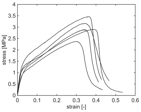
Figure 9.
Stress–strain curves of the tensile tests performed on selected spun mat, corrected to consider the actual solid cross-section. Five specimens, cut from the same electrospun mat, were tested.
From a scale-up perspective, some technical and economic challenges should be considered for the industrial implementation of the proposed electrospinning process. In line with other solution-based electrospinning approaches, large-scale production requires careful control of solvent evaporation and processing conditions to ensure uniform fiber morphology and reproducibility, particularly when multi-jet or needleless systems are employed. From an economic standpoint, the use of acetone and dimethyl carbonate is advantageous, as these solvents are inexpensive, readily available, and associated with lower health, safety, and disposal costs compared to conventional solvents such as DMF or chlorinated solvents. However, efficient solvent recovery and process optimization will be necessary to ensure economic viability at higher production throughput.
4. Conclusions
A green and sustainable electrospinning process for PLA was successfully developed using DMC as a biodegradable solvent in combination with acetone as a volatile co-solvent. Solubility and phase-stability tests identified PLA 4043D as the only grade capable of forming homogeneous and stable solutions in DMC/acetone mixtures across a broad composition range. The electrospinning parameters were optimized to ensure stable jet formation and uniform fiber deposition, identifying a PLA/DMC/AC solution (9/45.5/45.5 wt%) processed at 0.5 mL·h−1 and 15 kV as the most effective conditions. Under these optimized conditions, continuous and bead-free nanofibers with mean diameters of ~1 µm were produced. SEM analysis revealed a uniform nanoscale surface porosity generated by the differential evaporation rates of DMC and acetone. For the aforementioned conditions, XRD and DSC measurements confirmed the predominantly amorphous nature of the fibers, with crystallinity decreasing from ~44% in the pellets to ~12% in the electrospun mats. Mechanical tests performed on the highly porous structures (porosity ~ 78%) yielded an average tensile strength of 2.96 ± 0.40 MPa and an elastic modulus of 66.66 ± 16.47 MPa.
Overall, these results demonstrate that DMC-based binary solvent systems can reliably support PLA electrospinning, providing a less toxic and more sustainable alternative to traditional chlorinated solvents while ensuring stable processing and high fiber quality.
Author Contributions
Conceptualization, T.P., G.C., E.T., M.G.S. and M.V.; methodology, T.P. and G.C.; software, T.P. and G.C.; validation, T.P., G.C. and E.T.; formal analysis, T.P. and G.C.; investigation, T.P. and G.C.; resources, M.G.S. and M.V.; data curation, T.P., G.C. and E.T.; writing—original draft preparation, T.P., G.C. and E.T.; writing—review and editing, T.P., G.C., E.T., M.G.S. and M.V.; supervision, M.G.S. and M.V. All authors have read and agreed to the published version of the manuscript.
Funding
This research received no external funding.
Data Availability Statement
The raw data supporting the conclusions of this article will be made available by the authors upon request.
Acknowledgments
The authors would like to thank Martina Damizia and Benedetta De Caprariis for the help provided with the milling machine.
Conflicts of Interest
The authors declare no conflicts of interest.
References
- Castañeda-Rodríguez, S.; González-Torres, M.; Ribas-Aparicio, R.M.; Del Prado-Audelo, M.L.; Leyva-Gómez, G.; Gürer, E.S.; Sharifi-Rad, J. Recent Advances in Modified Poly (Lactic Acid) as Tissue Engineering Materials. J. Biol. Eng. 2023, 17, 21. [Google Scholar] [CrossRef] [PubMed]
- Khouri, N.G.; Bahú, J.O.; Blanco-Llamero, C.; Severino, P.; Concha, V.O.C.; Souto, E.B. Polylactic Acid (PLA): Properties, Synthesis, and Biomedical Applications—A Review of the Literature. J. Mol. Struct. 2024, 1309, 138243. [Google Scholar] [CrossRef]
- Yang, Z.; Yin, G.; Sun, S.; Xu, P. Medical Applications and Prospects of Polylactic Acid Materials. iScience 2024, 27, 111512. [Google Scholar] [CrossRef] [PubMed]
- Liu, H.; Zhang, J. Research Progress in Toughening Modification of Poly(Lactic Acid). J. Polym. Sci. Part B Polym. Phys. 2011, 49, 1051–1083. [Google Scholar] [CrossRef]
- Li, X.; Lin, Y.; Liu, M.; Meng, L.; Li, C. A Review of Research and Application of Polylactic Acid Composites. J. Appl. Polym. Sci. 2023, 140, e53477. [Google Scholar] [CrossRef]
- Chieng, B.W.; Ibrahim, N.A.; Yunus, W.M.Z.W.; Hussein, M.Z. Poly(Lactic Acid)/Poly(Ethylene Glycol) Polymer Nanocomposites: Effects of Graphene Nanoplatelets. Polymers 2014, 6, 93–104. [Google Scholar] [CrossRef]
- Peixoto, T.; Nunes, J.; Lopes, M.A.; Marinho, E.; Proença, M.F.; Lopes, P.E.; Paiva, M.C. Poly(Lactic Acid) Composites with Few Layer Graphene Produced by Noncovalent Chemistry. Polym. Compos. 2022, 43, 8409–8425. [Google Scholar] [CrossRef]
- Pokharel, A.; Falua, K.J.; Babaei-Ghazvini, A.; Nikkhah Dafchahi, M.; Tabil, L.G.; Meda, V.; Acharya, B. Development of Polylactic Acid Films with Alkali- and Acetylation-Treated Flax and Hemp Fillers via Solution Casting Technique. Polymers 2024, 16, 996. [Google Scholar] [CrossRef]
- Papadimitriou, L.; Manganas, P.; Ranella, A.; Stratakis, E. Biofabrication for Neural Tissue Engineering Applications. Mater. Today Bio 2020, 6, 100043. [Google Scholar] [CrossRef]
- Nikolova, M.P.; Chavali, M.S. Recent Advances in Biomaterials for 3D Scaffolds: A Review. Bioact. Mater. 2019, 4, 271–292. [Google Scholar] [CrossRef]
- Zarei, M.; Shabani Dargah, M.; Hasanzadeh Azar, M.; Alizadeh, R.; Mahdavi, F.S.; Sayedain, S.S.; Kaviani, A.; Asadollahi, M.; Azami, M.; Beheshtizadeh, N. Enhanced Bone Tissue Regeneration Using a 3D-Printed Poly(Lactic Acid)/Ti6Al4V Composite Scaffold with Plasma Treatment Modification. Sci. Rep. 2023, 13, 3139. [Google Scholar] [CrossRef] [PubMed]
- Sofokleous, P.; Chin, M.H.W.; Day, R. Phase-Separation Technologies for 3D Scaffold Engineering. In Functional 3D Tissue Engineering Scaffolds; Woodhead Publishing: London, UK, 2018; pp. 101–126. [Google Scholar]
- Szlek, D.B.; Fan, E.L.; Frey, M.W. Multifunctional, Flexible, Electrospun Lignin/PLA Micro/Nanofiber Mats from Softwood Kraft, Hardwood Alcell, and Switchgrass CELF Lignin. Fibers 2025, 13, 129. [Google Scholar] [CrossRef]
- Abdullah, K.; Molnár, K. The Influence of In Vitro Degradation on the Properties of Polylactic Acid Electrospun Fiber Mats. Fibers 2024, 13, 1. [Google Scholar] [CrossRef]
- Casasola, R.; Thomas, N.L.; Georgiadou, S. Electrospinning of Poly(Lactic Acid): Theoretical Approach for the Solvent Selection to Produce Defect-Free Nanofibers. J. Polym. Sci. Part B Polym. Phys. 2016, 54, 1483–1498. [Google Scholar] [CrossRef]
- Casasola, R.; Thomas, N.L.; Trybala, A.; Georgiadou, S. Electrospun Poly Lactic Acid (PLA) Fibres: Effect of Different Solvent Systems on Fibre Morphology and Diameter. Polymer 2014, 55, 4728–4737. [Google Scholar] [CrossRef]
- Karabulut, H.; Unal, S.; Ulag, S.; Ficai, A.; Ficai, D.; Gunduz, O. Mapping the Influence of Solvent Composition over the Characteristics of Polylactic Acid Nanofibers Fabricated by Electrospinning. ChemistrySelect 2024, 9, e202301142. [Google Scholar] [CrossRef]
- Tundo, P.; Selva, M. The Chemistry of Dimethyl Carbonate. Acc. Chem. Res. 2002, 35, 706–716. [Google Scholar] [CrossRef]
- Pyo, S.-H.; Park, J.H.; Chang, T.-S.; Hatti-Kaul, R. Dimethyl Carbonate as a Green Chemical. Curr. Opin. Green Sustain. Chem. 2017, 5, 61–66. [Google Scholar] [CrossRef]
- Oldal, D.G.; Topuz, F.; Holtzl, T.; Szekely, G. Green Electrospinning of Biodegradable Cellulose Acetate Nanofibrous Membranes with Tunable Porosity. ACS Sustain. Chem. Eng. 2023, 11, 994–1005. [Google Scholar] [CrossRef]
- da Silva Parize, D.D.; de Oliveira, J.E.; Foschini, M.M.; Marconcini, J.M.; Mattoso, L.H.C. Poly(Lactic Acid) Fibers Obtained by Solution Blow Spinning: Effect of a Greener Solvent on the Fiber Diameter. J. Appl. Polym. Sci. 2016, 133, 43379. [Google Scholar] [CrossRef]
- Da Silva Parize, D.D.; Foschini, M.M.; De Oliveira, J.E.; Klamczynski, A.P.; Glenn, G.M.; Marconcini, J.M.; Mattoso, L.H.C. Solution Blow Spinning: Parameters Optimization and Effects on the Properties of Nanofibers from Poly(Lactic Acid)/Dimethyl Carbonate Solutions. J. Mater. Sci. 2016, 51, 4627–4638. [Google Scholar] [CrossRef]
- Maldonado-Illescas, M.D.P.; Casares-López, J.M.; González-Martín, M.L.; Gallardo-Moreno, A.M.; Luque-Agudo, V. Toward Sustainable PLA Films by Replacing Chloroform for the Green Solvent Dimethyl Carbonate. Surf. Interfaces 2025, 59, 105843. [Google Scholar] [CrossRef]
- Capello, C.; Fischer, U.; Hungerbühler, K. What Is a Green Solvent? A Comprehensive Framework for the Environmental Assessment of Solvents. Green Chem. 2007, 9, 927. [Google Scholar] [CrossRef]
- Ciarleglio, G.; Toto, E.; Laurenzi, S.; Santonicola, M.G. Biocompatible PLA/nHAp Coatings for Titanium Implants Fabricated by Green Electrospinning. MRS Commun. 2025. [Google Scholar] [CrossRef]
- Olejnik, O.; Masek, A. Bio-Based Packaging Materials Containing Substances Derived from Coffee and Tea Plants. Materials 2020, 13, 5719. [Google Scholar] [CrossRef]
- Friné, V.-C.; Hector, A.-P.; Manuel, N.-D.S.; Estrella, N.-D.; Antonio, G.J. Development and Characterization of a Biodegradable PLA Food Packaging Hold Monoterpene–Cyclodextrin Complexes against Alternaria Alternata. Polymers 2019, 11, 1720. [Google Scholar] [CrossRef]
- Coltelli, M.-B.; Bertolini, A.; Aliotta, L.; Gigante, V.; Vannozzi, A.; Lazzeri, A. Chain Extension of Poly(Lactic Acid) (PLA)–Based Blends and Composites Containing Bran with Biobased Compounds for Controlling Their Processability and Recyclability. Polymers 2021, 13, 3050. [Google Scholar] [CrossRef]
- Yang, C.; Topuz, F.; Park, S.-H.; Szekely, G. Biobased Thin-Film Composite Membranes Comprising Priamine–Genipin Selective Layer on Nanofibrous Biodegradable Polylactic Acid Support for Oil and Solvent-Resistant Nanofiltration. Green Chem. 2022, 24, 5291–5303. [Google Scholar] [CrossRef]
- Ingeo Biopolymer 3251D Technical Data Sheet. Available online: https://www.natureworksllc.com/~/media/Files/NatureWorks/Technical-Documents/Technical-Data-Sheets/TechnicalDataSheet_3251D_injection-molding_pdf.pdf (accessed on 17 November 2025).
- Ingeo Biopolymer 4043D Technical Data Sheet. Available online: https://www.natureworksllc.com/~/media/Technical_Resources/Technical_Data_Sheets/TechnicalDataSheet_4043D_films_pdf.pdf (accessed on 17 November 2025).
- Luminy LX175 Technical Data Sheet. Available online: https://totalenergies-corbion.com//wp-content/uploads/2025/02/pds-luminy-lx175-20220722.pdf (accessed on 24 December 2025).
- Ciarleglio, G.; Russo, T.; Vella, S.; Toto, E.; Santonicola, M.G. Electrospray Fabrication of pH-Responsive Microspheres for the Delivery of Ozoile. Macromol. Symp. 2024, 413, 2400031. [Google Scholar] [CrossRef]
- Ciarleglio, G.; Pagani, L.; Ferri, L.; Toto, E.; Santonicola, M.G. Nanofibrous Coatings of Poly(Lactic Acid) and Nano-Hydroxyapatite for Enhanced Biocompatibility of Titanium Implants. Macromol. Symp. 2025, 414, e70248. [Google Scholar] [CrossRef]
- Li, D.; Xia, Y. Electrospinning of Nanofibers: Reinventing the Wheel? Adv. Mater. 2004, 16, 1151–1170. [Google Scholar] [CrossRef]
- Murphy, R.; Turcott, A.; Banuelos, L.; Dowey, E.; Goodwin, B.; Cardinal, K.O. SIMPoly: A Matlab-Based Image Analysis Tool to Measure Electrospun Polymer Scaffold Fiber Diameter. Tissue Eng. Part C Methods 2020, 26, 628–636. [Google Scholar] [CrossRef] [PubMed]
- ASTM E1356-08; Standard Test Method for Assignment of the Glass Transition Temperatures by Differential Scanning Calorimetry. ASTM International: West Conshohocken, PA, USA, 2014.
- Ciarleglio, G.; Pagani, L.; Toto, E.; Laurenzi, S.; Santonicola, M.G. Electrospun PLA/Nano-Hydroxyapatite Fiber Coatings for Improved Corrosion Protection of Titanium Implants. Surf. Interfaces 2025, 76, 107848. [Google Scholar] [CrossRef]
- Hernández Sánchez, F.; Molina Mateo, J.; Romero Colomer, F.J.; Salmerón Sánchez, M.; Gómez Ribelles, J.L.; Mano, J.F. Influence of Low-Temperature Nucleation on the Crystallization Process of Poly(l-Lactide). Biomacromolecules 2005, 6, 3283–3290. [Google Scholar] [CrossRef]
- Zhang, J.; Tashiro, K.; Tsuji, H.; Domb, A.J. Disorder-to-Order Phase Transition and Multiple Melting Behavior of Poly(l-Lactide) Investigated by Simultaneous Measurements of WAXD and DSC. Macromolecules 2008, 41, 1352–1357. [Google Scholar] [CrossRef]
- Puchalski, M.; Kwolek, S.; Szparaga, G.; Chrzanowski, M.; Krucińska, I. Investigation of the Influence of PLA Molecular Structure on the Crystalline Forms (α’ and α) and Mechanical Properties of Wet Spinning Fibres. Polymers 2017, 9, 18. [Google Scholar] [CrossRef]
- Baptista, C.; Azagury, A.; Baker, C.M.; Mathiowitz, E. The Characterization and Quantification of the Induced Mesophases of Poly-l-Lactic Acid. Polymer 2021, 226, 123822. [Google Scholar] [CrossRef]
- Stoclet, G.; Seguela, R.; Lefebvre, J.M.; Elkoun, S.; Vanmansart, C. Strain-Induced Molecular Ordering in Polylactide upon Uniaxial Stretching. Macromolecules 2010, 43, 1488–1498. [Google Scholar] [CrossRef]
- Stoclet, G.; Seguela, R.; Lefebvre, J.-M.; Rochas, C. New Insights on the Strain-Induced Mesophase of Poly(d,l-Lactide): In Situ WAXS and DSC Study of the Thermo-Mechanical Stability. Macromolecules 2010, 43, 7228–7237. [Google Scholar] [CrossRef]
- Stoclet, G.; Seguela, R.; Vanmansart, C.; Rochas, C.; Lefebvre, J.-M. WAXS Study of the Structural Reorganization of Semi-Crystalline Polylactide under Tensile Drawing. Polymer 2012, 53, 519–528. [Google Scholar] [CrossRef]
- Rezabeigi, E.; Wood-Adams, P.M.; Drew, R.A.L. Isothermal Ternary Phase Diagram of the Polylactic Acid-Dichloromethane-Hexane System. Polymer 2014, 55, 3100–3106. [Google Scholar] [CrossRef]
- Rissanen, M.; Puolakka, A.; Nousiainen, P.; Kellomäki, M.; Ellä, V. Solubility and Phase Separation of Poly(L,D-Lactide) Copolymers. J. Appl. Polym. Sci. 2008, 110, 2399–2404. [Google Scholar] [CrossRef]
- Ma, P.X. Biomimetic Materials for Tissue Engineering. Adv. Drug Deliv. Rev. 2008, 60, 184–198. [Google Scholar] [CrossRef] [PubMed]
- Thammawong, C.; Buchatip, S.; Petchsuk, A.; Tangboriboonrat, P.; Chanunpanich, N.; Opaprakasit, M.; Sreearunothai, P.; Opaprakasit, P. Electrospinning of Poly(l-Lactide-Co-Dl-Lactide) Copolymers: Effect of Chemical Structures and Spinning Conditions. Polym. Eng. Sci. 2014, 54, 472–480. [Google Scholar] [CrossRef]
- Megelski, S.; Stephens, J.S.; Chase, D.B.; Rabolt, J.F. Micro- and Nanostructured Surface Morphology on Electrospun Polymer Fibers. Macromolecules 2002, 35, 8456–8466. [Google Scholar] [CrossRef]
- Zhang, Y.; Lim, C.T.; Ramakrishna, S.; Huang, Z.-M. Recent Development of Polymer Nanofibers for Biomedical and Biotechnological Applications. J. Mater. Sci. Mater. Med. 2005, 16, 933–946. [Google Scholar] [CrossRef]
- Vadas, D.; Nagy, Z.K.; Csontos, I.; Marosi, G.; Bocz, K. Effects of Thermal Annealing and Solvent-Induced Crystallization on the Structure and Properties of Poly(Lactic Acid) Microfibres Produced by High-Speed Electrospinning. J. Therm. Anal. Calorim. 2020, 142, 581–594. [Google Scholar] [CrossRef]
- Tarani, E.; Pušnik Črešnar, K.; Zemljič, L.F.; Chrissafis, K.; Papageorgiou, G.Z.; Lambropoulou, D.; Zamboulis, A.N.; Bikiaris, D.; Terzopoulou, Z. Cold Crystallization Kinetics and Thermal Degradation of PLA Composites with Metal Oxide Nanofillers. Appl. Sci. 2021, 11, 3004. [Google Scholar] [CrossRef]
- Tarus, B.K.; Mwasiagi, J.I.; Fadel, N.; Al-Oufy, A.; Elmessiry, M. Electrospun Cellulose Acetate and Poly(Vinyl Chloride) Nanofiber Mats Containing Silver Nanoparticles for Antifungi Packaging. SN Appl. Sci. 2019, 1, 245. [Google Scholar] [CrossRef]
- Tarus, B.K.; Fadel, N.; Al-Oufy, A.; El-Messiry, M. Investigation of Mechanical Properties of Electrospun Poly (Vinyl Chloride) Polymer Nanoengineered Composite. J. Eng. Fibers Fabr. 2020, 15, 1558925020982569. [Google Scholar] [CrossRef]
- Rawal, A.; Singh, D.; Maurya, A.; Szenti, I.; Kukovecz, A.; Kudisonga, C.; Heitzmann, M. Tensile Strength of Continuous and Disordered Fibrous Mats: A Tale of Two-Length Scales. Macromol. Rapid Commun. 2025, 46, 2400943. [Google Scholar] [CrossRef]


Disclaimer/Publisher’s Note: The statements, opinions and data contained in all publications are solely those of the individual author(s) and contributor(s) and not of MDPI and/or the editor(s). MDPI and/or the editor(s) disclaim responsibility for any injury to people or property resulting from any ideas, methods, instructions or products referred to in the content. |
© 2025 by the authors. Licensee MDPI, Basel, Switzerland. This article is an open access article distributed under the terms and conditions of the Creative Commons Attribution (CC BY) license.