Article

Effect of Tea Tree Essential Oil@Chitosan Microcapsules on Surface Coating Properties of Pine Wood

1
Co-Innovation Center of Efficient Processing and Utilization of Forest Resources, Nanjing Forestry University, Nanjing 210037, China
2
College of Furnishings and Industrial Design, Nanjing Forestry University, Nanjing 210037, China
*
Author to whom correspondence should be addressed.
Coatings 2025, 15(8), 938; https://doi.org/10.3390/coatings15080938
Submission received: 30 June 2025 / Revised: 29 July 2025 / Accepted: 7 August 2025 / Published: 11 August 2025
(This article belongs to the Section Functional Polymer Coatings and Films)

Abstract

Pine wood has a natural, rustic, and environmentally friendly style and is used in a large number of applications in the furniture industry. However, its soft and porous texture makes it susceptible to bacteria, mould, and other micro-organisms. Pine wood was selected as the test substrate, and tea tree essential oil@chitosan (TTO@CS) microcapsules with emulsifier concentrations of 4%, 5%, and 6% were added to the waterborne topcoat at a content of 1%–9% (in 2% intervals) to investigate their effect on the surface coating properties of pine wood. With the increase in microcapsule content, there was an overall increase in colour difference and light loss rate of pine wood surface coating, and the reflectance showed an increase and then decrease. The overall performance of the pine wood surface coatings containing 7% of 13# microcapsules was found to be excellent: the antimicrobial activity of the coatings was 62.58% for Escherichia coli and 61.29% for Staphylococcus aureus after 48 h, and the antimicrobial activity of the coatings was 40.14% for Escherichia coli and 38.89% for Staphylococcus aureus after 4 months. The colour difference in the coating was 2.37, and the light loss was 63.71%. The reflectance value was found to be 0.6860, while the hardness was determined to be 2H and the adhesion class was categorised as one. The impact resistance class was determined to be three, while the roughness was measured at 1.320 μm. The waterborne coating on the surface of pine wood was modified by microencapsulation technology with the objective of enhancing the antimicrobial properties of pine wood and expanding its scope of application.

1. Introduction

Furnishings are subject to a range of loads during utilisation, which renders them vulnerable to impairment [1,2,3,4,5,6]. Wood, due to its inherent porous nature [7,8,9], exhibits a heightened propensity for damage. It has been established that variations in process parameter control and other factors can result in the formation of microscopic pores on the coating surface during the coating process [10,11]. This phenomenon renders furniture surfaces susceptible to the accumulation of bacteria, fungi, and other microorganisms, particularly in humid environments such as kitchens and bathrooms [12,13]. The antimicrobial treatment of wood has been shown to enhance its performance, extend its service life, improve hygiene and safety, maintain structural integrity, and increase its economic value [14,15,16]. In the contemporary context, there is an increasing societal emphasis on healthy living. This has resulted in a growing demand for antimicrobial-treated furniture, which meets consumers’ demands for a healthy home environment and aligns with sustainable development principles [17,18,19,20].
Waterborne coatings exhibit notable advantages, including environmental sustainability, safety, and ease of application [21,22,23]. Additionally, these coatings have demonstrated continuous advancements in weather resistance, adhesion, and aesthetic appeal, thereby becoming the preferred option within the coating market. Waterborne coatings have been shown to impart a variety of special functions to wood, including antibacterial, anti-mould, fire protection, fire retardant, and self-cleaning properties, amongst others [24,25,26,27]. Waterborne coatings encompass a range of formulations, including waterborne zinc-free coatings, waterborne epoxy coatings, waterborne acrylic coatings, and waterborne polyurethane coatings [28]. Waterborne acrylic coatings are constituted of acrylic polymers as the primary film-forming substances, in conjunction with functional additives, colouring components, and other raw materials. The coatings in question have been demonstrated to exhibit excellent resistance to chemical media, in addition to being ecologically friendly and economical [29,30]. It is evident that, in recent years, there has been a continuous growth trend in the industrial penetration of these coatings, with consideration given to the construction process, cost, and other aspects of improvement. Concurrently, there has been a persistent pursuit of enhanced performance. As a case in point, waterborne coatings are formulated to possess functions including the prevention of flash rust and the provision of an anti-static effect [31,32].
Microencapsulation technology has been employed in the development and production of coatings. For instance, colour-changing microcapsules can be incorporated into coatings, whereby the substrate undergoes a colour change in response to alterations in environmental conditions [33,34]. The development of special coating materials that exhibit insect and rodent resistance can be achieved through the encapsulation of biotoxic drugs or drugs with a noxious odour using microencapsulation technology, followed by their incorporation into the coatings [35]. Microencapsulation techniques also encompass antimicrobial encapsulation, a process that involves the encapsulation of antimicrobials. An encapsulation of antibiotics, antimicrobial peptides, metal ions, and natural extracts in microcapsules represents a technological approach that facilitates the protection, controlled release, and functionalisation of antimicrobial agents through the microcapsule walls [36,37]. This technique has been demonstrated to enhance the stability of antimicrobial agents, prolong their duration of action, mitigate adverse effects, and facilitate targeted release [38]. Li et al. [39] prepared antimicrobial microcapsules using xanthan gum and chitosan as the wall material, and cinnamon essential oil as the core material. The antimicrobial tests showed that the minimum inhibitory concentration of microcapsules against Escherichia coli (E. coli) and Staphylococcus aureus (S. aureus) was 2 mg mL−1. The incorporation of antimicrobial microcapsules within coatings, thereby endowing them with antimicrobial properties, represents a significant advancement in enhancing the efficacy of antimicrobial materials and expanding the functionality of waterborne coatings.
Pine wood is characterised by its natural appearance, rustic aesthetic, and environmental sustainability, which has led to its extensive utilisation within the furniture industry. However, the material possesses a soft and porous texture, which renders it susceptible to erosion by bacteria, moulds, and other microorganisms. The objective of this study is to develop an efficient pine wood surface protection technology based on an antibacterial microcapsule/water-based coating composite system, with a view to overcoming this deficiency. Pine wood was selected as the test substrate, and the present study systematically examined the introduction of homemade tea tree essential oil@chitosan (TTO@CS) antibacterial microcapsules (primarily performance-optimised samples 13#, 14#, and 15#) into water-based topcoats at gradient addition amounts (1%–9%, with 2% intervals). The pine wood substrate was subjected to an optimised double coating process, whereby it was surface-coated with two coats of primer and two coats of topcoat. The present study primarily explored and clarified the effects of two key preparation parameters, the amount of emulsifier utilised in the microcapsule synthesis process (4%, 5%, and 6%), and the quantity of TTO@CS microcapsules incorporated into the coating, on the comprehensive performance of the pine wood coating.

2. Methods and Test Materials

2.1. Test Materials

A substrate is composed of 50 mm × 50 mm × 6 mm pine wood, with the pine wood being 10-year-old Russian camphor pine. A Dulux brand waterborne varnish utilised in this study comprises two types of primer coatings and topcoat coatings. The reagents and equipment necessary for the preparation of TTO@CS microcapsules, along with the test apparatus utilised, are enumerated in Table 1. The raw materials utilised in the experimental procedure, along with their respective ratios, are enumerated in Table 2. The instruments employed in the mechanical tests are detailed in Table 3.

2.2. Microcapsule Screening Method

The preparation of TTO@CS microcapsules was undertaken through the design of L9(34) orthogonal experiments, with core–wall ratio, emulsifier concentration, mTween80:mSDBS, and oil–water ratio identified as the influencing factors. Orthogonal experiments were conducted to analyse the coating rate and yield, and it was found that emulsifier concentration was the most critical variable affecting the synthesis of TTO@CS microcapsules. In light of the key factors as determined by the orthogonal experiment, a single-factor optimisation experiment was subsequently designed to investigate the effect of concentration gradient changes from 1% to 6% (at 1% intervals) on the performance of TTO@CS microcapsules. A systematic evaluation of the performance (coating rate, yield, morphology, and particle size distribution) of microcapsules prepared at varying emulsifier concentrations (1%–6%) was conducted, leading to the selection of microcapsules exhibiting optimal performance in key performance indicators. These correspond to emulsifier concentrations of 13# (4%), 14# (5%), and 15# (6%). Subsequent systematic measurements of the key performance characteristics of the pine wood coating were conducted to investigate its impact on the coating performance of the pine wood surface [40].

2.3. Pine Wood Finishing Methods

The pine wood finish is applied by hand using a two-base, two-side coating method. In accordance with the stipulated coating volume, as outlined in the ‘Technical Specification for Construction of Waterborne Wood Coatings for Furniture Surface Coating’, and considering the loss error inherent in practical application, the total mass of the film applied to the surface of pine wood was determined to be 1.44 g, with the topcoat contributing 0.72 g and the primer 0.72 g. Subsequently, the prepared antimicrobial microcapsules were incorporated into the waterborne topcoat. The materials utilised in the waterborne coating process are enumerated in Table 4.
Initially, the surface of the pine wood should be sanded using 500-grit sandpaper to remove any burrs. Subsequently, the surface of the pine wood should be sanded using 800-grit sandpaper to remove any debris and impurities from the substrate. The primer was then applied in a uniform layer to the substrate’s surface using a brush. The substrate was allowed to level for a period of 30 min at room temperature. Thereafter, the substrate was transferred to an oven set at a temperature of 60 °C for the purpose of drying the coating. The oven was left to operate for a duration of time sufficient to ensure the coating had fully cured. Thereafter, the surface was subjected to a process of sanding with 800-grit sandpaper. The aforementioned steps of brushing, levelling, curing, and sanding were then repeated. The prepared antimicrobial microcapsules were added to the waterborne topcoat in quantity and mixed thoroughly. The brushing, levelling, and curing operations of the primer were then repeated. Following the complete curing of the surface of the coating, it was sanded with 1000# sandpaper. The second topcoat was then brushed on. Furthermore, the primer and topcoat devoid of microcapsules were meticulously brushed and coated onto pine wood to serve as control specimens.

2.4. Testing and Characterisation

The hardness of the coating was evaluated through a series of assessments utilising a portable coating hardness tester (Aipu Measuring Instrument Co., Ltd., Quzhou, China), in accordance with the standards outlined in GB/T 6739-2022 [41]. A pencil was embedded within the coating hardness tester, utilising an inclination angle of 45°, and the test was methodically executed in accordance with the order of the hardness level from soft to hard (commencing from 9B). The hardness level at which the pencil induced irreversible damage to the surface of the coating was employed as the resultant measurement of the coating’s hardness.
In accordance with the stipulations set out in GB/T 4893.9-2013 [42], the impact resistance of the coating on the substrate surface was subjected to rigorous testing. This assessment was conducted by employing a coating impactor (Jiaxin Measuring Instrument Co., Ltd., Dongguan, China), a specialised instrument designed to deliver controlled impacts to evaluate the performance of the coating under specific conditions. At a specified height of 50 mm, a weight of 1.0 kg was released to impact the coating, and the number of circles of rupture on the surface of the coating, observed with a magnifying glass, was used to assess the coating’s impact resistance grade. The level of impact resistance has been increased from 5 to 1.
In accordance with the stipulations set out in GB/T 4893.4-2013 [43], the adhesion performance of the coating on the substrate surface was evaluated through the implementation of a scribe test. The blade was cut at a 90° angle to the coating, and then the substrate was rotated 90° for the subsequent cutting process. The tape was applied to the cut and subsequently peeled off, after which the adhesion performance of the coating was assessed according to the degree of peeling of the coating. In this assessment, grade 0 represents the best adhesion.
In accordance with the provisions stipulated in GB/T 21866-2008 [44], the selection of E. coli and S. aureus as test strains was undertaken for the purpose of conducting coating antibacterial performance evaluation tests. Pine wood samples were coated with varying concentrations (1%–9%, in 2% increments) of 13#, 14#, or 15# microcapsule water-based coatings, which were then pre-sterilised. The agar medium should be prepared by weighing 24 g of agar and mixing it with 1000 mL of distilled water. The mixture should then be sterilised at 121 °C for 30 min. Subsequently, the bacterial strains must be transferred to the medium, which is then placed in a constant temperature and humidity chamber at 37 °C and 98% humidity. The incubation period was 18–20 h. Under aseptic conditions, the bacterial suspension (concentration: 106 CFU/mL) should be dispensed in a uniform manner onto the surface of each coated sample. The samples should then be immediately covered with a sterile, transparent polyethylene film. It is imperative to ensure that the bacterial solution is distributed evenly between the coating surface and the film, thereby forming a tight contact layer that prevents evaporation of the solution. The inoculated samples are then placed in the constant temperature and humidity incubator, where they are left to incubate for a period of 24 h. Following the incubation period, the elution solution must be prepared and the samples, along with the covering film, must be eluted. A quantity of 2 mL of the elution solution that has been rinsed over the coating and polyethylene film should be taken and spread evenly onto nutrient agar plates. The plates should then be inverted and placed at a temperature of 37 °C for a period of 48 h. Following this, the typical colonies formed on the plates should be enumerated. As stated in GB/T 4789.2-2022 [45], the enumeration of colonies in a given culture medium is to be conducted and documented by means of a colony counter. The antibacterial rate is calculated using the following formula: where R represents the antibacterial rate in percentage form, B represents the average number of colonies recovered after 48 h for the pure coating sample, and C represents the average number of bacteria recovered after 48 h for the antibacterial coating sample in colony-forming units CFU/piece.
R = B C B × 100 %

3. Results and Discussion

3.1. Microcapsule Microstructure and Chemical Composition Analysis

As illustrated in Figure 1, a microscopic image of microcapsules was presented. The microcapsule sample should be weighed out at 0.01 g and placed on a glass slide. Following this, an appropriate amount of anhydrous ethanol should be added as a dispersion medium, in order to spread the microcapsules evenly. The microcapsules should then be covered slowly with a cover slip. It is evident that the three distinct types of microcapsules are dispersed in a uniform manner, exhibiting no discernible agglomeration. It is evident that microcapsules 13# were characterised by relative dispersion and a wide variety of individual shapes. It has been established that microcapsules 14# form numerous small aggregates. It is evident that microcapsules 15# have been dispersed in a satisfactory manner, exhibiting minimal agglomeration. The presence of elevated concentrations of emulsifiers has been demonstrated to yield a number of notable outcomes. These include the augmentation of dispersing power, an increase in repulsive force or steric hindrance on the capsule surface, the prevention of capsule collision and agglomeration, and the facilitation of capsule aggregation.
Figure 2 presents the SEM image of microcapsules under low magnification in a single-factor experiment, Figure 3 shows the SEM image of microcapsules under high magnification, and Figure 4 presents the particle size distribution of microcapsules. It is evident that all three types of microcapsules are spherical in shape. Microcapsule 13# demonstrated the most uniform dispersion; however, the particle sizes exhibited significant variation, predominantly ranging from 4 to 8 μm, with microcapsules measuring 5–6 μm being the most prevalent. Microcapsule 14# was spherical in shape with a high surface smoothness, but there was some agglomeration, with particle sizes mainly distributed between 3 and 8 μm and a relatively concentrated particle size distribution, with microcapsules with a particle size of 4–5 μm being the most numerous. The 15# microcapsules exhibited a spherical morphology, accompanied by minimal agglomeration, with a substantial variation in particle size, predominantly distributed between 2 and 4 μm. In conclusion, the 13# and 14# microcapsules exhibited a relatively uniform particle size distribution and exhibited optimal microcapsule morphology.

3.2. Analysis of Micro-Morphology and Chemical Composition of Pine Wood Surface Coatings

As illustrated in Figure 5, a comparison is presented of the microstructural characteristics of the surface coatings on pine wood. The pine wood was divided into two groups: a blank control group, in which no microcapsules were added, and an experimental group, in which 7% TTO@CS microcapsules were added. SEM image analysis reveals that the coating of the blank group exhibits a uniform and smooth morphology. In contrast, the pine wood surface coating, containing 14# and 15# microcapsules, manifests a more pronounced roughness. This is attributable to the particulate microcapsules incorporated into the waterborne coating, resulting in an uneven dispersion phenomenon. Moreover, the microcapsules themselves exhibit agglomeration tendencies. With the progression of the coating curing process, these particulate microcapsules coalesce, giving rise to a raised surface texture on the coating. It is recommended that 13 # microcapsules be added to the pine wood surface coating, since these have been found to disperse more evenly and have a comparatively smaller particle size.
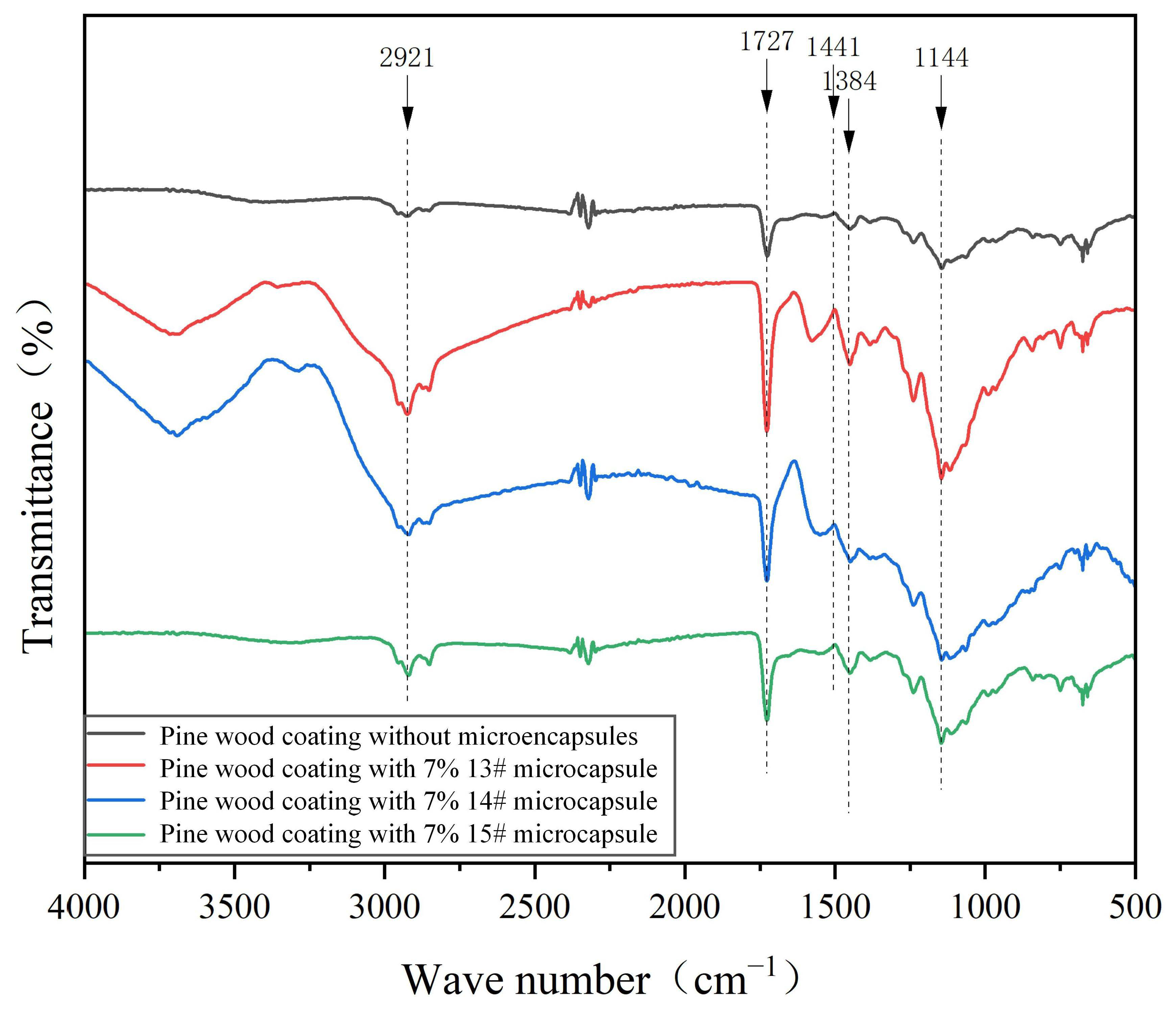
As illustrated in Figure 6, the infrared spectra of the pine wood surface coating in the absence of microcapsules and in the presence of 7% 13#, 14#, and 15# microcapsules are shown. The telescopic vibration peak of C=O in the waterborne coatings at 1727 cm−1, and the vibration peak of C-O in the waterborne coatings at 1144 cm−1, and the two characteristic peaks appeared in all four curves. This indicates that the addition of microcapsules did not result in a change to the main chemical composition of the waterborne coatings. The characteristic peak observed at 2921 cm−1 is attributed to the stretching vibration of saturated hydrocarbons (CH2) present within the wall of the chitosan. The peak at 1384 cm−1 is attributed to the bending vibration of the N-H group of amide II in the wall of the chitosan. The peak at 1441 cm−1 is attributed to the vibrational coupling of the C-O and O-H bonds of the alcohols contained within the core material of the microcapsule, which is composed of tea tree essential oil. The analysis of the coating samples containing microcapsules revealed the presence of three characteristic peaks, indicating the stability of the microcapsule’s internal chemical composition following its addition to the coating system. This observation suggests the absence of chemical reactions between the microcapsules and the waterborne coatings, thereby preserving the chemical properties and functional integrity of the components in the coating system.
In the 4000–3500 cm−1 region (O-H stretching vibration zone), samples 13# and 14# exhibit distinct broad absorption peaks, which may be associated with the presence of free hydroxyl groups on the surface or within the coating. As illustrated in sample 15, there is a marked decrease in the absorption intensity within this region. This finding suggests that the hydroxyl abundance on the surface or in the near-surface area of sample 15# is lower than that of other samples. In combination with the absence of significant O-H absorption in this region for sample 15#, it can be inferred that its coating surface exhibits stronger hydrophobic tendencies. The intensity of the C=O peak at 1727 cm−1 is strong in all coated samples, indicating that the main source of the strong peak at 1727 cm−1 is the carbonyl functional group of the water-based coating itself. The incorporation of microcapsules did not result in substantial alterations to the chemical milieu and the abundance of carbonyl groups within the base coating; consequently, the intensity of this peak remained comparable across all the coated specimens.
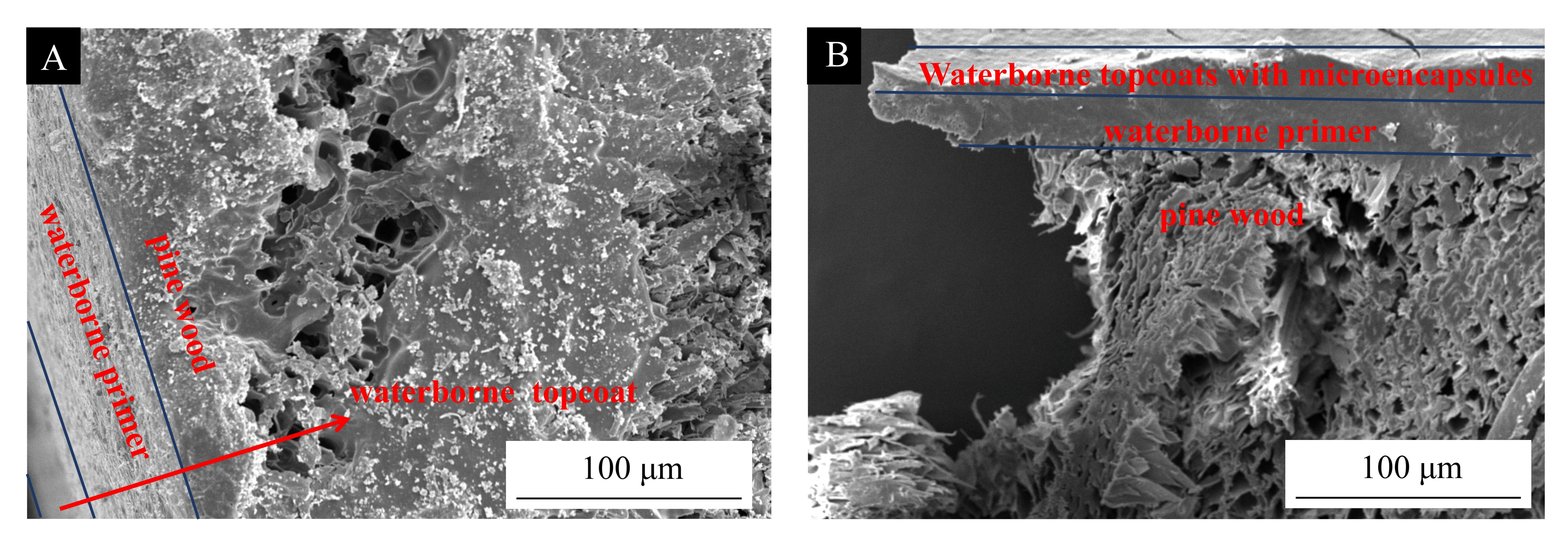
Figure 7 shows the cross-sectional microstructure of the coating on pine wood, as well as the interface between the coating and the wood. The surface of the pine wood is clearly visible to be coated with a water-based primer layer and a water-based topcoat layer. In the interface region, the primer has penetrated the wood to some extent, and microscopic structural changes have occurred at the interface between the primer and the wood. This indicates that the primer has performed a certain sealing and isolating function. At the same time, there is no evidence of deep penetration of large amounts of topcoat and microcapsules into the pine wood. Regardless of whether microcapsules were added, the cross-section SEM results showed no significant penetration of topcoat or microcapsules into the interior of the pine wood. This is related to limitations in the pore structure of the pine wood cell wall and the viscosity characteristics of the coating system. The high viscosity of the coating system makes it difficult to break through the wood cell wall’s physical barrier, resulting in the formation of a “shallow penetration–adhesion” interface structure on the wood surface.

3.3. Analysis of Antimicrobial Properties of Surface Coatings on Pine Wood

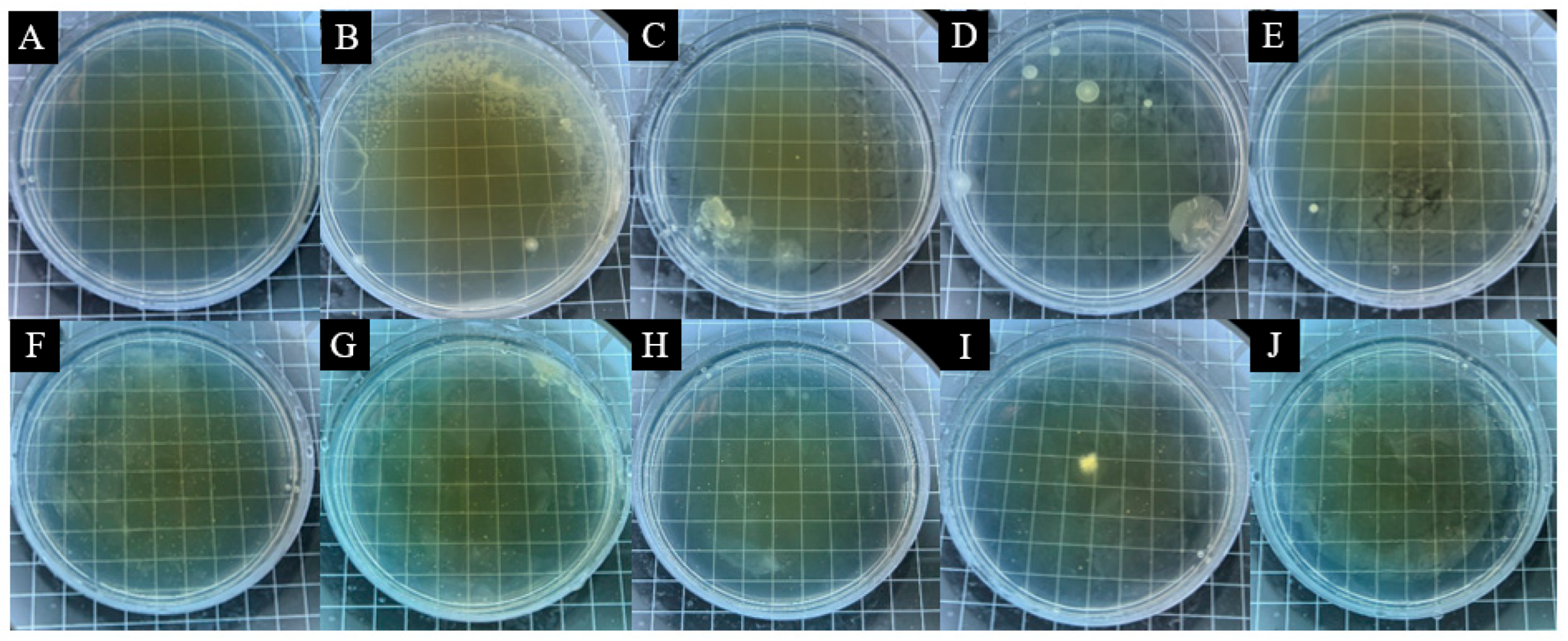
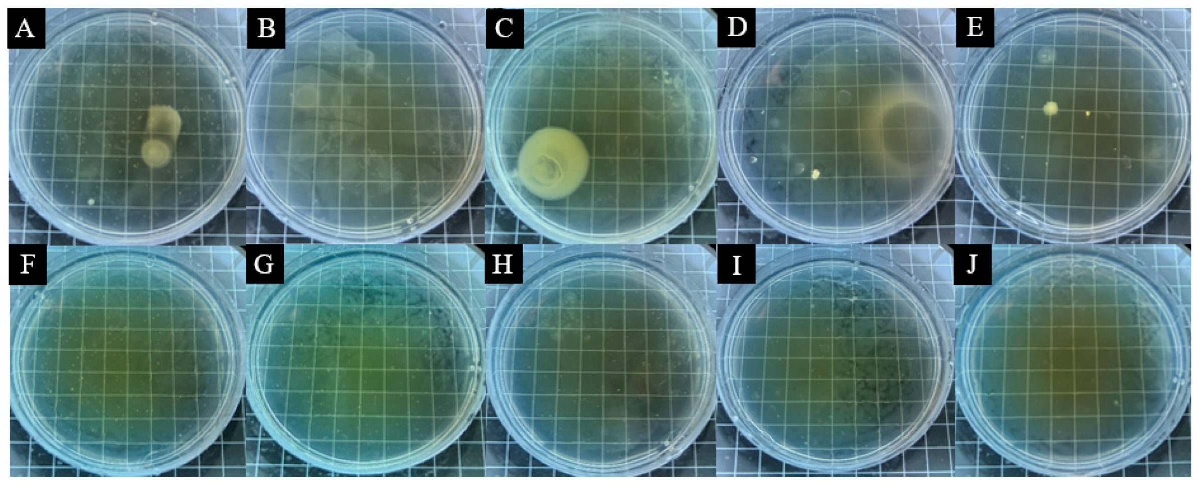
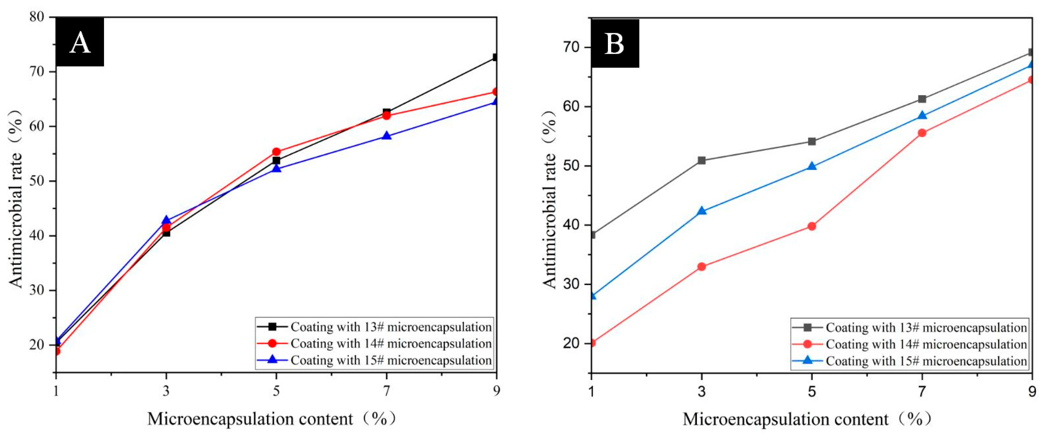
As illustrated in Table 5, the antimicrobial efficacy of waterborne coatings formulated with 13#, 14#, and 15# microcapsules at concentrations ranging from 1% to 9% (with a 2% interval) is evident. Figure 8, Figure 9 and Figure 10 illustrate the colony recovery plots of waterborne coatings with varying additions of microcapsules, respectively. As illustrated in Figure 11, the antimicrobial rate of waterborne coatings against E. coli and S. aureus at 1%–9% (2% interval) content demonstrates a clear trend. As the proportion of microcapsules increased from 1% to 9%, the antimicrobial rates of all three TTO@CS microcapsules against E. coli and S. aureus exhibited an upward trend. In the case of content levels below 5%, there was found to be negligible variation in the antimicrobial rates of the three microcapsules when confronted with E. coli. Furthermore, a consistent disparity was observed in the antimicrobial rates of the aforementioned microcapsules when confronted with E. coli at 7% and 9% content, with the highest antimicrobial rates of 13# microcapsules against E. coli, 62.58% and 72.64%, respectively, and the lowest antimicrobial rates of 15# microcapsules against E. coli being 58.18% and 64.47%, respectively. In the case of samples with a content of below 5%, the antimicrobial rates of the three microcapsules exhibited significant variation at the same content level. Furthermore, the antimicrobial rates of microcapsule 13# were consistently higher than those observed for microcapsules 2# and 3#. As the content of the microcapsules increased from 7% to 9%, the difference in the antimicrobial rate against S. aureus gradually decreased. The highest antimicrobial rate was found in microcapsules 13, which was 61.29% and 69.18%, respectively. When the emulsifier concentration of the added microcapsules was 4% (i.e., 13#), the highest antimicrobial rate of the coating was recorded as 72.64% against E. coli and 69.18% against S. aureus. When the coating contained 14# microcapsules, the prepared coating exhibited a more significant antimicrobial effect against both E. coli and S. aureus, with the highest recorded antibacterial rate of 66.35% against the former and 64.52% against the latter. When 9% 15# microcapsules were added, the antimicrobial performance of the coating against the two bacteria changed slightly, with an inhibition rate of 64.47% against E. coli and 67.03% against S. aureus. An increase in microcapsule content has been shown to result in the release of a greater quantity of tea tree essential oil, thereby increasing the local concentration and enhancing the antibacterial effect against E. coli and S. aureus. The incorporation of TTO@CS microcapsules within the waterborne coating system of pine wood has been demonstrated to enhance the bacterial inhibition efficacy of the coating. However, it is imperative to note that excessive microcapsule content can compromise the optical and mechanical characteristics of the coating. Therefore, it is essential to maintain optimal microcapsule concentrations to ensure the optimal balance between antibacterial performance and coating integrity.

3.4. Analysis of Optical Properties of Pine Wood Surface Coatings

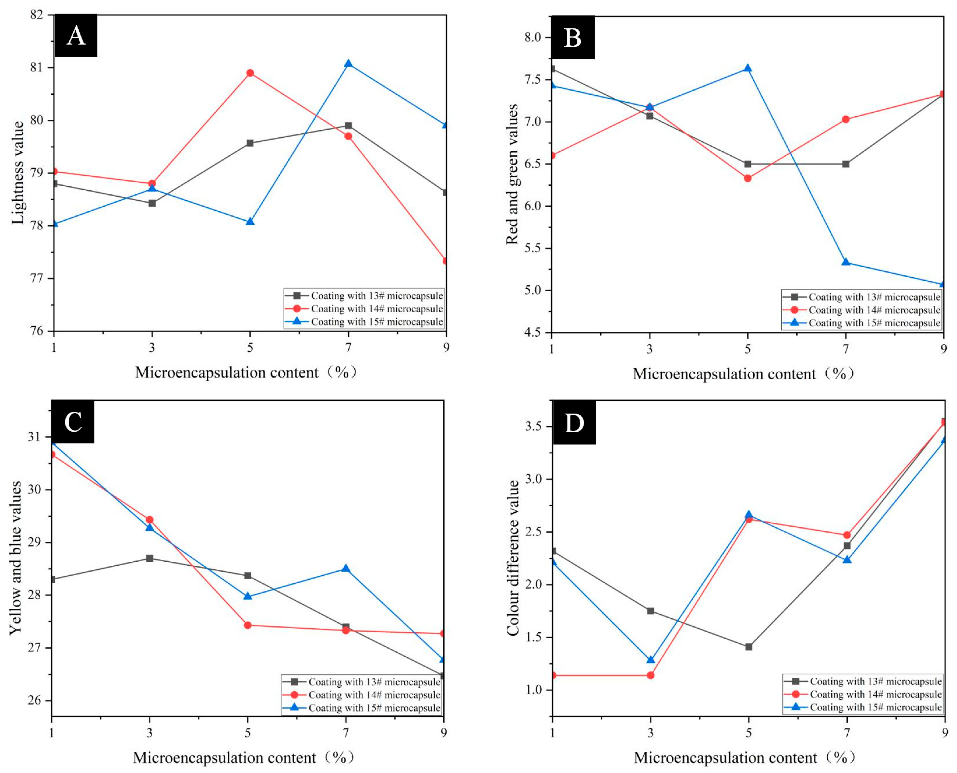
As illustrated in Table 6 and Figure 12, the results and trends of the colour difference values of the coatings with 4%, 5%, and 6% emulsifier concentration microcapsules at 1%–9% (with 2% intervals) content, respectively, are demonstrated. The incorporation of microcapsules did not exert a substantial influence on the chromaticity value and colour difference in pine wood surface coatings. In comparison with the coating devoid of microcapsules, the colour difference in pine wood surface coating exhibited an overall increasing trend with the increase in microcapsule content. The colour difference values for the coatings with 14# microcapsules were relatively small due to the lighter colour of the 14# microcapsules (5% emulsifier concentration). This minimal colour impact on the pine wood surface was demonstrated by the fact that the colour difference did not change significantly when added to the waterborne topcoat. In the instance of the coating with 13# microcapsules added to the pine wood surface, the minimum colour difference value was 1.41, when the microcapsule content was 5%. In the instance of the coating with 14# microcapsules added to the pine wood surface, the smallest colour difference values were 1.14 for both 1% and 3% microcapsule content, and 1.28 for the coating with 15# microcapsules added to the pine wood surface, when the microcapsule content was 3.0%. The colour difference values of the coatings all reached a maximum value of 3.55, 3.54, and 3.37, when the microcapsule content was 9%. As the content of microcapsules is increased, there is a concomitant decrease in the brightness value of the pine wood surface coating, although the decrease is minimal. The addition of microcapsules to the coating surface has been shown to result in the formation of protuberances, thereby compromising the coating’s capacity to reflect light, and consequently diminishing its brightness. The coating, comprising 13 # and 15 # microcapsules of red and green values, exhibits a decline in brightness. It has been demonstrated that the incorporation of 7% 15 # microcapsules into the coating results in a more pronounced decline in red and green values compared to the addition of 5% of the same microcapsules. With the increase in microcapsule content, a decrease in yellow and blue values was observed in pine wood surface coatings containing all three microcapsules.
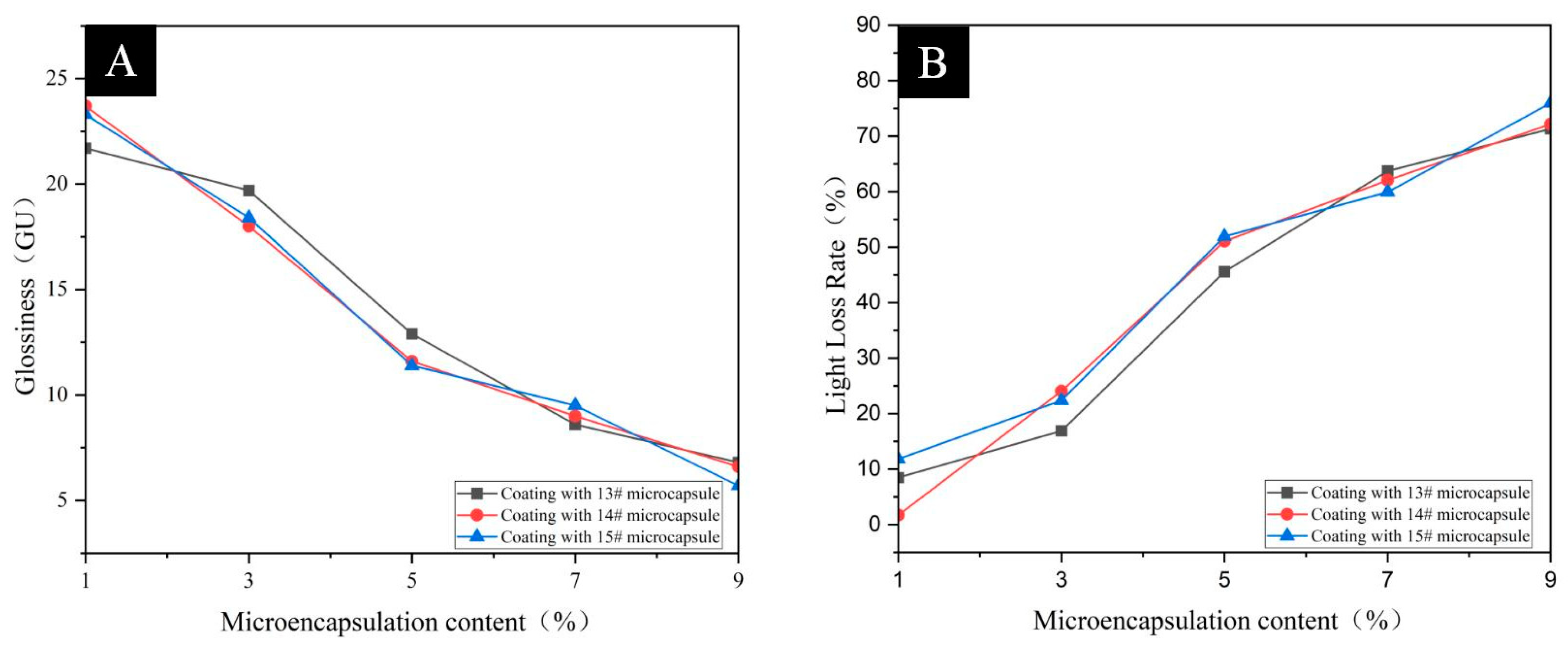
As illustrated in Table 7, the results of gloss and light loss for coatings with 13#, 14#, and 15# microcapsule at 1%–9% (2% interval) content are presented. As demonstrated in Figure 13, the trend of gloss and light loss for coatings with 13#, 14#, and 15# microcapsules at 60° incidence angle is shown for coatings with 1%–9% (2% interval) content. For all three microcapsules, the gloss of the coatings exhibited a marked decrease when the microcapsule content increased from 3% to 5%, and the change in gloss levelled off when the microcapsule content was in the range of 5%–9%. The addition of 13# microcapsule results in a marginal enhancement of the coating’s gloss, accompanied by an improvement in the optical properties of the microcapsules. When the content of microcapsules is 1%, the gloss of coatings containing three kinds of microcapsules all reach a maximum value of 21.7 GU, 23.7 GU, and 23.3 GU, respectively. It is imperative to note that the reduction in light loss from the wood surface coating is directly proportional to the enhancement in the optical performance of the coating. It was demonstrated that the loss of light rate of pine wood surface coating with three kinds of microcapsules increased in proportion to the increase in microcapsule content. An increase in microcapsule content from 1% to 9% resulted in a decline in glossiness of pine wood surface coatings containing TTO@CS microcapsules. This phenomenon can be attributed to the introduction of powdered microcapsules, leading to an increase in surface roughness of the coatings after curing. This, in turn, enhances the phenomenon of diffuse reflectance of the pine wood to light.
As illustrated in Table 8 and Figure 14, the impact of varying microcapsules and their incorporation levels on the reflectivity of pine wood surface coatings is demonstrated. It has been demonstrated that coatings with higher reflectance are capable of reflecting light within a certain range, whilst simultaneously absorbing less light and heat. This property enables them to attenuate heat, thereby preventing it from accumulating and warming on the coating surface. Consequently, this enhances the durability of the pine wood coating. The reflectance curves of the coatings with three kinds of microcapsules exhibited similar trends, indicating that the change in emulsifier concentration exerted a lesser effect on the reflectance of the coatings. In the coating with 13# microcapsules, the Rmax was 0.6860, at a microcapsule content of 7%, and the reflectance of the coating exhibited an increasing trend, followed by a decreasing trend, as the microcapsule content increased. In the coating with 14# microcapsules, Rmax was found to be 0.6780, at which time the microcapsule content was 5%, and with the increase in microcapsule content, the reflectivity of the coating showed an overall increasing trend. In the coating with 15# microcapsules, the Rmax was 0.6727, at which time the microcapsule content was 9.0%, and with the increase in microcapsule content, the reflectivity of the coating showed an overall increasing trend.

3.5. Analysis of Mechanical Properties of Pine Wood Surface Coatings

The hardness of the coating is a significant property that indicates the mechanical strength of the coating. It is also a crucial indicator of the quality of the coating product. It is evident that an increase in coating hardness is directly proportional to enhanced abrasion resistance, compression resistance, and liquid resistance. As illustrated in Table 9, the impact of varying microcapsules and their quantity on the hardness of the coating on the surface of pine wood is demonstrated. The hardness of the coating without added microcapsules was found to be HB. With the increase in microcapsule content, the hardness increased in all cases, from HB to 2H. The coating with added 13# microcapsules had a hardness of HB when the microcapsule content was 1%. When the microcapsule content was 3.0%, the hardness was H, and the coating had a hardness of 2H when the microcapsule content was 5%–9%. For the coatings with added 14# and 15# microcapsules, as the proportion of microcapsules in the mixture is increased from 1% to 3%, the hardness of the coating undergoes a transition from HB to H. Further increases in the microcapsule content, from 3% to 5% and from 5% to 7%, result in an additional shift in the hardness of the coating to 2H. This phenomenon can be attributed to the incorporation of microcapsules, which enhances the densification of the coating, thereby increasing its hardness.
The adhesion properties of coatings are instrumental in determining the bonding strength between the coating and the substrate interface, or between the coatings themselves. The presence of robust interfacial bonding is paramount in ensuring the long-term stability of the coating system. Conversely, a lack of sufficient interfacial adhesion can result in undesirable defects, such as the formation of bubbles, cracks, and delamination of the coating. As illustrated in Table 10, the effect of varying microcapsules, in addition to the quantity of added microcapsules, on the adhesion of pine wood surface coatings is demonstrated. The adhesion grade of the coating surface in the absence of microcapsules was determined to be 0. When the microcapsule content ranged from 1% to 7%, the adhesion grades of the coatings were all one, and when the microcapsule content was 9%, the adhesion of the coatings was two. The alteration in emulsifier concentration did not exert an influence on the coating adhesion. The incorporation of microcapsules resulted in a reduction in the adhesion of the coatings. This phenomenon can be attributed to the disruption of the homogeneity of the waterborne coatings caused by the microcapsules, which consequently led to a decrease in adhesion to the pine wood, thereby reducing the overall adhesion of the coatings.
The impact strength of the coating is defined as the resistance of the coating when impacted by an external force. It is a pivotal index for the evaluation of the coating’s impact and scratch resistance. As illustrated in Table 11, the impact of varying microcapsules and their addition amounts on the adhesion of coatings to pine wood surfaces is demonstrated. The impact resistance level of the coating without added microcapsules was determined to be 4. For the coatings containing 13# and 15# microcapsules, the impact resistance class of the coatings was assigned a value of four when the microcapsule content ranged from 1% to 5%, and a value of three when the microcapsule content ranged from 7% to 9%. For the coatings containing 14# microcapsules, the impact resistance class of the coatings was determined to be four when the microcapsule content ranged from 1% to 7%, and three when the microcapsule content ranged from 9%. It has been demonstrated that an elevated microcapsule content has the capacity to enhance the impact resistance of pine wood surface coatings. However, it is important to note that an excess of microcapsules can result in a deterioration of other properties. Due to the dense structure of the microcapsules, when employed as a solid powder filler agent in waterborne coatings, there is an enhancement of the mechanical strength of the pine wood surface coating, and an inhibition of the cracking of the coating due to stress.
The surface morphology of the coating is characterised by peaks and valleys on a microscopic scale, and its geometrical parameters reflect the degree of surface roughness. It has been determined that this degree of surface roughness not only determines the apparent quality of the coating, but also significantly affects its key indicators such as corrosion resistance and durability. As illustrated in Table 12, the impact of varying microcapsules and their quantity on the surface roughness of the coating on pine wood is demonstrated. The surface roughness of the coating without the incorporation of microcapsules was measured to be 0.356 μm. It was observed that the roughness of the coating increased in proportion to the number of microcapsules added. The roughness of the coatings with three kinds of microcapsules was found to be the least pronounced when the content of microcapsules was 1%, which was 0. The values obtained were 389 μm, 0.410 μm, and 0.487 μm, respectively. The surface roughness of the coatings with 13# microcapsules was found to be marginally lower than that of the coatings with the two other types of microcapsules. The surface roughness of the coating with 13# microcapsules is marginally lower than that of the coatings with the other two microcapsules because 13# microcapsules are more uniformly dispersed, and there are only a few irregularly shaped substances, but most of them show rounded spheres and have relatively small particle sizes.
The analysis of the test data on the mechanical properties of the surface coating of pine wood specimens indicates that the pine wood coating with the addition of 13# TTO@CS microcapsules demonstrates superior comprehensive mechanical properties, characterised by elevated hardness values and impact resistance grades. Additionally, this coating exhibits the lowest surface roughness index among the three specimen groups.

3.6. Analysis of Weather Resistance of Pine Wood Surface Coatings

The pine wood surface coating containing 7% of 13# microcapsules demonstrated superior performance. Following a four-month period of sample placement, a re-evaluation of the antibacterial performance was conducted to ascertain the long-term effectiveness of 13# microcapsules. As illustrated in Table 13, the antibacterial rate of the pine wood surface coating with 7% 13# microcapsules added is evident. Figure 15 provides a visual representation of the colony recovery of the pine wood surface coating with 7% 13# microcapsules added. As illustrated in Table 14, the performance of the pine wood surface coating containing 7% of 13# microcapsules was considered comprehensive. Following a period of four months, the pine wood surface coating, which contained 13# microcapsules, exhibited an antibacterial rate of 40.14% against E. coli and 38.89% against S. aureus. In comparison with the coating after 48 h, the antibacterial rate decreased. This phenomenon can be attributed to the prolonged exposure of the coating to the environment, resulting in the gradual evaporation or degradation of the effective antibacterial ingredients contained within the microcapsules. Consequently, there has been a decline in the concentration of active ingredients, thereby weakening the antibacterial effect.

4. Conclusions

The incorporation of TTO@CS microcapsules effectively imparts antimicrobial properties to the pine wood surface coating. The antimicrobial effects of the three groups of TTO@CS microcapsule surface coatings were found to be positively correlated with the microcapsule content when it was increased from 1% to 9% (at 2% intervals). The antimicrobial performance of the pine wood surface coating with 13# microcapsules was superior, with an antimicrobial rate of 72.64% against E. coli and 69.18% against S. aureus. The antibacterial rate of pine wood surface coating with 14# microcapsules was 66.35% and 64.52% for E. coli and S. aureus, respectively. The antimicrobial activity of the pine wood surface coating with 15# microcapsules was found to be 64.47% and 67.03% against E. coli and S. aureus, respectively. The surface coatings of pine wood with 7% 14# and 15# microcapsules exhibited increased surface roughness, while the surface of pine wood with 7% 13# microcapsules demonstrated a reduced propensity for projections. It has been demonstrated that an increase in microcapsule content results in a concomitant rise in colour difference in the pine wood surface coating in its entirety. At a microcapsule content of 9%, the colour difference in the coating attains its maximum value, while the colour difference in the coating with the addition of 14# microcapsule is comparatively negligible. It was observed that as the microcapsule content increased, there was a concomitant decrease in the brightness value of the pine wood surface coating. In addition, there was an increase in the rate of light loss, with a subsequent decrease in reflectivity. However, the mechanical properties of the coating were enhanced, particularly with regard to hardness and impact resistance. Conversely, surface adhesion was slightly reduced, while surface roughness was increased. The study concluded that the pine wood surface coating containing 7% 13# TTO@CS microcapsules exhibited excellent overall performance: there were relatively few bumps on the surface of the coating, and the antimicrobial rate of the coating was 62.58% for E. coli and 61.29% for S. aureus. The colour difference in the coating was found to be 2.37, with a gloss at 60° incidence angle of 8.6 GU. The loss of light was determined to be 63.71%, and the reflectance of the coating was measured to be 0.6860. The hardness was categorised as 2H, the adhesion class was designated as one, the impact resistance class was rated as three, and the roughness was measured to be 1.320 μm. The microcapsule coating developed in this study has achieved the advanced level reported in the literature with regard to its antibacterial performance (especially against E. coli), is significantly superior to unmodified pine wood coatings with regard to mechanical properties (hardness, impact resistance), and has better overall performance than some chemically modified substrates. The company’s core value lies in achieving a balance of multiple functions through a simple process, providing a coating solution for pine wood furniture that combines protection, aesthetics, and environmental friendliness. In the future, further optimisation of interface stability under high load conditions will be pursued through coupling agent modification and composite antimicrobial design.

Author Contributions

Conceptualization, methodology, validation, resources, and data management, N.Z.; supervision and writing—review and editing, Y.Z.; formal analysis and investigation, X.Y. All authors have read and agreed to the published version of the manuscript.

Funding

This project was partly supported by the Qing Lan Project and the Natural Science Foundation of Jiangsu Province (BK20201386).

Institutional Review Board Statement

Not applicable.

Informed Consent Statement

Not applicable.

Data Availability Statement

Data are contained within the article.

Conflicts of Interest

The authors declare no conflicts of interest.

References

  1. Hu, W.; Yang, Z.; Shi, N.; Yu, X. Experimental study on effects of the selected load parameters on fatigue life of the mortise-and-tenon furniture joint. Wood Mater. Sci. Eng. 2025, 19, 1–7. [Google Scholar] [CrossRef]
  2. Wang, C.; Li, J.; Wang, T.; Wang, X. Additive manufacturing of furniture corner guards based on thermoplastic polyurethane filament. BioResources 2025, 20, 5398–5406. [Google Scholar] [CrossRef]
  3. Liu, Y.; Hu, W.; Kasal, A.; Erdil, Y.Z. The state of the art of biomechanics applied in ergonomic furniture design. Appl. Sci. 2023, 13, 12120. [Google Scholar] [CrossRef]
  4. Xu, X.; Zhang, M.; Yue, X.; Xiong, X. Design of furniture mortise-and-tenon joints: A review of mechanical properties and design recommendations. Wood Mater. Sci. Eng. 2025, 1–15. [Google Scholar] [CrossRef]
  5. Yu, R.; Liu, Y.; Konukcu, C.A.; Hu, W. A method of simulating seat load for numerical analysis of wood chair structure. Wood Res-Slovak. 2024, 69, 432–444. [Google Scholar] [CrossRef]
  6. Hu, W.; Yu, R.; Yang, P. Characterizing roughness of wooden mortise and tenon considering effects of measured position and assembly condition. Forests 2024, 15, 1584. [Google Scholar] [CrossRef]
  7. Zhou, J.; Xu, W. Optimizing the Interface Compatibility of Transparent Wood for Green Phase-Change Thermal Storage. Wood Sci. Technol. 2025, 59, 45. [Google Scholar] [CrossRef]
  8. Yang, Y.; Xu, W.; Liu, X.; Wang, X. (Alice). Study on Permeability of Cunninghamia Lanceolata Based on Steam Treatment and Freeze Treatment. Wood Res. 2021, 66, 721–731. [Google Scholar] [CrossRef]
  9. Wu, S.; Tao, X.; Xu, W. Thermal Conductivity of Poplar Wood Veneer Impregnated with Graphene/Polyvinyl Alcohol. Forests 2021, 12, 777. [Google Scholar] [CrossRef]
  10. Liu, Q.; Gao, D.; Xu, W. Effect of Paint Process on the Performance of Modified Poplar Wood Antique. Coatings 2021, 11, 1174. [Google Scholar] [CrossRef]
  11. Li, S.; Hu, W. Study on mechanical strength of cantilever handrail joints for chair. BioResources 2023, 18, 209–219. [Google Scholar] [CrossRef]
  12. Fu, S.; Xiong, X.; Wan, R.; Zhang, M.; Xu, X. The Development and Future Challenges of China’s Furniture Industry. Drewno 2025, 68, 199709. [Google Scholar] [CrossRef]
  13. Hu, W.; Fu, W.; Zhao, Y. Optimal design of the traditional Chinese wood furniture joint based on experimental and numerical method. Wood Res-Slovak. 2024, 69, 50–59. [Google Scholar] [CrossRef]
  14. Xu, W.; Fang, X.Y.; Han, J.T.; Wu, Z.H. Effect of Coating Thickness on Sound Absorption Property of Four Wood Species Commonly Used for Piano Soundboards. Wood Fiber Sci. 2020, 52, 28–43. [Google Scholar] [CrossRef]
  15. Liu, Q.Q.; Gao, D.; Xu, W. Effect of Sanding Processes on the Surface Properties of Modified Poplar Coated by Primer Compared with Mahogany. Coatings 2020, 10, 856. [Google Scholar] [CrossRef]
  16. Liu, Q.Q.; Gao, D.; Xu, W. Effect of Polyurethane Non-Transparent Coating Process on Paint Film Performance Applied on Modified Poplar. Coatings 2022, 12, 39. [Google Scholar] [CrossRef]
  17. Liu, Q.Q.; Gu, Y.T.; Xu, W.; Lu, T.; Li, W.J.; Fan, H.B. Compressive Properties of Green Velvet Material Used in Mattress Bedding. Appl. Sci. 2021, 11, 11159. [Google Scholar] [CrossRef]
  18. Wan, R.Y.; Xiong, X.Q.; Fu, S.J.; Xiong, D.J.; Xu, X.T. Life cycle assessment and optimization scenarios of sofas: A case study in China. Int. J. Life Cycle Assess. 2025, 1–17. [Google Scholar] [CrossRef]
  19. Wang, C.; Li, J.Y.; Wang, T.Y.; Chu, Q.; Wang, X.W. Fused deposition 3D printing of bonsai tree guiding mold based on acrylonitrile-butadiene-styrene copolymer. BioResources 2024, 19, 5839–5846. [Google Scholar] [CrossRef]
  20. Wang, C.; Li, J.Y.; Wang, T.Y.; Chu, Q.; Wen, S.Q. Design and Rapid Prototyping of Packaging Liner for Rosewood Craft based on Gyroid Infill Structure. BioResources 2025, 20, 842–851. [Google Scholar] [CrossRef]
  21. Abolhasani, H.; Farzi, G.; Davoodi, A.; Vakili-Azghandi, M.; Das, O.; Neisiany, R.E. Development of self-healable acrylic water-based environmental-friendly coating as an alternative to chromates coatings. Prog. Org. Coat. 2023, 176, 107402. [Google Scholar] [CrossRef]
  22. Han, K.; Liu, J.; Hao, F.; Wang, J.; Yuan, J.; Pan, Z.; Pan, M. An adaptive waterborne fluorocarbon coatings with Anti-Flashing Rust, Antibiofouling, and Self-Repairing properties. Chem. Eng. J. 2024, 495, 153644. [Google Scholar] [CrossRef]
  23. Wu, A.; Sun, Y.; Wang, Z.; Cao, G.; Zhang, H. Polyaniline/cellulose nanocrystal nanorods integrated into waterborne polyurethane coatings for enhanced corrosion resistance. Colloids Surf. A-Physicochem. Eng. Asp. 2025, 713, 136531. [Google Scholar] [CrossRef]
  24. Mastouri, A.; Efhamisisi, D.; Tarmian, A.; Boukherroub, R.; Lexa, M.; Karami, E.; Frigione, M. Sustainable superhydrophobic and self-cleaning wood via wax within Epoxy/PDMS nano-composite coatings: Durability related to surface morphology. Prog. Org. Coat. 2024, 186, 107951. [Google Scholar] [CrossRef]
  25. Barthwal, S.; Uniyal, S.; Barthwal, S. Nature-inspired superhydrophobic coating materials: Drawing inspiration from nature for enhanced functionality. Micromachines 2024, 15, 391. [Google Scholar] [CrossRef]
  26. Lu, Q.X.; Cheng, R.F.; Jiang, H.Q.; Xia, S.W.; Zhan, K.; Yi, T.F.; Morrell, J.J.; Yang, L.; Wan, H.; Du, G.B. Superhydrophobic wood fabricated by epoxy/Cu2(OH)3Cl NPs/stearic acid with performance of desirable self-cleaning, anti-mold, dimensional stability, mechanical and chemical durability. Colloids Surf. A-Physicochem. Eng. Asp. 2022, 647, 1029162. [Google Scholar] [CrossRef]
  27. Jian, H.; Liang, Y.; Deng, C.; Xu, J.; Liu, Y.; Shi, J.; Wen, M.Y.; Park, H.J. Research progress on the improvement of flame retardancy, hydrophobicity, and antibacterial properties of wood surfaces. Polymers 2023, 15, 951. [Google Scholar] [CrossRef]
  28. Faccini, M.; Bautista, L.; Soldi, L.; Escobar, A.M.; Altavilla, M.; Calvet, M.; Domènech, A.; Domínguez, E. Environmentally friendly anticorrosive polymeric coatings. Appl. Sci. 2021, 11, 3446. [Google Scholar] [CrossRef]
  29. Lin, D.; Wang, X.; Zhang, M.; Yuan, S.; Xu, F.; Bao, D.; Wang, H. Development of a robust and eco-friendly waterborne anti-corrosion composite coating with multiple synergistic corrosion protections. Compos. Part B Eng. 2022, 232, 109624. [Google Scholar] [CrossRef]
  30. Chang, Y.; Yan, X.; Wu, Z. Application and prospect of self-healing microcapsules in surface coating of wood. Colloid Interface Sci. Commun. 2023, 56, 100736. [Google Scholar] [CrossRef]
  31. Wang, X.J.; Sun, W.P.; Li, W.G.; Zuo, C.L.; Jiang, Y.; Wang, S.X. Development of Waterborne Heavy-Duty Anticorrosive Coatings with Modified Nanoscale Titania. Coatings 2022, 12, 1651. [Google Scholar] [CrossRef]
  32. Tang, J.L.; Wang, Y.C.; He, M.J.; Huang, L.Q.; Wang, X.L.; Yu, J.Y. Electrothermochromic Fabrics with a Single-Layer Functional Coating Based on Silver Nanowires/Thermochromic Microcapsules/Waterborne Polyurethane Paints. ACS Appl. Mater. Interfaces 2024, 16, 70963–70972. [Google Scholar] [CrossRef]
  33. Xue, D.; Zhao, T. The electrothermal color-changing fabric based on high-sensitivity thermochromic microcapsules. Colloids Surf. A-Physicochem. Eng. Asp. 2023, 678, 132458. [Google Scholar] [CrossRef]
  34. Boh Podgornik, B.; Šandrić, S.; Kert, M. Microencapsulation for functional textile coatings with emphasis on biodegradability—A systematic review. Coatings 2021, 11, 1371. [Google Scholar] [CrossRef]
  35. Ahuja, A.; Rastogi, V.K. PFAS free, food-grade, water and grease-resistant coating based on crosslinked shellac for molded pulp products. Prog. Org. Coat. 2024, 196, 108734. [Google Scholar] [CrossRef]
  36. Becerril, R.; Nerín, C.; Silva, F. Encapsulation systems for antimicrobial food packaging components: An update. Molecules 2020, 25, 1134. [Google Scholar] [CrossRef]
  37. Rezić, I.; Somogyi Škoc, M.; Majdak, M.; Jurić, S.; Stracenski, K.S.; Vinceković, M. Functionalization of Polymer Surface with Antimicrobial Microcapsules. Polymers 2022, 14, 1961. [Google Scholar] [CrossRef]
  38. Bagheri Darvish, H.; Bahrami, A.; Jafari, S.M.; Williams, L. Micro/nanoencapsulation strategy to improve the efficiency of natural antimicrobials against Listeria monocytogenes in food products. Crit. Rev. Food Sci. Nutr. 2021, 61, 1241–1259. [Google Scholar] [CrossRef]
  39. Li, X.; Gao, Y.F.; Li, Y.; Liu, H.; Yang, Z.; Wu, H.; Hu, Y. Formation of cinnamon essential oil/xanthan gum/chitosan composite microcapsules basing on Pickering emulsions. Colloid Polym. Sci. 2022, 300, 1187–1195. [Google Scholar] [CrossRef]
  40. Zhu, Y.; Yan, X. Preparation of Tea Tree Essential Oil–Chitosan Microcapsules and Its Effect on the Properties of Water-Based Coating. Polymers 2025, 17, 849. [Google Scholar] [CrossRef]
  41. GB/T 6739-2022; Determination of Coating Hardness by Pencil Method for Coloured Paints and Varnishes. Standardization Administration of the People’s Republic of China: Beijing, China, 2022.
  42. GB/T 4893.9-2013; Physical and Chemical Performance Tests of the Paint Film on the Surface of Furniture—Part 9: Determination of Impact Resistance. Standardization Administration of the People’s Republic of China: Beijing, China, 2013.
  43. GB/T 4893.4-2013; Physical and Chemical Performance Tests of the Paint Film on the Surface of Furniture—Part 4: Determination of Adhesion by Cross-Cutting Method. Standardization Administration of the People’s Republic of China: Beijing, China, 2013.
  44. GB/T 21866-2008; Determination Method and Antibacterial Effect of Antibacterial Coatings (Paint Films). Standardization Administration of the People’s Republic of China: Beijing, China, 2008.
  45. GB/T 4789.2-2022; National Food Safety Standard—Microbiological Examination of Food—Aerobic Plate Count. Standardization Administration of the People’s Republic of China: Beijing, China, 2022.
Figure 1. OM images of TTO@CS microcapsules: (A) 13#, (B) 14#, and (C) 15# [40].
Coatings 15 00938 g001
Figure 2. SEM images of TTO@CS microcapsules at low magnification: (A) 13#, (B) 14#, and (C) 15# [40].
Coatings 15 00938 g002
Figure 3. SEM images of TTO@CS microcapsules at high magnification: (A) 13#, (B) 14#, and (C) 15# [40].
Coatings 15 00938 g003
Figure 4. Particle size distribution of TTO@CS microcapsules: (A) 13#, (B) 14#, and (C) 15# [40].
Coatings 15 00938 g004
Figure 5. SEM image of pine wood surface coating prepared with 7% microcapsules added: (A) without microcapsule, (B) 13#, (C) 14#, and (D) 15#.
Coatings 15 00938 g005
Figure 6. FTIR of coating on the surface of pine wood containing TTO@CS microcapsules.
Coatings 15 00938 g006
Figure 7. SEM image of the cross-section of the interface between the surface coating and pine wood: (A) without microcapsule and (B) with 7% of 14# microcapsule.
Coatings 15 00938 g007
Figure 8. 13# TTO@CS microcapsule coating colony recovery diagram for Escherichia coli: microcapsule content (A) 0% and (BF) represent 1%–9% (2% intervals). Staphylococcus aureus: (G) 0% and (HL) represent 1%–9% (2% intervals).
Coatings 15 00938 g008
Figure 9. 14# TTO@CS microcapsule coating colony recovery diagram for Escherichia coli: microcapsule content (AE) represent 1%–9% (2% intervals); Staphylococcus aureus: (FJ) represent 1%–9% (2% intervals).
Coatings 15 00938 g009
Figure 10. 15# TTO@CS microcapsule coating colony recovery diagram for Escherichia coli: microcapsule content (AE) represent 1%–9% (2% intervals); Staphylococcus aureus: (FJ) represent 1%–9% (2% intervals).
Coatings 15 00938 g010
Figure 11. Trend of antibacterial rate of TTO@CS microcapsule under different contents: (A) Escherichia coli and (B) Staphylococcus aureus.
Coatings 15 00938 g011
Figure 12. The chromaticity and colour difference in pine wood surface coating: (A) L, (B) a, (C) b, and (D) ΔE.
Coatings 15 00938 g012
Figure 13. The trend graphs of variations about pine surface coating: (A) glossiness and (B) light loss rate.
Coatings 15 00938 g013
Figure 14. The reflectance of pine wood surface coating: (A) 13#, (B) 14#, and (C) 15#.
Coatings 15 00938 g014
Figure 15. The 13# microcapsule coating colony recovery diagram, E. coli: (A) without microcapsules and (B) 13#; S. aureus: (C) without microcapsules and (D) 13#.
Coatings 15 00938 g015
Table 1. Test materials.
Test MaterialsPurityManufacturer
Tea tree essential oil-Wuhan Huaxiang Biotechnology Co., Ltd., Wuhan, China
ChitosanARSinopharm Chemical Reagent Co., Ltd., Shanghai, China
Tween-80ARSinopharm Chemical Reagent Co., Ltd., Shanghai, China
SDBSARShandong Xinjucheng Chemical Technology Co., Ltd., Jinan, China
Acetic acidARShandong Chengkai New Material Co., Ltd., Linyi, China
Sodium tripolyphosphateARTianjin Huasheng Chemical Reagent Co., Ltd., Tianjin, China
Dulux primer-Nanjing Jinyou Biotechnology Co., Ltd., Nanjing, China
Nutrient agar medium-Zhongshan Baimicrobial Technology Co., Ltd., Guangdong, China
Nutrient broth medium-Zhongshan Baimicrobial Technology Co., Ltd., Guangdong, China
Sodium chlorideARSinopharm Chemical Reagent Co., Ltd., Shanghai, China
Escherichia coli-Beijing Baocang Biotechnology Co., Ltd., Beijing, China
Staphylococcus aureus-Beijing Baocang Biotechnology Co., Ltd., Beijing, China
Table 2. Material for TTO@CS microcapsule single factor test.
Sample
(#)
CS (g)Acetic Acid
(mL)
Deionized
Water for
Acetic Acid (mL)
Tween80 (g)SDBS (g)Deionized Water for
Emulsifier (mL)
TTO (g)STPP (g)
132.0000.10099.0001.6006.400192.0002.4000.400
142.0000.10099.0002.0008.000190.0002.4000.400
152.0000.10099.0002.4009.600188.0002.4000.400
Table 3. Coating mechanical properties test equipment.
EquipmentModelManufacturer
Pencil hardness testerHT-6510PAipu Measuring Instrument Co., Ltd., Quzhou, China
Coating ImpactorQCJ-50Jiaxin Measuring Instrument Co., Ltd., Dongguan, China
Adhesion TesterQFH-AAipu Measuring Instrument Co., Ltd., Quzhou, China
Table 4. TTO@CS microcapsule dosage and coating dosage.
Microencapsulation Content (%)Primer Quality (g)Microencapsulation Content (%)Topcoat Quality (g)
00.72000.720
10.7200.0070.713
30.7200.0220.698
50.7200.0360.684
70.7200.0500.670
90.7200.0650.655
Table 5. The actual number of recovered viable colonies and antibacterial rate of Escherichia coli and Staphylococcus aureus on the coating surface.
SampleMicroencapsulation Content (%)Average Number of Bacteria
Recovered (CFU·Piece−1)
Antimicrobial Rate (%)
E. coliS. aureusE. coliS. aureus
Pine wood Surface Add 13# Coating of microcapsules0318279--
125317220.4438.35
318913740.5750.90
514712853.7754.12
711910862.5861.29
9878672.6469.18
Pine wood Surface Add 14# Coating of microcapsules0318279--
125822318.8720.07
318618741.5132.97
514216855.3539.78
712112461.9555.56
91079966.3564.52
Pine wood Surface Add 15# Coating of microcapsules0318279--
125220120.7527.96
318216142.7742.29
515214052.2049.82
713311658.1858.42
91139264.4767.03
Table 6. Chromaticity and colour difference value of pine wood surface coating.
SampleMicroencapsulation Content (%)Chromaticity Value ΔE
Lab
Coating without microencapsulation079.736.6029.77-
Pine wood Surface Add 13# Coating of microcapsules178.807.6328.302.32
378.437.0728.701.75
579.576.5028.371.41
779.906.5027.402.37
978.637.3326.473.55
Pine wood Surface Add 14# Coating of microcapsules179.036.6030.671.14
378.807.1729.431.14
580.906.3327.432.62
779.707.0327.332.47
977.337.3327.273.54
Pine wood Surface Add 15# Coating of microcapsules178.037.4330.902.21
378.707.1729.271.28
578.077.6327.972.66
781.075.3328.502.23
979.905.0726.773.37
Table 7. Glossiness and light loss rate of pine wood surface coating.
SampleMicroencapsulation Content (%)Glossiness (GU)Light Loss Rate (%)
20°60°85°20°60°85°
Coating without microencapsulation05.8023.7043.90---
Pine wood Surface Add 13# Coating of microcapsules15.6021.7037.603.458.4414.35
34.7019.7033.3018.9716.8824.15
53.5012.9023.4039.6645.5746.70
72.608.608.8055.1763.7179.95
92.506.806.4056.9071.3185.42
Pine wood Surface Add 14# Coating of microcapsules15.8023.7037.001.721.6915.72
34.3018.0029.3025.8624.0533.28
53.2011.6018.4044.8351.0558.09
73.009.002.1048.2862.0395.22
92.406.604.2058.6272.1590.43
Pine wood Surface Add 15# Coating of microcapsules15.5023.3043.905.1711.814.56
34.5018.4030.3022.4122.3630.98
53.0011.4014.3048.2851.9067.43
72.809.5011.4051.7259.9274.03
92.405.702.2058.6275.9594.99
Table 8. The reflectance of pine wood surface coating.
Microencapsulation Content (%)R
13#14#15#
00.64470.64470.6447
10.63880.65660.6042
30.64070.64060.6613
50.64650.67800.6530
70.68600.64930.6566
90.68030.65710.6727
Table 9. Hardness of pine wood surface coating.
Microencapsulation
Content (%)
Hardness
13#14#15#
0HBHBHB
1HBHBHB
3HHH
52HHH
72H2H2H
92H2H2H
Table 10. Adhesion grade of pine wood surface coating.
Microencapsulation
Content (%)
Adhesion (Level)
13#14#15#
0000
1111
3111
5111
7111
9222
Table 11. Impact resistance grade of pine wood surface coating.
Microencapsulation
Content (%)
Impact Resistance (Level)
13#14#15#
0444
1444
3444
5444
7343
9333
Table 12. Roughness of pine wood surface coating.
Microencapsulation
Content (%)
Roughness (μm)
13#14#15#
00.3560.3560.356
10.3890.4100.487
30.4870.4590.613
50.7580.6291.209
71.3201.2701.359
92.2794.9892.815
Table 13. Actual number of viable colonies and antibacterial rate of two bacteria on the coated surface of 13# microcapsules.
Sample (#)Average Number of Bacteria
Recovered (CFU·Piece−1)
Antimicrobial
Rate (%)
E. coliS. aureusE. coliS. aureus
0142162--
13859940.1438.89
Table 14. The 13# microcapsule comprehensive performance table.
Sample (#)Microencapsulation Content (%)Antibacterial Rate After 48 h (%) Antibacterial Rate After 4 Months (%)Optical PropertiesMechanical Properties
E. coliS. aureusE. coliS. aureus ΔE60° Light Loss Rate (%)RHardnessAdhesion (Level)Impact Resistance (Level)Roughness (μm)
13762.5861.2940.1438.892.3763.710.68602H131.320
Disclaimer/Publisher’s Note: The statements, opinions and data contained in all publications are solely those of the individual author(s) and contributor(s) and not of MDPI and/or the editor(s). MDPI and/or the editor(s) disclaim responsibility for any injury to people or property resulting from any ideas, methods, instructions or products referred to in the content.