Systematic Review

Meta-Analysis and Systematic Review of Phenotypic and Genotypic Antimicrobial Resistance and Virulence Factors in Vibrio parahaemolyticus Isolated from Shrimp

by
Varangkana Thaotumpitak
1,
Justice Opare Odoi
2,
Saran Anuntawirun
3 and
Saharuetai Jeamsripong
3,*
1
Department of Microbiology, Faculty of Public Health, Mahidol University, Bangkok 10400, Thailand
2
Animal Health Division, Animal Research Institute, Council for Scientific and Industrial Research, Accra P.O. Box AH20, Ghana
3
Research Unit in Microbial Food Safety and Antimicrobial Resistance, Department of Veterinary Public Health, Faculty of Veterinary Science, Chulalongkorn University, Bangkok 10330, Thailand
*
Author to whom correspondence should be addressed.
Antibiotics 2024, 13(4), 370; https://doi.org/10.3390/antibiotics13040370
Submission received: 23 February 2024 / Revised: 3 April 2024 / Accepted: 15 April 2024 / Published: 17 April 2024
(This article belongs to the Special Issue Antibiotics Resistance in Animals and the Environment)

Abstract

:
This systematic review and meta-analysis investigates the prevalence of Vibrio parahaemolyticus, its virulence factors, antimicrobial resistance (AMR), and its resistance determinants in shrimp. This study was conducted following the Preferred Reporting Items for Systematic Reviews and Meta-Analysis (PRISMA) guidelines, to identify and select relevant peer-reviewed articles published between January 2020 and December 2022. The search strategy involved multiple online databases, including Google Scholar, PubMed, ScienceDirect, and Scopus. The inclusion criteria focused on studies that examined V. parahaemolyticus prevalence, virulence factors, and AMR in shrimp from farms to retail outlets. A total of 32 studies were analyzed, revealing a pooled estimate prevalence of V. parahaemolyticus in shrimp at 46.0%, with significant heterogeneity observed. Subgroup analysis highlighted varying prevalence rates across continents, emphasizing the need for further investigation. Virulence factor analysis identified thermostable direct hemolysin (tdh) and tdh-related hemolysin (trh) as the most common. Phenotypic AMR analysis indicated notable resistance to glycopeptides, nitrofurans, and beta-lactams. However, the correlation between antimicrobial usage in shrimp farming and observed resistance patterns was inconclusive. Funnel plots suggested potential publication bias, indicating a need for cautious interpretation of findings. This study underscores the urgency of coordinated efforts to address AMR in V. parahaemolyticus to safeguard public health and to ensure sustainable aquaculture practices.

1. Introduction

Antimicrobial resistance (AMR) has emerged as a critical global health threat, fundamentally reshaping the management of infectious diseases and posing unprecedented challenges to healthcare systems worldwide [1]. The increasing resistance of a diverse array of bacteria responsible for infections in both human and animal populations to previously effective antimicrobial treatments has become a pressing concern [2]. This alarming phenomenon could imperil human health and undermine the sustainability of animal production, and this requires swift and coordinated actions to avert an imminent global health crisis.
Vibrio parahaemolyticus, a halophilic bacterium in the family Vibrionaceae, thrives within a temperature range from 10 °C to 44 °C, with an optimal growth temperature between 35 °C and 37 °C within marine and estuarine environments [3]. Studies have highlighted the role of this bacterium as a primary causative agent of acute hepatopancreatic necrosis disease in shrimp populations [4]. Further research suggests that V. parahaemolyticus can rapidly spread within shrimp tissues, affecting vital organs such as the gills, hepatopancreas, intestine, muscles, and hemolymph, exacerbating the challenges faced in shrimp farming [5]. V. parahaemolyticus has the potential to trigger devastating vibriosis outbreaks in shrimp aquaculture, leading to substantial economic losses for farmers worldwide [6]. Historically linked to seafood, such as shrimp, oysters, clams, and cockles, V. parahaemolyticus received recognition for its pathogenic potential in humans in 1950 following a major outbreak in Japan in which contaminated sardine with the bacterium led to 20 fatalities and 270 hospitalizations [7]. Subsequently, outbreaks stemming from the consumption of raw or undercooked seafood tainted with V. parahaemolyticus have been documented in various countries, including the USA, Thailand, Vietnam, China, Spain, and Chile [8,9,10,11,12,13]. The disease pathogenesis of V. parahaemolyticus infections is caused by virulence factors such as thermostable direct hemolysin (TDH) or TDH-related hemolysin (TRH), encoded by tdh and trh genes, respectively. These two factors are the main factors causing infections in shrimps as well as seafood-borne bacterial gastroenteritis in humans [14]. This emphasizes the significance of comprehending and managing V. parahaemolyticus infections to safeguard both the aquaculture industry and the general health of seafood consumers.
Currently, the primary approach to the treatment of V. parahaemolyticus infections involves the administration of antimicrobial agents. However, alarming reports have surfaced, highlighting the emergence of AMR strains of V. parahaemolyticus and increasing public health concerns [15]. The consequences of infections caused by pathogenic and resistant V. parahaemolyticus extend beyond the scope of healthcare, affecting economic and social aspects as well. Outbreaks involving resistant strains can disrupt the seafood industry, erode consumer trust, trigger trade restrictions, and cause economic losses. The interconnectedness between AMR, seafood trade, and economic stability necessitates comprehensive investigations to guide policy interventions and mitigate potential consequences.
The rise of AMR within V. parahaemolyticus strains adds further complexity to managing infections, requiring immediate action to mitigate potential impacts on human health and the sustainability of aquaculture systems. The consequences extend to both patient well-being and healthcare systems, leading to prolonged illnesses, treatment failures, and more severe clinical outcomes. Despite its importance, there remains a lack of comprehensive understanding regarding the prevalence of V. parahaemolyticus, its virulence factors, and AMR in shrimp, which serve as crucial reservoirs for this pathogen. This study seeks to bridge this gap by conducting a meta-analysis and systematic review of existing literature. The objective of this meta-analysis and comprehensive systematic review was to investigate the prevalence of V. parahaemolyticus, along with its virulence factors and AMR phenotype and genotype. This study offers valuable insights into the global occurrence and patterns of AMR in V. parahaemolyticus found in shrimp, thereby enhancing the international understanding of foodborne pathogens and AMR. Therefore, the findings from this study can collectively help formulate effective strategies involving various stakeholders to mitigate the impact of AMR in V. parahaemolyticus, safeguard public health, and establish resilient socioeconomic systems.

2. Results

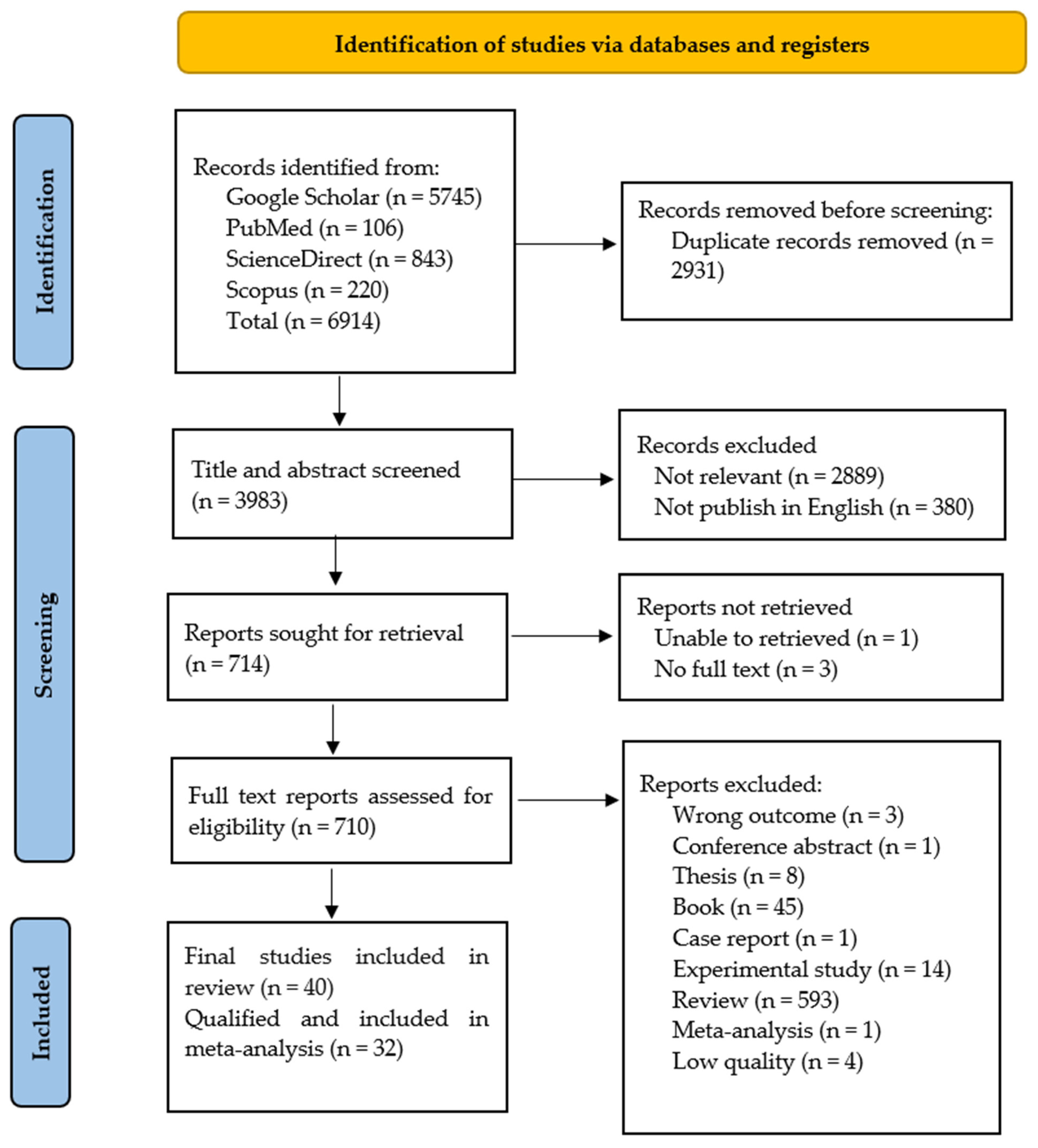
2.1. Summary of Literature Search

A total of 6914 articles were initially identified (Figure 1). Following the screening process, 710 articles were selected for further review. Finally, 40 articles were extensively reviewed. However, three of these articles contained few bacterial isolates (n~1–3 isolates each), one article presented varying and conflicting counts of isolates, and four articles did not specify the number of positive V. parahaemolyticus isolates [16,17,18,19,20,21,22,23].
Therefore, a total of 32 relevant studies from China (n = 8), Bangladesh (n = 4), Vietnam (n = 4), Malaysia (n = 4), India (n = 4), South Korea (n = 2), Cameroon (n = 1), Indonesia (n = 1), Iran (n = 1), Mexico (n = 1), Nigeria (n = 1), and the United Kingdom (n = 1) (Table 1) were selected. Shrimp species such as Litopenaeus vannamei, Penaeus vannamei, P. monodon, P. merguiensis, P. notialis, P. kerathurus, Parapaeopsis atlantica, Metapenaeus ensis, M. rosenbergii, and Oratosquilla oratoria were observed in this study.

2.2. Bacterial Isolation and Confirmation of V. parahaemolyticus Isolates

Bacterial isolation techniques were based on both national and international standards. The commonly available international standards for detection and confirmation of V. parahaemolyticus were the United States Food and Drug Administration Bacteriological Analytical Manual (U.S. FDA BAM), and the International Organization for Standardization (ISO 6222: 1999; ISO 4833-2: 2013; ISO 21872-1: 2017) [54,55,56]. The national standards included the Nation Standard Food Microbiological Examination V. parahaemolyticus (GB 4789.7-2013) and the Indonesia National Standard (SNI) 01-2332.5-2006, and available published papers, including Oanh et al. (2018), Blanco-Abad et al. (2009), Cowan and Steel’s Manual, Barrow and Feltham (1993), and Bergey (2005) in some studies [57,58,59,60,61,62]. Thiosulfate–citrate–bile salts–sucrose agar (TCBS) and Chromogenic agar were the most common media used for bacterial isolation.
For V. parahaemolyticus confirmation, colony morphology, Gram staining, and biochemical tests (namely, acetyl methyl carbinol production test, arginine dihydrolase test, catalase test, citrate test, dextrose utilization test, gelatinase test, growth at 0, 3, 6, 8, and 10% NaCl concentrations, growth at 4, 20, 30, 35, and 40 °C, halophilism test, hemolysis test, hydrogen sulfide production test, luminescent bacterial test, lysine decarboxylase test, methyl red (MR) test, Voges–Proskauer (VP) test, motility test, oxidase test, O-nitrophenyl beta-D-galactosidase (ONPG) test, ornithine decarboxylase test, indole test, oxidation fermentation (O-F) test, salt tolerance test, sulfur reduction (cysteine desulfurase) test, utilization of alpha ketoglutarate, urease test, triple sugar iron agar (TSI), as well as the commercial API 20E biochemical test kits (bioMérieux, Marcy-l’Etoile, France) were applied. In addition to PCR, MALDI-TOF and VITEK2 were employed by the studies for bacterial identification. Furthermore, 16S rRNA sequencing along with species-specific genes (tlh, tl, and toxR) detection were also utilized.

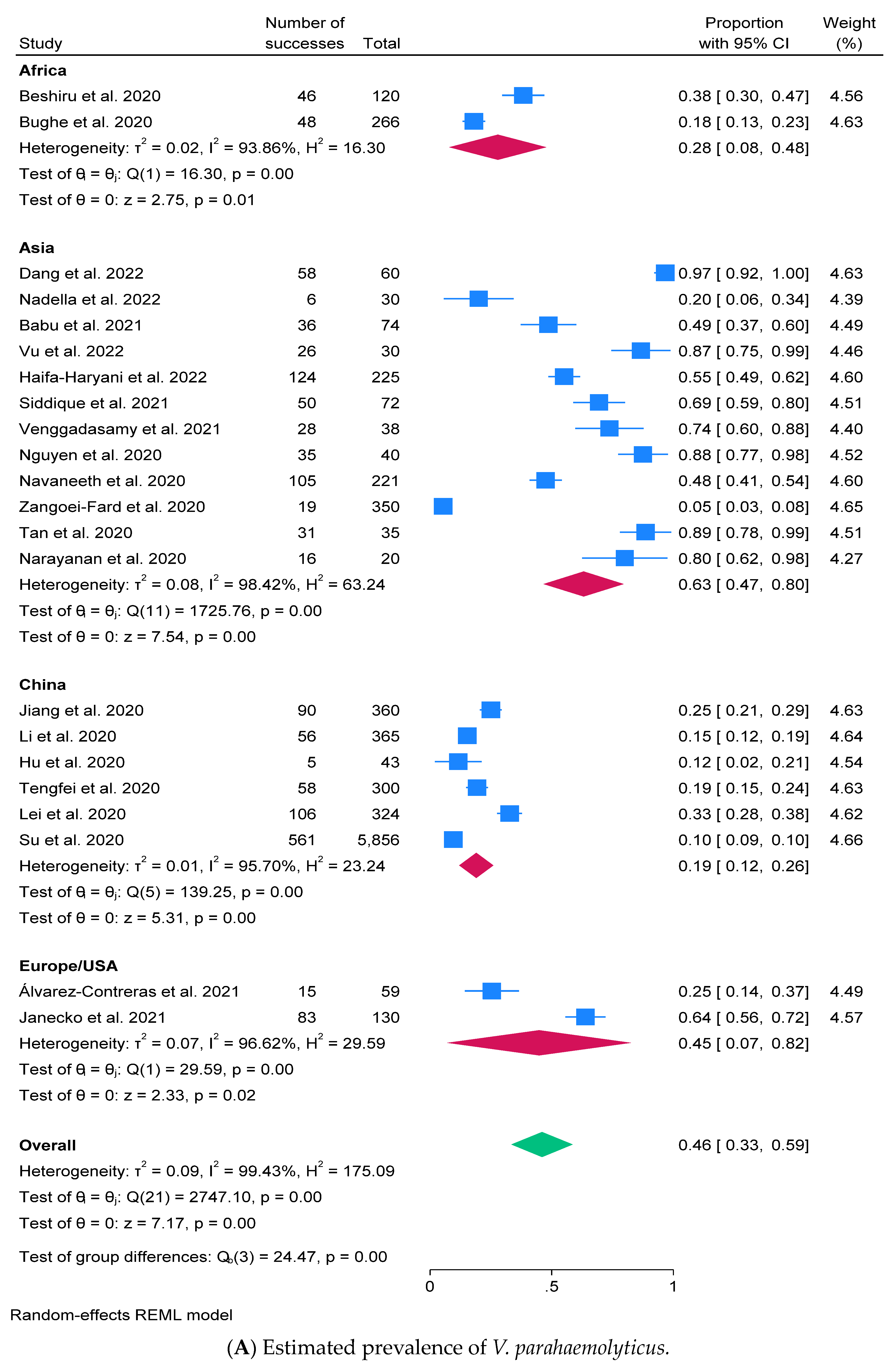
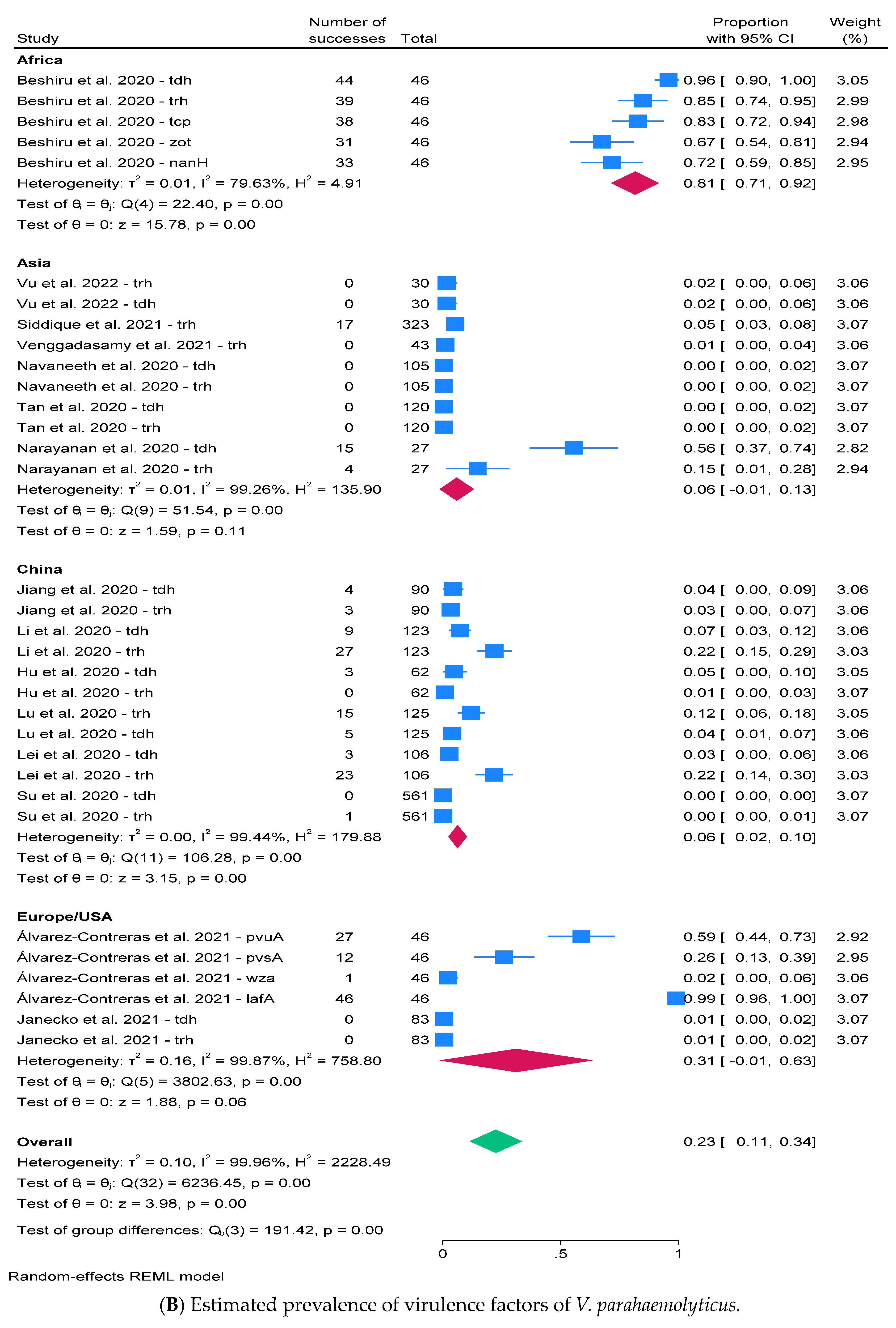
2.3. Prevalence of V. parahaemolyticus and Virulence Factors

The prevalence of V. parahaemolyticus in different anatomical parts of shrimp, such as hepatopancreas, hemolymph, gill, intestine, midgut, stomach, tissue or flesh, whole shrimp, and swab samples, were considered in the analysis. In addition to shrimp, some studies included other aquatic animals such as marine fish, oysters, crabs, shellfish, clams, mussels, tilapia, rui, and squid for examination. A subset of studies also included environmental samples, such as sediment, pond water, and reservoir water.
This comprehensive approach allowed for a robust examination of V. parahaemolyticus prevalence, considering both the random-effects model and the visualizations of study weights and bias assessment. The prevalence of V. parahaemolyticus was determined among 22 studies (22/32 = 68.8%) from 9018 samples/isolates, and the pooled estimate of V. parahaemolyticus prevalence in shrimp was 46.0% with a 95% C.I. (33.4–58.6%) and Q-statistic 2747.10 (Figure 2A). The overall heterogeneity was identified with tau2 = 0.088, I2 = 99.43%, H2 = 175.09, and p-value < 0.0001.
Although our study primarily provided descriptive presentations of the prevalence, virulence factors, and AMR of V. parahaemolyticus in shrimp populations, we recognize the potential advantages of conducting further analyses to extract additional insights from the dataset. As such, we have performed subgroup analyses to explore the relationship between variables more comprehensively and to identify potential determinants of V. parahaemolyticus prevalence and AMR in shrimp across different continents. Subgroup analysis was assessed based on continents—except China due to availability of several publications. The prevalence of V. parahaemolyticus was high in Asia (63.1% with C.I. = 46.7–79.5%), while China exhibited the lowest prevalence at 19.0% (C.I. = 12.0–26.0%). A funnel plot was employed for bias assessment, contributing to a comprehensive evaluation of the prevalence data (Figure 2C).
The analysis of virulence genes in V. parahaemolyticus within shrimp was conducted in 15 out of 32 articles (46.9%) from 3644 bacterial isolates. Virulence factors were limited to a specified set that included tdh, trh, pvuA, pvsA, wza, lafA, tcp, zot, nanH, pirA, and pirB. Among these, tdh and trh were identified as the most frequently detected virulence genes. The estimated prevalence of virulence factors was determined as 22.5% with a 95% C.I. of 11.4–33.6% (Figure 2B). The heterogeneity was observed using a Q value of 6236.45 with tau2 = 0.104, I2 = 99.96%, H2 = 2228.49, and p-value < 0.0001 (Figure 2B), and the bias assessment was indicated in a funnel plot (shown here in Figure 2D).

2.4. Antimicrobial Susceptibility Testing (AST)

Among 32 studies used (n = 29/32, 90.6%), the phenotypic antimicrobial susceptibility of V. parahaemolyticus was mostly tested by disk diffusion test (n = 23), broth microdilution method (n = 5), and both disk diffusion and broth microdilution (n = 1). The methods used for AST were followed by the Clinical and Laboratory Standards Institute (CLSI) (EP17, M45, and M100), European Committee on Antimicrobial Susceptibility Testing (EUCAST), and National Committee for Clinical Laboratory Standards (NCCLS). The interpretive criteria were as indicated according to the CLSI breakpoints and epidemiological cut-off values (ECOFFs) set by EUCAST.
Additionally, Escherichia coli ATCC 25922 (n = 12), V. parahaemolyticus ATCC 17802 (n = 2), both V. parahaemolyticus ATCC 17802 and 33847 (n = 2), both E. coli ATCC 25922 and V. parahaemolyticus ATCC 17802 (n = 1), both E. coli ATCC 25922 and Staphylococcus aureus ATCC 29213 (n = 1), V. parahaemolyticus DSM 11058 (n = 1), and V. parahaemolyticus (MTCC451, IMTCC, Chandigarh, India) (n = 1) were used as bacterial control strains, while 11 published studies did not mention any bacterial control strains.

2.5. Prevalence of AMR and Resistant Determinants

The occurrence of AMR in V. parahaemolyticus was investigated across 2164 isolates from 25 studies focusing on 16 antimicrobial classes. These antimicrobial classes comprised aminocyclitol, aminoglycosides, beta-lactams, carbapenems, cephalosporins, folate pathway inhibitors/sulfonamides, glycopeptides, lincosamides, macrolides, nitrofurans, oxazolidines, phenicols, polypeptides, quinolones, rifampicins, and tetracyclines (Table 2). A limited number of studies examined AMR in aminocyclitol (n = 1), glycopeptides (n = 3), lincosamides (n = 1), nitrofurans (n = 3), oxazolidines (n = 1), and rifamycins (n = 1).
The pooled estimate prevalence of phenotypic AMR in V. parahaemolyticus was categorized based on antimicrobial classes (Table 2). The predominant resistance phenotype examined was resistance to glycopeptides (i.e., vancomycin, novobiocin) (60.7% with 95% C.I. 19.6–101%, p < 0.0001), followed by nitrofurans (e.g., nitrofurantoin) (58.8% with 95% C.I. 17.8–99.8%, p < 0.0001), beta-lactams (e.g., amoxicillin, ampicillin, penicillin, etc.) (56.0% with 95% C.I. 44.6–67.5%, p < 0.0001), and polypeptides (e.g., bacitracin, colistin, polymyxin B) (31.2%, 95% C.I. 5.7–68.2%, p < 0.0001). However, exploration of resistance to carbapenems in China, folate pathway inhibitors/sulfonamides in Africa, Europe, and USA, and nitrofurans, aminocyclitol, and rifampicins at a global level was limited. The identification of bias was also conducted by examining funnel plots stratified by antimicrobial classes (see Figure S1).
Regarding analysis of AMR genes, one study from India conducted AMR genotypes of phenicols and tetracyclines [26]. Overall prevalence of phenicol resistance genes, including catI, catII, and florR, was estimated at 11.3% with a 95% C.I. of 3.3–19.3%, p = 0.865, while the higher prevalence was observed in tetracycline resistance genes (tetA, tetB, tetK, tetL, tetM, and tetS) at 18.1% with a 95% C.I. of 5.2–31.0%, p = 0.0191. In the same study, the estimated prevalence of mobile genetic elements was also observed to be 14.6% with 95% C.I. 3.5–25.7%, and the genes int1, int2, Tn1720, Tn1545, and Tn917 were examined. A bias assessment was conducted (Figure S2).

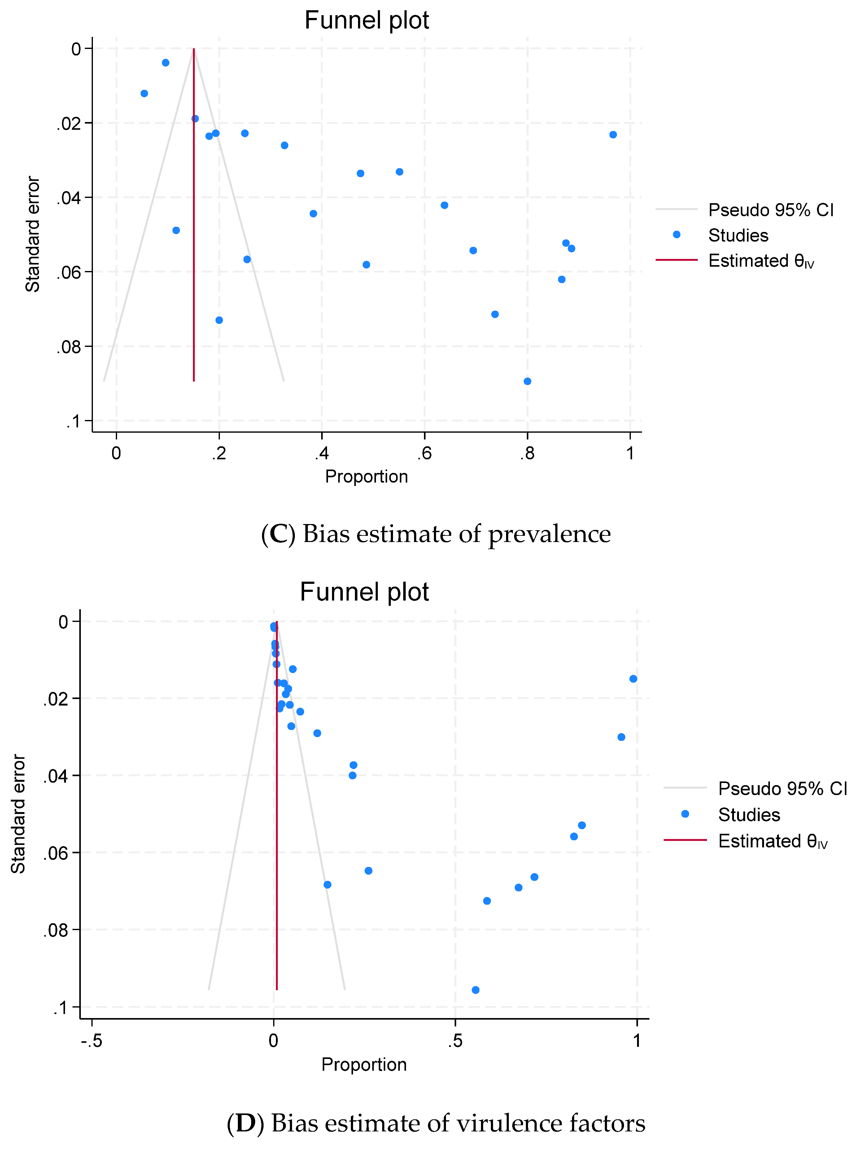
2.6. Publication Bias (Reporting Bias)

In this meta-analysis, publication bias was graphically assessed using visual funnel plots by the regression-based test of Egger at p < 0.05. On visual inspection, the funnel plot showed slight asymmetrical distribution in meta-analysis for V. parahaemolyticus, virulence factors, AMR, and their determinants (Figure 2C,D, Figures S1 and S2).

3. Discussion

This study comprehensively investigated the overall prevalence of V. parahaemolyticus, virulence factors, and AMR isolates in shrimp retrieved from 32 published articles. Given the considerable diversity in study designs and population characteristics across the included studies, a random-effects model was employed for meta-analysis. This study involved the inclusion of equal proportions of both retail and agricultural farms. The examination of subgroup meta-analysis was conducted with a focus on continents, revealing a heightened prevalence of V. parahaemolyticus in Asia at 57.0%, with China exhibiting the lowest prevalence at 19.0%. Nevertheless, there was a scarcity of studies from Europe and the USA. Shrimp presented a higher risk, with a prevalence rate of 3.2%, when contrasted with other decapod crustaceans such as lobster and crab, which had prevalence rates of 3.0% and 3.1%, respectively [63]. Consequently, it is imperative to conduct additional research to evaluate the prevalence of V. parahaemolyticus and its virulence factors, enhancing the representativeness of findings across these continents. The assessment of virulence factors in shrimp revealed that the predominant virulence factors detected were tdh and trh. Additionally, five studies performed serotyping for V. parahaemolyticus isolates, and the distribution of serotypes O3:K6, O5:KUT, O11:KUT, O3:KUT, O1:KUT, O3:K20, O1-O8, O10-O12, K25, K31, K64, and K68 was observed [33,42,44,48,49].
The primary technique used for AST involved a disk diffusion test, followed by a broth microdilution test. Notably, in South Korea, an automatic system such as Sensititre™ was used to perform broth microdilution [30,31]. The criteria for choosing antimicrobials for AST were not clearly outlined. However, it is recommended that antimicrobials should be selected based on AMR monitoring and surveillance guidelines by following CLSI standards for aquatic animals, humans, and animals and the OIE list of antimicrobial agents of veterinary importance [64,65,66]. While E. coli ATCC 25922 serves as the primary control strain for AST, about one-third of the studies did not explicitly mention the use of reference strains. This lack of clarity raises concerns about the reliability of the results in those studies, emphasizing the importance of proper reference controls in ensuring the accuracy of antimicrobial susceptibility assessments. In all the studies reviewed, the primary molecular method used for identifying resistance genes was predominantly PCR. However, the use of Whole-Genome Sequencing (WGS) was limited and not extensively utilized for this purpose.
This study observed that V. parahaemolyticus predominantly exhibited phenotypic resistance to glycopeptides, nitrofurans, and beta-lactams, a pattern similar to findings in a study on AMR Vibrio in marine bivalves [67]. Notably, these antimicrobials are not commonly used in shrimp farming practices. Contrasting results were found in studies on shrimp aquaculture in China and Vietnam, where oxytetracycline emerged as the primary antimicrobial and frequently used antimicrobial classes included tetracyclines, sulfonamides, and quinolones [68,69]. The inconsistency in these observations tentatively suggests that phenotypic AMR may not necessarily arise because of the selective pressure of antimicrobial applications on farms. Moreover, V. parahaemolyticus isolates with high resistance were not detectable in shrimp feed, implying a potential contribution from external sources of antimicrobials entering the farms. In human medicine, glycopeptides, nitrofurans, and beta-lactams are widely prescribed, and their resistance is observed in hospital wastewater worldwide [70]. To address the potential consumer risk associated with shrimp consumption, further studies are needed to investigate the sources of AMR contamination in shrimp farms, particularly focusing on environmental water.
Funnel plots were performed to evaluate publication bias and heterogeneity. Larger sample studies are clustered near the mean effect size at the top of the graph, contrasting with smaller-sample studies positioned at the bottom. The variability in effect size estimates within these smaller studies was influenced by sampling differences, evident in the graph’s distribution. Asymmetry in funnel plots indicated potential publication bias for the prevalence estimate of V. parahaemolyticus, virulence factors, and AMR. Notably, the funnel plot for V. parahaemolyticus prevalence suggests more heterogeneity with varied sample sizes than that from the occurrence of virulence factors. Regarding the funnel plot for V. parahaemolyticus prevalence, smaller studies with significant results were more likely to be published than those without significant virulence factors exhibited.
The previous studies referred to the CLSI standard for AST. Most of these studies adopted CLSI criteria originally developed for human and animal sources. Notably, only one CLSI document suggested nine antimicrobial classes for susceptibility tests specific to V. cholerae and other Vibrio spp. These classes included aminoglycosides, beta-lactams, carbapenems, cephalosporins, folate pathway inhibitors, macrolides, phenicols, quinolones, and tetracyclines with limited reference ranges [71]. Recent CLSI guidelines for bacteria isolated from aquatic animals do not provide any reference criteria for Vibrio spp. [65]. One study specifically examined the resistance of V. parahaemolyticus to medemycin, a newly developed macrolide antibiotic. This was because of the increased use of this drug due to its higher potency and low gastrointestinal irritation compared with other macrolides [72]. Results in our study suggested that harmonized protocols for antimicrobial resistance testing in aquaculture should be promptly initiated to achieve a valid global AMR assessment and prioritize urgent needs.
The limitations of this study were classified into two primary aspects: the review process and the evidence included in the review. The comprehensiveness of this study may be influenced by the chosen search strategies and time constraints. The potential introduction of bias could occur if other articles were not included in the study. Regarding the evidence incorporated in this study, the reliability of the included articles may exhibit variation concerning study design, sampling methods, and the approach to bacterial identification and selection criteria for performing AST. These variations could influence the overall robustness of the review and present challenges in the analysis of the data. Therefore, it is important to expand the pool of studies and continuously monitor the trends in V. parahaemolyticus and its resistant isolates. Further investigation in this regard is necessary to enhance our understanding and inform ongoing efforts to address potential public health implications.
In future research, it is essential to monitor and survey the trends in resistant V. parahaemolyticus isolates. This is crucial for evaluating the effectiveness of national and international frameworks or policies addressing AMR within the aquaculture sector. Such trend data derived from epidemiological studies will also contribute to a better understanding of the situation.

4. Materials and Methods

4.1. Study Selection

This systematic review was conducted following the Preferred Reporting Items for Systematic Reviews and Meta-Analysis (PRISMA) guidelines (Table S1) [67]. Different peer-reviewed journal articles published between 1 January 2020 and 31 December 2022 were selected based on inclusion and exclusion criteria. The prevalence of V. parahaemolyticus was calculated as the proportion of positive samples to the total number of samples, and the occurrence of virulence factors and AMR was expressed as the proportion of positive isolates to the total number of bacterial isolates. The primary focus of this study was on determining the prevalence of V. parahaemolyticus in shrimp from farms through retail outlets. Moreover, because of the significant losses in shrimp production caused by V. parahaemolyticus, only analyses involving this organism were taken into account, and data on any other aquatic animals were disregarded. This investigation specifically characterized the phenotypic and genotypic profiles of AMR, virulence factors, and resistance determinants in V. parahaemolyticus isolates obtained from these shrimp samples. To ensure the inclusion of publications, we implemented control measures involving cross-referencing between Web of Science (https://mjl.clarivate.com/search-results, accessed on 13 April 2024) and other databases. Furthermore, the words used in identification algorithms were clear and unique, increasing the likelihood of retrieving overlapping publications.

4.2. Eligibility Criteria

The articles that were considered eligible were peer-reviewed research papers published in reputable scientific journals listed in Google Scholar (https://scholar.google.com/, accessed on 13 April 2024), PubMed (https://pubmed.ncbi.nlm.nih.gov/, accessed on 13 April 2024), ScienceDirect (https://www.sciencedirect.com/, accessed on 13 April 2024), and Scopus (https://www.scopus.com/, accessed on 13 April 2024). All articles focused on pathogenicity and AMR characterization in V. parahaemolyticus as significant aspects of the study. The studies concerned strains of V. parahaemolyticus isolated from shrimp with exposure to AMR patterns, particularly phenotypic (e.g., disk diffusion and dilution methods) and genotypic (e.g., detection of resistance genes and determinants) assessments. Finally, the articles that were considered eligible were accessible for detailed analysis and reporting standards, including providing proper bacterial isolates; clear descriptions of the methods for bacterial isolation and confirmation, the method of antimicrobial susceptibility testing (AST), and the detection of virulence factors; and the results, discussions, and conclusions could be followed. These criteria were consistently applied during the article selection process.

4.3. Inclusion Criteria

This systematic review specifically targeted shrimp as the animal species of interest, with a primary focus on examining the presence of V. parahaemolyticus in shrimp. Included studies were those that examined the prevalence of virulence genes, AMR, or/and the associated resistance determinants in V. parahaemolyticus isolates derived from shrimp. Various methods were employed for the detection and confirmation of V. parahaemolyticus, including bacterial culture, biochemical tests, analytical profile index (API20E), and molecular techniques such as polymerase chain reaction (PCR) and Whole-Genome Sequencing. The AST method for detecting AMR phenotypes was also considered. Additionally, molecular techniques for identifying virulence genes, resistance genes, and their determinants were incorporated. The studies were limited to studies published in English.

4.4. Exclusion Criteria

Other aquatic animals, such as bivalves, fish, mollusks, and aquatic plants, were excluded from this study. Studies that did not report sample size, number of isolates, the method for bacterial isolation and confirmation, and AST were excluded from this study. Antimicrobials, including antifungals, antiseptics, and disinfectants, were also excluded from this study. Questionnaires, survey studies, and epidemiological studies without aquatic animals were also excluded. Studies that were not primary or original research but were based on review articles, abstracts, proceedings, short communications, correspondence, editorials, opinion articles, research theses, and book chapters were excluded. In addition, studies that were not published in the English language and were non-peer reviewed were not included.

4.5. Search Strategy

The search strategy was to explore the prevalence of bacteria, virulence factors, AMR, and resistant determinants among V. parahaemolyticus isolated from shrimp samples. Either the presence or absence of virulence factors, AMR, and resistant determinants of V. parahaemolyticus isolates was considered possible in the eligible criteria. The online databases searched were Google Scholar, PubMed, ScienceDirect, and Scopus. Searches on commercial websites, including Google and Yahoo, were performed using Boolean operators such as AND, OR, and NOT in conjunction to combine keywords in database searches (Table S2) [73].
The identification algorithm for the relevant literature was listed using the following keywords: V. parahaemolyticus, antimicrobial resistance, AMR, antibiotic resistance, AST, resistance profile, virulence factors (TDH, TRH, and other pathogenicity), and resistance determinants (integrons, integrative and conjugative elements (ICEs), and plasmid), specifically in shrimp, from farm to retail. Several keyword combinations were also used for each topic area to ensure as many articles as possible were captured within the specified timeframe and subject area.
This systematic review followed the PRISMA guidelines and a completed PRISMA 2020 checklist is provided (Table S1). Complete publications were reviewed by three veterinarians and one microbiologist. Any discrepancies or uncertainties were resolved through discussion and consensus among the reviewers. Additionally, we ensured the verification and credibility of the selected articles by cross-referencing them with reputable scientific journals listed in the databases. Subsequently, all articles that met the inclusion criteria were consolidated, and duplication detection was performed. Publications that met the inclusion criteria were classified by bacterial isolation and confirmation of V. parahaemolyticus from shrimp, number of total and positive samples and isolates, country, method of AST, and molecular detection of resistance genes and their determinants. All data with full details of the search strategy and results were independently extracted from materials and methods, results, discussions, figures, and tables by two individuals (V.T. and S.A.) and double-checked by the third investigator (S.J.), and the qualified articles were compiled in Microsoft Excel for further data analysis.

4.6. Data Collection and Assessment of Data Quality

Inclusion and exclusion criteria were used to detect the possible publications relevant to this study. The primary outcome measures were based on the prevalence of V. parahaemolyticus and contained virulence factors, phenotypic and genotypic AMR, and other genetic determinants. All eligible published articles underwent extraction, encompassing essential details such as the article title, author names, publication year, country, duration of data collection, shrimp species, shrimp status, total samples/bacterial isolates, number of positive samples/isolates, virulence genes, resistance phenotypes and genotypes, as well as their determinants. Additionally, other variables included were the detection and confirmation of V. parahaemolyticus, method for AST, bacterial control strains, and molecular techniques used for detection of resistance genes and their genetic determinants.

4.7. Statistical Analyses

The extracted data were stored in a Microsoft Excel spreadsheet and imported into STATA program version 18.0 (StataCorp, College Station, TX, USA). An observational study for one-sample binary outcome summaries of the prevalence of V. parahaemolyticus, virulence factors, AMR, and their determinants isolated from shrimp was examined. The prevalence was calculated based on the proportion of positive samples/isolates to number of total samples/isolates. The data are expressed as a pooled estimate of the prevalence of V. parahaemolyticus, virulence factors, resistance to each antimicrobial class, and determinants. Random-effects meta-analyses were conducted, and the summary weighted average proportion (effect size) was calculated based on the individual effect sizes and 95% confidence intervals (C.I.). Heterogeneity between studies of prevalence estimates among the studies was investigated using Q-statistics with I2 Index [74,75]. A possible source of heterogeneity among observed studies was assessed through subgroup analysis based on the continent of the source of samples.
The visual assessment of publication or dissemination bias was performed using a funnel plot for asymmetry with a pooled estimate. Egger’s tests were employed for detecting publication bias. Studies were included in the meta-analyses if they met the criteria, including at least 20 isolates for all analyzed studies and at least three studies for each meta-analysis. All analyses were two-tailed tests, with statistical significance set at a p-value < 0.05. The meta-analyses utilized a random-effects model in STATA software.

5. Conclusions

In conclusion, this comprehensive systematic review and meta-analysis has presented the prevalence of V. parahaemolyticus in shrimp, along with its associated virulence factors and AMR profiles. The study revealed a significant prevalence of V. parahaemolyticus in shrimp samples globally, with Asia exhibiting particularly high rates. Notably, virulence genes, such as tdh and trh, were frequently identified, indicating the potential pathogenicity of V. parahaemolyticus. Our study highlighted the levels of phenotypic AMR, revealing significant resistance against glycopeptides, nitrofurans, and beta-lactams. Furthermore, the investigation identified gaps in AMR surveillance and control measures, emphasizing the need for harmonized protocols and enhanced monitoring efforts. This finding showed the importance of ongoing research and collaborative interventions to address the growing threat of AMR in V. parahaemolyticus, to ensure public health safety, and to promote sustainable aquaculture practices.

Supplementary Materials

The following supporting information can be downloaded at: https://www.mdpi.com/article/10.3390/antibiotics13040370/s1, Table S1. Completed PRISMA 2020 checklist. Table S2. Search algorithms and an example of key terms used for identification of relevant publications in V. parahaemolyticus. Table S3: AMR and its determinants of V. parahaemolyticus isolated from shrimp among relevant studies (n = 32). Figure S1. Funnel plots of prevalence of AMR in V. parahaemolyticus in shrimp to assess potential bias stratified by antimicrobial classes: (A) aminoglycosides; (B) beta-lactamases; (C) carbapenems; (D) cephalosporins; (E) folate pathway inhibitors/sulfonamides; (F) glycopeptides; (G) macrolides; (H) nitrofurans; (I) phenicols; (J) polypeptides; (K) quinolones; (L) tetracyclines. A red vertical line represents the overall prevalence from meta-analysis, and the diagonal lines provide 95% C.I. Figure S2. The meta-analysis of prevalence of resistance determinants from V. parahaemolyticus isolates in shrimp using random-effects model with a 95% C.I.: (A) a forest plot representing the results in meta-analysis with size squares proportional to the weight assigned to the study; (B) a funnel plot of occurrence of resistance determinants in V. parahaemolyticus to assess potential bias.

Author Contributions

Conceptualization, S.J.; methodology and validation, V.T., J.O.O. and S.A.; formal analysis, V.T. and S.J.; investigation, S.J.; resources, S.J.; data curation, V.T. and S.A.; writing—original draft preparation, V.T., J.O.O. and S.A.; writing—review and editing, S.J.; supervision, S.J.; funding acquisition, S.J. All authors have read and agreed to the published version of the manuscript.

Funding

This study received support from Chulalongkorn University, grant number ReinUni_65_02_31_42, and the National Research Council of Thailand (NRCT) Project ID N42A660897.

Institutional Review Board Statement

Not applicable.

Informed Consent Statement

Not applicable.

Data Availability Statement

Data used in the manuscript for tables and figures are presented in the accompanying Supplementary Materials.

Conflicts of Interest

The authors declare no conflicts of interest.

References

  1. World Health Organization (WHO). Antimicrobial Resistance: Global Report on Surveillance. 2021. Available online: https://www.who.int/publications/i/item/9789241564748 (accessed on 5 December 2023).
  2. Prestinaci, F.; Pezzotti, P.; Pantosti, A. Antimicrobial resistance: A global multifaceted phenomenon. Pathog. Glob. Health 2015, 109, 309–318. [Google Scholar] [CrossRef]
  3. Boyd, E.F.; Cohen, A.L.; Naughton, L.M.; Ussery, D.W.; Binnewies, T.T.; Stine, O.C.; Parent, M.A. Molecular analysis of the emergence of pandemic Vibrio parahaemolyticus. BMC Microbiol. 2008, 8, 110. [Google Scholar] [CrossRef]
  4. Hong To, T.T.; Yanagawa, H.; Khanh Thuan, N.; Hiep, D.M.; Cuong, D.V.; Khai, L.T.L.; Taniguchi, T.; Kubo, R.; Hayashidani, H. Prevalence of Vibrio parahaemolyticus causing acute hepatopancreatic necrosis disease of shrimp in shrimp, molluscan shellfish and water samples in the Mekong Delta, Vietnam. Biology 2020, 9, 312. [Google Scholar] [CrossRef]
  5. Khimmakthong, U.; Sukkarun, P. The spread of Vibrio parahaemolyticus in tissues of the Pacific white shrimp Litopenaeus vannamei analyzed by PCR and histopathology. Microb. Pathog. 2017, 113, 107–112. [Google Scholar] [CrossRef] [PubMed]
  6. Abdel-Latif, H.M.; Yilmaz, E.; Dawood, M.A.; Ringø, E.; Ahmadifar, E.; Yilmaz, S. Shrimp vibriosis and possible control measures using probiotics, postbiotics, prebiotics, and synbiotics: A review. Aquaculture 2022, 551, 737951. [Google Scholar] [CrossRef]
  7. Fujino, T.; Okuno, Y.; Nakada, D.; Aoyama, A.; Mukai, T.; Ueho, T. On the bacteriological examination of shirasu food poisoning. Med. J. Osaka Univ. 1953, 4, 299–304. [Google Scholar]
  8. Iwamoto, M.; Ayers, T.; Mahon, B.E.; Swerdlow, D.L. Epidemiology of seafood-associated infections in the United States. Clin. Microbiol. Rev. 2010, 23, 399–411. [Google Scholar] [CrossRef]
  9. Janekrongtham, C.; Dejburum, P.; Sujinpram, S.; Rattanathumsakul, T.; Swaddiwudhipong, W. Outbreak of seafood-related food poisoning from undetectable Vibrio parahaemolyticus-like pathogen, Chiang Mai Province, Thailand, December 2020. Trop. Med. Int. Health 2022, 27, 92–98. [Google Scholar] [CrossRef]
  10. Chowdhury, A.; Ishibashi, M.; Thiem, V.D.; Tuyet, D.T.N.; Tung, T.V.; Chien, B.T.; von Seidlein, L.; Canh, D.G.; Clemens, J.; Trach, D.D.; et al. Emergence and serovar transition of Vibrio parahaemolyticus pandemic strains isolated during a diarrhea outbreak in Vietnam between 1997 and 1999. Microbiol. Immunol. 2004, 48, 319–327. [Google Scholar] [CrossRef]
  11. Li, Y.J.; Yang, Y.F.; Zhou, Y.J.; Zhang, R.H.; Liu, C.W.; Liu, H.; Li, X.G.; Chen, W.; Chen, Y.; Wu, Y.N. Estimating the burden of foodborne gastroenteritis due to nontyphoidal Salmonella enterica, Shigella and Vibrio parahaemolyticus in China. PLoS ONE 2022, 17, e0277203. [Google Scholar] [CrossRef]
  12. Martinez-Urtaza, J.; Trinanes, J.; Abanto, M.; Lozano-Leon, A.; Llovo-Taboada, J.; Garcia-Campello, M.; Pousa, A.; Powell, A.; Baker-Austin, C.; Gonzalez-Escalona, N. Epidemic dynamics of Vibrio parahaemolyticus illness in a hotspot of disease emergence, Galicia, Spain. Emerg. Infect. Dis. 2018, 24, 852–859. [Google Scholar] [CrossRef]
  13. Harth, E.; Matsuda, L.; Hernández, C.; Rioseco, M.L.; Romero, J.; González-Escalona, N.; Martínez-Urtaza, J.; Espejo, R.T. Epidemiology of Vibrio parahaemolyticus outbreaks, southern Chile. Emerg. Infect. Dis. 2009, 15, 163–168. [Google Scholar] [CrossRef]
  14. Raghunath, P. Roles of thermostable direct hemolysin (TDH) and TDH-related hemolysin (TRH) in Vibrio parahaemolyticus. Front. Microbiol. 2015, 5, 805. [Google Scholar] [CrossRef]
  15. Elmahdi, S.; DaSilva, L.V.; Parveen, S. Antibiotic resistance of Vibrio parahaemolyticus and Vibrio vulnificus in various countries: A review. Food Microbiol. 2016, 57, 128–134. [Google Scholar] [CrossRef]
  16. Prabina, D.; Swaminathan, T.R.; Mohandas, S.P.; Anjana, J.C.; Manjusha, K.; Preena, P.G. Investigation of antibiotic-resistant vibrios associated with shrimp (Penaeus vannamei) farms. Arch. Microbiol. 2023, 205, 41. [Google Scholar] [CrossRef]
  17. Khan, M.; Paul, S.I.; Rahman, M.M.; Lively, J.A. Antimicrobial resistant bacteria in shrimp and shrimp farms of Bangladesh. Water 2022, 14, 3172. [Google Scholar] [CrossRef]
  18. Paria, P.; Behera, B.K.; Mohapatra, P.K.D.; Parida, P.K. Virulence factor genes and comparative pathogenicity study of tdh, trh and tlh positive Vibrio parahaemolyticus strains isolated from Whiteleg shrimp, Litopenaeus vannamei (Boone, 1931) in India. Infect. Genet. Evol. 2021, 95, 105083. [Google Scholar] [CrossRef]
  19. Yasin, A.; Begum, K.; Eshik, M.E.; Punom, N.J.; Ahmmed, S.; Rahman, M.S. Molecular identification and antibiotic resistance patterns of diverse bacteria associated with shrimp PL nurseries of Bangladesh: Suspecting Acinetobacter venetianus as future threat. PeerJ 2022, 10, e12808. [Google Scholar] [CrossRef]
  20. Zhang, X.; Sun, J.; Chen, F.; Qi, H.; Chen, L.; Sung, Y.Y.; Huang, Y.; Lv, A.; Hu, X. Phenotypic and genomic characterization of a Vibrio parahaemolyticus strain causing disease in Penaeus vannamei provides insights into its niche adaptation and pathogenic mechanism. Microb. Genom. 2021, 7, 000549. [Google Scholar] [CrossRef]
  21. Yu, L.H.; The, C.S.J.; Yap, K.P.; Ung, E.H.; Thong, K.L. Comparative genomic provides insight into the virulence and genetic diversity of Vibrio parahaemolyticus associated with shrimp acute hepatopancreatic necrosis disease. Infect. Genet. Evol. 2020, 83, 104347. [Google Scholar] [CrossRef]
  22. Quang, H.T.; Lan, T.T.; Hai, T.T.H.; Yen, P.T.H.; Van, T.Q.K.; Tung, H.T.; Binh, M.N.; Son, N.K.H.; Linh, N.Q.; Tram, N.D.Q. Genetic diversity and toxic genes analysis of vibrio spp. isolated from white leg shrimp and marine fishes cultured in Tam G iang lagoon in Thua Thien Hue province, Vietnam. Indian J. Sci. Technol. 2020, 13, 1412–1422. [Google Scholar] [CrossRef]
  23. Han, J.E.; Choi, S.-K.; Han, S.-H.; Lee, S.C.; Jeon, H.J.; Lee, C.; Kim, K.Y.; Lee, Y.S.; Park, S.C.; Rhee, G.; et al. Genomic and histopathological characteristics of Vibrio parahaemolyticus isolated from an acute hepatopancreatic necrosis disease outbreak in Pacific white shrimp (Penaeus vannamei) cultured in Korea. Aquaculture 2020, 524, 735284. [Google Scholar] [CrossRef]
  24. Mulya, M.A.; Pasaribu, F.H.; Afiff, U.; Yuhana, M. Characterization and molecular detection of pathogenicity and antibiotic resistance genes in Vibrio parahaemolyticus isolated from pacific white shrimp. J. Akuakultur Indones. 2022, 21, 81–92. [Google Scholar] [CrossRef]
  25. Dang, T.H.O.; Truong, Q.P.; Nguyen, T.P. Antibacterial resistance of Vibrio parahaemolyticus isolated from shrimp farms located in east coastal region of the Mekong Delta, Vietnam. CTU J. Sci. 2022, 14, 1–7. [Google Scholar] [CrossRef]
  26. Nadella, R.K.; Panda, S.K.; Badireddy, M.R.; Kurcheti, P.P.; Raman, R.P.; Mothadaka, M.P. Multi-drug resistance, integron and transposon-mediated gene transfer in heterotrophic bacteria from Penaeus vannamei and its culture environment. Environ. Sci. Pollut. Res. 2022, 29, 37527–37542. [Google Scholar] [CrossRef]
  27. Babu, B.; Sathiyaraj, G.; Mandal, A.; Kandan, S.; Biju, N.; Palanisamy, S.; You, S.; Nisha, R.G.; Prabhu, N.M. Surveillance of disease incidence in shrimp farms located in the east coastal region of India and in vitro antibacterial efficacy of probiotics against Vibrio parahaemolyticus. J. Invertebr. Pathol. 2021, 179, 107536. [Google Scholar] [CrossRef]
  28. Vu, T.T.T.; Hoang, T.T.H.; Fleischmann, S.; Pham, H.N.; Lai, T.L.H.; Cam, T.T.H.; Truong, L.O.; Dac Cam Phung, V.P.L.; Alter, T. Quantification and antimicrobial resistance of Vibrio parahaemolyticus in retail seafood in Hanoi, Vietnam. J. Food Protect. 2022, 85, 786–791. [Google Scholar] [CrossRef]
  29. Haifa-Haryani, W.O.; Amatul-Samahah, M.A.; Azzam-Sayuti, M.; Chin, Y.K.; Zamri-Saad, M.; Natrah, I.; Amal, M.N.A.; Satyantini, W.H.; Ina-Salwany, M.Y. Prevalence, antibiotics resistance and plasmid profiling of Vibrio spp. isolated from cultured shrimp in Peninsular Malaysia. Microorganisms 2022, 10, 1851. [Google Scholar] [CrossRef]
  30. Kim, D.H.; Rajapaksha, L.G.T.G.; Gunasekara, C.W.R.; Wimalasena, S.H.M.P.; Pathirana, H.N.K.S.; Kim, S.R.; Seo, B.J.; Heo, G.J.; Shin, G.W. Phylogenetic relationships and antibiotic resistance of Vibrio parahaemolyticus isolates related to acute hepatopancreatic necrosis disease in Korea. Aquaculture 2021, 545, 737253. [Google Scholar] [CrossRef]
  31. Mok, J.S.; Cho, S.R.; Park, Y.J.; Jo, M.R.; Ha, K.S.; Kim, P.H.; Kim, M.J. Distribution and antimicrobial resistance of Vibrio parahaemolyticus isolated from fish and shrimp aquaculture farms along the Korean coast. Mar. Pollut. Bull. 2021, 171, 112785. [Google Scholar] [CrossRef]
  32. Jin, J.; Zhou, Y.; Zhang, Z.; Wang, H.; Hou, W.; Wang, H.; Li, R.; Zhou, M. Characteristics of antimicrobial-resistant Vibrio parahaemolyticus strains and identification of related antimicrobial resistance gene mutations. Foodborne Pathog. Dis. 2021, 18, 873–879. [Google Scholar] [CrossRef]
  33. Siddique, A.B.; Moniruzzaman, M.; Ali, S.; Dewan, M.N.; Islam, M.R.; Islam, M.S.; Amin, M.B.; Mondal, D.; Parvez, A.K.; Mahmud, Z.H. Characterization of pathogenic Vibrio parahaemolyticus isolated from fish aquaculture of the southwest coastal area of Bangladesh. Front. Microbiol. 2021, 12, 635539. [Google Scholar] [CrossRef]
  34. Álvarez-Contreras, A.K.; Quiñones-Ramírez, E.I.; Vázquez-Salinas, C. Prevalence, detection of virulence genes and antimicrobial susceptibility of pathogen Vibrio species isolated from different types of seafood samples at “La Nueva Viga” market in Mexico City. Antonie Leeuwenhoek 2021, 114, 1417–1429. [Google Scholar] [CrossRef]
  35. Janecko, N.; Bloomfield, S.J.; Palau, R.; Mather, A.E. Whole genome sequencing reveals great diversity of Vibrio spp. in prawns at retail. Microb. Genom. 2021, 7, 000647. [Google Scholar] [CrossRef]
  36. Venggadasamy, V.; Tan, L.T.H.; Law, J.W.F.; Ser, H.L.; Letchumanan, V.; Pusparajah, P. Incidence, antibiotic susceptibility and characterization of Vibrio parahaemolyticus isolated from seafood in Selangor, Malaysia. Prog. Microbes Mol. Biol. 2021, 4, 1–34. [Google Scholar] [CrossRef]
  37. Nguyen, T.P.Y.; Nguyen, T.N.; Nguyen, T.B.V.; Nguyen, V.C.; Le, T.T.C.; Huynh, N.T.; Chu, V.T.; Nguyen, D.T.; Nguyen, P.H.L.; James, C.; et al. Antimicrobial residues, non-typhoidal Salmonella, Vibrio spp. and associated microbiological hazards in retail shrimps purchased in Ho Chi Minh city (Vietnam). Food Control 2020, 107, 106756. [Google Scholar]
  38. Navaneeth, K.A.; Bhuvaneswari, T.; Rajan, J.J.S.; Alavandi, S.V.; Vijayan, K.K.; Otta, S.K. Characterization of Vibrio parahaemolyticus isolates from shrimp farms of Southeast coast of India with special reference to Acute Hepatopancreatic Necrosis Disease (AHPND) status. Aquaculture 2020, 518, 734813. [Google Scholar] [CrossRef]
  39. Jiang, H.; Yu, T.; Yang, Y.; Yu, S.; Wu, J.; Lin, R.; Li, Y.; Fang, J.; Zhu, C. Co-occurrence of antibiotic and heavy metal resistance and sequence type diversity of Vibrio parahaemolyticus isolated from Penaeus vannamei at freshwater farms, seawater farms, and markets in Zhejiang province, China. Front. Microbiol. 2020, 11, 1294. [Google Scholar] [CrossRef]
  40. Beshiru, A.; Okareh, O.T.; Okoh, A.I.; Igbinosa, E.O. Detection of antibiotic resistance and virulence genes of Vibrio strains isolated from ready-to-eat shrimps in Delta and Edo States, Nigeria. J. Appl. Microbiol. 2020, 129, 17–36. [Google Scholar] [CrossRef]
  41. Rahman, M.S.; Eshik, M.E.; Punom, N.J.; Abedin, M.; Begum, M.K. Diversity of Vibrio species and their antibiotic resistance patterns in black tiger shrimp Penaeus monodon Fabricius, 1798 cultured in South-West region of Bangladesh. Asian Fish. Sci. 2020, 33, 330–340. [Google Scholar] [CrossRef]
  42. Li, Y.; Xie, T.; Pang, R.; Wu, Q.; Zhang, J.; Lei, T.; Xue, L.; Wu, H.; Wang, J.; Ding, Y.; et al. Food-borne Vibrio parahaemolyticus in China: Prevalence, antibiotic susceptibility, and genetic characterization. Front. Microbiol. 2020, 16, 1670. [Google Scholar] [CrossRef] [PubMed]
  43. Zangoei-Fard, S.; Rahimi, E.; Shakerian, A. Incidence and phenotypic pattern of antibiotic resistance of Vibrio species isolated from seafood samples caught from the Persian Gulf. Egypt. J. Vet. Sci. 2020, 51, 337–347. [Google Scholar] [CrossRef]
  44. Hu, Y.; Li, F.; Zheng, Y.; Jiao, X.; Guo, L. Isolation, molecular characterization and antibiotic susceptibility pattern of Vibrio parahaemolyticus from aquatic products in the Southern Fujian coast, China. J. Microbiol. Biotechnol. 2020, 30, 856–867. [Google Scholar] [CrossRef] [PubMed]
  45. Lu, Y.; Yang, L.; Meng, J.; Zhao, Y.; Song, Y.; Zhu, Y.; Ou, J.; Pan, Y.; Liu, H. Microevolution of Vibrio parahaemolyticus isolated from clinical, acute hepatopancreatic necrosis disease infecting shrimps, and aquatic production in China. Microb. Environ. 2020, 35, ME19095. [Google Scholar] [CrossRef] [PubMed]
  46. Bughe, R.N.; Oben, B.O.; Nji, A.M.; Chedjou, J.P.K.; Mbange, A.E.; Ali, I.M.; Oben, P.M.; Mbacham, W.F. Occurrence and antibiotics susceptibility of Vibrio sp. From Penaeid shrimps from Kribi Coastal Water, Cameroon. Int. J. Adv. Res. Biol Sci. 2020, 7, 96–111. [Google Scholar]
  47. Tan, C.W.; Rukayadi, Y.; Hasan, H.; Thung, T.Y.; Lee, E.; Rollon, W.D.; Hara, H.; Kayali, A.Y.; Nishibuchi, M.; Radu, S. Prevalence and antibiotic resistance patterns of Vibrio parahaemolyticus isolated from different types of seafood in Selangor, Malaysia. Saudi J. Biol. Sci. 2020, 27, 1602–1608. [Google Scholar] [CrossRef] [PubMed]
  48. Tengfei, X.; Quan, Y.; Xiong, T.; Jing, Z.; Xujun, H. Prevalence, antibiotic susceptibility and characterization of Vibrio parahaemolyticus isolates in China. FEMS Microbiol. Lett. 2020, 367, fnaa136. [Google Scholar]
  49. Lei, T.; Jiang, F.; He, M.; Zhang, J.; Zeng, H.; Chen, M.; Pang, R.; Wu, S.; Wei, L.; Wang, J.; et al. Prevalence, virulence, antimicrobial resistance, and molecular characterization of fluoroquinolone resistance of Vibrio parahaemolyticus from different types of food samples in China. Int. J. Food Microbiol. 2020, 317, 108461. [Google Scholar] [CrossRef] [PubMed]
  50. Amin, A.A.; Shipa, S.A.; Naser, N.; Miah, F. Surveillance of Vibrio spp. in Penaeus monodon collected from shrimp pond of Satkhira, Bangladesh. J. Zoo. Res. 2020, 2, 1–8. [Google Scholar] [CrossRef]
  51. Narayanan, S.V.; Joseph, T.C.; Peeralil, S.; Koombankallil, R.; Vaiyapuri, M.; Mothadaka, M.P.; Lalitha, K.V. Tropical shrimp aquaculture farms harbour pathogenic Vibrio parahaemolyticus with high genetic diversity and carbapenem resistance. Mar. Pollut. Bull. 2020, 160, 111551. [Google Scholar] [CrossRef]
  52. Amatul-Samahah, M.A.; Muthukrishnan, S.; Omar, W.H.H.W.; Ikhsan, N.F.M.; Ina-Salwany, M.Y.  Vibrio sp. Associated with acute hepatopancreatic necrosis disease (AHPND) found in penaeid shrimp pond from east cost of peninsular Malaysia. J. Environ. Biol. 2020, 41, 1160–1170. [Google Scholar] [CrossRef]
  53. Su, C.; Chen, L. Virulence, resistance, and genetic diversity of Vibrio parahaemolyticus recovered from commonly consumed aquatic products in Shanghai, China. Mar. Pollut. Bull. 2020, 160, 111554. [Google Scholar] [CrossRef] [PubMed]
  54. ISO 6222: 1999; Water Quality—Enumeration of Culturable Micro-Organisms-Colony Count by Inoculation in a Nutrient Agar Culture Medium, 2nd ed. International Organization for Standardization: Geneva, Switzerland, 1999.
  55. ISO 4833–2:2013; Microbiology of the Food Chain—Horizontal Method for the Enumeration of Microorganisms—Part 2: Colony Count at 30 °C by the Surface Plating Technique. International Organization for Standardization: Geneva, Switzerland, 2013.
  56. ISO 21872–1:2017; Microbiology of the Food Chain—Horizontal Method for the Determination of Vibrio spp.—Part 1: Detection of Potentially Enteropathogenic Vibrio parahaemolyticus, Vibrio cholerae and Vibrio vulnificus. International Organization for Standardization: Geneva, Switzerland, 2017.
  57. GB 4789.7-2013; National Food Safety Standard—Food Microbiological Examination—Vibrio parahaemolyticus. National Health and Family Planning Commission of China: Beijing, China, 2013.
  58. SNI 01-2332.5-2006; Cara uji Mikrobiologi—Bagian 5: Penentuan Vibrio parahaemolitycus Pada Produk Perikanan. Badan Standar Nasional: Jakarta, Indonesia, 2006.
  59. Oanh, D.T.H.; Nguyen, T.N.; Tran, V.T.; Bondad-Reantaso, M.G. Identification and characterization of Vibrio bacteria isolated from shrimp infected with early mortality syndrome/acute hepatopancreatic necrosis syndrome (EMS/AHPNS) in Vietnam. Asian Fish. Sci. 2018, 31S, 283–292. [Google Scholar] [CrossRef]
  60. Blanco-Abad, V.; Ansede-Bermejo, J.; Rodriguez-Castro, A.; Martinez-Urtaza, J. Evaluation of different procedures for the optimized detection of Vibrio parahaemolyticus in mussels and environmental samples. Int. J. Food Microbiol. 2009, 129, 229–236. [Google Scholar] [CrossRef] [PubMed]
  61. Barrow, G.I.; Feltham, R.K.A. Cowan and Steel’s Manual for the Identification of Medical Bacteria, 3rd ed.; Cambridge University Press: London, UK, 1993. [Google Scholar]
  62. Bergey, D.H.; Holt, J.G. Bergey’s Manual of Systemic Bacteriology, Volume 2: The Proteobacteria, Part B: The Gammaproteobacteria; Springer: New York, NY, USA, 2005. [Google Scholar]
  63. Raissy, M.; Moumeni, M.; Ansari, M.; Rahimi, E. Occurrence of Vibrio spp. in lobster and crab from the Persian Gulf. J Food Saf. 2012, 32, 198–203. [Google Scholar] [CrossRef]
  64. OIE. List of Antimicrobials of Veterinary Importance. Available online: https://www.oie.int/fileadmin/Home/eng/Internationa_Standard_Setting/docs/pdf/OIE_list_antimicrobials.pdf (accessed on 5 December 2023).
  65. Clinical and Laboratory Standards Institute (CLSI). Methods for Antimicrobial Broth Dilution and Disk Diffusion Susceptibility Testing of Bacteria Isolated from Aquatic Animals, 2nd ed.; CLSI Guideline VET03; Clinical and Laboratory Standard Institute: Wayne, PA, USA, 2022. [Google Scholar]
  66. Clinical and Laboratory Standards Institute (CLSI). Performance Standards for Antimicrobial Susceptibility Testing, 30th ed.; CLSI Guideline M100-S30; Clinical and Laboratory Standard Institute: Wayne, PA, USA, 2020. [Google Scholar]
  67. Albini, E.; Orso, M.; Cozzolino, F.; Sacchini, L.; Leoni, F.; Magistrali, C.F. A systematic review and meta-analysis on antimicrobial resistance in marine bivalves. Front. Microbiol. 2022, 13, 1040568. [Google Scholar] [CrossRef]
  68. Li, F.; Huang, J.; Wang, M.; Chen, L.; Xiao, Y. Sources, distribution and dynamics of antibiotics in Litopenaeus vannamei farming environment. Aquaculture 2021, 545, 737200. [Google Scholar] [CrossRef]
  69. Luu, Q.H.; Nguyen, T.B.T.; Nguyen, T.L.A.; Do, T.T.T.; Dao, T.H.T.; Padungtod, P. Antibiotics use in fish and shrimp farms in Vietnam. Aquac. Rep. 2021, 20, 100711. [Google Scholar] [CrossRef]
  70. Zhang, S.; Huang, J.; Zhao, Z.; Cao, Y.; Li, B. Hospital wastewater as a reservoir for antibiotic resistance genes: A meta-analysis. Front. Public Health 2020, 8, 574968. [Google Scholar] [CrossRef]
  71. Clinical Laboratory Standard Institute (CLSI). Methods for Antimicrobial Dilution and Disk Susceptibility Testing of Infrequently Isolated or Fastidious Bacteria, 3rd ed.; CLSI document M45; Clinical and Laboratory Standard Institute: Wayne, PA, USA, 2016. [Google Scholar]
  72. Singhal, M.; Agrawal, M.; Bhavna, K.; Gondkar, K.S.; Sethiya, N.K.; Joshi, K.; Kumar, R.; Rai, U.; Bhargava, S.; Rana, V.S. Macrolide antibiotics. In Antibiotics—Therapeutic Spectrum and Limitations, 1st ed.; Academic Press: Cambridge, MA, USA, 2023; pp. 167–181. [Google Scholar]
  73. Page, M.J.; McKenzie, J.E.; Bossuyt, P.M.; Boutron, I.; Hoffmann, T.C.; Mulrow, C.D.; Shamseer, L.; Tetzlaff, J.M.; Akl, E.A.; Brennan, S.E.; et al. The PRISMA 2020 statement: An updated guideline for reporting systematic reviews. Br. Med. J. 2021, 372, n71. [Google Scholar] [CrossRef]
  74. Bangar, Y.C.; Singh, B.; Dohare, A.K.; Verma, M.R. A systematic review and meta-analysis of prevalence of subclinical mastitis in dairy cows in India. Trop. Anim. Health Prod. 2014, 47, 291–297. [Google Scholar] [CrossRef] [PubMed]
  75. Higgins, J.P.; Thompson, S.G. Quantifying heterogeneity in a meta-analysis. Stat. Med. 2002, 21, 1539–1558. [Google Scholar] [CrossRef] [PubMed]
Figure 1. A PRISMA flow chart of the study selection process and the literature search. Different databases were assessed to search for eligible studies of V. parahaemolyticus using predefined search strategies.
Antibiotics 13 00370 g001
Figure 2. The meta-analysis of prevalence of V. parahaemolyticus and virulence factors in shrimp using random-effects model with a 95% C.I. A forest plot of (A) prevalence of V. parahaemolyticus [19,25,26,27,28,29,31,33,34,35,36,37,38,39,40,41,42,43,44,46,47,48,49,51,53] and (B) prevalence of virulence factors [4,24,28,33,34,35,36,38,39,40,42,44,45,47,49,51,52,53] representing the results in meta-analysis with size squares proportional to the weight assigned to the study; a funnel plot of the prevalence estimates of V. parahaemolyticus (C) and virulence factors (D) to assess potential bias. A red vertical line represents the overall prevalence from meta-analysis, and the diagonal lines provide 95% C.I. The prevalence with ranges of individual virulence factors is represented by the light blue square with line, while the average prevalence is presented by the red diamond. The overall heterogeneity of virulence factors is indicated by the green diamond.
Antibiotics 13 00370 g002aAntibiotics 13 00370 g002bAntibiotics 13 00370 g002c
Table 1. Descriptive summary of prevalence of V. parahaemolyticus and virulence genes among relevant studies (n = 32).
IDAuthor and Published YearCountryStudy PeriodBacteriaVirulence
TSPS P (%)GeneTSPSP (%)
1Mulya et al. (2022) [24]IndonesiaNANA12NAtdh
trh
12
12
12
0
100.0
0
2Dang et al. (2022) [25]VietnamNA605896.7NANANANA
3Nadella et al. (2022) [26]IndiaJanuary 2017–December 201830620.0NANANANA
4Babu et al. (2021) [27]IndiaFebruary 2014–July 2015743648.6 *NANANANA
5Vu et al. (2022) [28]VietnamMay 2020–October 2020302686.7tdh
trh
30
30
0
0
0
0
6Haifa-Haryani et al. (2022) [29]MalaysiaMarch 2019–March 202122512455.1NANANANA
7Kim et al. (2021) [30]South KoreaAutumn 2016NA48NANANANANA
8Mok et al. (2021) [31]South KoreaApril 2018–November 201811654.5 **NANANANA
9Jin et al. (2021) [32]China2015–2016180NANANANANANA
10Siddique et al. (2021) [33]BangladeshMay 2017–April 2018725069.4trh323175.3
11Álvarez-Contreras et al. (2021) [34]MexicoAugust 2017–February 2018591525.4pvuA
pvsA
wza
lafA
46
46
46
46
27
12
1
46
58.7
26.1
2.2
100.0
12Janecko et al. (2021) [35]United KingdomMay 2018–April 20191308363.8 *tdh
trh
83
83
0
0
0
0
13Venggadasamy et al. (2021) [36]MalaysiaNA382873.7trh4300
14Yasin et al. (2021) [19]BangladeshNA1317.7NANANANA
15Nguyen et al. (2020) [37]VietnamMarch 2018–June 2018403587.5NANANANA
16Navaneeth et al. (2020) [38]IndiaApril 2013–March 201722110547.5tdh
trh
105
105
0
0
0
0
17Jiang et al., 2020 [39]China2017–20193609025.0tdh
trh
90
90
4
3
4.4
3.3
18Beshiru et al., 2020 [40]NigeriaNovember 2016–December 20171204638.3tdh
trh
tcp
zot
nanH
46
46
46
46
46
44
39
38
31
33
95.7
84.8
82.6
67.4
71.7
19Rahman et al., 2020 [41]BangladeshNA16425.0NANANANA
20Li et al., 2020 [42]ChinaSeptember 2015–March 20163655615.3tdh
trh
123
123
9
27
7.3
22.0
21Zangoei-Fard et al., 2020 [43]IranOctober 2017–October 2018350195.4NANANANA
22Hu et al., 2020 [44]ChinaJune 2018–October 201843511.6tdh
trh
62
62
3
0
4.8
0
23Lu et al., 2020 [45]ChinaApril 2014–December 2015NANANAtdh
trh
125
125
15
5
12.0
4.0
24Bughe et al., 2020 [46]CameroonMay 2014–April 20152664818.1NANANANA
25Tan et al., 2020 [47]MalaysiaJane 2018–June 2018353188.6tdh
trh
120
120
0
0
0
0
26Hong To et al., 2020 [4]Vietnam2015–2017NANANApirA
pirB
12
12
10
12
83.3
100
27Tengfei et al., 2020 [48]ChinaJanuary 2017–December 20193005819.3NANANANA
28Lei et al., 2020 [49]ChinaJune 2014–June 201532410632.7tdh
trh
106
106
3
23
2.8
21.7
29Amin et al., 2020 [50]BangladeshNANANANANANANANA
30Narayanan et al., 2020 [51]IndiaNA201680tdh
trh
27
27
15
4
55.6
14.8
31Amatul-Samahah et al., 2020 [52]MalaysiaMay 2017NANANAtdh
trh
pirA
pirB
2
2
2
2
0
0
2
2
0
0
100
100
32Su et al., 2020 [53]ChinaJuly 2017–August 201758565619.6tdh
trh
561
561
0
1
0
0.2
TS: total samples; PS: positive samples; P: prevalence; NA: data not available; *: data based on number of isolates; **: data based on number of farms.
Table 2. Summary of pooled prevalence of AMR V. parahaemolyticus stratified by antimicrobial classes.
Antimicrobial ClassAntimicrobials Total Tested IsolatesPooled
Prevalence
(%)
95% C.I.tau2I2 (%)H2 (%)p-Value
Aminocyclitol *Spectinomycin561NA
AminoglycosidesAmikacin; gentamicin; kanamycin; piperacillin; streptomycin; netilmicin; neomycin; tobramycin504921.714.0–29.30.07099.57230.44<0.0001
Africa2641.70.2–3.20.00.111.000.017
Asia153715.15.5–24.70.05299.51204.6<0.0001
China311033.718.3–49.10.09899.63272.35<0.0001
Europe/USA13838.020.3–55.70.02080.675.170.004
Beta-lactamsAmoxicillin; ampicillin; ampicillin-sulbactam; carbenicillin; penicillin; amoxicillin/clavulanic acid; carboxybenzicillin; oxacillin; piperacillin; piperacillin/tazobactam; ticarcillin384256.044.6–67.50.14599.77442.14<0.0001
Africa27050.621.5–79.70.12997.8746.91<0.0001
Asia 186152.234.4–70.10.18299.87755.17<0.0001
China161963.746.1–81.30.10399.53213.54<0.0001
Europe/USA9263.9−5.4–133.20.24799.07107.85<0.0001
CarbapenemsImipenem; meropenem; tebipenem8240.70.01–1.300.051.000.001
Africa 9215.07.70–22.200.031.000.561
Asia 6420.60–1.200.011.000.059
China *900.5−1.0–2.10NANA-
CephalosporinsCefamandole; cefazolin; cefepime; cefixime; cefoperazone; cefotaxime; cefoxitin; cefradine; ceftazidime; ceftiofur; ceftizoxime; ceftriaxone; cefuroxime; cephalexin; cephalothin; cephazolin448123.816.2–31.30.08399.7336.890.210
Africa 13215.4−1.5–32.20.02090.4310.45<0.0001
Asia 255225.014.7–35.20.08999.71340.82<0.0001
China165916.26.1–26.30.04799.55221.02<0.0001
Europe/USA13863.512.7–114.30.19999.18122.35<0.0001
Folate pathway inhibitors/sulfonamidesSulfadiazine; sulfamethoxazole; sulfisoxazole; trimethoprim; trimethoprim-sulfamethoxazole235626.113.9–38.30.07599.55224.17<0.0001
Africa * 9236.726.9–46.60.00.01.00.386
Asia 65224.14.8–43.40.10499.72359.28<0.0001
China156629.49.0–49.80.06499.16124.03<0.0001
Europe/USA *468.70.6–16.80.0---
GlycopeptidesVancomycin; novobiocin21960.719.6–1010.12998.1253.21<0.0001
Asia15760.9−10.2–1320.26098.8990.44<0.0001
China *6259.719.6–71.90.0---
Lincosamides *Clindamycin62NA
MacrolidesAzithromycin; erythromycin; medemycin85722.15.0–39.30.08299.47189.31<0.0001
Africa *4615.24.8–25.60.0---
Asia28341.39.3–73.30.12998.8486.40<0.0001
China5284.90.5–9.40.00289.619.63<0.0001
Nitrofurans Nitrofurantoin26058.817.8–99.80.12798.0350.76<0.0001
Asia * 12487.979.5–96.30.0---
China *9019.412.4–26.30.0---
Europe/USA *4670.049.9–90.10.0---
OxazolidinesFurazolidone62NA
PhenicolsChloramphenicol; florfenicol 206214.53.9–25.10.06399.76415.43<0.0001
Africa 8610.6−9.4–30.70.01990.5210.550.001
Asia 6991.10.3–1.90.05.401.060.007
China118532.84.8–60.80.14299.84634.79<0.0001
Europe/USA925.0−4.4–14.40.04275.774.130.004
PolypeptidesBacitracin; colistin;
polymyxin B
38131.2−5.7–68.20.21399.911058.5<0.0001
Africa 921.7−0.9–4.30.00.031.000.328
Asia 13790.974.3–107.50.01393.5215.44<0.0001
China1520.6−0.6–1.90.00.351.000.858
QuinolonesCiprofloxacin; enrofloxacin; levofloxacin; nalidixic acid; norfloxacin; ofloxacin;
pefloxacin
33954.42.3–6.50.00496.0225.15<0.0001
Africa 801.2−1.2–3.60.00.01.001.000
Asia 18193.11.7–4.50.00181.065.28<0.0001
China14964.90–9.70.00998.2155.79<0.0001
Rifampicins *Rifampicin561NA
TetracyclinesDoxycycline; minocycline; oxytetracycline; tetracycline26256.02.0–10.00.01198.7177.68<0.0001
Asia 11888.21.1–15.40.02399.24131.64<0.0001
China14371.60.6–2.50.040.701.690.008
NA: data not available; *: the observed data based on a single study.
Disclaimer/Publisher’s Note: The statements, opinions and data contained in all publications are solely those of the individual author(s) and contributor(s) and not of MDPI and/or the editor(s). MDPI and/or the editor(s) disclaim responsibility for any injury to people or property resulting from any ideas, methods, instructions or products referred to in the content.

