

Antibacterial Activity of Oregano (Origanum vulgare L.) Essential Oil Vapors against Microbial Contaminants of Food-Contact Surfaces


Abstract
1. Introduction
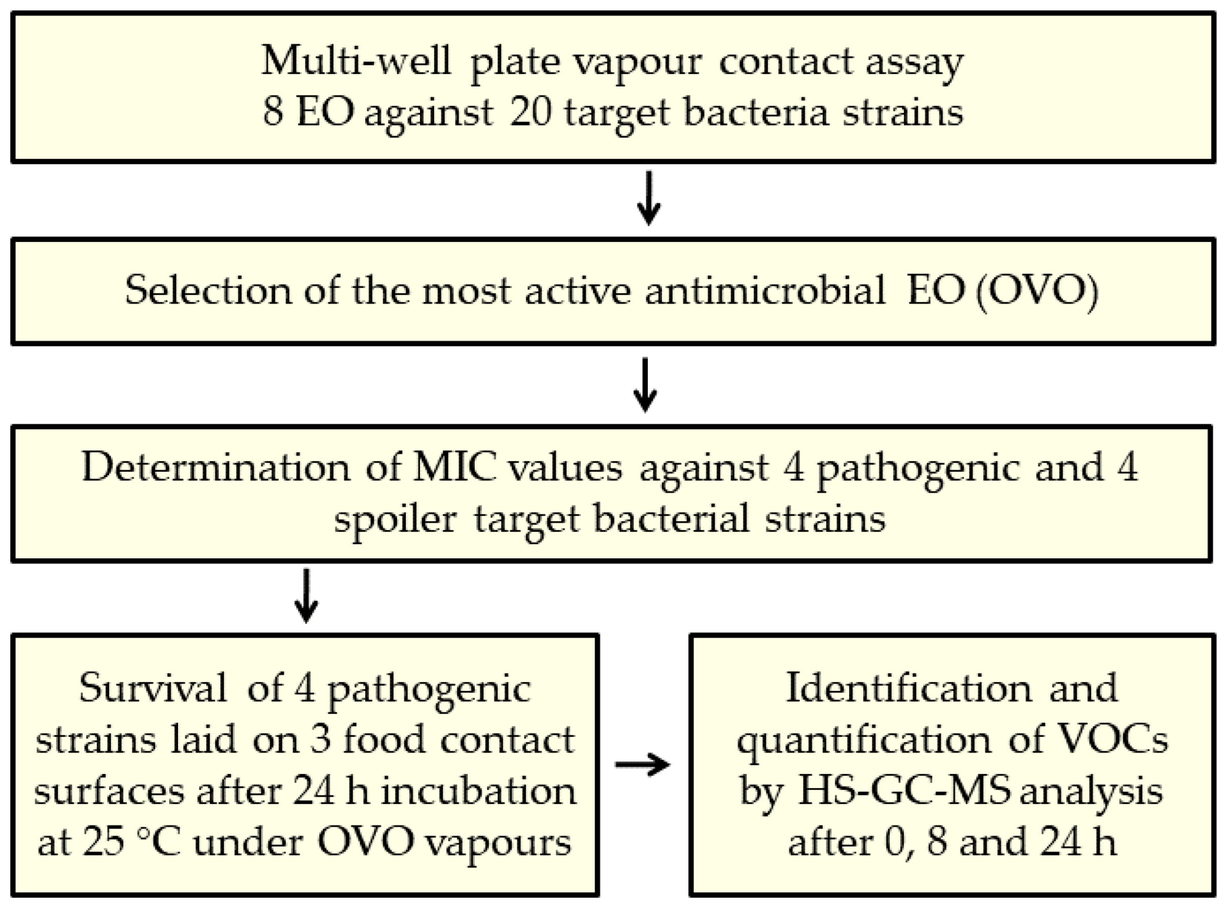
2. Results
2.1. Antibacterial Activity of EOs in Vapor Phase
2.2. Determination of MIC Values
2.3. Antibacterial Activity of OVO Vapors on Food-Contact Surfaces
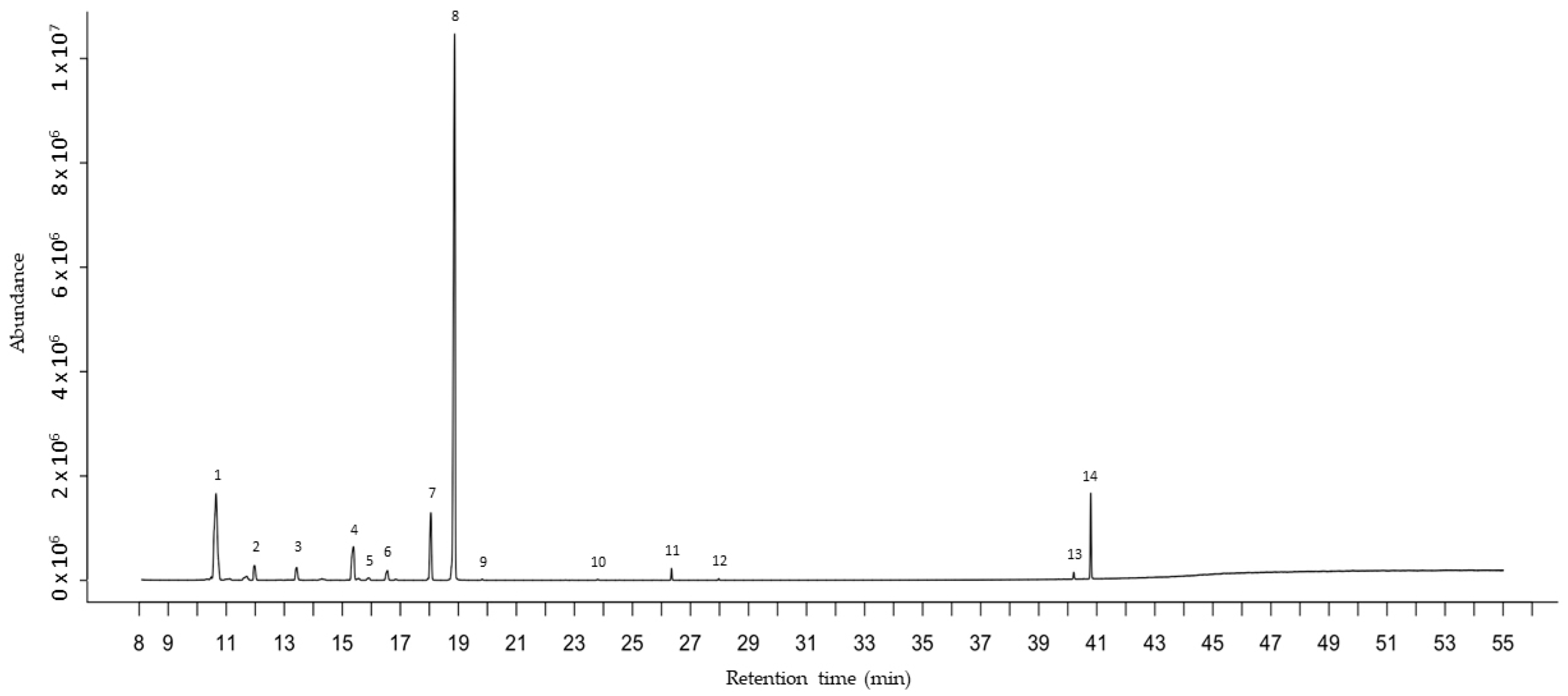
2.4. HS-GC-MS Analysis
3. Discussion
4. Materials and Methods
4.1. Chemical Reagents and Essential Oils
4.2. Bacterial Strains and Growth Conditions
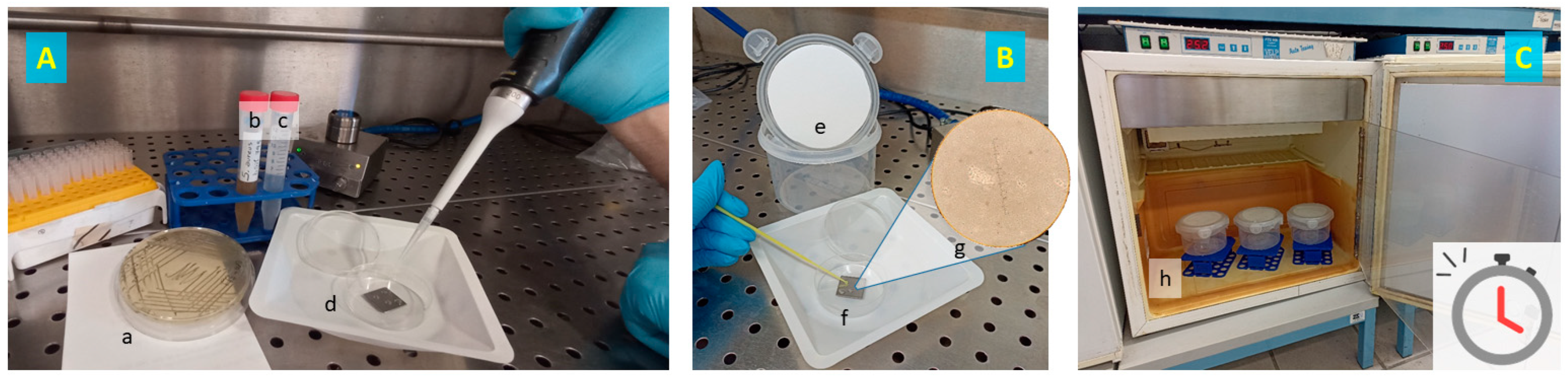
4.3. Vapor Contact Assay
4.4. Application of EOs on Food-Contact Surfaces
4.5. Headspace-Gas-Chromatography-Mass-Spectrometry (HS-GC-MS) Analysis
4.6. Statistical Analysis
5. Conclusions
Supplementary Materials
Author Contributions
Funding
Institutional Review Board Statement
Informed Consent Statement
Data Availability Statement
Conflicts of Interest
References
- Pandey, A.K.; Kumar, P.; Singh, P.; Tripathi, N.N.; Bajpai, V.K. Essential Oils: Sources of Antimicrobials and Food Preservatives. Front. Microbiol. 2017, 7, 2161. [Google Scholar] [CrossRef]
- Pinto, L.; Cefola, M.; Bonifacio, M.; Cometa, S.; Bocchino, C.; Pace, B.; De Giglio, E.; Palumbo, M.; Sada, A.; Logrieco, A.; et al. Effect of red thyme oil (Thymus vulgaris L.) vapours on fungal decay, quality parameters and shelf-life of oranges during cold storage. Food Chem. 2021, 336, 127590. [Google Scholar] [CrossRef]
- Pinto, L.; Bonifacio, M.A.; De Giglio, E.; Cometa, S.; Logrieco, A.F.; Baruzzi, F. Unravelling the Antifungal Effect of Red Thyme Oil (Thymus vulgaris L.) Compounds in Vapour Phase. Molecules 2020, 25, 4761. [Google Scholar] [CrossRef]
- Iseppi, R.; Tardugno, R.; Brighenti, V.; Benvenuti, S.; Sabia, C.; Pellati, F.; Messi, P. Phytochemical Composition and In Vitro Antimicrobial Activity of Essential Oils from the Lamiaceae Family against Streptococcus agalactiae and Candida albicans Biofilms. Antibiotics 2020, 9, 592. [Google Scholar] [CrossRef]
- Khammassi, M.; Ayed, R.B.; Loupasaki, S.; Amri, I.; Hanana, M.; Hamrouni, L.; Jamoussi, B.; Khaldi, A. Chemical diversity of wild fennel essential oils (Foeniculum vulgare Mill.): A source of antimicrobial and antioxidant activities. South Afr. J. Bot. 2023, 153, 136–146. [Google Scholar] [CrossRef]
- Galovičová, L.; Čmiková, N.; Vukovic, N.; Vukic, M.; Kowalczewski, P.Ł.; Bakay, L.; Kačániová, M. Chemical Composition, Antioxidant, Antimicrobial, Antibiofilm and Anti-Insect Activities of Jasminum grandiflorum Essential Oil. Horticulturae 2022, 8, 953. [Google Scholar] [CrossRef]
- Lee, G.; Kim, Y.; Kim, H.; Beuchat, L.R.; Ryu, J.H. Antimicrobial activities of gaseous essential oils against Listeria monocytogenes on a laboratory medium and radish sprouts. Int. J. Food Microbiol. 2018, 265, 49–54. [Google Scholar] [CrossRef]
- Cho, Y.; Kim, H.; Beuchat, L.R.; Ryu, J.H. Synergistic activities of gaseous oregano and thyme thymol essential oils against Listeria monocytogenes on surfaces of a laboratory medium and radish sprouts. Food Microbiol. 2020, 86, 103357. [Google Scholar] [CrossRef]
- Seo, H.S.; Beuchat, L.R.; Kim, H.; Ryu, J.H. Development of an experimental apparatus and protocol for determining antimicrobial activities of gaseous plant essential oils. Int. J. Food Microbiol. 2015, 215, 95–100. [Google Scholar] [CrossRef] [PubMed]
- Pinto, L.; Tapia-Rodríguez, M.R.; Baruzzi, F.; Ayala-Zavala, J.F. Plant antimicrobials for food quality and safety: Recent views and future challenges. Foods 2023, 12, 2315. [Google Scholar] [CrossRef] [PubMed]
- Ríos-Castillo, A.G.; Ripolles-Avila, C.; Rodríguez-Jerez, J.J. Evaluation of bacterial population using multiple sampling methods and the identification of bacteria detected on supermarket food contact surfaces. Food Control 2020, 119, 107471. [Google Scholar] [CrossRef]
- Rossi, F.; Amadoro, C.; Conficoni, D.; Giaccone, V.; Colavita, G. Occurrence, Diversity of Listeria spp. Isolates from Food and Food-Contact Surfaces and the Presence of Virulence Genes. Microorganisms 2020, 8, 294. [Google Scholar] [CrossRef]
- Maes, S.; Heyndrickx, M.; Vackier, T.; Steenackers, H.; Verplaetse, A.; Reu, K.D. Identification and spoilage potential of the remaining dominant microbiota on food contact surfaces after cleaning and disinfection in different food industries. J. Food Prot. 2019, 82, 262–275. [Google Scholar] [CrossRef]
- de Candia, S.; Morea, M.; Baruzzi, F. Eradication of high viable loads of Listeria monocytogenes contaminating food-contact surfaces. Front. Microbiol. 2015, 6, 733. [Google Scholar] [CrossRef]
- Gabriel, A.A.; Ballesteros, M.L.P.; Rosario, L.M.D.; Tumlos, R.B.; Ramos, H.J. Elimination of Salmonella enterica on common stainless steel food contact surfaces using UV-C and atmospheric pressure plasma jet. Food Control 2018, 86, 90–100. [Google Scholar] [CrossRef]
- Falcó, I.; Verdeguer, M.; Aznar, R.; Sánchez, G.; Randazzo, W. Sanitizing food contact surfaces by the use of essential oils. Inn. Food Sci. Emerg. Technol. 2019, 51, 220–228. [Google Scholar] [CrossRef]
- Rossi, C.; Chaves-López, C.; Serio, A.; Casaccia, M.; Maggio, F.; Paparella, A. Effectiveness and mechanisms of essential oils for biofilm control on food-contact surfaces: An updated review. Crit. Rev. Food Sci. Nutr. 2020, 62, 2172–2191. [Google Scholar] [CrossRef] [PubMed]
- Lin, C.M.; Sheu, S.R.; Hsu, S.C.; Tsai, Y.H. Determination of bactericidal efficacy of essential oil extracted from orange peel on the food contact surfaces. Food Control 2010, 21, 1710–1715. [Google Scholar] [CrossRef]
- Engel, J.B.; Heckler, C.; Tondo, E.C.; Daroit, D.J.; da Silva Malheiros, P. Antimicrobial activity of free and liposome-encapsulated thymol and carvacrol against Salmonella and Staphylococcus aureus adhered to stainless steel. Int. J. Food Microbiol. 2017, 252, 18–23. [Google Scholar] [CrossRef]
- dos Santos Rodrigues, J.B.; de Carvalho, R.J.; de Souza, N.T.; de Sousa Oliveira, K.; Franco, O.L.; Schaffner, D.; de Souza, E.L.; Magnani, M. Effects of oregano essential oil and carvacrol on biofilms of Staphylococcus aureus from food-contact surfaces. Food Control 2017, 73, 1237–1246. [Google Scholar] [CrossRef]
- Garzoli, S.; Turchetti, G.; Giacomello, P.; Tiezzi, A.; Laghezza Masci, V.; Ovidi, E. Liquid and Vapour Phase of Lavandin (Lavandula × intermedia) Essential Oil: Chemical Composition and Antimicrobial Activity. Molecules 2019, 24, 2701. [Google Scholar] [CrossRef] [PubMed]
- Ács, K.; Balázs, V.L.; Kocsis, B.; Bencsik, T.; Böszörményi, A.; Horváth, G. Antibacterial activity evaluation of selected essential oils in liquid and vapour phase on respiratory tract pathogens. BMC Complement. Altern. Med. 2018, 18, 227. [Google Scholar] [CrossRef] [PubMed]
- Clerck, C.D.; Maso, S.D.; Parisi, O.; Dresen, F.; Zhiri, A.; Jijakli, M.H. Screening of Antifungal and Antibacterial Activity of 90 Commercial Essential Oils against 10 Pathogens of Agronomical Importance. Foods 2020, 9, 1418. [Google Scholar] [CrossRef] [PubMed]
- Kloucek, P.; Smid, J.; Frankova, A.; Kokoska, L.; Valterova, I.; Pavela, R. Fast screening method for assessment of antimicrobial activity of essential oils in vapour phase. Food Res. Int. 2012, 47, 161–165. [Google Scholar] [CrossRef]
- Feyaerts, A.F.; Mathé, L.; Luyten, W.; Tournu, H.; Dyck, K.V.; Broekx, L.; Dijck, P.V. Assay and recommendations for the detection of vapour-phase-mediated antimicrobial activities. Flavour. Fragr. J. 2017, 32, 347–353. [Google Scholar] [CrossRef]
- Houdkova, M.; Rondevaldova, J.; Doskocil, I.; Kokoska, L. Evaluation of antibacterial potential and toxicity of plant volatile compounds using new broth microdilution volatilization method and modified MTT assay. Fitoterapia 2017, 118, 56–62. [Google Scholar] [CrossRef] [PubMed]
- Antih, J.; Houdkova, M.; Urbanova, K.; Kokoska, L. Antibacterial Activity of Thymus vulgaris L. Essential Oil Vapours and Their GC/MS Analysis Using Solid-Phase Microextraction and Syringe Headspace Sampling Techniques. Molecules 2021, 26, 6553. [Google Scholar] [CrossRef] [PubMed]
- Netopilova, M.; Houdkova, M.; Urbanova, K.; Rondevaldova, J.; Kokoska, L. Validation of qualitative broth volatilization checkerboard method for testing of essential oils: Dual-column GC–FID/MS analysis and in vitro combinatory antimicrobial effect of Origanum vulgare and Thymus vulgaris against Staphylococcus aureus in liquid and vapor phases. Plants 2021, 10, 393. [Google Scholar] [CrossRef] [PubMed]
- Soni, K.A.; Oladunjoye, A.; Nannapaneni, R.; Schilling, M.W.; Silva, J.L.; Mikel, B.; Bailey, R.H. Inhibition and inactivation of Salmonella Typhimurium biofilms from polystyrene and stainless steel surfaces by essential oils and phenolic constituent carvacrol. J. Food Protect. 2013, 76, 205–212. [Google Scholar] [CrossRef]
- Rhoades, J.; Gialagkolidou, K.; Gogou, M.; Mavridou, O.; Blatsiotis, N.; Ritzoulis, C.; Likotrafiti, E. Oregano essential oil as an antimicrobial additive to detergent for hand washing and food contact surface cleaning. J. Appl. Microbiol. 2013, 115, 987–994. [Google Scholar] [CrossRef][Green Version]
- Sengun, I.Y.; Senturk, S.; Gul, S.; Kilic, G. Potential of essential oil combinations for surface and air disinfection. Lett. Appl. Microbiol. 2021, 72, 526–534. [Google Scholar] [CrossRef]
- Russell, A.D. Bacterial resistance to disinfectants: Present knowledge and future problems. J. Hosp. Infect. 1999, 43, 57–68. [Google Scholar] [CrossRef]
- Langsrud, S.; Sidhu, M.S.; Heir, E.; Holck, A.L. Bacterial disinfectant resistance—A challenge for the food industry. Int. Biodeterior. Biodegrad. 2003, 51, 283–290. [Google Scholar] [CrossRef]
- Sibanyoni, J.J.; Tabit, F.T. An assessment of the hygiene status and incidence of foodborne pathogens on food contact surfaces in the food preparation facilities of schools. Food Control 2019, 98, 94–99. [Google Scholar] [CrossRef]
- Gounadaki, A.S.; Skandamis, P.N.; Drosinos, E.H.; Nychas, G.J.E. Microbial ecology of food contact surfaces and products of small-scale facilities producing traditional sausages. Food Microbiol. 2008, 25, 313–323. [Google Scholar] [CrossRef]
- Pu, H.; Xu, Y.; Lin, L.; Sun, D.W. Biofilm formation of Pectobacterium carotovorum subsp. carotovorum on polypropylene surface during multiple cycles of vacuum cooling. Int. J. Food Sci. Technol. 2021, 56, 3495–3506. [Google Scholar] [CrossRef]
- Otter, J.A.; French, G.L. Survival of nosocomial bacteria and spores on surfaces and inactivation by hydrogen peroxide vapor. J. Clin. Microbiol. 2009, 47, 205–207. [Google Scholar] [CrossRef]
- Chaibenjawong, P.; Foster, S.J. Desiccation tolerance in Staphylococcus aureus. Arch. Microbiol. 2011, 193, 125–135. [Google Scholar] [CrossRef]
- Jawad, A.; Heritage, J.; Snelling, A.M.; Gascoyne-Binzi, D.M.; Hawkey, P.M. Influence of Relative Humidity and Suspending Menstrua on Survival of Acinetobacter spp. on Dry Surfaces. J. Clin. Microbiol. 1996, 34, 2881–2887. [Google Scholar] [CrossRef]
- Hansen, L.T.; Vogel, B.F. Desiccation of adhering and biofilm Listeria monocytogenes on stainless steel: Survival and transfer to salmon products. Int. J. Food Microbiol. 2011, 146, 88–93. [Google Scholar] [CrossRef]
- Kuruwita, D.P.; Jiang, X.; Darby, D.; Sharp, J.L.; Fraser, A.M. Persistence of Escherichia coli O157: H7 and Listeria monocytogenes on the exterior of three common food packaging materials. Food Control 2020, 112, 107153. [Google Scholar] [CrossRef]
- Adator, E.H.; Cheng, M.; Holley, R.; McAllister, T.; Narvaez-Bravo, C. Ability of Shiga toxigenic Escherichia coli to survive within dry-surface biofilms and transfer to fresh lettuce. Int. J. Food Microbiol. 2018, 269, 52–59. [Google Scholar] [CrossRef]
- Wißmann, J.E.; Kirchhoff, L.; Brüggemann, Y.; Todt, D.; Steinmann, J.; Steinmann, E. Persistence of Pathogens on Inanimate Surfaces: A Narrative Review. Microorganisms 2021, 9, 343. [Google Scholar] [CrossRef]
- Verran, J.; Airey, P.; Packer, A.; Whitehead, K.A. Microbial retention on open food contact surfaces and implications for food contamination. In Advances in Applied Microbiology; Laskin, A.I., Sariaslani, S., Gadd, G.M., Eds.; Academic Press: Cambridge, MA, USA, 2008; Volume 64, pp. 223–246. [Google Scholar] [CrossRef]
- Oulahal, N.; Brice, W.; Martial, A.; Degraeve, P. Quantitative analysis of survival of Staphylococcus aureus or Listeria innocua on two types of surfaces: Polypropylene and stainless steel in contact with three different dairy products. Food Control 2008, 19, 178–185. [Google Scholar] [CrossRef]
- Park, S.H.; Kang, D.H. Influence of surface properties of produce and food contact surfaces on the efficacy of chlorine dioxide gas for the inactivation of foodborne pathogens. Food Control 2017, 81, 88–95. [Google Scholar] [CrossRef]
- Nakas, A.; Giannarelli, G.; Fotopoulos, I.; Chainoglou, E.; Peperidou, A.; Kontogiannopoulos, K.N.; Tsiaprazi-Stamou, A.; Varsamis, V.; Gika, H.; Hadjipavlou-Litina, D.; et al. Optimizing the distillation of greek Oregano—Do process parameters affect bioactive aroma constituents and in vitro antioxidant activity? Molecules 2023, 28, 971. [Google Scholar] [CrossRef]
- Freitas, P.R.; de Araújo, A.C.J.; dos Santos Barbosa, C.R.; Muniz, D.F.; da Silva, A.C.A.; Rocha, J.E.; de Morais Oliveira-Tintino, C.D.; Ribeiro-Filho, J.; da Silva, L.E.; Confortin, C.; et al. GC-MS-FID and potentiation of the antibiotic activity of the essential oil of Baccharis reticulata (ruiz & pav.) pers. and α-pinene. Ind. Crop. Prod. 2020, 145, 112106. [Google Scholar] [CrossRef]
- Wang, C.Y.; Chen, Y.W.; Hou, C.Y. Antioxidant and antibacterial activity of seven predominant terpenoids. Int. J. Food Prop. 2019, 22, 230–238. [Google Scholar] [CrossRef]
- Allenspach, M.; Steuer, C. α-Pinene: A never-ending story. Phytochemistry 2021, 190, 112857. [Google Scholar] [CrossRef]
- Cristani, M.; D’Arrigo, M.; Mandalari, G.; Castelli, F.; Sarpietro, M.G.; Micieli, D.; Venuti, V.; Bisignano, G.; Saija, A.; Trombetta, D. Interaction of four monoterpenes contained in essential oils with model membranes: Implications for their antibacterial activity. J. Agric. Food Chem. 2007, 55, 6300–6308. [Google Scholar] [CrossRef]
- Aulia, A.F.; Anugraha, R.P.; Kuswandi, K. Isobaric Vapor–Liquid Equilibrium of Citronellal+ Geraniol and Citronellal+ Citronellol Binary Systems at 16.0 and 32.0 kPa. J. Chem. Eng. Data 2023, 68, 1646–1653. [Google Scholar] [CrossRef]
- Rodrigues, V.H.S.; Almeida, R.N.; Vargas, R.M.; Cassel, E. Vapor pressure and vapor-liquid equilibrium data for eugenol/caryophyllene binary system at low pressures by experimental and predictive methods. J. Chem. Thermodyn. 2022, 168, 106725. [Google Scholar] [CrossRef]
- Baruzzi, F.; Cefola, M.; Carito, A.; Vanadia, S.; Calabrese, N. Changes in bacterial composition of zucchini flowers exposed to refrigeration temperatures. Sci. World J. 2012, 2012, 127805. [Google Scholar] [CrossRef]
- Pinto, L.; Ippolito, A.; Baruzzi, F. Control of spoiler Pseudomonas spp. on fresh cut vegetables by neutral electrolyzed water. Food Microbiol. 2015, 50, 102–108. [Google Scholar] [CrossRef]
- Baruzzi, F.; Pinto, L.; Quintieri, L.; Carito, A.; Calabrese, N.; Caputo, L. Efficacy of lactoferricin B in controlling ready-to-eat vegetable spoilage caused by Pseudomonas spp. Int. J. Food Microbiol. 2015, 215, 179–186. [Google Scholar] [CrossRef]
- Pinto, L.; Malfeito-Ferreira, M.; Quintieri, L.; Silva, A.C.; Baruzzi, F. Growth and metabolite production of a grape sour rot yeast-bacterium consortium on different carbon sources. Int. J. Food Microbiol. 2019, 296, 65–74. [Google Scholar] [CrossRef]
- Pinto, L.; Caputo, L.; Quintieri, L.; de Candia, S.; Baruzzi, F. Efficacy of gaseous ozone to counteract postharvest table grape sour rot. Food Microbiol. 2017, 66, 190–198. [Google Scholar] [CrossRef]
- Zellner, B.D.A.; Bicchi, C.; Dugo, P.; Rubiolo, P.; Dugo, G.; Mondello, L. Linear retention indices in gas chromatographic analysis: A review. Flavour Frag. J. 2008, 23, 297–314. [Google Scholar] [CrossRef]


| Target Strains | GO | LEO | LO | OVO | RO | RTO | SOO | TTO | |
|---|---|---|---|---|---|---|---|---|---|
| Spoilers | Acetobacter malorum LMG 1746 | 0* | 5 | 5 | 0* | 5 | 0* | 5 | 0 |
| A. syzygii LMG 21419 | 0* | 5 | 5 | 5 | 5 | 0* | 5 | 0* | |
| Dickeya dadantii LMG 25991 | 5 | 3 | 0* | 0* | 0* | 0* | 5 | 0* | |
| Enterobacter aerogenes ITEM 17998 | 5 | 5 | 5 | 5 | 5 | 3 | 5 | 5 | |
| Erwinia persicina ITEM 17997 | 5 | 5 | 5 | 5 | 5 | 3 | 5 | 5 | |
| Gluconobacter oxydans LMG 1408 | 0* | 5 | 5 | 5 | 5 | 0* | 5 | 0 | |
| Pantoea agglomerans LMG 2565 | 5 | 5 | 5 | 5 | 5 | 5 | 5 | 5 | |
| Pectobacterium carotovorum subsp. actinidiae LMG 26003 | 5 | 3 | 5 | 0* | 0 | 0* | 5 | 0* | |
| Pec. carotovorum subsp. carotovorum LMG 2404 | 5 | 3 | 5 | 0* | 0 | 0* | 5 | 0* | |
| Pseudomonas chicorii ITEM 17298 | 5 | 5 | 0* | 3 | 5 | 3 | 5 | 3 | |
| Pse. fluorescens ITEM 19245 | 5 | 5 | 5 | 0 | 0 | 0 | 5 | 0 | |
| Pse. fluorescens NCPPB 1964T | 5 | 5 | 5 | 3 | 5 | 3 | 5 | 0 | |
| Pse. marginalis pv. marginalis LMG 2210 | 5 | 5 | 5 | 5 | 5 | 3 | 5 | 3 | |
| Pse. putida ITEM 17297 | 5 | 3 | 5 | 0 | 5 | 0 | 5 | 5 | |
| Serratia marcescens ITEM 17999 | 5 | 3 | 5 | 5 | 5 | 5 | 5 | 3 | |
| Pathogens | Escherichia coli ATCC 35401 | 5 | 5 | 5 | 0* | 5 | 5 | 5 | 5 |
| Listeria monocytogenes DSM 20600 | 5 | 5 | 5 | 0* | 5 | 5 | 5 | 5 | |
| Pse. aeruginosa DSM 939 | 5 | 5 | 5 | 5 | 5 | 5 | 5 | 5 | |
| Salmonella enterica ATCC 13311 | 5 | 5 | 5 | 0* | 5 | 5 | 5 | 3 | |
| Staphylococcus aureus DSM 799 | 5 | 5 | 5 | 0* | 5 | 0* | 5 | 5 |
| Target Strain | MIC of OVO | |
|---|---|---|
| as v/v in n-Hexane v/v | in µg cm−3 Air | |
| A. malorum LMG 21419 | 40% | 753.6 |
| D. dadantii LMG 25991 | 5% | 94.2 |
| E. coli ATCC 35401 | 40% | 753.6 |
| L. monocytogenes DSM 20600 | 20% | 376.8 |
| Pec. carotovorum subsp. actinidiae LMG 26003 | 40% | 753.6 |
| Pec. carotovorum subsp. carotovorum LMG 2404 | 10% | 188.4 |
| Sal. enterica ATCC 13311 | 40% | 753.6 |
| Sta. aureus DSM 799 | 20% | 376.8 |
| Strain | Initial Load | Treatment | Stainless Steel | Polypropylene | Glass |
|---|---|---|---|---|---|
| Escherichia coli ATCC 35401 | 6.55 ± 0.02 A | Air | 6.98 ± 0.20 Aa | 6.97 ± 0.12 Aa | 6.76 ± 0.12 Aa |
| OVO 1 | n.d. Bb | n.d. Bb | n.d. Bb | ||
| Listeria monocytogenes DSM 20600 | 6.80 ± 0.20 A | Air | 6.79 ± 0.17 Aa | 6.72 ± 0.10 Aa | 6.74 ± 0.12 Aa |
| OVO 2 | n.d. Bb | n.d. Bb | n.d. Bb | ||
| Salmonella enterica ATCC 13311 | 6.72 ± 0.10 A | Air | 6.94 ± 0.02 Aa | 7.12 ± 0.06 Aa | 6.44 ± 0.19 Aa |
| OVO 1 | n.d. Bb | n.d. Bb | n.d. Bb | ||
| Staphylococcus aureus DSM 799 | 6.74 ± 0.09 A | Air | 7.81 ± 0.14 Ba | 7.66 ± 0.02 Ba | 7.45 ± 0.07 Ba |
| OVO 2 | n.d. Bb | n.d. Bb | 3.23 ± 0.02 Bb |
| Compounds | LRIlt/LRIsp a | LOD b (ng mL−1) | 377 µg cm−3 Air | 754 µg cm−3 Air | ||
|---|---|---|---|---|---|---|
| Mean c Concentration (ng mL−1) | Mean c Composition (%) | Mean c Concentration (ng mL−1) | Mean c Composition (%) | |||
| α-Pinene d | 1020/1021 | 9 | 1009a ± 96 | 62 | 1763b ± 69 | 63 |
| p-Cymene d | 1270/1275 | 0.17 | 343a ± 17 | 21 | 582b ± 59 | 21 |
| β-Myrcene d | 1160/1167 | 0.76 | 63a ± 4 | 3.9 | 132b ± 18 | 4.7 |
| Camphene d | 1062/1067 | 1.5 | 62a ± 8 | 3.8 | 113b ± 7 | 4.0 |
| β-Pinene d | 1120/1109 | 1.6 | 29a ± 5 | 1.8 | 61b ± 5 | 2.2 |
| γ-Terpinene d | 1250/1251 | 0.10 | 29a ± 2 | 1.8 | 56b ± 8 | 2.0 |
| Carvacrol d | 2225/2225 | 0.24 | 46a ± 1 | 2.9 | 48a ± 6 | 1.7 |
| R-Limonene d | 1200/1203 | 0.50 | 12a ± 1 | 0.72 | 19a ± 2 | 0.69 |
| Linalool d | 1551/1551 | 0.40 | 16a ± 2 | 1.0 | 14a ± 1 | 0.51 |
| α-Terpinene d | 1180/1183 | 1.3 | 4.7a ± 0.1 | 0.29 | 7.5b ± 0.7 | 0.27 |
| o-Cymene d | 1298/1309 | 0.73 | 2.7a ± 0.1 | 0.17 | 3.1a ± 0.1 | 0.11 |
| Thymol d | 2198/2193 | 0.19 | 2.8a ± 0.1 | 0.17 | 2.9a ± 0.4 | 0.10 |
| Thymol methyl ether e | 1599/1616 | 0.12 | 0.5a ± 0.1 | traces f | 0.7a ± 0.1 | traces f |
| 1-Octen-3-ol d | 1450/1450 | 0.13 | 0.38a ± 0.05 | traces f | 0.34a ± 0.04 | traces f |
| Total terpenes | 1620 | 2802 | ||||
Disclaimer/Publisher’s Note: The statements, opinions and data contained in all publications are solely those of the individual author(s) and contributor(s) and not of MDPI and/or the editor(s). MDPI and/or the editor(s) disclaim responsibility for any injury to people or property resulting from any ideas, methods, instructions or products referred to in the content. |
© 2024 by the authors. Licensee MDPI, Basel, Switzerland. This article is an open access article distributed under the terms and conditions of the Creative Commons Attribution (CC BY) license (https://creativecommons.org/licenses/by/4.0/).
Share and Cite
Pinto, L.; Cervellieri, S.; Netti, T.; Lippolis, V.; Baruzzi, F. Antibacterial Activity of Oregano (Origanum vulgare L.) Essential Oil Vapors against Microbial Contaminants of Food-Contact Surfaces. Antibiotics 2024, 13, 371. https://doi.org/10.3390/antibiotics13040371
Pinto L, Cervellieri S, Netti T, Lippolis V, Baruzzi F. Antibacterial Activity of Oregano (Origanum vulgare L.) Essential Oil Vapors against Microbial Contaminants of Food-Contact Surfaces. Antibiotics. 2024; 13(4):371. https://doi.org/10.3390/antibiotics13040371
Chicago/Turabian StylePinto, Loris, Salvatore Cervellieri, Thomas Netti, Vincenzo Lippolis, and Federico Baruzzi. 2024. "Antibacterial Activity of Oregano (Origanum vulgare L.) Essential Oil Vapors against Microbial Contaminants of Food-Contact Surfaces" Antibiotics 13, no. 4: 371. https://doi.org/10.3390/antibiotics13040371
APA StylePinto, L., Cervellieri, S., Netti, T., Lippolis, V., & Baruzzi, F. (2024). Antibacterial Activity of Oregano (Origanum vulgare L.) Essential Oil Vapors against Microbial Contaminants of Food-Contact Surfaces. Antibiotics, 13(4), 371. https://doi.org/10.3390/antibiotics13040371