Abstract
Moraxella species are Gram-negative coccobacilli that typically colonize the flora of the human upper respiratory tract and have low pathogenic potential. There are limited case reports implicating the organisms as the cause of endocarditis, bacteremia, septic arthritis, ocular infection, and meningitis. In cases of keratitis and conjunctivitis, Moraxella nonliquefaciens is not commonly isolated from the ocular surface. We present a case of a diabetic patient who developed late-onset bleb-related endophthalmitis caused by M. nonliquefaciens 4 years after glaucoma filtering surgery. Within one day, the patient presented with an acutely fulminant course with sudden visual loss, redness, and ocular pain. Appropriate antibiotic treatment and early vitrectomy resulted in a favorable final visual acuity of 20/100, which was his vision prior to infection. The use of Matrix-Assisted Laser Desorption Ionization–Time of Flight Mass spectrometry (MALDI-TOF MS) enabled the rapid identification of the organism. Endophthalmitis caused by M. nonliquefaciens should be considered in patients who underwent glaucoma filtering surgery with antifibrotic agents.
1. Introduction
Bacterial endophthalmitis, which can lead to significant loss of vision, is a concerning complication of ocular surgery. Most cases of endophthalmitis are exogenous and result from ocular surgical procedures, especially cataract surgery. The Endophthalmitis Vitrectomy Study defined acute-onset postoperative endophthalmitis as infections that occur within six weeks after the surgery, whereas delayed-onset postoperative endophthalmitis was defined as infections that occur after a period greater than six weeks following the surgery. In post-cataract patients, delayed-onset cases of endophthalmitis are mostly caused by fungal or indolent bacterial infections, such as Cutibacterium acnes. These cases typically present as a persistent low-grade inflammation in the anterior chamber. Patients present with decreased vision in the affected eye, and half of them also have eye pain, which is usually mild. However, the time courses for endophthalmitis following cataract surgery and filtration surgery are distinct from each other. Late-onset endophthalmitis associated with filtering bleb, which occurs in an eye that has remained stable for several months or years after surgery, is less common, but carries a high degree of risk [1]. It can develop many years after the procedure without warning and progress rapidly. Additionally, the organisms responsible for this condition are often more virulent than those typically associated with post-cataract endophthalmitis. The frequency of late-onset endophthalmitis after glaucoma surgery ranges from 0.1% to 1.5% [2]. Streptococci are the most common pathogens involved in bleb-related endophthalmitis; other major pathogens include Staphylococcus aureus, Hemophilus influenzae, enterococci, and Moraxella catarrhalis [3].
Moraxella nonliquefaciens, a Gram-negative coccobacillus, is part of the normal microbiota of the human respiratory tract and is considered to have low pathogenic potential [4]. Ocular infections due to M. nonliquefaciens are usually limited to eye surface and present as conjunctivitis, keratitis, and corneal abscess [5,6]. Herein, we report an unusual case of late-onset bleb-related endophthalmitis caused by M. nonliquefaciens.
2. Case Report
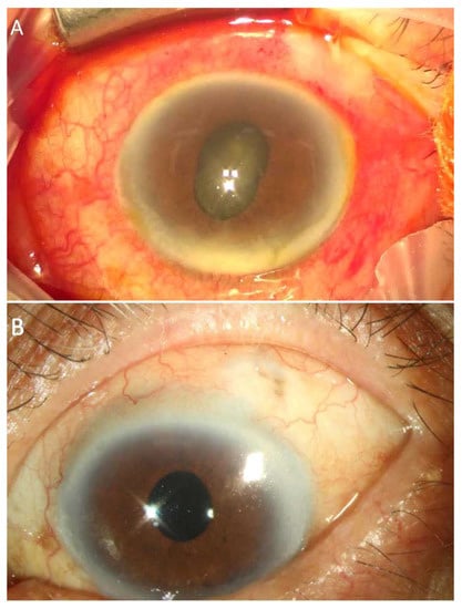
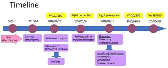
A 72-year-old diabetic male presented to our emergency department with a 1-day history of red eye and sudden visual loss in his left eye. He had a known history of advanced primary angle closure glaucoma, which was treated with bilateral laser iridectomy and bilateral cataract extraction with a posterior chamber intraocular lens implantation 10 years prior. Left eye trabeculectomy with mitomycin C was performed 4 years prior. His right eye had no light perception due to end-stage chronic angle closure glaucoma. Slit-lamp biomicroscopic examination of left eye showed opacified bleb surrounded by hyperemic conjunctiva and cornea edema. The filtering bleb was congested, and a 2 mm hypopyon and vitreous clouding were observed (Figure 1A). No fundal red reflex was observed. Intraocular pressure was 24 mm Hg and visual acuity decreased to light perception. The fundus was invisible and B-scan ultrasonography showed increased echogenicity in the vitreous cavity with no evidence of the posterior hyaloid detachment. Following aqueous and vitreous tapping, emergent pars plana vitrectomy was performed. Multiple yellow-white vitreous condensations were removed and the fundus showed diffuse retinal hemorrhages as well as retinal vasculitis. Intravitreal vancomycin (1 mg/0.1 mL), ceftazidime (2.25 mg/0.1 mL), and dexamethasone (0.4 mg/0.1 mL) were administrated at the end of surgery. Gram stains from vitreous revealed Gram-negative coccobacillus. Both bacterial cultures from aqueous and vitreous fluids showed M. nonliquefaciens growth, which was confirmed by Matrix-Assisted Laser Desorption Ionization–Time of Flight Mass spectrometry (MALDI-TOF MS). In vitro antibiotic susceptible testing showed that the M. nonliquefaciens isolate was sensitive to amoxicillin-clavulanic acid, cefuroxime, ceftriaxone, and trimethoprim-sulfamethoxazole. One month later, his visual acuity recovered to 20/200. The patient achieved a best-corrected visual acuity of 20/100, which was his vision prior to infection, during his 6-month follow-up (Figure 1B). Figure 2 illustrates his clinical progression.

Figure 1.
(A). Anterior segment revealed opacified bleb surrounded by hyperemic conjunctiva, corneal edema, hypopyon, and fibrinous reaction. (B). Six months later, anterior segment showed resolution of endophthalmitis without anatomical sequelae.

Figure 2.
Clinical course.
3. Discussion
Moraxella species are oxidase-positive, aerobic Gram-negative cocci. They are easily identified by their classic appearance as brick-shaped diplobacilli, which are distinct from other Gram-negative bacteria. Moraxella spp. are a part of the normal flora of the human respiratory and genital tracts and are considered to have low pathogenic potential. Among Moraxella spp., the most clinically relevant microorganism from this genus is M. catarrhalis, which can cause otitis media in children and exacerbations of chronic obstructive pulmonary disease [7,8]. Other members of the Moraxella genus, such as M. lacunata, M. osloensis, and M. nonliquefaciens rarely cause disease.
Ocular infections due to Moraxella is low and usually limited to eye surface. The epidemiology of Moraxella keratitis has been studied in detail all over the world; this condition accounts for 2 to 3% of all cases of bacterial corneal ulcers between 2010 and 2018 [9,10,11]. Indeed, Moraxella may be considered an emerging pathogen, as a ninefold increase in the incidence of Moraxella keratitis was recently reported in Ireland [12]. A similar trend has also been observed in the USA and England, where Moraxella species appear to be on the rise, as an increase of 6% has been observed during the last decade [13]. The prevalence of Moraxella spp. in endophthalmitis is also increasing. In a large retrospective series at Bascom Palmer Eye Institute between 1991 and 2000, only 1.3% (10 of 757) of endophthalmitis cases revealed Moraxella species infection [14]. A subsequent French multicenter prospective study performed between 2004 and 2005 revealed that Moraxella spp. accounted for 6.5% (7 of 108) of the bacterial spectrum [15]. It is possible that these findings are indicative of alterations in the ocular microbiota profiles, which may be more accurately detected using new molecular diagnostic techniques that enhance the sensitivity and accuracy of pathogen identification.
M. nonliquefaciens is considered a part of the normal human flora of the upper respiratory tract, with few infections, such as osteomyelitis, meningitis, and endocarditis, being mentioned in the literature [16,17]. Ocular infections due to M. nonliquefaciens have rarely been characterized. The first report of ocular infection due to M. Nonliquefaciens, by Ebright et al., was concerned with a post-renal transplant, immunocompromised patient whose infection was believed to have been caused by minor trauma of the cornea from a contact lens [18]. Loube et al. reported two cases of M. nonliquefaciens endophthalmitis following trabeculectomy. Symptoms included pain, inflammation, and a rapid decrease in visual acuity [19]. A review of the literature shows only six other reported cases of late-onset bleb-related M. nonliquefaciens endophthalmitis and these infections occurred within a 2-month to 15-year period following surgery (Table 1) [1,16,19,20,21,22].

Table 1.
Trabeculectomy-related Endophthalmitis caused by Moraxella nonliquefaciens in literature and present case.
The majority of cases of late-onset endophthalmitis after glaucoma filtering surgery demonstrate a devastating visual outcome. The most common presenting complaints of bleb-related endophthalmitis include ocular pain (71%) and redness (53%), which typically appear within three days of presentation. Other common symptoms include blurred vision (35%), tearing (12%), purulent discharge (12%), and photophobia (10%). Findings include marked intraocular inflammation and, often, a bleb infiltrate [23]. Staphylococcus and Streptococcus species are the main pathogens. Streptococcus produces exotoxins causing a fulminant infection with poor visual outcome [24,25]. Other causes of bleb-related endophthalmitis are H. influenzae, Acinetobacter, and M. catarrhalis, among other bacteria. M. nonliquefaciens is generally considered to be of low virulence and is sensitive to most antibiotics.
A filtering bleb is a small sclera defect, created by glaucoma surgery, that allows excess aqueous humor to be absorbed into the systemic circulation. However, only a thin layer of conjunctiva separates the aqueous from the ocular surface in the location of the bleb, and this carries a risk of developing endophthalmitis. A large study from Japan reported the risk of endophthalmitis to be 1.3% per patient-year, and that it could occur between 1 month and 8 years following glaucoma surgery, with an average onset of 2 years. Leaking blebs increase endophthalmitis risk nearly fivefold [26]. Our patient presented with a thin opacified bleb with marked congestion, which is suspected to be an infection of the bleb preceding endophthalmitis. Several factors are known to be significantly associated with the development of infection following late-onset glaucoma surgery, with major factors including an inferiorly located bleb, the presence of bleb leakage, diabetes mellitus, and the use of antiproliferative agents, usually mitomycin or 5-fluorouracil (5-FU), concomitantly with surgery [25,27]. The most significant risk factor for endophthalmitis was found to be bleb leakage. Patients with a history of bleb leakage were 4.71 times more likely to develop an infection. According to Solus et al., bleb-related infections occurred more frequently in eyes with a limbus-based flap than in those with a fornix-based flap [28]. The initial stage of endophthalmitis development involves bacteria migrating across the filtering bleb. Therefore, patients must be educated to promptly report any signs of ocular surface infection, such as redness, pain, or discharge, to their clinic. Early detection of bleb infection can be facilitated through timely intervention. Bleb leakage must be repaired or treated as soon as it is detected.
In the past decade, antifibrotic agents such as mitomycin C and 5-FU have been increasingly used in glaucoma filtering surgery as they can help achieve a further decrease in intraocular pressure and improve the functional success of trabeculectomy with a high risk of surgical failure. Histologic studies have shown that the use of antifibrotic agents is associated with blebs that are less vascularized, have a thinner epithelium, and have more atrophic stroma. Because of the increased use of antifibrotic agents and the resulting thin, cystic, avascular blebs, there is growing concern about the increased risk of bleb-related endophthalmitis [29].
Identifying Moraxella species through routine laboratory techniques has been challenging. It is important to identify the species of Moraxella, as each species has a different antibiotic sensitivity. Many isolates cannot be identified by 16S rRNA sequencing alone [15]. With the use of a diagnostic tool, MALDI-TOF MS, we were able to rapidly identify the bacteria in our case. This ensured that our patient received appropriate therapy based on published antibiotic susceptibility data. A commercial system, Biolog GenIII (Hayward, CA, USA), can also be used to identify bacteria to genus and species [4,30]. Next-generation sequencing has been shown to be a promising technology for the identification of emerging pathogens and rare infectious diseases. These new sequencing technologies may enable the implementation of rapid and accurate genotypic drug susceptibility testing prior to the administration of antimicrobial therapy [31].
M. nonliquefaciens is not commonly implicated in human disease, so detailed susceptibility patterns are not fully known. Generally, Moraxella species are more susceptible to penicillin, ceftazidime (100%), cefazolin (98%), fluoroquinolones (100%) (ciprofloxacin, ofloxacin, and moxifloxacin), aminoglycosides (94–100%) (gentamicin, tobramycin, and amikacin), and polymyxin B (99–100%), but are less susceptible to trimethoprim (11%) [4]. A recent study reported increased resistance in clinical strains to macrolides [32]. M. nonliquefaciens, unlike M. catarrhalis, was found to be susceptible to vancomycin. This is the motivation for treating the present case with a direct intravitreal injection of vancomycin and ceftazidime, which resulted in a favorable outcome.
Endophthalmitis is a medical emergency as delayed treatment may result in permanent vision loss. The treatment strategy of endophthalmitis depends on the visual function of the patient and the extent of the inflammation. Surgical debridement of the vitreous humor is recommended for patients with fulminant endophthalmitis who present with severe vision loss or rapidly deteriorating vision. Early, prompt vitrectomy involves sample collection for microbiologic investigation and cleaning the vitreous cavity. Fortified, broad-spectrum antibiotics can be used not only at the end of vitrectomy but also during the surgery, and the anterior chamber and the vitreous cavity can also be washed with antibiotics. Vancomycin and ceftazidime or amikacin are the current drugs of choice. The injection is performed after a sample of vitreous humor is taken for Gram staining and culturation. A repeat intravitreal antibiotic injection may be administered based on culture results. Intravitreal steroid injection, which can reduce inflammation, is sometimes given as adjunctive therapy to preserve tissue integrity. Awareness of endophthalmitis risk in patients who have undergone glaucoma filtering surgery with antifibrotic agents is necessary. Prompt Gram staining is imperative for the early identification of the offending pathogen.
Author Contributions
Conceptualization, S.-C.S. and K.-J.C.; formal analysis, S.-C.S. and K.-J.C.; investigation, S.-C.S. and K.-J.C.; writing—review and editing, S.-C.S. and K.-J.C. All authors have read and agreed to the published version of the manuscript.
Funding
This research received no external funding.
Institutional Review Board Statement
The study was conducted according to the guidelines of the Declaration of Helsinki and approved by the Institutional Review Board of Chang Gung Memorial Hospital (201900614B0C601, 10 August 2019) in Taoyuan, Taiwan.
Informed Consent Statement
Not applicable.
Data Availability Statement
Not applicable.
Acknowledgments
The authors sincerely thank Alexandria Wang (Columbia University, Mailman School of Public Health) for participating in the technical editing of the manuscript.
Conflicts of Interest
The authors declare no conflict of interest.
References
- Mandelbaum, S.; Forster, R.K.; Gelender, H.; Culbertson, W. Late onset endophthalmitis associated with filtering blebs. Ophthalmology 1985, 92, 964–972. [Google Scholar] [CrossRef] [PubMed]
- Kirwan, J.F.; Lockwood, A.J.; Shah, P.; Macleod, A.; Broadway, D.C.; King, A.J.; McNaught, A.I.; Agrawal, P. Trabeculectomy in the 21st century: A multicenter analysis. Ophthalmology 2013, 120, 2532–2539. [Google Scholar] [CrossRef] [PubMed]
- Durand, M.L. Bacterial and Fungal Endophthalmitis. Clin. Microbiol. Rev. 2017, 30, 597–613. [Google Scholar] [CrossRef]
- LaCroce, S.J.; Wilson, M.N.; Romanowski, J.E.; Newman, J.D.; Jhanji, V.; Shanks, R.M.Q.; Kowalski, R.P. Moraxella nonliquefaciens and M. osloensis Are Important Moraxella Species That Cause Ocular Infections. Microorganisms 2019, 7, 163. [Google Scholar] [CrossRef]
- Cobo, F.; Rodriguez-Granger, J.; Sampedro, A.; Navarro-Mari, J.M. Corneal infection due to Moraxella nonliquefaciens. Enferm. Infecc. Microbiol. Clin. (Engl. Ed.) 2019, 37, 351–352. [Google Scholar] [CrossRef]
- Hoarau, G.; Merabet, L.; Brignole-Baudouin, F.; Mizrahi, A.; Borderie, V.; Bouheraoua, N. Moraxella keratitis: Epidemiology and outcomes. Eur. J. Clin. Microbiol. Infect. Dis. 2020, 39, 2317–2325. [Google Scholar] [CrossRef]
- Cools, P.; Nemec, A.; Kämpfer, P.; Vaneechoutte, M. Acinetobacter, Chryseobacterium, Moraxella, and Other Nonfermentative Gram-negative Rods; ASM Press: Washington, DC, USA, 2019. [Google Scholar]
- van den Munckhof, E.H.A.; Hafkamp, H.C.; de Kluijver, J.; Kuijper, E.J.; de Koning, M.N.C.; Quint, W.G.V.; Knetsch, C.W. Nasal microbiota dominated by Moraxella spp. is associated with respiratory health in the elderly population: A case control study. Respir Res. 2020, 21, 181. [Google Scholar] [CrossRef] [PubMed]
- Das, S.; Constantinou, M.; Daniell, M.; Taylor, H.R. Moraxella keratitis: Predisposing factors and clinical review of 95 cases. Br. J. Ophthalmol. 2006, 90, 1236–1238. [Google Scholar] [CrossRef] [PubMed]
- Durrani, A.F.; Faith, S.C.; Kowalski, R.P.; Yu, M.; Romanowski, E.; Shanks, R.; Dhaliwal, D.; Jhanji, V. Moraxella Keratitis: Analysis of Risk Factors, Clinical Characteristics, Management, and Treatment Outcomes. Am. J. Ophthalmol. 2019, 197, 17–22. [Google Scholar] [CrossRef] [PubMed]
- Zafar, H.; Tan, S.Z.; Walkden, A.; Fullwood, C.; Au, L.; Brahma, A.; Carley, F. Clinical Characteristics and Outcomes of Moraxella Keratitis. Cornea 2018, 37, 1551–1554. [Google Scholar] [CrossRef] [PubMed]
- McSwiney, T.J.; Knowles, S.J.; Murphy, C.C. Clinical and microbiological characteristics of Moraxella keratitis. Br. J. Ophthalmol. 2019, 103, 1704–1709. [Google Scholar] [CrossRef] [PubMed]
- Tan, S.Z.; Walkden, A.; Au, L.; Fullwood, C.; Hamilton, A.; Qamruddin, A.; Armstrong, M.; Brahma, A.K.; Carley, F. Twelve-year analysis of microbial keratitis trends at a UK tertiary hospital. Eye 2017, 31, 1229–1236. [Google Scholar] [CrossRef]
- Berrocal, A.M.; Scott, I.U.; Miller, D.; Flynn, H.W., Jr. Endophthalmitis caused by Moraxella species. Am. J. Ophthalmol. 2001, 132, 788–790. [Google Scholar] [CrossRef] [PubMed]
- Cornut, P.L.; Chiquet, C.; Bron, A.; Romanet, J.P.; Lina, G.; Lafontaine, P.O.; Benito, Y.; Pechinot, A.; Burillon, C.; Vandenesch, F.; et al. Microbiologic identification of bleb-related delayed-onset endophthalmitis caused by Moraxella species. J. Glaucoma. 2008, 17, 541–545. [Google Scholar] [CrossRef] [PubMed]
- Laukeland, H.; Bergh, K.; Bevanger, L. Posttrabeculectomy endophthalmitis caused by Moraxella nonliquefaciens. J. Clin. Microbiol. 2002, 40, 2668–2770. [Google Scholar] [CrossRef]
- Kao, C.; Szymczak, W.; Munjal, I. Meningitis due to Moraxella nonliquefaciens in a paediatric patient: A case report and review of the literature. JMM Case Rep. 2017, 4, e005086. [Google Scholar] [CrossRef]
- Ebright, J.R.; Lentino, J.R.; Juni, E. Endophthalmitis caused by Moraxella nonliquefaciens. Am. J. Clin. Pathol. 1982, 77, 362–363. [Google Scholar] [CrossRef]
- Lobue, T.D.; Deutsch, T.A.; Stein, R.M. Moraxella nonliquefaciens endophthalmitis after trabeculectomy. Am. J. Ophthalmol. 1985, 99, 343–345. [Google Scholar] [CrossRef]
- Sherman, M.D.; York, M.; Irvine, A.R.; Langer, P.; Cevallos, V.; Whitcher, J.P. Endophthalmitis caused by beta-lactamase-positive Moraxella nonliquefaciens. Am. J. Ophthalmol. 1993, 115, 674–676. [Google Scholar] [CrossRef]
- Schmidt, M.E.; Smith, M.A.; Levy, C.S. Endophthalmitis caused by unusual gram-negative bacilli: Three case reports and review. Clin. Infect. Dis. 1993, 17, 686–690. [Google Scholar] [CrossRef]
- Diaz Barron, A.; Hervas Hernandis, J.M.; Sanz Gallen, L.; Lopez Montero, A.; Gil Hernandez, I.; Duch-Samper, A.M. Delayed-onset bleb-related endophthalmitis caused by Moraxella nonliquefaciens. Arch. Soc. Esp. Oftalmol. (Engl. Ed.) 2020, 95, 559–564. [Google Scholar] [CrossRef] [PubMed]
- Lehmann, O.J.; Bunce, C.; Matheson, M.M.; Maurino, V.; Khaw, P.T.; Wormald, R.; Barton, K. Risk factors for development of post-trabeculectomy endophthalmitis. Br. J. Ophthalmol. 2000, 84, 1349–1353. [Google Scholar] [CrossRef] [PubMed]
- Jacobs, D.J.; Leng, T.; Flynn, H.W., Jr.; Shi, W.; Miller, D.; Gedde, S.J. Delayed-onset bleb-associated endophthalmitis: Presentation and outcome by culture result. Clin. Ophthalmol. 2011, 5, 739–744. [Google Scholar] [CrossRef]
- Yassin, S.A. Bleb-related infection revisited: A literature review. Acta Ophthalmol. 2016, 94, 122–134. [Google Scholar] [CrossRef] [PubMed]
- Yamamoto, T.; Kuwayama, Y.; Kano, K.; Sawada, A.; Shoji, N. Clinical features of bleb-related infection: A 5-year survey in Japan. Acta Ophthalmol. 2013, 91, 619–624. [Google Scholar] [CrossRef]
- Rai, P.; Kotecha, A.; Kaltsos, K.; Ruddle, J.B.; Murdoch, I.E.; Bunce, C.; Barton, K. Changing trends in the incidence of bleb-related infection in trabeculectomy. Br. J. Ophthalmol. 2012, 96, 971–975. [Google Scholar] [CrossRef]
- Solus, J.F.; Jampel, H.D.; Tracey, P.A.; Gilbert, D.L.; Loyd, T.L.; Jefferys, J.L.; Quigley, H.A. Comparison of limbus-based and fornix-based trabeculectomy: Success, bleb-related complications, and bleb morphology. Ophthalmology 2012, 119, 703–711. [Google Scholar] [CrossRef]
- Yamamoto, T.; Sawada, A.; Mayama, C.; Araie, M.; Ohkubo, S.; Sugiyama, K.; Kuwayama, Y.; Collaborative Bleb-Related Infection, I.; Treatment Study, G. The 5-year incidence of bleb-related infection and its risk factors after filtering surgeries with adjunctive mitomycin C: Collaborative bleb-related infection incidence and treatment study 2. Ophthalmology 2014, 121, 1001–1006. [Google Scholar] [CrossRef]
- Murray, P.R. What is new in clinical microbiology-microbial identification by MALDI-TOF mass spectrometry: A paper from the 2011 William Beaumont Hospital Symposium on molecular pathology. J. Mol. Diagn. 2012, 14, 419–423. [Google Scholar] [CrossRef]
- Wang, H.; Lu, Z.; Bao, Y.; Yang, Y.; de Groot, R.; Dai, W.; de Jonge, M.I.; Zheng, Y. Clinical diagnostic application of metagenomic next-generation sequencing in children with severe nonresponding pneumonia. PLoS ONE 2020, 15, e0232610. [Google Scholar] [CrossRef]
- Nonaka, S.; Matsuzaki, K.; Kazama, T.; Nishiyama, H.; Ida, Y.; Koyano, S.; Sonobe, K.; Okamura, N.; Saito, R. Antimicrobial susceptibility and mechanisms of high-level macrolide resistance in clinical isolates of Moraxella nonliquefaciens. J. Med. Microbiol. 2014, 63, 242–247. [Google Scholar] [CrossRef] [PubMed]
Disclaimer/Publisher’s Note: The statements, opinions and data contained in all publications are solely those of the individual author(s) and contributor(s) and not of MDPI and/or the editor(s). MDPI and/or the editor(s) disclaim responsibility for any injury to people or property resulting from any ideas, methods, instructions or products referred to in the content. |
© 2023 by the authors. Licensee MDPI, Basel, Switzerland. This article is an open access article distributed under the terms and conditions of the Creative Commons Attribution (CC BY) license (https://creativecommons.org/licenses/by/4.0/).