Trends and Perspectives in Immunosensors for Determination of Currently-Used Pesticides: The Case of Glyphosate, Organophosphates, and Neonicotinoids


Abstract
1. Introduction
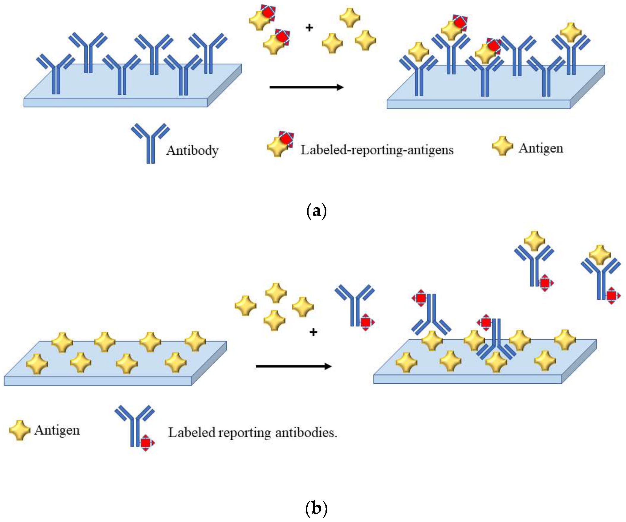
2. Immunosensor Development: Practical Tips and New Trends
3. Immunosensors for Pesticide Determination
3.1. Glyphosate
3.1.1. Optical Immunosensor
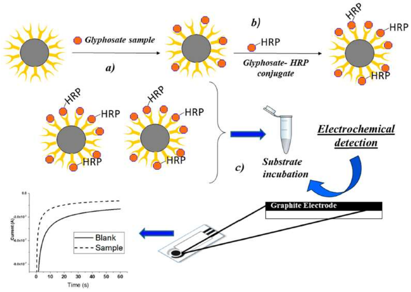
3.1.2. Electrochemical Immunosensor
3.2. Carbamates and Organophosphates
3.2.1. Optical Immunosensor
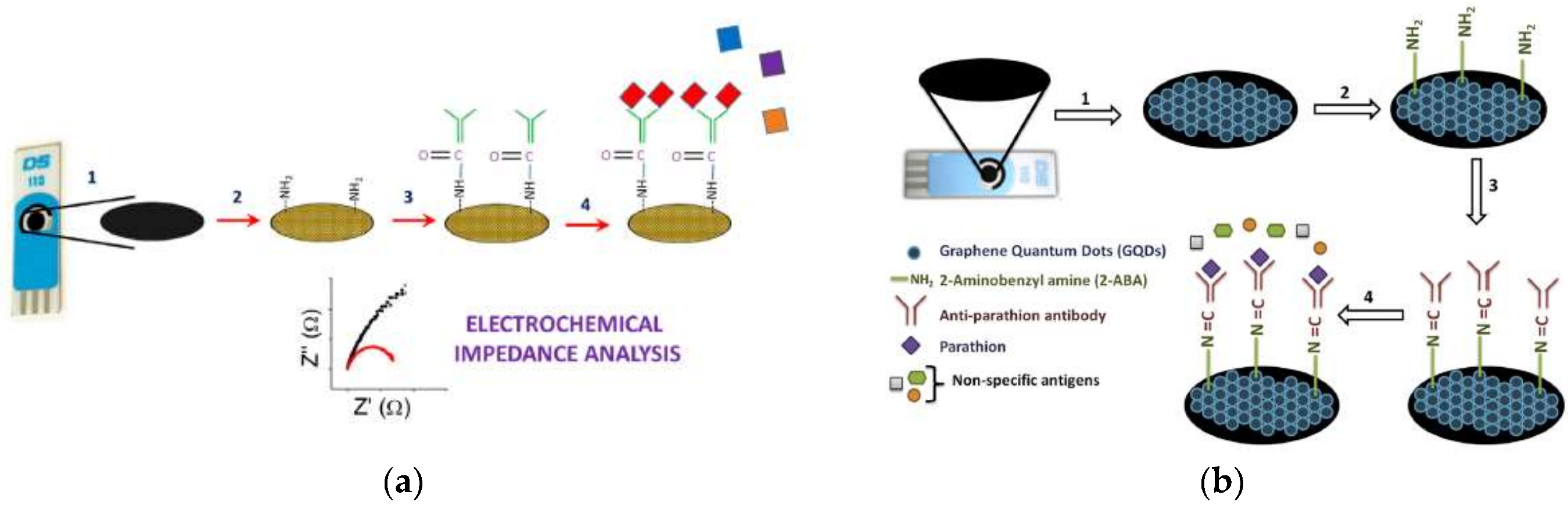
3.2.2. Electrochemical Immunosensor
3.3. Neonicotinoids
3.3.1. Optical Immunosensors
3.3.2. Electrochemical Immunosensor
4. General Consideration and Conclusions
Funding
Acknowledgments
Conflicts of Interest
References
- Storck, V.; Karpouzas, D.G.; Martin-Laurent, F. Towards a better pesticide policy for the European Union. Sci. Total Environ. 2017, 575, 1027–1033. [Google Scholar] [CrossRef] [PubMed]
- Kim, K.H.; Kabir, E.; Jahan, S.A. Exposure to pesticides and the associated human health effects. Sci. Total Environ. 2017, 575, 525–535. [Google Scholar] [CrossRef] [PubMed]
- Landrigan, P.J.; Fuller, R.; Acosta, N.J.R.; Adeyi, O.; Arnold, R.; Basu, N.; Baldé, A.B.; Bertollini, R.; Bose-O’Reilly, S.; Boufford, J.I.; et al. The Lancet Commission on pollution and health. Lancet 2017, 391, 462–512. [Google Scholar] [CrossRef]
- Steingrímsdóttir, M.M.; Petersen, A.; Fantke, P. A screening framework for pesticide substitution in agriculture. J. Clean. Prod. 2018, 192, 306–315. [Google Scholar] [CrossRef]
- World Health Organization (WHO). Manual on Development and Use of FAO and WHO Specifications for Pesticides, 1st ed.; World Health Organization: Rome, Italy, 2016. [Google Scholar]
- Tadeo, J.L.; Sánchez-Brunete, C.; González, L. Pesticides: Classification and Properties. In Analysis of Pesticides in Food and Environmental Samples; Taylor & Francis: Milton Park, UK, 2008. [Google Scholar]
- World Health Organization (WHO). The Who Recommended Classification of Pesticides by Hazard and Guidelines to Classification 2009; World Health Organization: Rome, Italy, 2010; pp. 1–60. [Google Scholar]
- Capoferri, D.; della Pelle, F.; del Carlo, M.; Compagnone, D. Affinity Sensing Strategies for the Detection of Pesticides in Food. Foods 2018, 7, 148. [Google Scholar] [CrossRef] [PubMed]
- Lewis, K.A.; Tzilivakis, J.; Warner, D.J.; Green, A. An international database for pesticide risk assessments and management. Hum. Ecol. Risk Assess. 2016, 22, 1050–1064. [Google Scholar] [CrossRef]
- Rasmussen, J.J.; Wiberg-Larsen, P.; Baattrup-Pedersen, A.; Cedergreen, N.; McKnight, U.S.; Kreuger, J.; Jacobsen, D.; Kristensen, E.A.; Friberg, N. The legacy of pesticide pollution: An overlooked factor in current risk assessments of freshwater systems. Water Res. 2015, 84, 25–32. [Google Scholar] [CrossRef]
- Zhen, M.; Song, B.; Liu, X.; Chandankere, R.; Tang, J. Biochar-mediated regulation of greenhouse gas emission and toxicity reduction in bioremediation of organophosphorus pesticide-contaminated soils. Chin. J. Chem. Eng. 2018, 26, 2592–2600. [Google Scholar] [CrossRef]
- Barghi, M.; Jin, X.; Lee, S.; Jeong, Y.; Yu, J.P.; Paek, W.K.; Moon, H.B. Accumulation and exposure assessment of persistent chlorinated and fluorinated contaminants in Korean birds. Sci. Total Environ. 2018, 645, 220–228. [Google Scholar] [CrossRef]
- Koivisto, E.; Santangeli, A.; Koivisto, P.; Korkolainen, T.; Vuorisalo, T.; Hanski, I.K.; Loivamaa, I.; Koivisto, S. The prevalence and correlates of anticoagulant rodenticide exposure in non-target predators and scavengers in Finland. Sci. Total Environ. 2018, 642, 701–707. [Google Scholar] [CrossRef]
- Ruiz-Suárez, N.; Camacho, M.; Boada, L.D.; Henríquez-Hernández, L.A.; Rial, C.; Valerón, P.F.; Zumbado, M.; González, M.A.; Luzardo, O.P. The assessment of daily dietary intake reveals the existence of a different pattern of bioaccumulation of chlorinated pollutants between domestic dogs and cats. Sci. Total Environ. 2015, 530–531, 45–52. [Google Scholar] [CrossRef] [PubMed]
- Ernst, F.; Alonso, B.; Colazzo, M.; Pareja, L.; Cesio, V.; Pereira, A.; Márquez, A.; Errico, E.; Segura, A.M.; Heinzen, H.; et al. Occurrence of pesticide residues in fish from south American rainfed agroecosystems. Sci. Total Environ. 2018, 631–632, 169–179. [Google Scholar] [CrossRef] [PubMed]
- Abdelhameed, R.M.; El-Zawahry, M.; Emam, H.E. Efficient removal of organophosphorus pesticides from wastewater using polyethylenimine-modified fabrics. Polymer 2018, 155, 225–234. [Google Scholar] [CrossRef]
- Carrère, M.; DeMaria, F.; Drogué, S. Maximum residual levels of pesticides and public health: Best friends or faux amis? Agric. Econ. 2018, 49, 111–118. [Google Scholar] [CrossRef]
- WHO. Codex Alimentarius. Codex Alimentarius. 2018. Available online: http://www.fao.org/fao-who-codexalimentarius/en/ (accessed on 7 November 2018).
- WHO. Codex Alimentarius: Pesticides Index. Codex Alimentarius. 2018. Available online: http://www.fao.org/fao-who-codexalimentarius/codex-texts/dbs/pestres/pesticides/en/ (accessed on 13 November 2018).
- Hird, S. Analysis of pesticides by chromatographic technqiues coupled with mass spectrometry. In Analysis of Pesticides in the Environment, 1st ed.; Tadeo, J.L., Ed.; CRC Press: Boca Raton, FL, USA, 2008; pp. 59–95. [Google Scholar]
- Raina, R. Chemical Analysis of Pesticides Using GC/MS, GC/MS/MS, and LC/MS/MS. In Pesticides-Strategies Por Pesticides Analysis; Stoytcheva, M., Ed.; IntechOpen: Rijeka, Croatia, 2011; p. 28. [Google Scholar]
- Kumar, P.; Kim, K.-H.; Deep, A. Recent advancements in sensing techniques based on functional materials for organophosphate pesticides. Biosens. Bioelectron. 2015, 70, 469–481. [Google Scholar] [CrossRef] [PubMed]
- Akyüz, D.; Keleş, T.; Biyiklioglu, Z.; Koca, A. Electrochemical pesticide sensors based on electropolymerized metallophthalocyanines. J. Electroanal. Chem. 2017, 804, 53–63. [Google Scholar] [CrossRef]
- Liu, S.; Zheng, Z.; Li, X. Advances in pesticide biosensors: Current status, challenges, and future perspectives. Anal. Bioanal. Chem. 2013, 405, 63–90. [Google Scholar] [CrossRef] [PubMed]
- Ding, J.; Sun, X.; Guo, Y.; Jia, H.; Qiao, L.; Wang, X. A Portable Pesticide Residues Detection Instrument Based on Impedance Immunosensor. Sens. Transducers 2014, 172, 27–33. [Google Scholar]
- Guo, Y.; Sun, X.; Liu, X.; Sun, X.; Zhao, G.; Chen, D.; Wang, X. A miniaturized portable instrument for rapid determination pesticides residues in vegetables and fruits. IEEE Sens. J. 2015, 15, 4046–4052. [Google Scholar] [CrossRef]
- Palchetti, I.; Laschi, S.; Marrazza, G.; Mascini, M. Electrochemical imaging of localized sandwich DNA hybridization using scanning electrochemical microscopy. Anal. Chem. 2007, 79, 7206–7213. [Google Scholar] [CrossRef]
- Cervera-Chiner, L.; Juan-Borrás, M.; March, C.; Arnau, A.; Escriche, I.; Montoya, Á.; Jiménez, Y. High Fundamental Frequency Quartz Crystal Microbalance (HFF-QCM) immunosensor for pesticide detection in honey. Food Control 2018, 92, 1–6. [Google Scholar] [CrossRef]
- Arduini, F.; Cinti, S.; Scognamiglio, V.; Moscone, D. Nanomaterials in electrochemical biosensors for pesticide detection: Advances and challenges in food analysis. Microchim. Acta 2016, 183, 2063–2083. [Google Scholar] [CrossRef]
- Yan, X.; Li, H.; Su, X. Review of optical sensors for pesticides. TrAC Trends Anal. Chem. 2018, 103, 1–20. [Google Scholar] [CrossRef]
- Verma, N.; Bhardwaj, A. Biosensor Technology for Pesticides—A review. Appl. Biochem. Biotechnol. 2015, 175, 3093–3119. [Google Scholar] [CrossRef] [PubMed]
- Mehrotra, P. Biosensors and their applications—A review. J. Oral Biol. Craniofac. Res. 2016, 6, 153–159. [Google Scholar] [CrossRef] [PubMed]
- Zhao, G.; Wang, H.; Liu, G. Advances in Biosensor-Based Instruments for Pesticide Residues Rapid Detection. Int. J. Electrochem. Sci. 2015, 10, 9790–9807. [Google Scholar]
- Bettazzi, F.; Laschi, S.; Mascini, M. One-shot screen-printed thylakoid membrane-based biosensor for the detection of photosynthetic inhibitors in discrete samples. Anal. Chim. Acta 2007, 589, 14–21. [Google Scholar] [CrossRef]
- Felix, F.S.; Angnes, L. Electrochemical immunosensors—A powerful tool for analytical applications. Biosens. Bioelectron. 2018, 102, 470–478. [Google Scholar] [CrossRef]
- Ricci, F.; Adornetto, G.; Palleschi, G. A review of experimental aspects of electrochemical immunosensors. Electrochim. Acta 2012, 84, 74–83. [Google Scholar] [CrossRef]
- Gopinath, S.C.B.; Tang, T.H.; Citartan, M.; Chen, Y.; Lakshmipriya, T. Current aspects in immunosensors. Biosens. Bioelectron. 2014, 57, 292–302. [Google Scholar] [CrossRef]
- Kokkinos, C.; Economou, A.; Prodromidis, M.I. Electrochemical immunosensors: Critical survey of different architectures and transduction strategies. TrAC Trends Anal. Chem. 2016, 79, 88–105. [Google Scholar] [CrossRef]
- Palchetti, I. Affinity biosensors for tumor-marker analysis. Bioanalysis 2014, 6, 3417–3435. [Google Scholar] [CrossRef] [PubMed]
- Jiao, S.; Liu, P.; Liu, Y.; Zou, R.; Zhao, Y.; Liu, Y.; Zhu, G.; Guo, Y. Binding properties of broad-specific monoclonal antibodies against three organophosphorus pesticides by a direct surface plasmon resonance immunosensor. Anal. Bioanal. Chem. 2018, 410, 7263–7273. [Google Scholar] [CrossRef] [PubMed]
- Flajnik, M.F.; Deschacht, N.; Muyldermans, S. A Case of Convergence: Why Did a Simple Alternative to Canonical Antibodies Arise in Sharks and Camels? PLoS Biol. 2011, 9, e1001120. [Google Scholar] [CrossRef] [PubMed]
- Sun, Z.; Lv, J.; Liu, X.; Tang, Z.; Wang, X.; Xu, Y.; Hammock, B.D. Development of a Nanobody-AviTag Fusion Protein and Its Application in a Streptavidin-Biotin-Amplified Enzyme-Linked Immunosorbent Assay for Ochratoxin A in Cereal. Anal. Chem. 2018, 90, 10628–10634. [Google Scholar] [CrossRef] [PubMed]
- Tang, X.; Li, P.; Zhang, Q.; Zhang, Z.; Zhang, W.; Jiang, J. Time-Resolved Fluorescence Immunochromatographic Assay Developed Using Two Idiotypic Nanobodies for Rapid, Quantitative, and Simultaneous Detection of Aflatoxin and Zearalenone in Maize and Its Products. Anal. Chem. 2017, 89, 11520–11528. [Google Scholar] [CrossRef] [PubMed]
- Huo, J.; Li, Z.; Wan, D.; Li, D.; Qi, M.; Barnych, B.; Vasylieva, N.; Zhang, J.; Hammock, B.D. Development of a Highly Sensitive Direct Competitive Fluorescence Enzyme Immunoassay Based on a Nanobody-Alkaline Phosphatase Fusion Protein for Detection of 3-Phenoxybenzoic Acid in Urine. J. Agric. Food Chem. 2018, 66, 11284–11290. [Google Scholar] [CrossRef]
- Mascini, M.; Palchetti, I.; Tombelli, S. Nucleic acid and peptide aptamers: Fundamentals and bioanalytical aspects. Angew. Chem. Int. Ed. 2012, 51, 1316–1332. [Google Scholar] [CrossRef]
- Baydemir, G.; Bettazzi, F.; Palchetti, I.; Voccia, D. Strategies for the development of an electrochemical bioassay for tnf-Alpha detection by using a non-immunoglobulin bioreceptor. Talanta 2016, 151, 141–147. [Google Scholar] [CrossRef]
- Peltomaa, R.; López-Perolio, I.; Benito-Peña, E.; Barderas, R.; Moreno-Bondi, M.C. Application of bacteriophages in sensor development. Anal. Bioanal. Chem. 2016, 408, 1805–1828. [Google Scholar] [CrossRef]
- Bettazzi, F.; Marrazza, G.; Minunni, M.; Palchetti, I.; Scarano, S. Chapter One—Biosensors and Related Bioanalytical Tools. In Past, Present and Future Challenges of Biosensors and Bioanalytical Tools in Analytical Chemistry: A Tribute to Professor Marco Mascini 77; Palchetti, I., Hansen, P.-D., Barceló, D.B.T.-C.A.C., Eds.; Elsevier: Amsterdam, The Netherlands, 2017; pp. 1–33. [Google Scholar]
- Piro, B.; Reisberg, S. Recent advances in electrochemical immunosensors. Sensors 2017, 17, 794. [Google Scholar] [CrossRef]
- Voccia, D.; Bettazzi, F.; Baydemir, G.; Palchetti, I. Alkaline-Phosphatase-Based Nanostructure Assemblies for Electrochemical Detection of microRNAs. J. Nanosci. Nanotechnol. 2015, 15, 3378–3384. [Google Scholar] [CrossRef]
- Centi, S.; Silva, E.; Laschi, S.; Palchetti, I.; Mascini, M. Polychlorinated biphenyls (PCBs) detection in milk samples by an electrochemical magneto-immunosensor (EMI) coupled to solid-phase extraction (SPE) and disposable low-density arrays. Anal. Chim. Acta 2007, 594, 9–16. [Google Scholar] [CrossRef]
- Gooding, J.J. Advances in interfacial design for electrochemical biosensors and sensors: Aryl diazonium salts for modifying carbon and metal electrodes. Electroanalysis 2008, 20, 573–582. [Google Scholar] [CrossRef]
- Yáñez-Sedeño, P.; Campuzano, S.; Pingarrón, J.M. Integrated affinity biosensing platforms on screen-printed electrodes electrografted with diazonium salts. Sensors 2018, 18, 675. [Google Scholar] [CrossRef] [PubMed]
- Bettazzi, F.; Enayati, L.; Sánchez, I.C.; Motaghed, R.; Mascini, M.; Palchetti, I. Electrochemical bioassay for the detection of TNF-α using magnetic beads and disposable screen-printed array of electrodes. Bioanalysis 2013, 5, 11–19. [Google Scholar] [CrossRef] [PubMed]
- Bettazzi, F.; Martellini, T.; Shelver, W.L.; Cincinelli, A.; Lanciotti, E.; Palchetti, I. Development of an Electrochemical Immunoassay for the Detection of Polybrominated Diphenyl Ethers (PBDEs). Electroanalysis 2016, 28, 1817–1823. [Google Scholar] [CrossRef]
- Romanelli, S.; Bettazzi, F.; Martellini, T.; Shelver, W.L.; Cincinelli, A.; Galarini, R.; Palchetti, I. Evaluation of a QuEChERS-like extraction approach for the determination of PBDEs in mussels by immuno-assay-based screening methods. Talanta 2017, 170, 540–545. [Google Scholar] [CrossRef] [PubMed]
- Bettazzi, F.; Natale, A.R.; Torres, E.; Palchetti, I. Glyphosate Determination by Coupling an Immuno-Magnetic Assay with Electrochemical Sensors. Sensors 2018, 18, 2965. [Google Scholar] [CrossRef] [PubMed]
- Martini, E.; Merola, G.; Tomassetti, M.; Campanella, L. Agent orange herbicides, organophosphate and triazinic pesticides analysis in olive oil and industrial oil mill waste effluents using new organic phase immunosensors. Food Chem. 2015, 169, 358–365. [Google Scholar] [CrossRef]
- Talan, A.; Mishra, A.; Eremin, S.A.; Narang, J.; Kumar, A.; Gandhi, S. Ultrasensitive electrochemical immuno-sensing platform based on gold nanoparticles triggering chlorpyrifos detection in fruits and vegetables. Biosens. Bioelectron. 2018, 105, 14–21. [Google Scholar] [CrossRef]
- Mehta, J.; Vinayak, P.; Tuteja, S.K.; Chhabra, V.A.; Bhardwaj, N.; Paul, A.K.; Kim, K.H.; Deep, A. Graphene modified screen printed immunosensor for highly sensitive detection of parathion. Biosens. Bioelectron. 2016, 83, 339–346. [Google Scholar] [CrossRef]
- Zhao, W.W.; Xu, J.J.; Chen, H.Y. Photoelectrochemical Immunoassays. Anal. Chem. 2018, 90, 615–627. [Google Scholar] [CrossRef]
- Bettazzi, F.; Palchetti, I. Photoelectrochemical genosensors for the determination of nucleic acid cancer biomarkers. Curr. Opin. Electrochem. 2018, 12, 51–59. [Google Scholar] [CrossRef]
- Bettazzi, F.; Laschi, S.; Voccia, D.; Gellini, C.; Pietraperzia, G.; Falciola, L.; Pifferi, V.; Testolin, A.; Ingrosso, C.; Placido, T.; et al. Ascorbic acid-sensitized Au nanorods-functionalized nanostructured TiO2 transparent electrodes for photoelectrochemical genosensing. Electrochim. Acta 2018, 276, 389–398. [Google Scholar] [CrossRef]
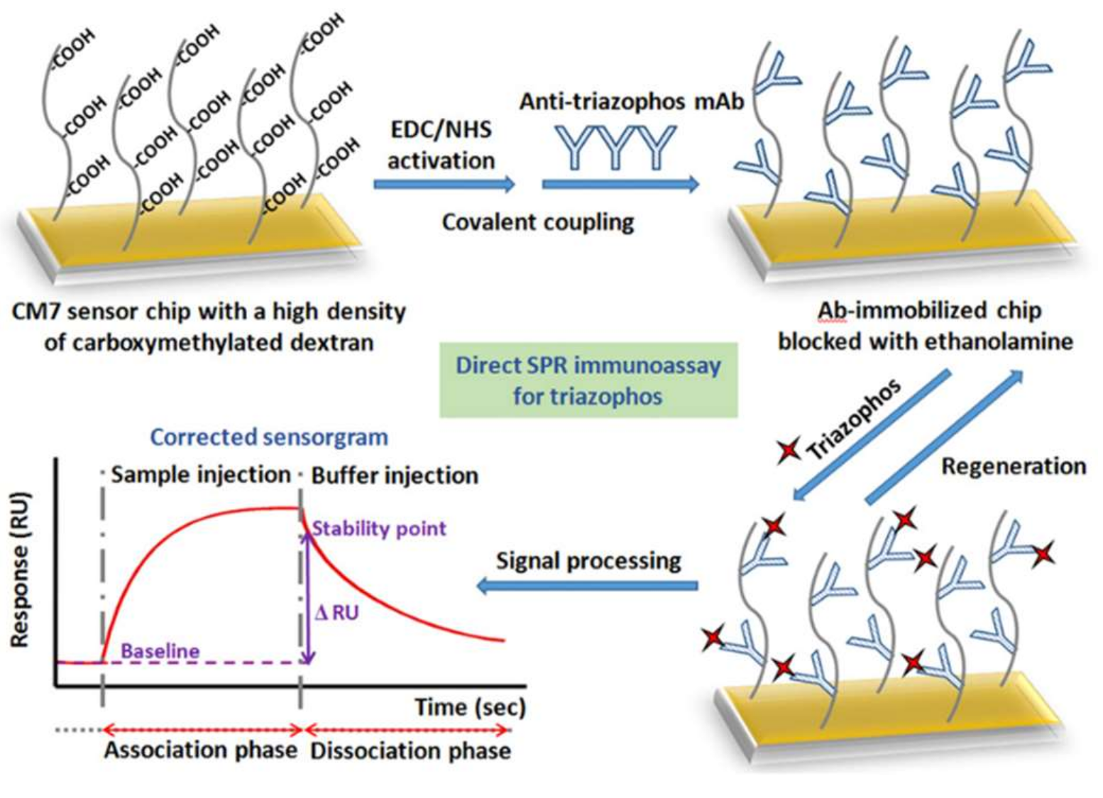
- Guo, Y.; Liu, R.; Liu, Y.; Xiang, D.; Liu, Y.; Gui, W.; Li, M.; Zhu, G. A non-competitive surface plasmon resonance immunosensor for rapid detection of triazophos residue in environmental and agricultural samples. Sci. Total Environ. 2018, 613–614, 783–791. [Google Scholar] [CrossRef]
- Székács, A.; Darvas, B. Forty Years with Glyphosate. In Herbicides; Hasaneen, M.N.A.E.-G., Ed.; IntechOpen: Rijeka, Croatia, 2012. [Google Scholar]
- Firdous, S.; Iqbal, S.; Anwar, S.; Jabeen, H. Identification and analysis of 5-enolpyruvylshikimate-3-phosphate synthase (EPSPS) gene from glyphosate-resistant Ochrobactrum intermedium Sq20. Pest Manag. Sci. 2018, 74, 1184–1196. [Google Scholar] [CrossRef]
- Briefs, I.S.A.A.A. Global Status of Commercialized Biotech/GM Crops in 2017: Biotech Crop Adoption Surges as Economic Benefits Accumulate in 22 Years. 2017, pp. 1–153. Available online: https://www.isaaa.org/resources/publications/briefs/53/download/isaaa-brief-53-2017.pdf (accessed on 7 November 2018).
- Mertens, M.; Höss, S.; Neumann, G.; Afzal, J.; Glyphosate, W.R. Glyphosate, a chelating agent–relevant for ecological risk assessment? Environ. Sci. Pollut. Res. 2018, 25, 5298–5317. [Google Scholar] [CrossRef]
- Bus, J.S. IARC use of oxidative stress as key mode of action characteristic for facilitating cancer classification: Glyphosate case example illustrating a lack of robustness in interpretative implementation. Regul. Toxicol. Pharmacol. 2017, 86, 157–166. [Google Scholar] [CrossRef]
- Davoren, M.J.; Schiestl, R.H. Glyphosate-based herbicides and cancer risk: A post-IARC decision review of potential mechanisms, policy and avenues of research. Carcinogenesis 2018, 39, 1207–1215. [Google Scholar] [CrossRef]
- Tarazona, J.V.; Court-Marques, D.; Tiramani, M.; Reich, H.; Pfeil, R.; Istace, F.; Crivellente, F. Glyphosate toxicity and carcinogenicity: A review of the scientific basis of the European Union assessment and its differences with IARC. Arch. Toxicol. 2017, 91, 2723–2743. [Google Scholar] [CrossRef]
- IARC. IARC Monographs Volume 112: Evaluation of Five Organophosphate Insecticides and Herbicides; IARC: Lyon, France, 2015. [Google Scholar]
- ECHA. Glyphosate Not Classified as a Carcinogen by ECHA. 2017. Available online: https://echa.europa.eu/-/glyphosate-not-classified-as-a-carcinogen-by-echa (accessed on 7 November 2018).
- EFSA. Conclusion on the peer review of the pesticide risk assessment of the active substance glyphosate. EFSA J. 2015, 13, 4302. [Google Scholar]
- FAO. Pesticides Database Search: Glyphosate. In Codex Alimentarius; FAO: Rome, Italy, 2018. [Google Scholar]
- T. C. of the E. Union. Council Directive 98/83/EC of 3 November 1998 on the quality of water intended for human consumption. Off. J. Eur. Commun. 1998, L330, 32–54. [Google Scholar]
- USEPA. EPA 816-F-09-004. National Primary Drinking Water Regulations. Usepa 2009, 1, 7. [Google Scholar]
- SEGOB. NOM-201-SSA1-2015. Productos y Servicios. Agua y Hielo para Consumo Humano, Envasados y a Granel. Especificaciones Sanitarias. Diario Oficial de la Federación. 2015. Available online: http://dof.gob.mx/nota_detalle.php?codigo=5420977&fecha=22/12/2015 (accessed on 13 November 2018).
- H. Canada. Guidelines for Canadian Drinking Water Quality: Guideline Technical Document—Glyphosate. 1995, pp. 1–5. Available online: http://ceqg-rcqe.ccme.ca/download/en/182/ (accessed on 13 November 2018).
- Winfield, T.W.; Bashe, W.J.; Baker, T.V. Method 547 Determination of Glyphosate in Drinking Water by Direct-Aqueous-Injection Hplc, Post-Column Derivatization, and Fluorescence Detection. Technol. Appl. 1990, 1, 1–16. [Google Scholar]
- Byer, J.D.; Struger, J.; Klawunn, P.; Todd, A.; Sverko, E. Low Cost Monitoring of Glyphosate in Surface waters Using the ELSA Method an Evaluation. Environ. Sci. Technol. 2008, 42, 6052–6057. [Google Scholar] [CrossRef]
- Mörtl, M.; Németh, G.; Juracsek, J.; Darvas, B.; Kamp, L.; Rubio, F.; Székács, A. Determination of glyphosate residues in Hungarian water samples by immunoassay. Microchem. J. 2013, 107, 143–151. [Google Scholar] [CrossRef]
- Osten, J.R.; Dzul-Caamal, R. Glyphosate residues in groundwater, drinking water and urine of subsistence farmers from intensive agriculture localities: A survey in Hopelchén, Campeche, Mexico. Int. J. Environ. Res. Public Health 2017, 14, 595. [Google Scholar] [CrossRef]
- Sanchís, J.; Kantiani, L.; Llorca, M.; Rubio, F.; Ginebreda, A.; Fraile, J.; Garrido, T.; Farré, M. Determination of glyphosate in groundwater samples using an ultrasensitive immunoassay and confirmation by on-line solid-phase extraction followed by liquid chromatography coupled to tandem mass spectrometry. Anal. Bioanal. Chem. 2012, 402, 2335–2345. [Google Scholar] [CrossRef]
- Zhao, J.; Pacenka, S.; Wu, J.; Richards, B.K.; Steenhuis, T.; Simpson, K.; Hay, A.G. Detection of glyphosate residues in companion animal feeds. Environ. Pollut. 2018, 243, 1113–1118. [Google Scholar] [CrossRef]
- Rubio, F.; Guo, E.; Kamp, L. Survey of Glyphosate Residues in Honey, Corn and Soy Products. J. Environ. Anal. Toxicol. 2014, 5, 1–8. [Google Scholar]
- González-Martínez, M.Á.; Brun, E.M.; Puchades, R.; Maquieira, Á.; Ramsey, K.; Rubio, F. Glyphosate immunosensor. Application for water and soil analysis. Anal. Chem. 2005, 77, 4219–4227. [Google Scholar] [CrossRef]
- Lee, H.U.; Shin, H.Y.; Lee, J.Y.; Song, Y.S.; Park, C.; Kim, S.W. Quantitative detection of glyphosate by simultaneous analysis of UV spectroscopy and fluorescence using DNA-labeled gold nanoparticles. J. Agric. Food Chem. 2010, 58, 12096–12100. [Google Scholar] [CrossRef]
- Lee, H.U.; Jung, D.U.; Lee, J.H.; Song, Y.S.; Park, C.; Kim, S.W. Detection of glyphosate by quantitative analysis of fluorescence and single DNA using DNA-labeled fluorescent magnetic core-shell nanoparticles. Sens. Actuator B Chem. 2013, 177, 879–886. [Google Scholar] [CrossRef]
- Noori, J.; Dimaki, M.; Mortensen, J.; Svendsen, W. Detection of Glyphosate in Drinking Water: A Fast and Direct Detection Method without Sample Pretreatment. Sensors 2018, 18, 2961. [Google Scholar] [CrossRef]
- Songa, E.A.; Arotiba, O.A.; Owino, J.H.O.; Jahed, N.; Baker, P.G.L.; Iwuoha, E.I. Electrochemical detection of glyphosate herbicide using horseradish peroxidase immobilized on sulfonated polymer matrix. Bioelectrochemistry 2009, 75, 117–123. [Google Scholar] [CrossRef]
- Vaghela, C.; Kulkarni, M.; Haram, S.; Aiyer, R.; Karve, M. A novel inhibition based biosensor using urease nanoconjugate entrapped biocomposite membrane for potentiometric glyphosate detection. Int. J. Biol. Macromol. 2018, 108, 32–40. [Google Scholar] [CrossRef]
- Dalefield, R. Insecticides and Acaricides. In Veterinary Toxicology for Australia and New Zealand; Elsevier Inc.: Amsterdam, The Netherlands, 2017; pp. 87–109. [Google Scholar]
- Alloway, B.J. Land Contamination and Reclamation. In Understanding our Environment. An Introduction to Environmental Chemistry and Pollution, 3rd ed.; Royal Society of Chemistry: London, UK, 2007; pp. 199–236. [Google Scholar]
- Atwood, D.; Paisley-Jones, C. Pesticides Industry Sales and Usage 2008–2012; FAO: FAO: Rome, Italy, November 2017; p. 24. [Google Scholar]
- Organophosphates, A.M. Carbamate. In Encyclopedia of Food Safety, 1st ed.; Motarjemi, Y., Moy, G.G., Todd, E.C.D., Eds.; Elsevier Inc.: Amsterdam, The Netherlands, 2015; pp. e1236–e1239. [Google Scholar]
- Georgiadis, G.; Mavridis, C.; Belantis, C.; Zisis, I.E.; Skamagkas, I.; Fragkiadoulaki, I.; Heretis, I.; Tzortzis, V.; Psathakis, K.; Tsatsakis, A.; et al. Nephrotoxicity issues of organophosphates. Toxicology 2018, 406–407, 129–136. [Google Scholar] [CrossRef]
- Rowe, C.; Gunier, R.; Bradman, A.; Harley, K.G.; Kogut, K.; Parra, K.; Eskenazi, B. Residential proximity to organophosphate and carbamate pesticide use during pregnancy, poverty during childhood, and cognitive functioning in 10-year-old children. Environ. Res. 2016, 150, 128–137. [Google Scholar] [CrossRef]
- Dhouib, I.B.; Annabi, A.; Jallouli, M.; Marzouki, S.; Gharbi, N.; Elfazaa, S.; Lasram, M.M. Carbamates pesticides induced immunotoxicity and carcinogenicity in human: A review. J. Appl. Biomed. 2016, 14, 85–90. [Google Scholar] [CrossRef]
- Gupta, R.C.; Sachana, M.; Mukherjee, I.M.; Doss, R.B.; Malik, J.K.; Milatovic, D. Organophosphates and Carbamates. In Veterinary Toxicology-Basic and Clinical Principles, 3rd ed.; Academic Press: Cambridge, MA, USA, 2018; pp. 495–508. [Google Scholar]
- FAO. Maximum Residue Limits for Processed or Ready-To-Eat; FAO-WHO: Rotterdam, The Netherlands, 2003. [Google Scholar]
- IARC. List of Classifications. 2018, Volumes 1–115. Available online: https://monographs.iarc.fr/wp-content/uploads/2018/09/List_of_Classifications.pdf (accessed on 7 November 2018).
- Tang, X.; Liang, B.; Yi, T.; Palchetti, I.; Manco, G.; Liu, A. Cell surface display of organophosphorus hydrolase for sensitive spectrophotometric detection of p-nitrophenol substituted organophosphates. Enzyme Microb. Technol. 2014, 55, 107–112. [Google Scholar] [CrossRef]
- Porzio, E.; Bettazzi, F.; Mandrich, L.; del Giudice, I.; Restaino, O.F.; Laschi, S.; Febbraio, F.; de Luca, V.; Borzacchiello, M.G.; Carusone, T.M.; et al. Innovative Biocatalysts as Tools to Detect and Inactivate Nerve Agents. Sci. Rep. 2018, 8, 1–14. [Google Scholar] [CrossRef]
- Hernandez, S.; Palchetti, I.; Mascini, M. Determination of anticholinesterase activity for pesticides monitoring using a thiocholine sensor. Int. J. Environ. Anal. Chem. 2000, 78, 263–278. [Google Scholar] [CrossRef]
- Cagnini, A.; Palchetti, I.; Mascini, M.; Turner, A.P.F. Ruthenized screen-printed choline oxidase-based biosensors for measurement of anticholinesterase activity. Mikrochim. Acta 1995, 121, 155–166. [Google Scholar] [CrossRef]
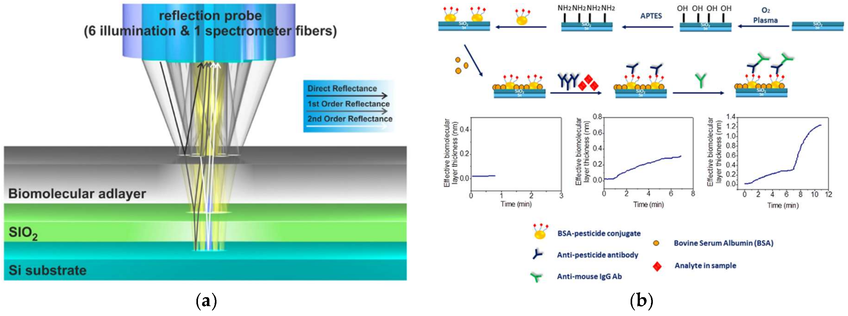
- Koukouvinos, G.; Τsialla, Z.; Petrou, P.S.; Misiakos, K.; Goustouridis, D.; Moreno, A.U.; Fernandez-Alba, A.R.; Raptis, I.; Kakabakos, S.E. Fast simultaneous detection of three pesticides by a White Light Reflectance Spectroscopy sensing platform. Sens. Actuators B Chem. 2017, 238, 1214–1223. [Google Scholar] [CrossRef]
- Mauriz, E.; Calle, A.; Montoya, A.; Lechuga, L.M. Determination of environmental organic pollutants with a portable optical immunosensor. Talanta 2006, 69, 359–364. [Google Scholar] [CrossRef]
- Martini, E.; Tomassetti, M.; Campanella, L. Determination of traces of several pesticides in sunflower oil using organic phase immuno electrodes (OPIEs). Talanta 2015, 132, 503–512. [Google Scholar] [CrossRef]
- Mehta, J.; Bhardwaj, N.; Bhardwaj, S.K.; Tuteja, S.K.; Vinayak, P.; Paul, A.K.; Kim, K.H.; Deep, A. Graphene quantum dot modified screen printed immunosensor for the determination of parathion. Anal. Biochem. 2017, 523, 1–9. [Google Scholar] [CrossRef]
- Wang, W.H.; Han, Z.J.; Liang, P.J.; Guo, D.Q.; Xiang, Y.J.; Tian, M.X.; Song, Z.L.; Zhao, H.R. Co3O4/PAn magnetic nanoparticle-modified electrochemical immunosensor for chlorpyrifos. Dig. J. Nanomater. Biostruct. 2017, 12, 1–9. [Google Scholar]
- Qiao, L.; Guo, Y.; Sun, X.; Jiao, Y.; Wang, X. Electrochemical immunosensor with NiAl-layered double hydroxide/graphene nanocomposites and hollow gold nanospheres double-assisted signal amplification. Bioprocess Biosyst. Eng. 2015, 38, 1455–1468. [Google Scholar] [CrossRef]
- Cao, Y.; Sun, X.; Guo, Y.; Zhao, W.; Wang, X. An electrochemical immunosensor based on interdigitated array microelectrode for the detection of chlorpyrifos. Bioprocess Biosyst. Eng. 2015, 38, 307–313. [Google Scholar] [CrossRef]
- Jia, H.; Guo, Y.; Sun, X.; Wang, X. An electrochemical immunosensor based on microfluidic chip for detection of chlorpyrifos. Int. J. Electrochem. Sci. 2015, 10, 8750–8758. [Google Scholar]
- Liu, L.; Xu, D.; Hu, Y.; Liu, S.; Wei, H.; Zheng, J.; Wang, G.; Hu, X.; Wang, C. Construction of an impedimetric immunosensor for label-free detecting carbofuran residual in agricultural and environmental samples. Food Control 2015, 53, 72–80. [Google Scholar] [CrossRef]
- Han, E.; Zhou, L.-N.; Yan, J.-K.; Cai, J.-R.; Tian, Q.-R.; Zeng, N. Label-free electrochemical immunosensor for sensitive detection of pesticide residue in vegetables. Mod. Food Sci. Technol. 2014, 30, 268–273. [Google Scholar]
- Ihara, M.; Matsuda, K. Neonicotinoids: Molecular mechanisms of action, insights into resistance and impact on pollinators. Curr. Opin. Insect Sci. 2018. [Google Scholar] [CrossRef]
- Blake, R.J.; Copping, L.G. Are neonicotinoids killing bees? Pest Manag. Sci. 2017, 73, 1293–1294. [Google Scholar] [CrossRef]
- Sparks, T.C.; Nauen, R. IRAC: Mode of action classification and insecticide resistance management. Pestic. Biochem. Physiol. 2015, 121, 122–128. [Google Scholar] [CrossRef]
- Zhang, Q.; Li, Z.; Chang, C.H.; Lou, J.L.; Zhao, M.R.; Lu, C. Potential human exposures to neonicotinoid insecticides: A review. Environ. Pollut. 2018, 236, 71–81. [Google Scholar] [CrossRef]
- Rico, A.; Arenas-Sánchez, A.; Pasqualini, J.; García-Astillero, A.; Cherta, L.; Nozal, L.; Vighi, M. Effects of imidacloprid and a neonicotinoid mixture on aquatic invertebrate communities under Mediterranean conditions. Aquat. Toxicol. 2018, 204, 130–143. [Google Scholar] [CrossRef]
- Han, W.; Tian, Y.; Shen, X. Human exposure to neonicotinoid insecticides and the evaluation of their potential toxicity: An overview. Chemosphere 2018, 192, 59–65. [Google Scholar] [CrossRef]
- Casida, J.E. Neonicotinoids and Other Insect Nicotinic Receptor Competitive Modulators: Progress and Prospects. Annu. Rev. Entomol. 2018, 63, 125–144. [Google Scholar] [CrossRef]
- European Food Safety Authority. Q&A: Conclusions on Neonicotinoids 2018; European Food Safety Authority: Parma, Italy, 2018; pp. 1–2. [Google Scholar]
- European Commision. COMMISSION REGULATION (EU) No 491/2014. Off. J. Eur. Union L 2014, 146, 1–91. [Google Scholar]
- Mitchell, E.A.D.; Mulhauser, B.; Mulot, M.; Aebi, A. A worldwide survey of neonicotinoids in honey. Science 2017, 358, 109–111. [Google Scholar] [CrossRef]
- Zhang, Q.; Wang, X.; Li, Z.; Jin, H.; Lu, Z.; Yu, C.; Huang, Y.F.; Zhao, M. Simultaneous determination of nine neonicotinoids in human urine using isotope-dilution ultra-performance liquid chromatography–tandem mass spectrometry. Environ. Pollut. 2018, 240, 647–652. [Google Scholar] [CrossRef]
- E. Commission. Legislation 132. Off. J. Eur. Union 2018, 61, 31–40. Available online: https://eur-lex.europa.eu/legal-content/EN/TXT/?uri=OJ:L:2018:132:TOC (accessed on 1 September 2018).
- Si, F.; Zou, R.; Jiao, S.; Qiao, X.; Guo, Y.; Zhu, G. Inner filter effect-based homogeneous immunoassay for rapid detection of imidacloprid residue in environmental and food samples. Ecotoxicol. Environ. Saf. 2018, 148, 862–868. [Google Scholar] [CrossRef]
- Lee, K.L.; You, M.L.; Tsai, C.H.; Lin, E.H.; Hsieh, S.Y.; Ho, M.H.; Hsu, J.C.; Wei, P.K. Nanoplasmonic biochips for rapid label-free detection of imidacloprid pesticides with a smartphone. Biosens. Bioelectron. 2016, 75, 88–95. [Google Scholar] [CrossRef]
- Hirakawa, Y.; Yamasaki, T.; Harada, A.; Iwasa, S.; Narita, H.; Miyake, S. Development of an immunosensor based on surface plasmon resonance for simultaneous residue analysis of three pesticides—boscalid, clothianidin, and nitenpyram—In vegetables. Anal. Sci. 2018, 34, 533–539. [Google Scholar] [CrossRef]
- Oliveira, A.E.F.; Bettio, G.B.; Pereira, A.C. An Electrochemical Sensor Based on Electropolymerization of ß-Cyclodextrin and Reduced Graphene Oxide on a Glassy Carbon Electrode for Determination of Neonicotinoids. Electroanalysis 2018, 30, 1918–1928. [Google Scholar] [CrossRef]
- Urbanová, V.; Bakandritsos, A.; Jakubec, P.; Szambó, T.; Zbořil, R. A facile graphene oxide based sensor for electrochemical detection of neonicotinoids. Biosens. Bioelectron. 2017, 89, 532–537. [Google Scholar] [CrossRef]
- Madianos, L.; Tsekenis, G.; Skotadis, E.; Patsiouras, L.; Tsoukalas, D. A highly sensitive impedimetric aptasensor for the selective detection of acetamiprid and atrazine based on microwires formed by platinum nanoparticles. Biosens. Bioelectron. 2018, 101, 268–274. [Google Scholar] [CrossRef]
- Verdian, A. Apta-nanosensors for detection and quantitative determination of acetamiprid—A pesticide residue in food and environment. Talanta 2018, 176, 456–464. [Google Scholar] [CrossRef]
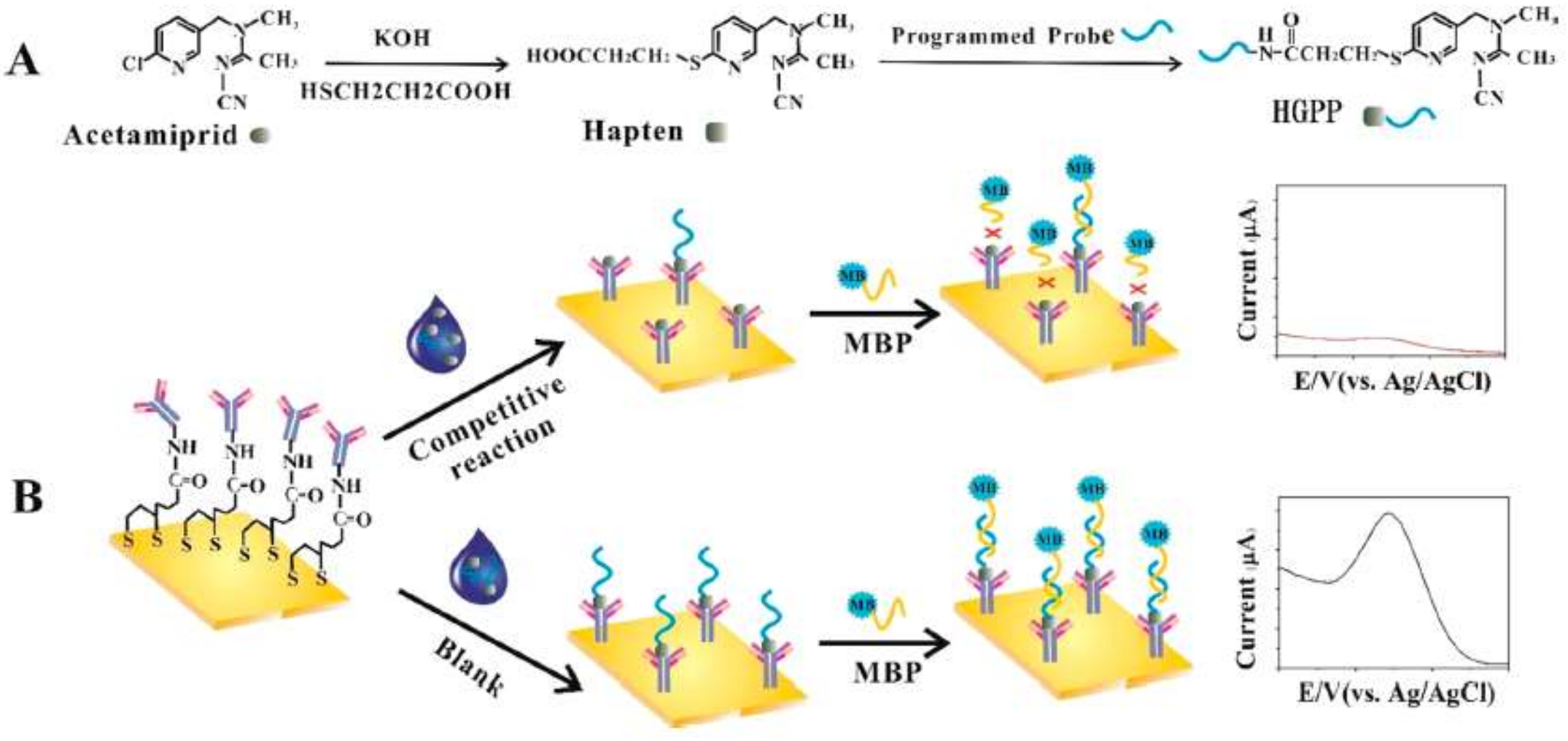
- Xu, X.; Guo, Y.; Wang, L.; He, K.; Guo, Y.; Wang, X.; Gunasekaran, S. Hapten-Grafted Programmed Probe as a Corecognition Element for a Competitive Immunosensor to Detect Acetamiprid Residue in Agricultural Products. J. Agric. Food Chem. 2018, 66, 7815–7821. [Google Scholar] [CrossRef]
- Giri, B.; Pandey, B.; Neupane, B.; Ligler, F.S. Signal amplification strategies for microfluidic immunoassays. TrAC Trends Anal. Chem. 2016, 79, 326–334. [Google Scholar] [CrossRef]
- Jamshaid, T.; Neto, E.T.T.; Eissa, M.M.; Zine, N.; Kunita, M.H.; El-Salhi, A.E.; Elaissari, A. Magnetic particles: From preparation to lab-on-a-chip, biosensors, microsystems and microfluidics applications. TrAC Trends Anal. Chem. 2016, 79, 344–362. [Google Scholar] [CrossRef]


| Immunosensor | Compound Detected | Detection Limit | Linear Range | Matrix | Reference |
|---|---|---|---|---|---|
| Magnetic nanoparticles-modified | Chlorpyrifos | 0.01 µg/mL | 0.01–10 µg/mL | Agricultural products | [111] |
| Graphene nanocomposites and hollow gold nanospheres | Chlorpyrifos | 0.052 ng/mL | 2–150 µg/mL | Vegetable samples | [112] |
| Interdigitated array microelectrodes | Chlorpyrifos | 0.014 ng/mL | 0.001–10 µg/mL | Chives, lettuce and cabbage | [113] |
| Microfluidic chip | Chlorpyrifos | 0.5 ng/mL | 0.5–500 ng/mL | Chives, lettuce and pakchoi | [114] |
| Gold electrode | Carbofuran | 0.1 ng/mL | 0.1–1000 ng/mL | Agricultural and environmental samples | [115] |
| Glassy carbon electrode | Carbofuran | 1 ng/mL | 0.001–100 µg/mL | Vegetable samples | [116] |
© 2019 by the authors. Licensee MDPI, Basel, Switzerland. This article is an open access article distributed under the terms and conditions of the Creative Commons Attribution (CC BY) license (http://creativecommons.org/licenses/by/4.0/).
Share and Cite
Reynoso, E.C.; Torres, E.; Bettazzi, F.; Palchetti, I. Trends and Perspectives in Immunosensors for Determination of Currently-Used Pesticides: The Case of Glyphosate, Organophosphates, and Neonicotinoids. Biosensors 2019, 9, 20. https://doi.org/10.3390/bios9010020
Reynoso EC, Torres E, Bettazzi F, Palchetti I. Trends and Perspectives in Immunosensors for Determination of Currently-Used Pesticides: The Case of Glyphosate, Organophosphates, and Neonicotinoids. Biosensors. 2019; 9(1):20. https://doi.org/10.3390/bios9010020
Chicago/Turabian StyleReynoso, Eduardo C., Eduardo Torres, Francesca Bettazzi, and Ilaria Palchetti. 2019. "Trends and Perspectives in Immunosensors for Determination of Currently-Used Pesticides: The Case of Glyphosate, Organophosphates, and Neonicotinoids" Biosensors 9, no. 1: 20. https://doi.org/10.3390/bios9010020
APA StyleReynoso, E. C., Torres, E., Bettazzi, F., & Palchetti, I. (2019). Trends and Perspectives in Immunosensors for Determination of Currently-Used Pesticides: The Case of Glyphosate, Organophosphates, and Neonicotinoids. Biosensors, 9(1), 20. https://doi.org/10.3390/bios9010020