1. Introduction
Droplet-based microfluidics has become a central platform for miniaturized biochemical assays and high-throughput experimentation. By compartmentalizing reactions into monodisperse, picolitre–nanoliter droplets dispersed in an immiscible carrier fluid, droplet systems dramatically reduce reagent consumption, improve reaction kinetics and enable large numbers of parallel experiments that would be impractical in conventional microtubes or microplates [
1,
2,
3]. Early reviews by Teh et al. and Theberge et al. summarized how monodisperse droplets can serve as isolated microreactors for synthesis, enzyme screening and single-cell studies, and highlighted their potential to transform analytical chemistry and biology [
4,
5]. Subsequent work has expanded these ideas across pharmaceutical development, microbiology, single-cell omics and synthetic biology, establishing droplet microfluidics as an indispensable tool in contemporary life science research [
6,
7,
8].
Digital nucleic-acid assays are among the most widely adopted applications of droplet technology. In digital PCR (dPCR) and droplet digital PCR (ddPCR), a bulk sample is partitioned into thousands to millions of nanoliter or sub-nanoliter compartments, each behaving as a yes/no microreaction whose positivity reflects the presence of one or more target molecules [
9]. Counting fluorescent-positive droplets and invoking Poisson statistics enables absolute quantification of DNA or RNA copies without external calibration curves [
10]. Over the last decade, a rich ecosystem of droplet- and chamber-based dPCR devices has emerged, including highly integrated chips for oncology and infectious disease testing [
11]. More recently, loop-mediated isothermal amplification (LAMP) has been combined with droplet partitioning to create digital LAMP (dLAMP) platforms [
12], which leverage the rapid, single-temperature amplification and inhibitor tolerance of LAMP while preserving the absolute quantification advantages of digital read-out [
13,
14,
15]. These dLAMP approaches span droplet array chips, membrane-based devices and fully integrated cartridges for continuous partitioning, incubation and fluorescence detection [
16].
Parallel to nucleic-acid detection, droplet microfluidics has also advanced microbiology. Encapsulating individual bacteria in droplets allows digital colony-forming unit (CFU) counting and single-cell phenotyping over wide dynamic ranges [
17]. Scheler and co-workers optimized a droplet digital CFU (ddCFU) assay that quantifies bacterial loads over six orders of magnitude by fitting the fraction of growth-positive droplets to a Poisson model [
15]. Kao et al. later implemented a gravity-driven droplet platform for digital enumeration of bacteria and antibiotic susceptibility testing, underscoring how digital microbiology can deliver precise counts and minimum inhibitory concentration measurements in a single workflow [
18]. More recently, microfluidic “digital plating” platforms have begun to bridge droplet-based assays with classical agar-plate microbiology, enabling controlled deposition of droplets onto solid media for robust CFU analysis and colony isolation [
19,
20,
21]. These developments collectively demonstrate that droplet techniques can support both molecular diagnostics and culture-based microbiology within unified frameworks.
The mechanisms and efficiency of droplet or bubble formation in microchannels have been studied in depth, and a broad set of strategies has been developed to improve breakup stability, throughput, and size control [
22,
23]. Representative passive approaches rely on interfacial shear within canonical geometries such as T-junctions, flow-focusing, and co-flow configurations, where carefully designed channel constraints promote reproducible pinch-off [
24]. For example, Shen demonstrated that a simple geometric modification—introducing a glass capillary into a microchannel—can create local confinement that enhances droplet separation and controllability, yielding a more stable droplet stream [
25]. Beyond purely passive designs, active or hybrid methods further expand the controllable parameter space: a Physics of Fluids study showed that applying an electric field to flow-focusing or T-junction microchannels can reshape the interface and alter the effective shear conditions, thereby increasing breakup efficiency and droplet generation frequency [
26]. Meanwhile, work reported in the International Journal of Multiphase Flow introduced a redesigned T-junction capable of alternating one-to-one droplet formation [
27], where precise tuning of flow rates and geometry enabled sequential droplet production from different phases, highlighting the strong coupling between channel design, hydrodynamics, and multiphase partitioning.
Despite these advances, conventional droplet microfluidic implementations remain technically demanding in practice. Most systems still depend on microfabricated chips incorporating flow-focusing or T-junction structures, driven by syringe or pressure pumps and supported by ancillary valves, tubing, and control electronics [
28]. Achieving stable, monodisperse droplet generation typically requires tight regulation of flow rates, surfactant formulation, and surface wetting conditions; even modest fluctuations in pump performance or gradual channel fouling can rapidly compromise droplet uniformity [
29]. As a result, the operational complexity and infrastructure requirements continue to limit accessibility to laboratories with microfabrication capabilities and experienced users, and they hinder adoption in low-resource or point-of-care scenarios. This barrier is particularly evident in microbiology and teaching laboratories, where the assays themselves may be conceptually straightforward, yet the need for specialized chips and external pumping hardware remains a major obstacle to broader dissemination [
24].
To address these issues, considerable effort has gone into simplifying droplet hardware and flow control. Several groups have developed pump-free or hand-powered droplet generators based on negative pressure, gravity, spring-driven syringes or vacuum-assisted actuation [
30]. For example, Chen et al. reported a hand-held, power-free microfluidic device that uses a pre-evacuated syringe to drive flow through a droplet-generation chip, producing monodisperse sub-nanoliter droplets with coefficients of variation below 8% [
31]. Han et al. designed a wind-up mechanical pump that maintains nearly constant syringe flow without electricity, while Fajrial et al. introduced a frugal gravity-assisted pump built from commodity components [
32]. Other minimal droplet generators integrate microchannel networks with small negative-pressure sources or laptop-controlled valves to reduce footprint and setup complexity [
33]. These approaches substantially lower system cost and improve portability, yet they still depend on microfabricated channels and chip-to-world connections that can be prone to leakage, clogging and sample loss.
Droplet generation strategies in microfluidics span a broad spectrum, from chip-free handheld systems to highly integrated, actively controlled lab-on-a-chip platforms, with substantial differences in actuation principles, integration level, and required infrastructure. On the chip-free side, droplets can be generated directly from simple capillaries or pipette tips by applying acoustic, mechanical, or centrifugal excitation to destabilize a liquid jet and drive controlled breakup without the need for enclosed microchannels. Representative examples include vibrating sharp-tip capillary systems, in which piezoelectric actuation induces acoustic streaming and produces droplets with finely tunable diameters and high throughput—features that are particularly attractive for quantitative workflows such as digital PCR—albeit at the cost of bespoke actuators and careful alignment between the vibration source and the nozzle geometry [
34]. Similarly, acoustically activated nozzles can generate and dispense microdroplets via acoustic streaming at the capillary interface, enabling excellent size control and rates up to ~2000 droplets/s, but typically requiring customized transducers and control electronics to ensure reproducible operation [
35]. These chip-free approaches reduce fabrication burden, minimize dead volume in tubing, and simplify cleaning, yet the dependence on specialized hardware and alignment procedures can still limit adoption outside specialist laboratories.
At the opposite end of the spectrum are integrated, chip-based active droplet generators, such as microvalve platforms, which can precisely coordinate droplet generation, splitting, and merging on a single microfluidic chip to support complex fluidic sequencing and high-throughput screening. For example, Agnihotri et al. demonstrated a multi-site microvalve system that achieves programmable droplet operations through on-chip valves, but this level of functionality generally relies on microfabrication, external pressure controllers, and sophisticated control hardware [
36].
The toothbrush-driven droplet generator presented here occupies a practical niche between these extremes in terms of actuation simplicity, infrastructure requirements, and achievable performance. By repurposing a consumer-grade toothbrush motor and pairing it with a simple fluidic interface, it enables chip-free droplet generation with high accessibility and minimal dependence on external equipment. Unlike vibrating-capillary or acoustofluidic systems, it does not require bespoke electronics or precise actuator–nozzle alignment; unlike microvalve-based platforms, it eliminates microfabricated chips and external pumping hardware. The trade-off is that throughput and droplet size uniformity are presently more modest than those achieved by specialized high-performance platforms. Nevertheless, the ultra-low cost, ease of use, and integration of actuation and handling into a single handheld unit make this approach well suited for resource-limited settings, rapid field deployment, and point-of-care digital workflows where simplicity and accessibility are prioritized.
In parallel with these engineering efforts, there is growing interest in “frugal science” and repurposed consumer hardware for laboratory applications, where low-cost devices based on toys, optical drives or smartphones are adapted as centrifuges, microscopes and analyzers for resource-limited settings [
37]. In the context of droplet workflows, however, most existing frugal devices still operate upstream or downstream of chip-based generators rather than replacing the generator itself.
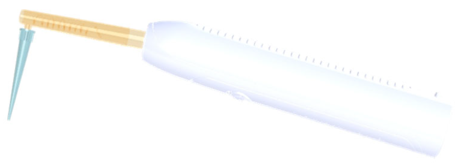
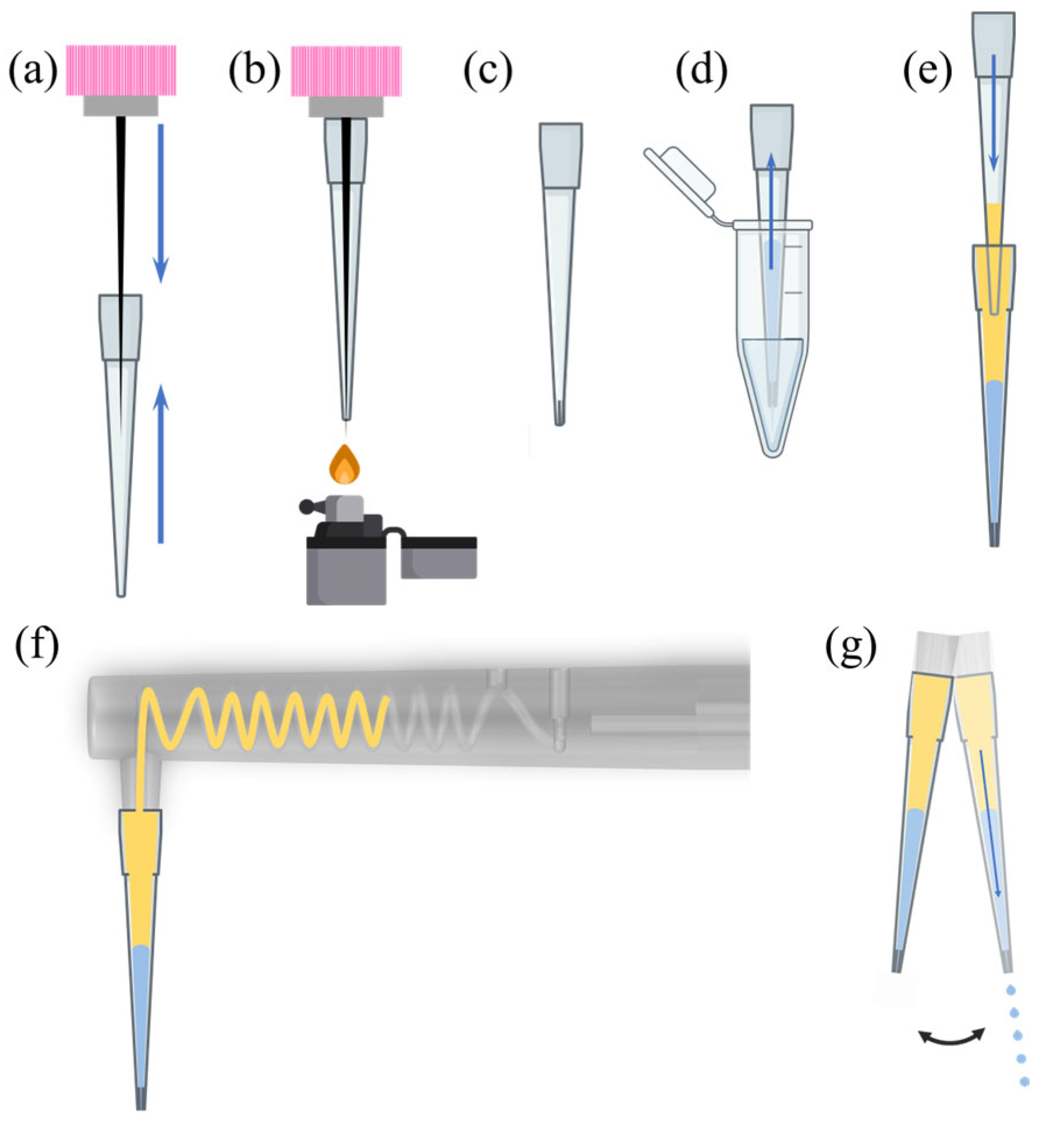
Here we propose a different approach: a handheld vibrational droplet generator that repurposes a commercial electric toothbrush as both actuator and handle. A disposable, heat-modified pipette tip is mounted on a compact 3D-printed adapter attached to the toothbrush shaft. By tuning a small counterweight on the shaft, the strong, well-defined vibrations produced by the toothbrush are converted into a controlled lateral oscillation at the nozzle, yielding a single stable jet that breaks into nearly monodisperse water-in-oil droplets at rates of ~100 droplets s−1. In contrast to conventional microfluidic generators, our system is entirely chip-free and pump-free: there are no microchannels, valves or external pressure sources, and the only wetted component is the disposable pipette tip. This configuration minimizes dead volume, avoids cross-contamination between assays, and confines all fabrication to 3D-printed parts that can be readily reproduced.
We further show that droplets produced by this toothbrush-based generator are compatible with both nucleic-acid amplification and microbiological culture. As proof of concept, we perform droplet digital LAMP using a feline parvovirus detection kit and demonstrate that the fraction of fluorescent-positive droplets decreases systematically with template dilution, consistent with Poisson statistics. In a complementary application, we use the same device as a droplet-based spreader to distribute diluted Escherichia coli suspensions over LB agar plates, achieving uniform colony distributions that are suitable for rapid CFU estimation. Together, these results suggest that a simple, repurposed toothbrush can support “lab-in-hand” droplet workflows that bridge digital molecular assays and classical plate microbiology, while avoiding the fabrication and operational complexity of conventional microfluidic platforms.
3. Results
3.1. Vibration Tuning and Jet Formation Regimes
Without counterweight tuning, the toothbrush’s built-in amplitude levels provide only coarse control over the vibration transmitted to the pipette tip. At the lowest setting, the aqueous phase forms a large pendant droplet at the nozzle that detaches only occasionally, producing millimeter-scale droplets and very low throughput. When the amplitude is suddenly increased to the highest setting, liquid is expelled violently in multiple directions, leading to unstable jets and a wide spread of droplet sizes.
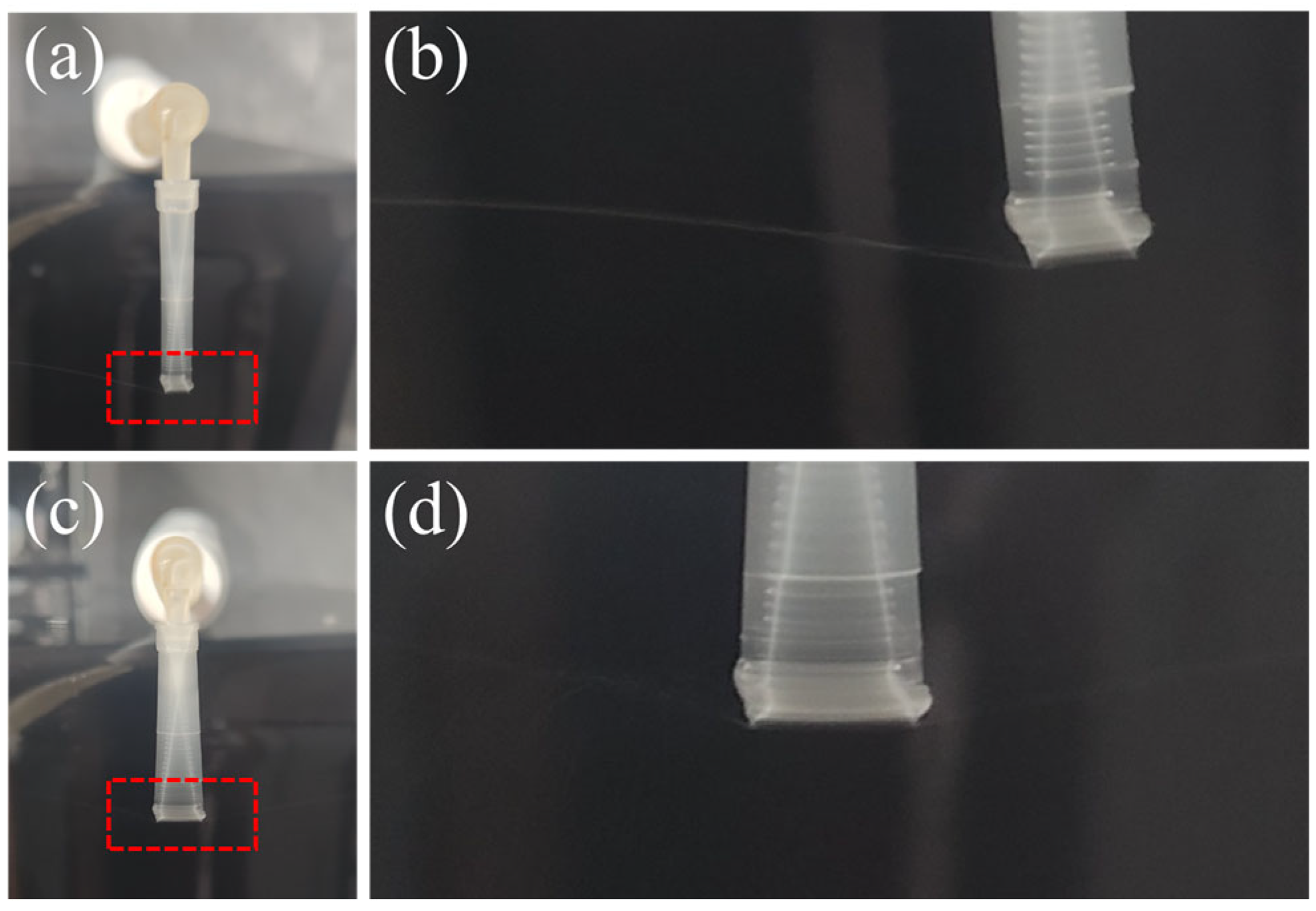
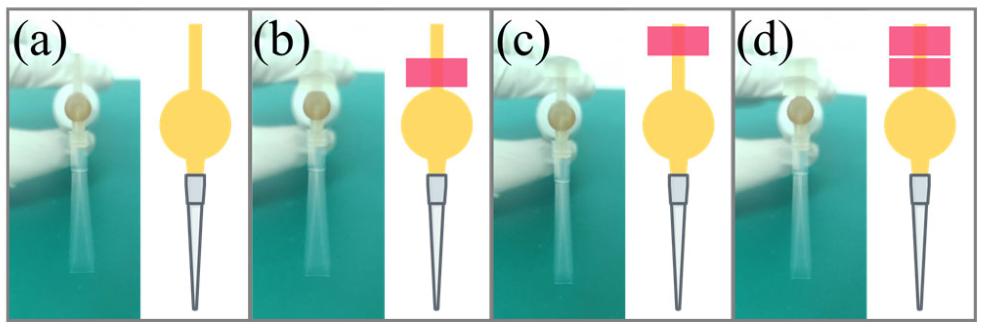
Adding an adjustable counterweight to the toothbrush shaft effectively smooths these transitions. By gradually increasing the counterweight mass and shifting its position, we attenuated the amplitude to a regime where the pipette tip undergoes a controlled lateral oscillation. In this window, a single, narrow jet emerges from the nozzle and breaks into droplets at a nearly constant distance from the tip (
Figure 3a,b). If the amplitude is further increased, the jet splits into multiple branches (
Figure 3c,d), again broadening the droplet size distribution.
Thus, the counterweight transforms the toothbrush from a two-level actuator into a tunable vibrational source and is essential for robust single-jet operation across different tip geometries, as depicted in
Figure 4.
3.2. Droplet Size Control and Monodispersity
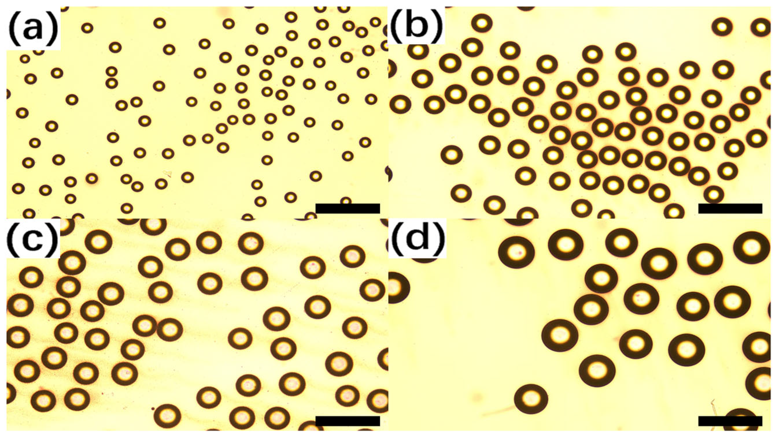
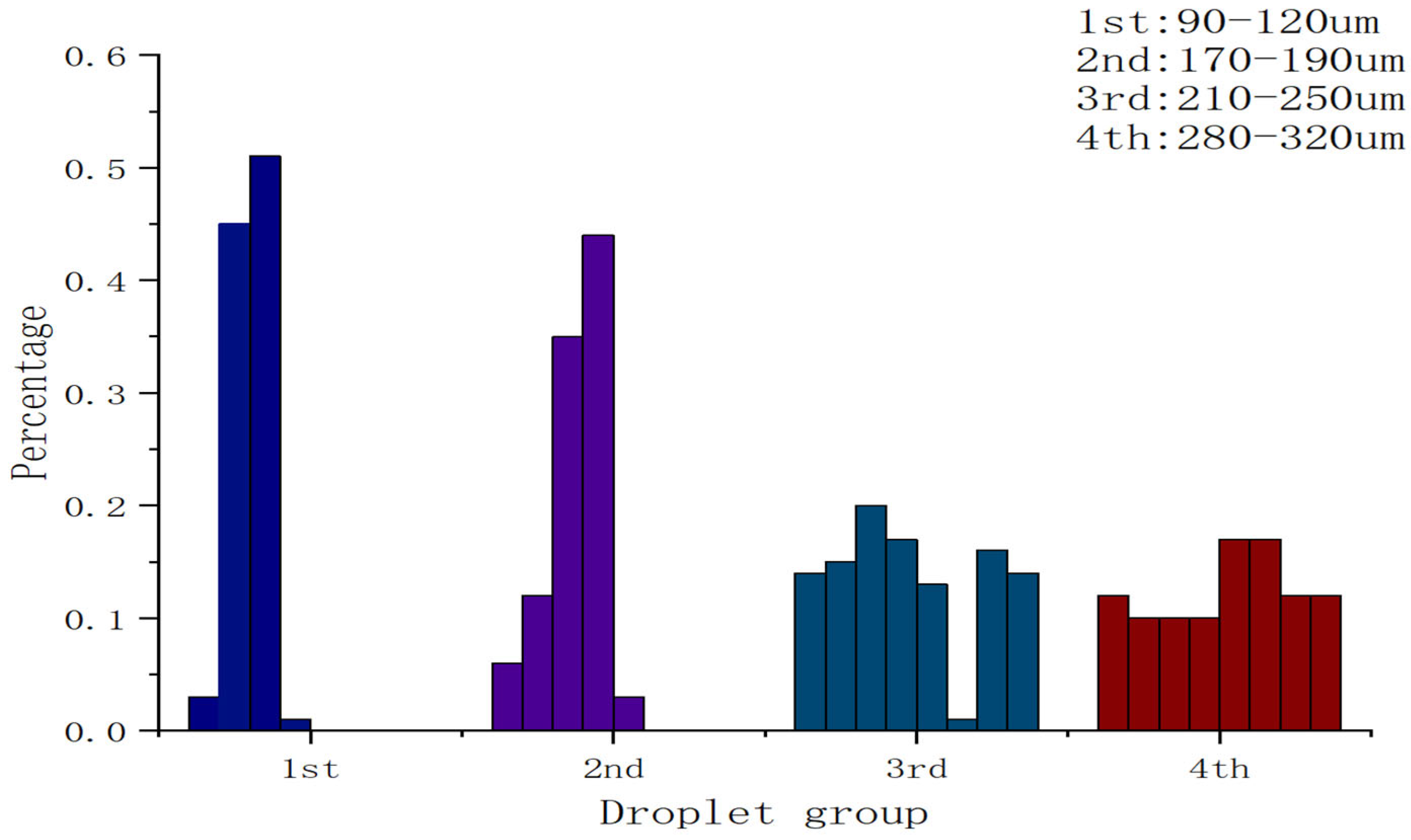
Figure 5 shows representative droplet fields generated using four pipette tips with different nozzle apertures. As expected from capillary-breakup physics, droplet diameter increases with nozzle size: tips with ~90, 180, 220 and 300 µm apertures produced droplets centered around approximately 100, 180, 220 and 300 µm, respectively. Histograms constructed from >300 droplets per condition (
Figure 5) reveal narrow distributions with coefficients of variation of (3–5%) for the optimized operating point. Meanwhile, for each nozzle aperture, droplets with different size distributions were generated, as analyzed in
Figure 6.
In the droplet-generation experiments, the nozzle aperture was approximately 90 µm and enabled stable droplet production. Based on multiple experimental runs, the average generation rate under the specified operating conditions was ~106 droplets per second. Each run could sustain stable jetting for tens of seconds; therefore, the total number of droplets produced in a single experiment typically exceeded 1000. The droplet diameter was mainly distributed in the range of 90–110 µm.
Supplementary Video S1 demonstrates the droplet generation process and shows droplets falling into the oil phase.
To evaluate how the droplet count varied with operation time, we used the same device with this nozzle and performed droplet-generation runs for 5, 10, 15, and 20 s. As shown in
Table 2. Droplets were then imaged and counted in selected local fields of view under a microscope, and the total droplet number was estimated accordingly. Owing to the current limitations in imaging and counting, the reported droplet counts should be regarded as approximate; the actual number may vary with experimental conditions and fluctuations in device performance.
Across four runs (5–20 s), the estimated droplet generation rate ranged from ~85 to ~115 droplets s−1 (mean ≈ 106 droplets s−1).
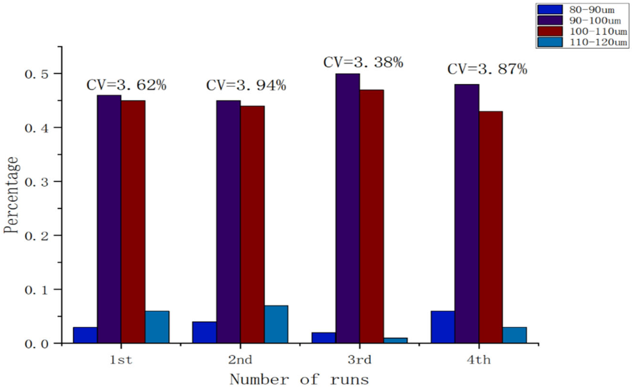
Because droplets generated using the ~90 µm nozzle aperture were the most stable, we conducted multiple experiments using different devices fitted with nozzles of the same nominal aperture to examine variations in droplet size. In all experiments, droplets were generated in a Petri dish for a fixed duration of 5 s, and regions with relatively high droplet density were selected for microscopic observation. Across four experimental runs, the droplet diameter coefficient of variation (CV) was 3.62%, 3.94%, 3.38%, and 3.87%, respectively. The mean CV was approximately 3.70%, indicating good monodispersity (low size polydispersity) and high run-to-run consistency in droplet size. It should be noted that, due to differences in experimental conditions and device performance, the total number of droplets varied across experiments. The size distribution is summarized in
Figure 7.
We also evaluated droplet stability at room temperature and under LAMP incubation conditions. At room temperature, no coalescence was observed over at least 2 h, and mean diameter decreased by less than 5%, consistent with minor evaporation or slight oil uptake. When emulsions were heated to 65 °C for 45 min in the ABIL EM180/isopropyl palmitate formulation, droplet counts and diameters remained unchanged within the measurement uncertainty, confirming that the droplets withstand isothermal reaction conditions.
Given the simplicity of the device and manual tip fabrication, this level of monodispersity is sufficient for many digital assay and encapsulation tasks.
3.3. Proof-of-Concept Droplet Digital LAMP
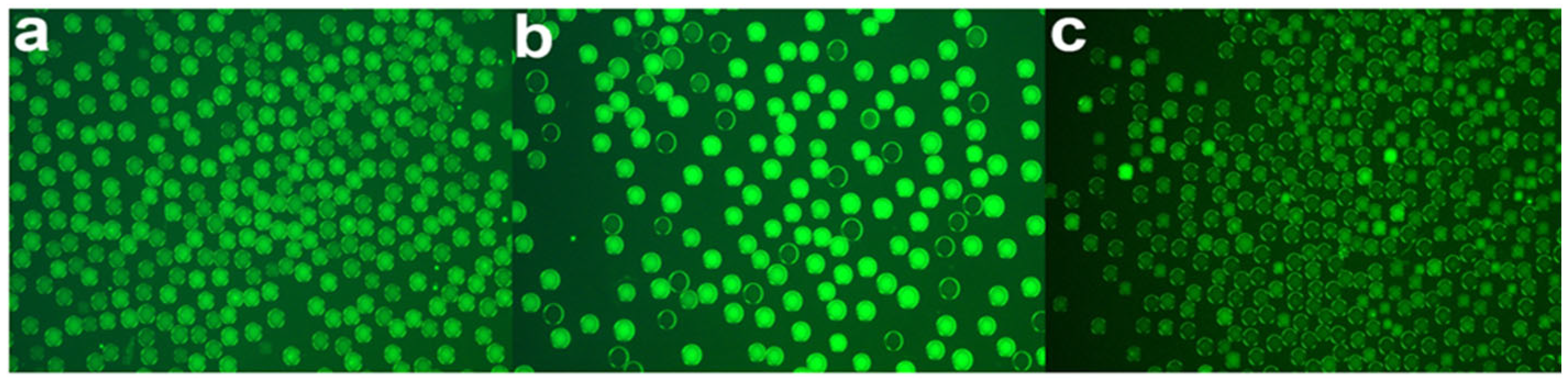
We next evaluated the compatibility of the handheld droplet generator with nucleic-acid amplification by performing droplet-based digital LAMP using the feline parvovirus kit. Emulsified reaction droplets at three template concentrations (normal, 5-fold dilution, 25-fold dilution) were incubated at 65 °C for 45 min and imaged under fluorescence (
Figure 8a–c).
Even without quantitative analysis, visual inspection clearly shows a stepwise decrease in the fraction of fluorescence-positive (bright) droplets with increasing template dilution: at the undiluted condition, nearly all droplets are positive; at 5× dilution, the number of positive droplets is markedly reduced; and at 25× dilution, only sparse positive droplets are observed (
Figure 8a–c). We then applied the image-analysis workflow described in
Section 2.4 to automatically segment droplets, extract their contours, and perform counting to obtain quantitative readouts.
To further assess reproducibility, we performed multiple independent replicate experiments for each template concentration under identical operating conditions, including independent droplet generation and reaction preparation. For each replicate, the same analysis pipeline was used to extract the total droplet count n, the number of positive droplets h, and the positive fraction
p =
h/
n, from which the mean copy number per droplet was estimated using the Poisson model,
λ = −
ln(1 −
p).
Table 3 summarizes the total droplet counts and positive fractions across conditions.
Based on these data, using
, the estimated mean copy numbers per droplet for Batch 1 at the three concentrations were approximately 5.2, 1.56, and 0.0806, corresponding to relative copy numbers of 1, 0.2, and 0.04, respectively. Fitting a linear model of
vs. the logarithm of the relative concentration
yielded
indicating a good correlation between measured mean copy number and expected dilution factor over the explored range.
Overall, λ decreased monotonically with increasing dilution, with acceptable inter-run variability, further supporting the compatibility of droplets generated by the handheld device with digital LAMP and the reproducibility of the readout.
We emphasize that this experiment is intentionally limited in scope: only one droplet size and three concentrations were tested, with a single batch per condition. The goal here is not to fully validate a quantitative FPV assay but to demonstrate that (i) droplets generated by the handheld device are compatible with isothermal amplification chemistry, (ii) the droplets remain stable under incubation, and (iii) differences in template concentration can be resolved via differences in the fraction of positive droplets.
A more comprehensive analytical characterization, including limit-of-detection, linear dynamic range, inter-run variability and comparison with benchtop droplet systems, is left for future work.
3.4. E. coli Plate Dispersion for CFU Counting
To assess the utility of the droplet generator for microbiological applications, we used it to distribute
E. coli suspensions onto LB agar plates.
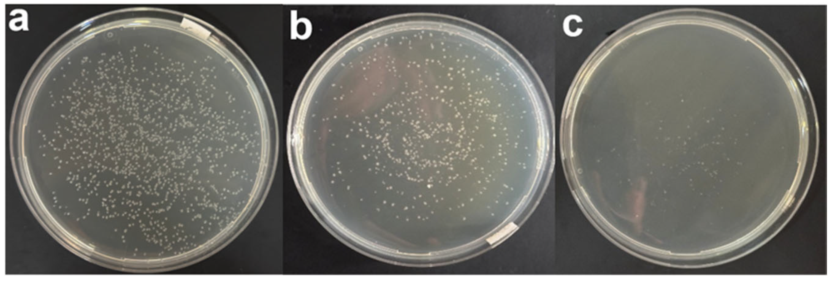
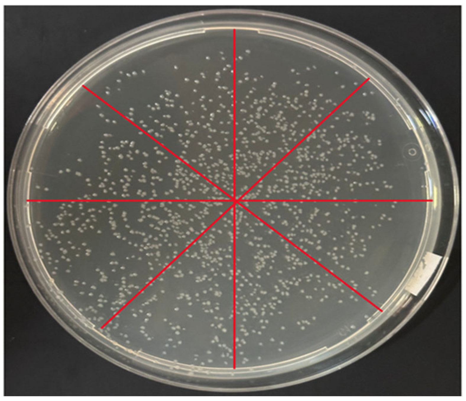
Figure 9 shows representative bright-field images for the three tested concentrations (1×, 10×, 100×). At the highest concentration, colonies are numerous and relatively close to each other; at 10× dilution, colony density is reduced and individual colonies are easily distinguished; at 100× dilution, colonies are sparse but evenly scattered across the plate.
Across repeated inoculation runs, we adjusted the swaying motion to achieve uniform coverage. The sector-based analysis was performed on representative plates to quantify spatial uniformity, and
Table 4 summarizes colony counts from triplicate plates per dilution. To quantitatively characterize the spatial distribution of colonies, we employed a sector-based partitioning method in preliminary CFU plate assays: each agar plate was divided into 4, 6, 8, or 16 radial sectors of equal angle, and an appropriate partitioning scale (8 divisions) was selected based on the overall colony coverage and density (
Figure 9). Colony counts were then obtained for each sector to characterize the plate-wide spatial distribution, yielding values of 231, 233, 235, 237, 247, 249, 251, and 253, respectively.
To further quantify distribution uniformity, we calculated the coefficient of variation (CV = standard deviation/mean × 100%) of the sector-wise colony counts. Under the optimized operating conditions, the CV across sectors was approximately ~3.6%, indicating that the droplet-dispensing approach can produce a relatively uniform and reproducible colony distribution on agar plates.
Qualitatively, the droplet-based inoculation offers two benefits compared with manual streaking with an inoculation loop:
Uniformity. Because the device emits many small droplets over the plate area in a short time, spatial distribution of bacteria is more uniform, and there are fewer regions of overcrowding or empty space.
Labor saving and reproducibility. Inoculation requires only holding the device over the plate for a fixed time (8 s in our experiments), reducing operator-to-operator variability in streaking patterns and hands-on effort.
For full quantitative CFU analysis, colony counts should be obtained from multiple plates per dilution. As shown in
Table 4. In the current proof-of-concept experiment, plates at each dilution level yielded approximately:
From these values, CFU mL−1 can be calculated in the usual manner by accounting for the plated volume and dilution factor. A linear relationship between log10(CFU mL−1) and log10 (dilution) with coefficient of determination R2 = 0.99337 supports the suitability of the device for rapid CFU estimation.
Although these experiments are preliminary and based mainly on photographic evidence, they demonstrate that the vibrational droplet generator can indeed break up a bacterial suspension into many micro-droplets that land on the agar surface and grow into discrete, well-separated colonies.
Figure 10 shows experiments with samples at different dilution concentrations.
4. Discussion
4.1. Advantages and Potential Applications
The present work shows that a simple, handheld, toothbrush-based device can produce reasonably monodisperse droplets and support both nucleic-acid amplification and microbial culture workflows. Key advantages include:
Accessibility and cost. All components are low-cost and widely available. This dramatically lowers the barrier to entry for labs that wish to explore droplet-based methods without investing in microfabrication or pump systems.
Simplicity of operation. Generating droplets is as simple as loading a pipette tip, mounting it on the adapter and turning on the toothbrush. No flow-rate tuning or complex tubing is required.
Flexibility of droplet size. By modifying only the pipette tip aperture and vibration amplitude, the same actuator can generate droplets suitable for digital assays (~100 µm) or for plate dispersion (~100–300 µm).
Compatibility with conventional workflows. Oil-based droplet emulsions can be imaged under standard microscopes, and sprayed droplets on LB plates integrate seamlessly with traditional CFU counting and colony picking procedures.
These features make the device attractive not only for resource-limited settings but also for educational labs, where students can directly see how mechanical vibration governs droplet formation and how digital assays work.
4.2. Future Applications
Although this work primarily focuses on demonstrating the performance of a toothbrush-vibration-driven handheld droplet generator for producing stable water-in-oil droplets, and on its proof-of-concept applications in digital LAMP and rapid CFU enumeration—highlighting the potential of such an ultra-minimal device in resource-limited settings—readers may also wish to understand how droplets generated by this front-end module could be interfaced with more mature microfluidic droplet-manipulation components to support more complex, multi-step analytical workflows. In practice, droplet microfluidics encompasses not only droplet generation but also downstream operations such as droplet splitting, merging, routing, trapping, and on-chip reagent addition, which are essential for building coherent and automated biochemical analysis pipelines.
In this context, Agnihotri et al. provided a systematic review of droplet splitting mechanisms in microfluidics, categorizing passive and active splitting strategies and analyzing how interfacial forces, flow conditions, and geometrical design work together to control volume partitioning and continuous operation [
38]. This body of work offers both theoretical and practical guidance for implementing volume allocation and re-allocation once externally generated droplets are introduced into channel networks. Likewise, the review by Fergola et al. comprehensively covered droplet generation and manipulation strategies, including passive microchannel geometries, active actuation using electric/magnetic/acoustic fields, and downstream operations such as droplet trapping, immobilization, and sorting. These methods complement the upstream generation unit proposed here and provide a useful design framework for constructing an integrated “toolbox” for droplet-based microfluidic analysis [
39].
In addition, reviews such as that by Xi et al. have summarized a range of active droplet-sorting techniques—including electric-, magnetic-, and acoustic-based approaches—that enable real-time selection and routing of droplets in microchannels based on internal signals or markers. Such capabilities are particularly important for multiplexed reaction pathways, sample selection, and closed-loop feedback workflows [
40].
Building on these studies, one can envision using the toothbrush-driven generator as a general-purpose front-end module to produce droplets that are subsequently transferred into PDMS or thermoplastic microfluidic chip networks for downstream operations. For example:
Splitting and recombination: By incorporating T- or Y-junction splitters and implementing passive/active splitting units described by Agnihotri et al., a single larger droplet could be divided into smaller sub-droplets to adjust reaction volume or effective concentration.
Merging and reagent addition: Using droplet docking interfaces or connector structures, droplets from different sources could be merged within microchannels while additional reagents are introduced, enabling stepwise reactions or combinatorial screening.
Routing and sorting: Electrically, magnetically, or acoustically actuated sorting modules could classify droplets based on internal reaction readouts (e.g., fluorescence intensity) and direct them to different outlets or detection zones, providing a foundation for high-throughput analyses and feedback control.
Such integration strategies would enable multi-step workflows (e.g., sequential reagent addition, serial dilution, and condition screening), more advanced digital assays (including preparation steps for sequencing or protein analysis), and high-throughput combinatorial screening. By coupling a toothbrush-based front-end droplet generator with standard downstream microfluidic modules, future work could establish an ultra-low-cost, user-friendly, and scalable droplet analysis platform that leverages the strengths of droplet microfluidics in bioanalysis, clinical diagnostics, and synthetic biology, while retaining the simplicity and field-deployability of handheld droplet generation.
4.3. Current Limitations
Several limitations of the present prototype should be acknowledged:
Device-to-device and batch variability. Because pipette tip apertures are created manually by heat-shrinking around microneedles, aperture diameters vary between tips. This leads to differences in droplet size and sometimes in the onset of stable jetting. Similarly, minor differences in how the counterweight is attached can change the effective vibration amplitude. Producing standardised tips (e.g., moulded or laser-drilled) and a more reproducible counterweight mechanism would improve uniformity.
Droplet motion and imaging challenges. Droplets are generated into an open Petri dish filled with oil. During and shortly after generation, continuous vibration causes droplets to move and occasionally float upwards or downwards in the depth of the dish. This motion complicates time-lapse imaging and quantitative tracking of individual droplets. In this work we mitigated the issue by stopping the vibration and allowing droplets to settle before imaging, but for live observation of reaction dynamics a shallower observation chamber or density-matched oil could be beneficial.
Limited quantitative validation. The LAMP and E. coli plate experiments were deliberately designed as proof-of-concept demonstrations. Sample numbers are small, and there is little replication or comparison with established benchtop methods. While the observed trends are encouraging, rigorous analytical performance (limit of detection, accuracy, precision, robustness) has not yet been established.
Lack of integrated temperature control. For nucleic-acid amplification, an external oven was used to maintain 65 °C. Integration of a simple heating module, or use of an existing incubator while retaining handheld droplet generation, would streamline workflows.
4.4. Broader Relevance to Biosensing and Single-Cell Heterogeneity
The integration of microfluidics with biosensing technologies offers substantial potential for next-generation lab-on-a-chip platforms and point-of-care (POC) diagnostics. Luka et al. (2015) reviewed how such integration combines biorecognition elements with microfluidic channels to reduce sample and reagent volumes, enhance mass transport and mixing, and consolidate sample preparation and detection within a single platform, thereby significantly improving sensitivity, real-time capability, and multiplexing potential [
41]. This review also highlighted the diversity of continuous-flow, droplet-based, and digital microfluidic systems, as well as their prospective applications in precision agriculture, environmental monitoring, and clinical diagnostics.
Droplet-based digital analysis (e.g., ddPCR and ddLAMP) is one of the core applications driving this integrated trend. By partitioning a sample into a large number of small-volume compartments (such as droplets), digital assays enable statistical quantification at the single-target level, thereby markedly improving quantitative accuracy and dynamic range. Related work has shown that a portable system combining a vibrating sharp-tip capillary with digital isothermal amplification can generate tunable droplet sizes without external pumps or complex microfabricated structures, and can achieve digital nucleic-acid detection over a range of ~2 to ~6000 copies/µL [
42]. Using a simple signal generator and a low-power vibration mechanism, this approach illustrates how low-cost droplet generators can be embedded into quantitative biosensing workflows and deliver high-dynamic-range digital amplification under resource-limited conditions.
Beyond nucleic-acid quantification, microfluidic systems have also been widely used for single-cell and heterogeneity analyses, revealing cell-to-cell behavioral differences that are masked by population-averaged measurements. For example, in reproductive biology, researchers have used droplet microfluidics to encapsulate individual sperm cells and track their dynamics in compartments of different sizes, thereby uncovering how microenvironmental constraints influence motility. Conceptually, this mirrors how droplet compartmentalization and amplification in digital assays isolate single target molecules or CFUs to reveal otherwise hidden functional heterogeneity.
4.5. Comparison with Portable and Chip-Free Droplet Generators
To more comprehensively situate this work within the landscape of existing portable and chip-free droplet generation technologies, it is necessary to compare key performance metrics, including droplet size range, throughput (droplet generation rate), system complexity, and dependence on external equipment or microfabricated chips. In He et al., a vibrating sharp-tip capillary approach leveraged acoustic streaming to enable efficient droplet generation without an external pressure source [
36]. This system allowed real-time tuning of droplet diameters from ~6.77 µm to 661 µm within a single setup, and generated highly monodisperse droplets (CV < 4%) at rates up to ~5000 droplets/s. The actuation power was below 60 mW, and the overall device could be driven by a battery-powered, low-cost signal generator. While this method substantially outperforms the present toothbrush-driven device in throughput and size tunability, it still requires a bespoke piezoelectric actuator and precise alignment of the glass capillary tip, which increases experimental setup and tuning complexity.
Trossbach et al. reported a portable negative-pressure-driven droplet generator assembled entirely from commercial components, designed to mitigate the reliance of many microfluidic systems on custom fabrication and costly pump hardware [
33]. Although this platform still depends on microfabricated channels and fluidic connections—and typically requires external control (e.g., a laptop) to regulate negative pressure—it demonstrated stable, size-tunable water-in-oil droplet generation within a chip, highlighting the design trade-offs between portability and system integration.
Earlier work by Chen et al. presented a handheld, power-free microfluidic droplet generator for applications such as single-cell genomic analysis and digital PCR, capable of producing monodisperse droplets [
31]. While advantageous in cost and the absence of external power, this design still relies on microchannel chip structures and manual control, and thus remains more device-dependent than fully chip-free approaches.
By contrast, the toothbrush-driven droplet generator offers a distinctive advantage in that it integrates both the actuation mechanism and the droplet generation unit into a single consumer-grade device, eliminating the need for microfabricated chips, external pumps, dedicated signal generators, or complex alignment procedures. Under typical operating conditions, it can continuously generate droplets at an order-of-magnitude rate (e.g., ~100 droplets/s with CV ~3–5%). Although this throughput is lower than that of the vibrating-capillary system reported by He et al., the toothbrush-based approach offers unique benefits in experimental simplicity, ease of use, and accessibility. Compared with chip-dependent solutions (e.g., Trossbach and Chen), it avoids barriers associated with microstructure fabrication and chip-to-world interconnections, making it potentially more deployable in resource-limited settings and rapid field-testing scenarios.
For an intuitive comparison,
Table 5 below provides a concise summary table:
4.6. Differences in Device Performance Discussion
Device-to-device variability is an inherent consideration for this prototype and was briefly noted in the Abstract and Conclusions; however, it was not discussed in sufficient detail in the main text. Here, “device-to-device variability” may arise from (i) differences among electric toothbrush units (even within the same model) and/or (ii) assembly-related variability when the nozzle/adapter is mounted and remounted on a given device. To preliminarily assess these effects, we tested multiple electric toothbrushes across different brands and models and characterized their vibration frequencies. While all devices operated within a broadly similar range (≈20,000–40,000 strokes per minute), we observed measurable differences in both frequency and amplitude, which can influence droplet size and generation rate. For instance, a Philips Sonicare toothbrush (sonic vibration, ~31,000 strokes per minute, ≈517 Hz) provided relatively stable droplet generation in our setup. Nonetheless, subtle unit-to-unit differences (e.g., motor performance and assembly tolerances) may still shift droplet formation behavior, underscoring the need for more systematic, quantitative evaluation of how such variability impacts performance.