Review

Arsenic Toxicity on Metabolism and Autophagy in Adipose and Muscle Tissues

1
Department of Biochemistry and the Redox Biology Center, University of Nebraska-Lincoln, Lincoln, NE 68588, USA
2
Department of Cell and Regenerative Biology, University of Wisconsin School of Medicine and Public Health, Madison, WI 53705, USA
3
Department of Neurology, Institute for Cell Engineering, The Johns Hopkins University School of Medicine, Baltimore, MD 21205, USA
4
Laboratory of Immunology, Office of Biotechnology Products, Center for Drug Evaluation and Research, United States Food and Drug Administration, Silver Spring, MD 20993, USA
5
Department of Microbiology and Immunology, Sidney Kimmel Medical College and Jefferson College of Life Sciences, MD-PhD Program, Thomas Jefferson University, Philadelphia, PA 19107, USA
6
Department of Nutrition, University of Massachusetts, Amherst, MA 01003, USA
7
Department of Nutrition and Health Sciences, University of Nebraska-Lincoln, Lincoln, NE 68583, USA
8
School of Veterinary Medicine and Biomedical Sciences and the Redox Biology Center, University of Nebraska-Lincoln, Lincoln, NE 68583, USA
9
Department of Biological Systems Engineering, University of Nebraska-Lincoln, Lincoln, NE 68583, USA
10
Department of Food Science and Technology, Nebraska Food for Health Center, University of Nebraska-Lincoln, Lincoln, NE 68588, USA
*
Author to whom correspondence should be addressed.
These authors contributed equally to this work.
Antioxidants 2022, 11(4), 689; https://doi.org/10.3390/antiox11040689
Submission received: 22 February 2022 / Revised: 28 March 2022 / Accepted: 29 March 2022 / Published: 31 March 2022

Abstract

:
Arsenic, a naturally occurring metalloid derived from the environment, has been studied worldwide for its causative effects in various cancers. However, the effects of arsenic toxicity on the development and progression of metabolic syndrome, including obesity and diabetes, has received less attention. Many studies suggest that metabolic dysfunction and autophagy dysregulation of adipose and muscle tissues are closely related to the development of metabolic disease. In the USA, arsenic contamination has been reported in some ground water, soil and grain samples in major agricultural regions, but the effects on adipose and muscle tissue metabolism and autophagy have not been investigated much. Here, we highlight arsenic toxicity according to the species, dose and exposure time and the effects on adipose and muscle tissue metabolism and autophagy. Historically, arsenic was used as both a poison and medicine, depending on the dose and treatment time. In the modern era, arsenic intoxication has significantly increased due to exposure from water, soil and food, which could be a contributing factor in the development and progression of metabolic disease. From this review, a better understanding of the pathogenic mechanisms by which arsenic alters metabolism and autophagy regulation could become a cornerstone leading to the development of therapeutic strategies against arsenic-induced toxicity and metabolic disease.

1. Introduction: Arsenic’s Impacts on Food and Human Health in the USA

US citizens consume significant amounts of agricultural products (e.g., rice grain from California or vegetables and fruits from Michigan) produced from the states with high exposure levels of arsenic (As) contamination [1,2,3]. This could correlate with the metabolic dysfunction caused by As accumulation in adipose (fat) and muscle tissues of the human body. For example, in Nebraska, the highest concentrations of As in groundwater, measured at over 20 μg/L (ppb), were found in the panhandle, southwestern region and Republican River valley [4], which belong to the Ogallala Aquifer in the Great Plains [5]. Nebraska is one of the largest beef-producing states, along with having high levels of corn and soybean production [6]. According to the United States Department of Agriculture (USDA) and Food and Drug Administration (FDA), As levels in beef, corn and soybean are quite low and no health concern has been found for most of Nebraska. Since cow livestock are fed with corn and soybeans and consume water from underground sources, there is still a possibility of a certain level of As accumulation in beef, corn and soybeans due to As contents in water and soil in highly As-contaminated areas. Indeed, multiple studies indicate that corn and soybeans are very susceptible to As uptake in highly As-contaminated areas [7,8]. Since beef mostly consists of fat and muscle tissues, measurements of As accumulation traveling from water and soil through to corn and soybeans, beef and finally to humans are increasingly sought, although there is no accurate database available due to various technical limitations in the measurement of live animals and humans in highly As-contaminated areas. These data relate to the USA as a whole, as the prevalence of As contamination in food production poses a significant concern due to the impacts on human health.
As, a group 1A carcinogen, is a worldwide public health concern; exposure causes lung, bladder and non-melanoma skin cancers in humans [9,10,11]. However, in contrast to extensive research on its carcinogenicity, considerably less studies are available on the impacts of As exposure on the onset and progression of other prevalent diseases, such as metabolic and degenerative diseases [12,13,14]. This is a critical knowledge gap, as several studies have suggested an association between chronic exposure of high-dose As and metabolic complications such as obesity and type 2 diabetes mellitus (T2DM), including from human studies in Michigan and population studies in high-arsenic areas (≥150 µg/L or ppm in drinking water) in Chile, Taiwan and Bangladesh [1,2,3,15,16,17,18].
A recent study estimated that about 2.1 million people in the USA alone are exposed to wells or underground water that is high in As [19]. Excess body weight and obesity combined make up the second leading cause of death in the USA [20]. The combined medical costs associated with the treatment of preventable obesity-associated diseases are estimated to increase by $48–$66 billion per year by 2030 in the USA [21,22]. It is important to note that obesity is often associated with dysplasia, lipodystrophy and malfunction of adipose tissue [17,23]. Disruption of glucose metabolism and accumulation of oxidative stress in the muscle [24,25] also significantly contribute to the development of metabolic diseases. Nevertheless, studies on the effects of As on adipose and muscle tissue metabolism, which are tightly linked to obesity and metabolic diseases, are limited. A better understanding of As’s instigating roles in metabolic dysfunction in adipose and muscle tissues could lead to innovative prevention and treatment strategies for people with obesity and metabolic diseases living in As-contaminated areas.

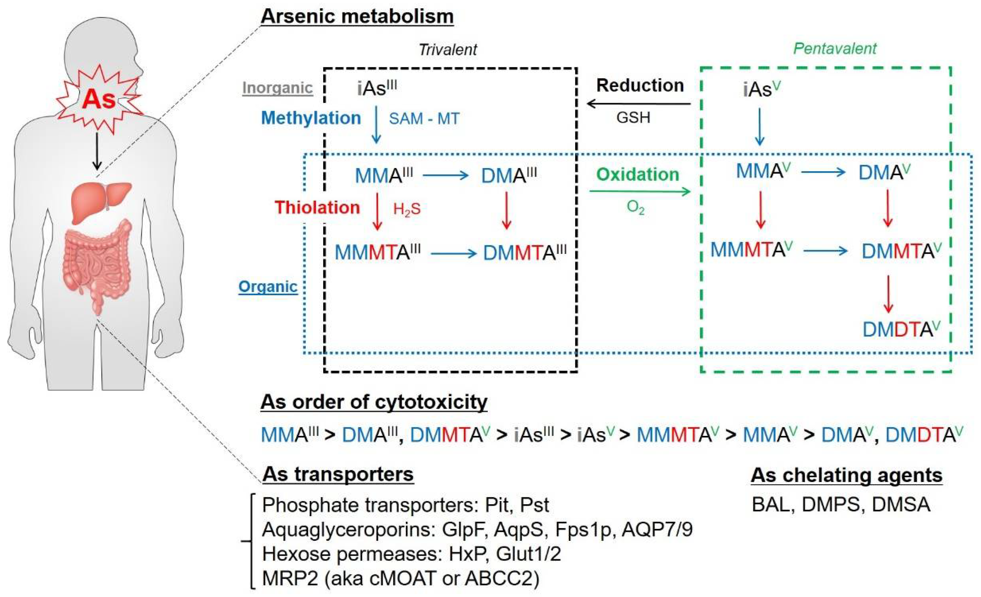
2. As Biochemistry and Metabolism: Biotransformation of As in the Body

As is a metalloid with an atomic number of 33 that is naturally found in the environment. As compounds can be classified into inorganic, organic and gas states. Inorganic As is generally considered more toxic than the organic form, while the gas form of As (Arsine) is the most toxic arsenical when acutely exposed in high concentrations [26]. As originates from the earth’s crust and is present at high levels in groundwater [27]. Humans can be exposed to As most significantly from contaminated groundwater by drinking, and indirectly at lower amounts from food (crops, fish, meat and dairy products prepared with As-contaminated water), industrial processes (alloying agents, metal adhesives, wood preservatives, pesticides, pharmaceuticals, etc.) or tobacco via cigarette smoking [28]. As can enter into the human body via ingestion through the gastrointestinal (GI) tract, inhalation through the lungs and dermal absorption. Absorbed inorganic As is metabolized in the human body with a half-life of two to four days [29], and approximately 60–80% of As is excreted in the urine through the kidneys within a few days of ingestion [30]. After absorption through the GI tract and lungs, As is taken up by red and white blood cells in the bloodstream first before reaching the liver [31].
Accumulated As in the body can generate ROS and epigenetic dysregulation, which promotes carcinogenesis. One study reported that the Rac family small GTPase 1 (Rac1) signaling pathway mediates As-induced generation of reactive oxygen species (ROS), which are well-known second messengers for cellular pathogenic effects by damaging protein, DNAs and organelles. Rac1 is an essential subunit of NADPH oxidase isoform Nox2, which is required for As-stimulated vessel remodeling during carcinogenesis [32,33]. Recent work found the links between As exposure to epigenetic dysregulation and progression of the pathology. During carcinogenesis, As exposure induces a loss of DNA methylation, which increases gene expression [34,35]. Arsenic-induced malignant transformations in human urothelial cells are associated with an aberrant DNA methylation increased by As exposure [36]. Therefore, the detoxification and secretion of absorbed As out of the body is the critical process in As biotransformation in order to remedy the toxicity.
The most efficient detoxification process of As in the human body is through methylation. As methylation is a biotransformation conserved from bacteria to humans to modulate As toxicity and carcinogenicity [37,38,39]. This process is catalyzed through S-adenosylmethionine (SAM)—dependent methyltrasferase (MT), which is encoded by the arsM gene [37,40]. Arsenate (AsV) can be reduced to arsenite (AsIII) or methylated in the liver via enzymatic transfer of the methyl group from SAM to methyl arsonate (MMAV) and dimethyl arsenate (DMAV). However, methylation of inorganic As such as monomethylarsonous acid (MMAIII) and dimethylarsinous acid (DMAIII) would be toxic when interacting with cellular proteins and DNA [41,42]. When the methylating capacity of the liver is exceeded, As can be retained for two to four weeks or longer in soft tissues, including adipose and muscle tissues, as well as in keratin-rich tissues, including hair, nail and skin tissues [43,44].
As can be reduced by the antioxidant glutathione (GSH). As binding to the thiol (SH-) group is very strong and the reduction is initiated by shifts in the carbon atom bonded to the sulfhydryl group of the cysteinyl residue of GSH [45]. Dietary selenium (Se) compounds and α–tocopherol (Vitamin E) protect human cells and mice from As-induced mutagenesis and carcinogenesis [46].
Methylated trivalent inorganic As in the liver is released into the bloodstream and excreted in urine via transport through cell membranes. The groups of uptake and efflux channels facilitate As species transport, namely prokaryotic phosphate transporters (Pit and Pts); GlpF (glycerol facilitator protein) from Escherichia coli; AqpS (aquaporin) from Sinorhizobium meliloti); HxP (hexose permease) and Fps1p (yeast aquaglyceroporin) from Saccharomyces cerevisiae; and mouse aquaporin (AQP) 7, rat and human AQP9, glucose transporter (GLUT) 1 from mammals [47,48]. Additionally in mammals, GLUT2 and multidrug-resistance-associated protein (MRP) 2 play an important role in the efflux of dimethylarsenic metabolites (DMAs) [49,50]. These studies indicate that As transporters in each species have evolutionarily significant differences both in selectivity and transport rates of arsenicals.
Recently, there has been an emerging interest in the thiolation of As, which is a naturally occurring chemical reaction via adsorption on iron oxides and precipitation on iron sulfide minerals [51] and was found in geothermal waters in Yellowstone National Park [52] and in soil pore waters in rice fields [53,54]. The increasing synthesis and detection of thioarsenic species such as monomethylmonothioarsonous acid (MMMTAIII), dimethylmonothioarsinous acid (DMMTAIII), monomethylmonothioarsonic acid (MMMTAV), dimethylmonothioarsinic acid (DMMTAV) and dimethyldithioarsinic acid (DMDTAV) suggest the importance of these As metabolites in terms of cellular toxicity and therapeutic effects [55,56,57]. DMMTAV is the most cytotoxic thioarsenic radical, similar to DMAIII in mammals [58]. Since the effects of thioarsenic exposure on human health are controversial, analytical and diagnostic methods for thioarsenicals are urgently needed.
Upon recent and chronic exposure to doses less than acute toxic concentrations (10 to 1000 ppb), 24 h urine collection is the most useful laboratory test. Other biomarkers such as measurement of the liver transaminase level or complete blood count (CBC) can be performed for clinical assessment of As exposure according to the guidelines from the Agency for Toxic Substances and Disease Registry (ATSDR, https://www.atsdr.cdc.gov/toxprofiledocs/index.html, reviewed and accessed on 22 February 2022.) under the Centers for Disease Control (CDC) in the USA. Patients with suspected acute As intoxication and poisoning generally require gut decontamination, hemodynamic stabilization and fluid and electrolyte replacement in an intensive care setting. As-chelating agents such as dimercaprol (2,3-dimercaptopropanol, also known as British anti-Lewisite or BAL), 2-3-dimercapto-1-propanesulfonate (DMPS) or meso 2,3-dimercaptosuccinic acid (DMSA) can be administered within hours of As absorption to prevent As-induced toxicity in human body [59,60,61].
We briefly reviewed the As biotransformation and clinical treatment methods in humans. Since As-associated diseases such as cancer and metabolic diseases may have a long latency period, many As-exposed patients remain asymptomatic for years. Therefore, the understanding of As metabolism and the clinical manifestation based on the As exposure route, dose, chemical form or species, frequency, intensity, duration and elapsed time from As-exposed individuals or patients would be critical for prevention and treatment of As-associated toxicity and diseases (Figure 1).

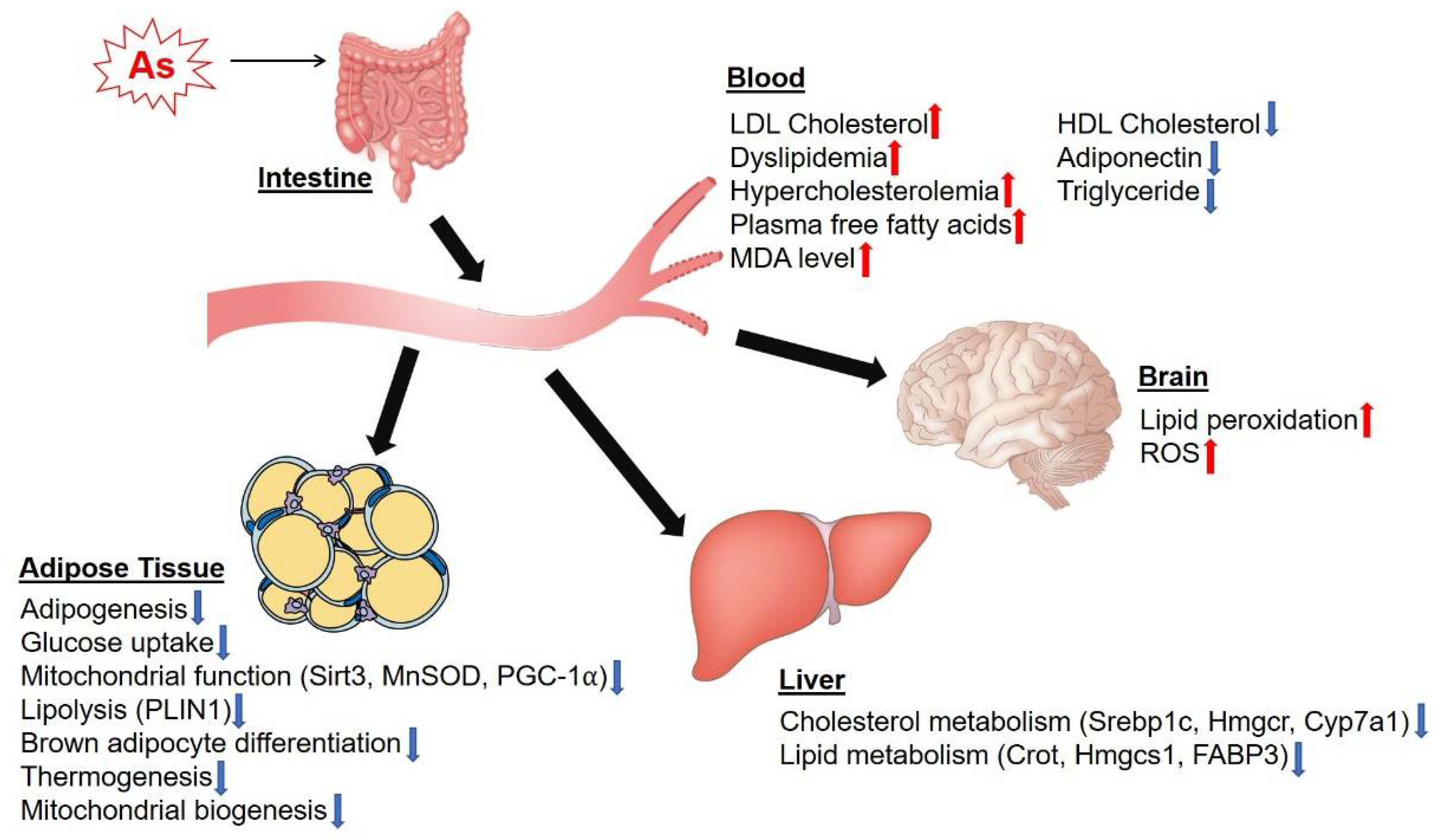
3. Effects of as on Lipid Metabolism and Adipocyte Function

As accumulates in various tissues in the body, including liver, lung, brain, heart, kidney, eye and spleen tissues [62]. Chronic exposure to high levels of As has been associated with metabolic diseases such as cardiovascular diseases, hepatotoxicity, diabetes, obesity and cancers [17,63]. Lipid metabolism plays an important role in chronic diseases and metabolic syndrome [64,65]. A 15-year birth cohort study showed that early-life As exposure caused higher atherogenic low-density lipoprotein (LDL) cholesterol levels in Taiwanese adolescents [66]. As induced dyslipidemia and hypercholesterolemia in male albino rats after 12 weeks of 150 mg/L (ppm) As exposure in drinking water [67], but this study used a very high dose and would be expected to be associated with acute liver toxicity. This would not be relevant to chronic effects in humans at environmental exposure levels—even at relatively high exposure levels. As inhibited high-density lipoprotein (HDL) cholesterol and increased plasma-free fatty acids. The expression levels of cholesterol metabolism genes, including sterol regulatory element-binding protein 1 (Srebp1c), 3-hydroxy-3-methylglutaryl-CoA reductase (Hmgcr) and cytochrome P450 family 7 subfamily A member 1 (Cyp7a1), were significantly decreased in the liver samples of mice exposed to 0.25 ppm As in drinking water for two weeks [68]. In addition, serum cholesterol and lipid levels were significantly increased in mice treated with 1 ppm As for two weeks [68]. Furthermore, lipid peroxidation was increased in the brains of rats exposed to 25 ppm of As compared to the control group, suggesting that As induced ROS production in the rat brain [69]. High-dose exposure of As also disrupts lipid metabolism in the adult zebrafish liver, as revealed by decreased mRNA expression of carnitine O-octanoyl transferase (Crot) and 3-hydroxy-e-methylglutaryl-CoA synthase 1 (Hmgcs1) and increased mRNA expression of fatty acid binding protein-3 (FABP3) [70]. As exposure promotes atherosclerosis in the aorta of rats by decreasing the HDL cholesterol/LDL cholesterol ratio [71]. The level of an indicator of lipid peroxidation, malondialdehyde (MDA), was increased in the serum of rats exposed to 3 ppm As in drinking water throughout development until the adult stage [72]. Additionally, serum adiponectin, triglyceride and HDL cholesterol levels were significantly decreased in mice after 18 weeks of exposure to 50 ppm As in their drinking water [73]. Collectively, these studies demonstrate that As can affect lipid metabolism as well as cholesterol metabolism at acute high-dose exposure levels.
As plays a role in adipocyte differentiation and adipose tissue dysfunction and lipodystrophy [74]. As inhibited adipogenesis and glucose uptake in various adipocyte cell types, including 3T3-L1 preadipocytes, mouse-adipose-derived stromal vascular fraction cells and human adipose tissue-derived stem cells [75]. The level of mitochondrial oxidative stress response protein, sirtuin (Sirt) 3, was decreased in As-treated 3T3-L1 adipocytes and C2C12 myotubes, leading to mitochondrial damage [76]. Moreover, the expression levels of Sirt3 targets, including manganese superoxide dismutase (MnSOD) and peroxisome-proliferator-activated receptor-gamma coactivator (PGC)-1α, were decreased in As-treated 3T3-L1 and C2C12 cells, indicating that As regulates ROS and its related metabolism in adipocytes [76]. As also inhibits the differentiation of C3H10T1/2 mouse adipose stem cells, resulting in inhibition of lipid accumulation [77]. Chronic exposure to As reduces the adipose tissue expression of perilipin (PLIN1), which regulates lipid storage and lipolysis [78]. Moreover, As also regulated browning and brown adipocyte function. As reduced adipocyte differentiation, lipogenesis, thermogenesis and mitochondrial biogenesis in murine brown adipocytes and brown adipose tissue (BAT) [79,80]. Mice exposed to 5 ppm As in their drinking water showed cold intolerance, as revealed by the whitening of BAT [80]. In addition, the expression levels of genes that regulate thermogenesis, mitochondrial function, adipogenesis and lipolysis were decreased in BAT samples from mice exposed to 5 ppm As for 17 weeks in their drinking water [80]. Taken together, these results provide evidence that As inhibits adipogenesis, mitochondrial function and BAT thermogenic functions (Figure 2).

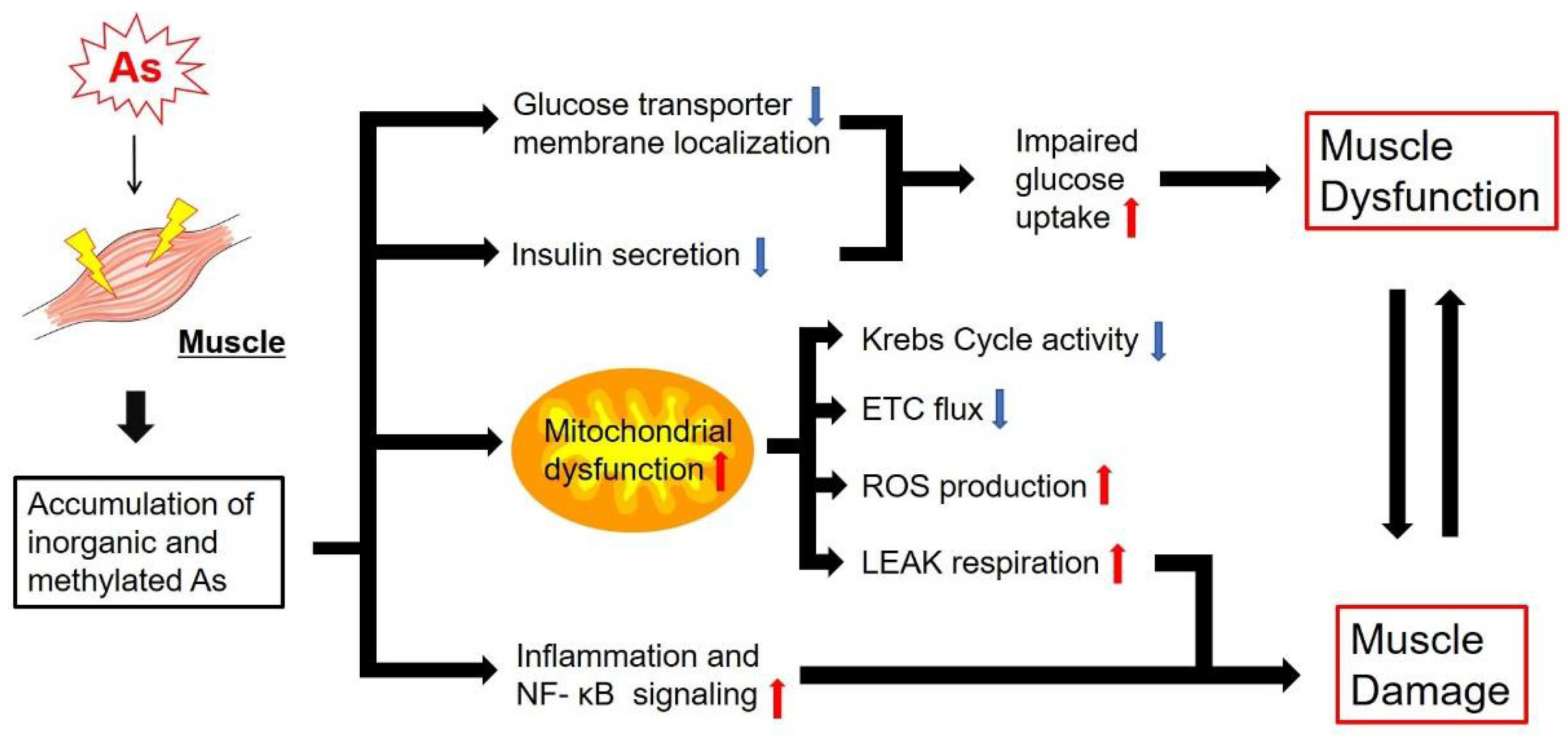
4. Effects of As on Glucose Metabolism, Mitochondrial Energy Metabolism and Muscle Function

Most of the As studies in muscle focused on fish meat (e.g., tuna, tilapia, killifish) as a food source [81,82,83,84]. Arsenite-induced oxidative stress induced autophagy as a defense mechanism in chicken muscle [85]. In a mouse study, up to 0.5 ppm of As trioxide (As2O3) caused myoatrophic effects with decreased muscle mass and endurance [86]. In a recent human study, muscular atrophy was observed as a side effect in acute promyelocytic leukemia patients treated with As trioxide [87]. While this study used doses of As that exceeded those found in environmental exposure, this is important to acknowledge because it shows the connection between As and muscle atrophy in a human model. Arsenic exposure increases the reductions in skeletal muscle mass [88] and increases the genetic risk factors in muscle atrophy and cardiovascular disease [89] in humans. A chronic low-dose As treatment (0.25–1 µM of As trioxide) causes C2C12 myotube atrophy by inhibiting the AKT pathway [90]. Since humans consume low doses of As chronically through As-contaminated food sources such as fish and chicken, a quantitative analysis of As accumulation in skeletal muscle in humans and how those chronically accumulated As exposures can affect the glucose metabolism in muscle tissue is required.
As exposure inhibits myogenesis by inhibiting the β-catenin pathway [91]. As interferes with gluconeogenesis in muscle by inhibiting glucose transporters and suppressing regulatory genes in glucose metabolism [92]. Thus, As can substantially interfere with glycolytic and mitochondrial energy generation. However, the exact mechanism explaining the function of As as an antagonist or synergist in glucose homeostasis in muscle tissue remains to be understood. Biologically relevant oxidation states of As, namely trivalent (AsIII) and pentavalent (AsV) forms, are interconvertible and detoxified through methylation in mammals [93,94]. Both oxidation states may have different chemistries and interactions with glucose metabolism and energy metabolism pathways in adipose and muscle tissues, although the relevance of As metabolism in adipose and muscle tissues with human health outcomes requires further study. While As poisoning and exposure may seem to be a rare and relatively insignificant issue, in reality hundreds of millions of people are chronically exposed to elevated levels of As [95] through contaminated drinking water [96], crops and livestock [97]. This chronic exposure can cause a substantial disease burden in exposed populations [98], with a prevalence of As-mediated disease directly relating to the level of exposure [95]. It is worthy of note that there is a threshold for As-related diseases. One study suggests that the low exposure range of iAs over 150 ppb would increase the risk of lung and bladder cancers [99]. Large epidemiology studies in Japan and the USA show no increased risk of cardiovascular disease with even lower exposure levels of iAs under 100 ppb related to amount of rice consumed, the grain with the highest As levels [100]. There are no threshold data available for human studies in metabolic disease, such as obesity and diabetes, meaning the levels of chronic iAs contamination required to influence the emergence of these diseases in a population could be much lower than for cancer.
In addition to impacting a host of biological systems, an important aspect of As-mediated disease is its ability to mimic the symptoms of diabetes through the interactions with glucose metabolism [101]. In vitro studies have demonstrated that treatment with sub-toxic levels (1 mM, Table 1) of AsIII, in both its inorganic and metabolized mono- and di-methyl species, significantly reduced insulin mediated glucose uptake in 3T3-L1 adipocytes by interfering with the localization of insulin-responsive glucose transporter 4 (Glut 4) to the cell membrane [102]. Additional in vitro studies found that longer term exposure of 3T3-L1 adipocytes to lower levels of inorganic AsIII (iAsIII) significantly reduced insulin-mediated glucose uptake in levels as low as 0.5 µM [76]. These findings are recapitulated in in vivo mouse models [101], and additional studies indicate that AsIII may inhibit the secretion of insulin from pancreatic cells [103]. A proposed mechanism for the reduced insulin secretion is via the inhibition of mitochondrial metabolism in pancreatic β-cells [104]. A prospective cohort in humans exposed to very high As levels indicated that the diabetes risk increased by 3% for every 1 ppb of As present in drinking water [105]. In 2001, the USA Environmental Protection Agency (EPA) revised the acceptable level of As in drinking water down from 50 ppb to 10 ppb. As is also capable of directly impairing glucose metabolism. Some evidence suggests that inorganic AsV (iAsV) can directly interact with glycolytic pathways via substitution with phosphate [106], although this study was conducted using only enzymes and substrates and required iAsV to be at twice the concentration of phosphate to observe any effect, while arsenate levels would never be that high in vivo as that would be lethal, so the biological significance of As on glycolysis is not clear.
Clearer evidence exists relating As to mitochondrial dysfunction. When isolated rat mitochondria were incubated with 10 to 100 µM of iAsIII, mitochondrial respiratory complexes I and II were inhibited, leading to excess ROS production, membrane potential collapse and release of cytochrome c [107]. These findings were reproduced with in vivo rat studies of chronic As exposure, inhibiting the activity of complexes I, II and IV [108,109], while the mRNA levels of mitochondrial genes in complexes I and IV were downregulated following chronic As exposure [109]. In vivo studies in rats exposed to levels of As commonly found in contaminated drinking water for 30 days found significantly reduced activity levels of several key enzymes of the citric acid cycle, including isocitrate dehydrogenase (ICDH) and succinate dehydrogenase (SDH) [108]. The inhibition of mitochondrial complexes following As exposure has important implications, as the ROS produced by As-exposed mitochondria has been shown to be the major route of As-mediated mutagenesis at cytotoxic concentration (0.25 μg/mL, Table 1) [110]. Thus, the generation of mitochondrial ROS by As can be linked to the role of As as a carcinogen [111]. Most of the current cell culture and animal experiments used lethal concentrations (Table 1 shows the doses and durations of As administration). Nearly all of the mitochondrial effects of As are at acutely toxic, poisonous and lethal concentrations. While these studies were conducted at high As concentrations, they provide valuable insight into the basic mechanisms of how chronic lower level As exposure might cause or worsen metabolic diseases.
As exposure also poses important implications for muscle function. Mice exposed to 100 ppb of iAsIII for five weeks showed decreased muscle function, damage to muscle progenitor cells and a significant increase in non-phosphorylating LEAK respiration [112]. This indicates that the reduced muscle performance stemming from As may be a result of mitochondrial abnormalities. As is further implicated in muscle damage by inhibiting muscle repair, increasing nuclear factor kappa light-chain enhancer of activated B cells (NF- κB) inflammation signaling and resulting in increased healing time and fibrosis [113]. In addition to direct damage to muscle by As, the tendency for As to mimic diabetes can also impact muscle tissue [114]. Muscle atrophy can stem directly from the neuropathy caused by diabetes [115], and a reduction in isometric muscle performance was observed in diabetic patients during hyperglycemia [116]. Diabetes is also associated with an increase in intramuscular fat storage, which is in turn associated with reduced muscle strength [117].
In summary, As is associated with significant alterations to glucose metabolism and mitochondrial function with clear ability to deleteriously impact skeletal muscle function. A better understanding of how environmental As can impact muscle metabolism and function would be essential for developing a therapeutic method for humans suffering from As-mediated metabolic diseases (Figure 3).

5. Effects of as on Autophagy in Adipose and Muscle Tissues

Several studies indicated that As is known to have detrimental effects on adipose and muscle tissues of fish, animals, livestock, experimental rodents and humans, as discussed above. In the case of adipose tissue, many research groups have already evaluated the effects of As using various adipocytes such as 3T3-L1, 3T3-F442A, C3H 10T1/2, HIB1B and so on [77,79,118,119,120,121] (Table 1). Additionally, several animal experiments were also performed with As exposure [17,79,118] (Table 1). Most of these studies showed the inhibitory effects of As administration on the growth and differentiation of the adipocytes. Although many genes and signaling pathways have been suggested as possible causes, including peroxisome-proliferator-activated receptor gamma (PPARγ), CCAAT/enhancer-binding protein-alpha (C/EBPα), cyclin-dependent kinase inhibitor 1A (p21), protein kinase B (Akt/PKB), sirtuin 3 (SIRT3)-forkhead box O3 (FOXO3a), mitogen-activated protein kinase (MAPK) cascade and C/EBP homologous protein (CHOP10)-G protein coupled receptor (GPCR) pathways, the role of As in adipose tissue remains unclear [17,74]. Muscle is also known to be another target tissue of As exposure, although this is much less studied even than adipose tissue. One study showed that the levels of glucose transporters 1 and 4 (GLUT1/4) and activation of inositol phospholipid 3-kinase (PI3K) increased with 0.5 mM As treatment in L6 rat skeletal muscle cells [118,122]. In our previous study, we observed the accumulation of As in a dose-dependent manner in mouse adipose tissue [79]. However, in the case of muscle tissue, another study reported that there was no As accumulation in muscle tissue when mice were exposed to As [17,112]. Although As did not accumulate in the muscle tissue of mice, they found impaired muscle function and increased mitochondrial oxygen consumption through the isolated primary muscle cells from mice after As exposure of 100 μg/L for 5 weeks [112]. Interestingly, in our previous studies, we also measured the oxygen consumption rate in response to high-dose As treatment in adipocytes (10 μM) and in adipose tissue of mice (10 mg/kg/d). However, the oxygen consumption was significantly decreased in the adipocytes when the treated As concentration was increased, and there was no difference in mice when compared with the control group [79]. The working mechanisms of As in adipose and muscle tissues are unique, presumably due to sensitivity to As and differences in cellular adaptivity regarding As accumulation. Muscle weakness is a major factor contributing to decreased functional mobility and is a strong predictor of mortality. Muscle weakness and wasting are also some of the most common pathologic clinical symptoms of environmental exposure to As [86,88]. However, the mechanisms and molecular pathogenesis in the etiology of As-induced muscle morbidity are relatively unknown. In addition, although the contribution of As exposure to stem cell function is important for development and has been increasingly investigated, there is an unmet need to know whether and how environmental toxicants such as As affect adult stem cell behavior. It is important to resolve how stem cell vulnerability to environmental contaminants affects the ability of otherwise healthy tissues to respond to acute injury. An enhanced understanding of the mechanisms underlying the clinical symptoms of skeletal muscle dysfunction induced by As is crucial for the design of strategies and policies to prevent or reduce muscle injury.
Autophagy, a topic of the 2016 Nobel Prize in Physiology or Medicine [123], is one of the major cellular degradation systems as a catabolic process of lysosomes for cellular life preservation [124,125,126]. Degenerated proteins, organelles and cytoplasmic wastes are removed by the autophagy system [127]. The objects for removal are isolated in the vesicles called autophagosomes, which form double-membraned structures in the cells. After the vesicle is combined with the lysosomes, autophagolysosomes are formed and then degraded by lysosomal enzymes [128,129]. The degraded materials are used to make energy for cell survival or to generate new organelles [130]. Thus, autophagy is a substantial intracellular recycling system that is used to protect cells from the accumulated toxic proteins and to maintain intracellular homeostasis [124]. Autophagy defects have been implicated in various metabolic diseases and cancer [131,132]. Autophagy is also necessary for proper differentiation of adipose tissue [133], and obese patients showed an abnormally high level of autophagy gene expression in white adipose tissue analyzed via bariatric surgery [134]. This could be a compensatory effect from the deficiency in autophagy activity and the elevation of the apoptosis pathway in obese patients [135,136]. Autophagy promotes muscle wasting and sarcopenia, a loss of muscle mass upon aging, because of protein breakdown. However, autophagy actually maintains muscle mass and protects against age-related muscle degeneration and dysfunction [137]. Increased basal autophagy protects against sarcopenia by degrading misfolded proteins and organelles. Although there are strong correlations between autophagy and metabolism, there is a knowledge gap on how autophagy regulates lipid and glucose metabolism in adipose and muscle tissues.
It is already well known that autophagy is closely connected in adipogenesis through studies in autophagy-deficient cell lines and mouse models, such as Atg5 and Atg7 knockouts [138]. Although impaired autophagy function from the embryo stage was fatal in mice after birth [139], in another sense autophagy-related gene knockout mice showed antiobesity and antidiabetic phenotypes in adipocytes [138]. Ironically, this depends on the conditions, but autophagy shows ambivalence. Although there is a lack of definitive research supporting the causal relationship between autophagy and As in adipose and muscle tissues, it has been shown that there is a close correlation between As toxicity and autophagy function [79,85,140,141]. In one research group, the upregulated autophagy-related proteins such as microtubule-associated protein 1 light chain 3 (LC3)-II and sequestosome 1 (p62) were observed in mouse skeletal muscle upon As exposure (4 μM or 4 mg/L in drinking water for 12 weeks) [141]. In another study, the accumulated As (Table 1) in chicken skeletal muscle induced intracellular oxidative damage and was linked to the autophagy activation by PI3K–AKT–mechanistic target of rapamycin kinase (mTOR) pathway inhibition and mitochondrial dysfunction [85,140]. In our previous studies, we also showed that As exposure induced oxidative stress and suppressed adipogenesis and mitochondrial biogenesis, respiration and thermogenesis via autophagy inhibition in adipocyte cell culture and adipose tissue of mice. The cause of the autophagy inhibition by As was associated with the suppressed sestrin-2 (SESN2) and unc-51-like autophagy-activating kinase 1 (ULK1) in BAT of mice [79].
The acute exposure to As trioxide (50 μM for 24 h) induced autophagy as a result of blocking the apoptosis pathway in H9c2 rat cardiomyoblastoma cells [142]. Acute autophagy induction in As exposure in mammalian cells is known to be significantly induced by ROS [143]. Contrary to studies in cardiomyocytes, multiple studies have demonstrated autophagy inhibition by As in NIH3T3 fibroblasts. One study reported that short-term treatment of 1 μM As for 4 h began to block autophagy flux by activating the Nrf2 pathway in a p62-dependent manner in NIH3T3 fibroblasts [144]. Another study observed that 2 μM As (III) treatment up to 48 h caused autophagy dysfunction by inhibiting SNARE complex formation in NIH3T3 fibroblasts [145].
We have summarized the effects of As exposure on autophagy induction and function in adipose and muscle tissues. Although autophagy activation or inhibition by As is dependent on the As dose and length of treatment, cell type and mouse species, chronic As accumulation beyond the physiological threshold leads to autophagy dysregulation accompanied by an impaired antioxidant defense system, damaged intracellular organelles with metabolic dysfunction and unbalanced metabolic homeostasis in adipose and muscle tissues. Thus, autophagy dysregulation caused by chronic As exposure in adipose and muscle tissues contributes to the development of metabolic disease in humans (Figure 4).

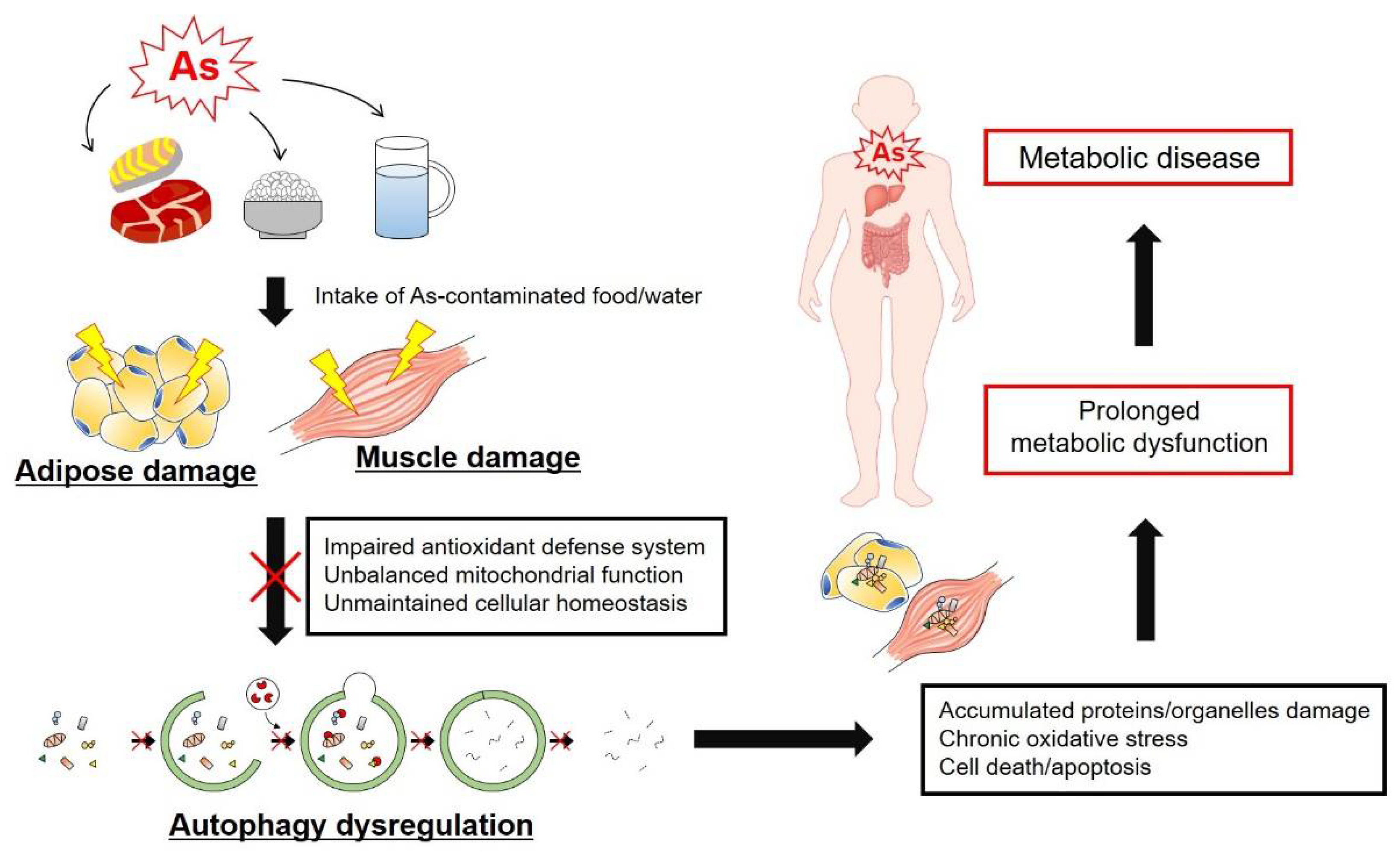
6. Conclusion: Perspective on the Connection between As Exposure and Metabolic Disease

Historically, iAs, due to its odorless and tasteless properties, was used to kill many kings and emperors, and is known as the “poison of kings and the king of poisons” [146]. In BC 55, the Roman emperor Nero poisoned his step-brother Tiberius Britannicus with As to secure his position as Roman emperor. In contrast, in ancient Greece and China, Hippocrates and Chinese physicians used As as a treatment for certain types of disease, such as ulcers and abscesses, as well as for remedies against As-induced poisoning as a “poison against a poison” medicine [147]. Since then, As has been used as a chemotherapeutic method to cure syphilis, parasites and leukemia [148,149]. As has been used as a poison as well as a medicine, highlighting that the right amount of As in treatment could be beneficial for human health. In that sense, we could not exclude the possibility that chronic low-dose (less than 10 ppb) As exposure in adipose and muscle tissues would be beneficial as a hormesis effect for inducing the oxidative defense mechanism or inhibiting apoptosis differently from the harmful effects of high-dose As exposure in the liver, urinary tract, lungs and skin. In this review, we have summarized the effects of As exposure on metabolism and autophagy in adipose and muscle tissues, which are major target tissues in metabolic syndrome. Although the effects of acute and chronic As exposure have identified action mechanisms in adipose and muscle tissues, the effects of indirect As exposure from food or directly from the environment remain controversial and require further research in the USA. In conclusion, studies on the effects of As in metabolic function and in autophagy activity in adipose and muscle tissues could be beneficial for the group of people who are suffering from metabolic syndrome and diseases in highly As-contaminated areas worldwide.
Table 1. Summary of the maximum dose and duration of As treated or administered in the research papers that are referenced in this review.
SamplesDose and Duration of AsReference
Cell culture3T3-L1 preadipocytes, mouse-adipose-derived stromal vascular fraction cells and human adipose tissue-derived stem cells5 μM (sodium arsenite), 1 μM (trivalent monomethylated arsenic), or 2 μM (trivalent dimethylated arsenic) in medium, 2 days with DMI (differentiation media) [75]
3T3-L1 adipocytes1 mM, sodium arsenite (NaAsO2), methylarsine oxide and iododimethylarsine in medium, until full differentiation for several days[102]
3T3-L13 μM, arsenic trioxide (As2O3) in medium, during differentiation for 24 h[119]
3T3-F442A0.5 μM/L, arsenic trioxide in medium, 3 days[120]
Adipose-tissue-derived primary human mesenchymal stem cells (hMSCs)1 μM, sodium arsenite in medium, 72 h[78]
C2C12 myoblast cells and 3T3-L12 μM, sodium arsenite in medium, 8 weeks[76]
C2C12 myotubules1 μM, arsenic trioxide in medium, 48 h[90]
C3H 10T1/2 adipocytes6 μM, sodium arsenite in medium, 2 months[77,121]
EJ-1, human bladder cancer cellsUp to 1 mM (iASIII, iASV, MMAV, MMMTAV, DMAV, DMAIII,DMMTAV or DMDTAV) in medium, 24 h[58]
H9c2 rat cardiomyoblastoma cells50 μM, arsenic trioxide in medium, 24 h[142]
HaCaT human keratinocytes25 μM, sodium arsenite in medium, 24 h[35]
HEK293, NIH3T3, BEAS-2B1 μM, sodium arsenite in medium, 24 h[144]
HIB1B adipocytes10 μM, sodium arsenite in medium, 6 days[79]
Human–hamster hybrid AL cells0.25 μg/mL, sodium arsenite in medium, 60 days[110]
INS-1 832/13 beta cells2 μM (sodium arsenite, dimethyl arsenite), 0.5 μM (mono-methyl arsenite) in medium, 24 h[104]
L6 rat skeletal muscle cells0.5 mM, sodium arsenite in medium, 30 min[123]
NIH3T3, HeLa cells2 μM, sodium arsenite in medium, 48 h[145]
P19 mouse embryonic stem cells1 μM, sodium arsenite in medium, 5 days until full differentiation[91]
PAEC, porcine aortic endothelial cells5 μM, sodium arsenite in medium, 2 h[32]
Pancreatic eyelets (ex vivo) of B6 mice0.1 μM (methylarsonite, dimethylarsinite) in medium, 48 h[103]
Primary SECs, Liver sinusoidal endothelial cells5 μM, sodium arsenite in medium, 8 h[33]
UROtsa, Non-tumorigenic human urothelial cell lines1 μM, sodium arsenite in medium, 12 weeks
50 nM, MMA (III) in medium, 24 weeks
[36]
AnimalsChicken skeletal muscle (in vivo)2.5 mg/kg/day, until reach 30 mg/kg, H3AsO3 in diet [85]
Chickens2.5 mg/kg, As2O3 in diet, 12 weeks[140]
Hamster LD50112 μmol/kg and 29.3 μmol/kg (MMAIII) in diet[42]
Mice (C57BL/6 aka B6, male and female)10 mg/kg/d, sodium arsenite in gavage, 9 days[79]
Mice (B6, male)45 ppm, sodium arsenite in drinking water, 48 weeks[34]
Mice (B6, male)50 ppm, sodium arsenite in drinking water, 18 weeks[73]
Mice (B6, male)50 ppm, sodium arsenate in drinking water, 8 weeks[101]
Mice (B6, female)20 ppm, sodium arsenite and sodium arsenate dibasic heptahydrate (1:1 ratio) in drinking water, 17 weeks[80]
Mice (C57BL/6J, male) eye250 ppm for 1 month or 50 ppm for 6 months (sodium arsenite) in drinking water[62]
Mice (B6) skeletal muscle4 μM or 4 mg/L, arsenic trioxide in drinking water, 12 weeks[141]
Mice (C57BL/6Tac)100 μg/L, sodium arsenite in drinking water, 5 weeks[78]
Mice (C57BL/6Tac)100 μg/L, trivalent arsenite (AsIII) in drinking water, 5 weeks[112]
Mice (C57BL/6NTac male)100 μg/L, sodium metaarsenite (NaAsO2) in drinking water, 5 weeks[113]
Mice (C57BL/6Ai p47phox knockout)250 ppb, sodium arsenite in drinking water, 2 weeks[33]
Mice (CrL:Sk1-hrBD, weanling female hairless mice) skin5 mg/L, sodium arsenite in drinking water, 26 weeks[46]
Mice (sciatic nerve denervated)0.5 ppm, arsenic trioxide in drinking water, 4 weeks[86]
Mice (specific pathogen free female)1 ppm, sodium arsenite in drinking water, 2 weeks[68]
Rat (Winstar, Albino male)150 ppm (sodium arsenite), 200 ppm (sodium arsenate) in drinking water, 12 weeks[67]
Rat (Albino male)25 ppm, sodium arsenite in drinking water, 8 weeks[69]
Rat (Albino male)133 μg/mL (arsenic trioxide) in drinking water, 8 weeks[71]
Rat (Albino pregnant)13 ppm, sodium arsenite, from gestation, lactation through full adulthood in drinking water[72]
Rat liver tissue in test tube40 μM sodium arsenate uptake upto 30 min[106]
Rat liver mitochondria100 μM, sodium arsenite, 5 min[107]
Rat liver mitochondria25 ppm, sodium arsenite, 12 weeks[109]
Rat (Winstar, TR)0.5 mg/kg, 1 mmol, 0.4 mmol (MMA), 1 mmol (DMA), injection through bile duct cannulation less than 5 s[50]
Zebrafish500 ppb, sodium arsenite in water, 7 days[70]
Human patientsArsenic biomarkers in human1–2 mg/kg (LD50 of iAs), accumulated arsenic from food, water, air and soil[29]
Hair, nails and skin scales of arsenic-exposed patients in West Bengal, IndiaArsenic patients exposed to above 50 mg/L in drinking groundwater[44]
Human (2–14 years old adolescents) arsenicosis patientsUpto 31.6 μg/L total inorganic arsenic exposure, upto 14 years[66]
Human acute promyelocytic leukaemia (APL) patientsAll patients were treated with intravenous infusion of 10 mg of As2O3 (10 mL, 0.1% solution) over 2–8 h daily for 28 d and the cycle was repeated at intervals of 2 weeks until complete remission (CR) was achieved. After CR, they were maintained with several cycles of As2O3 at intervals of 2–3 months.[87]
Human cardiovascular disease patients100 μg/L of arsenic above or below in meta-analysis[95]
Human patients (arsenic exposure)Chronic exposure of arsenic in Bandladeshi (above 10 μg/L exposure) for 9 days[98]
Human patients (Diabetes)Geographical tracing (0.1 μg/L detection limit of arsenic) in Denmark for mean follow-up for 9.7 years[105]

Author Contributions

Conceptualization, S.-H.R., J.B., Y.J. and J.F.M.; Writing—Original Draft Preparation, S.-H.R., J.B., Y.J. and J.F.M.; Writing—Review and Editing, S.C., J.Y., S.K.N., R.F. and H.-S.S.; Visualization, S.-H.R., J.B., Y.J. and J.F.M.; Supervision and Project Administration, S.-H.R.; Funding Acquisition, S.-H.R., J.B., J.F.M. and H.-S.S. All authors have read and agreed to the published version of the manuscript.

Funding

This work was supported by National Science Foundation (NSF)-REU site in the form of Training in Redox Biology (DBI-1757951) grant to J.F.M.; the National Institute of Health (NIH)/National Heart, Lung and Blood Institute (NHLBI) grant under Ruth L. Kirschstein National Research Service Award (NRSA) (T32 HL007936) from the University of Wisconsin Cardiovascular Research Center to J.B.; the University of Nebraska Collaboration Initiative Team Formation and Publication Grant to S.-H.R. and Samuel M. Cohen; the University of Nebraska Collaboration Initiative Team Seed Grant to S.-H.R. and H.-S.S.; and the University of Nebraska ARD/ORED/BIOC grants and Nebraska Tobacco Settlement Biomedical Research enhancement funds to S.-H.R. and H.-S.S. This research was supported in part by the Nebraska Center for the Prevention of Obesity Diseases (NPOD) Program Seed Grant from the National Institute of General Medical Sciences (NIGMS) of the NIH under award number P20 GM104320.

Institutional Review Board Statement

Not applicable.

Informed Consent Statement

Not applicable.

Data Availability Statement

Data are available within the article.

Acknowledgments

We thank Samuel M. Cohen (University of Nebraska Medical Center) for comments and extensive editing of the manuscript.

Conflicts of Interest

The authors declare no conflict of interest.

References

  1. Meliker, J.R.; Wahl, R.L.; Cameron, L.L.; Nriagu, J.O. Arsenic in drinking water and cerebrovascular disease, diabetes mellitus, and kidney disease in Michigan: A standardized mortality ratio analysis. Environ. Health 2007, 6, 4. [Google Scholar] [CrossRef] [PubMed] [Green Version]
  2. Steinmaus, C.; Yuan, Y.; Liaw, J.; Smith, A.H. Low-level population exposure to inorganic arsenic in the United States and diabetes mellitus: A reanalysis. Epidemiology 2009, 20, 807–815. [Google Scholar] [CrossRef] [PubMed]
  3. Navas-Acien, A.; Silbergeld, E.K.; Pastor-Barriuso, R.; Guallar, E. Arsenic exposure and prevalence of type 2 diabetes in US adults. JAMA 2008, 300, 814–822. [Google Scholar] [CrossRef] [PubMed] [Green Version]
  4. Gosselin, D.C.; Klawer, L.M.; Joeckel, R.M.; Harvey, F.E.; Reade, A.R.; McVey, K. Arsenic in Groundwater and Rural Public Water Supplies in Nebraska, USA. Great Plains Res. 2006, 16, 137–148. [Google Scholar]
  5. Cohen, S. Agricultural Chemical News. Groundw. Monit. Remediat. 1989, 9, 57–64. [Google Scholar] [CrossRef]
  6. NDA. Nebraska Department of Agriculture Report; NDA: Lincoln, NE, USA, 2020. [Google Scholar]
  7. Parsons, J.G.; Martinez-Martinez, A.; Peralta-Videa, J.R.; Gardea-Torresdey, J.L. Speciation and uptake of arsenic accumulated by corn seedlings using XAS and DRC-ICP-MS. Chemosphere 2008, 70, 2076–2083. [Google Scholar] [CrossRef] [PubMed] [Green Version]
  8. Armendariz, A.L.; Talano, M.A.; Travaglia, C.; Reinoso, H.; Wevar Oller, A.L.; Agostini, E. Arsenic toxicity in soybean seedlings and their attenuation mechanisms. Plant Physiol. Biochem. 2016, 98, 119–127. [Google Scholar] [CrossRef]
  9. Palma-Lara, I.; Martinez-Castillo, M.; Quintana-Perez, J.C.; Arellano-Mendoza, M.G.; Tamay-Cach, F.; Valenzuela-Limon, O.L.; Garcia-Montalvo, E.A.; Hernandez-Zavala, A. Arsenic exposure: A public health problem leading to several cancers. Regul. Toxicol. Pharm. 2019, 110, 104539. [Google Scholar] [CrossRef]
  10. Karagas, M.R.; Gossai, A.; Pierce, B.; Ahsan, H. Drinking Water Arsenic Contamination, Skin Lesions, and Malignancies: A Systematic Review of the Global Evidence. Curr. Environ. Health Rep. 2015, 2, 52–68. [Google Scholar] [CrossRef]
  11. Naujokas, M.F.; Anderson, B.; Ahsan, H.; Aposhian, H.V.; Graziano, J.H.; Thompson, C.; Suk, W.A. The broad scope of health effects from chronic arsenic exposure: Update on a worldwide public health problem. Environ. Health Perspect. 2013, 121, 295–302. [Google Scholar] [CrossRef]
  12. Jomova, K.; Jenisova, Z.; Feszterova, M.; Baros, S.; Liska, J.; Hudecova, D.; Rhodes, C.J.; Valko, M. Arsenic: Toxicity, oxidative stress and human disease. J. Appl. Toxicol. 2011, 31, 95–107. [Google Scholar] [CrossRef]
  13. Abdul, K.S.; Jayasinghe, S.S.; Chandana, E.P.; Jayasumana, C.; De Silva, P.M. Arsenic and human health effects: A review. Environ. Toxicol. Pharm. 2015, 40, 828–846. [Google Scholar] [CrossRef] [PubMed]
  14. Kuo, C.C.; Moon, K.A.; Wang, S.L.; Silbergeld, E.; Navas-Acien, A. The Association of Arsenic Metabolism with Cancer, Cardiovascular Disease, and Diabetes: A Systematic Review of the Epidemiological Evidence. Environ. Health Perspect. 2017, 125, 087001. [Google Scholar] [CrossRef] [PubMed] [Green Version]
  15. Castriota, F.; Acevedo, J.; Ferreccio, C.; Smith, A.H.; Liaw, J.; Smith, M.T.; Steinmaus, C. Obesity and increased susceptibility to arsenic-related type 2 diabetes in Northern Chile. Environ. Res. 2018, 167, 248–254. [Google Scholar] [CrossRef] [PubMed]
  16. Maull, E.A.; Ahsan, H.; Edwards, J.; Longnecker, M.P.; Navas-Acien, A.; Pi, J.; Silbergeld, E.K.; Styblo, M.; Tseng, C.H.; Thayer, K.A.; et al. Evaluation of the association between arsenic and diabetes: A National Toxicology Program workshop review. Environ. Health Perspect. 2012, 120, 1658–1670. [Google Scholar] [CrossRef] [PubMed] [Green Version]
  17. Farkhondeh, T.; Samarghandian, S.; Azimi-Nezhad, M. The role of arsenic in obesity and diabetes. J. Cell Physiol. 2019, 234, 12516–12529. [Google Scholar] [CrossRef] [PubMed]
  18. Spratlen, M.J.; Grau-Perez, M.; Best, L.G.; Yracheta, J.; Lazo, M.; Vaidya, D.; Balakrishnan, P.; Gamble, M.V.; Francesconi, K.A.; Goessler, W.; et al. The Association of Arsenic Exposure and Arsenic Metabolism With the Metabolic Syndrome and Its Individual Components: Prospective Evidence From the Strong Heart Family Study. Am. J. Epidemiol. 2018, 187, 1598–1612. [Google Scholar] [CrossRef] [PubMed]
  19. Ayotte, J.D.; Medalie, L.; Qi, S.L.; Backer, L.C.; Nolan, B.T. Estimating the High-Arsenic Domestic-Well Population in the Conterminous United States. Environ. Sci. Technol. 2017, 51, 12443–12454. [Google Scholar] [CrossRef]
  20. Preston, S.H.; Vierboom, Y.C.; Stokes, A. The role of obesity in exceptionally slow US mortality improvement. Proc. Natl. Acad. Sci. USA 2018, 115, 957–961. [Google Scholar] [CrossRef] [Green Version]
  21. Wang, Y.C.; McPherson, K.; Marsh, T.; Gortmaker, S.L.; Brown, M. Health and economic burden of the projected obesity trends in the USA and the UK. Lancet 2011, 378, 815–825. [Google Scholar] [CrossRef]
  22. Kim, D.D.; Basu, A. Estimating the Medical Care Costs of Obesity in the United States: Systematic Review, Meta-Analysis, and Empirical Analysis. Value Health 2016, 19, 602–613. [Google Scholar] [CrossRef] [PubMed] [Green Version]
  23. Ro, S.H.; Jang, Y.; Bae, J.; Kim, I.M.; Schaecher, C.; Shomo, Z.D. Autophagy in Adipocyte Browning: Emerging Drug Target for Intervention in Obesity. Front. Physiol. 2019, 10, 22. [Google Scholar] [CrossRef] [PubMed]
  24. Wells, G.D.; Noseworthy, M.D.; Hamilton, J.; Tarnopolski, M.; Tein, I. Skeletal muscle metabolic dysfunction in obesity and metabolic syndrome. Can. J. Neurol. Sci. 2008, 35, 31–40. [Google Scholar] [CrossRef] [PubMed] [Green Version]
  25. Marette, A.; Liu, Y.; Sweeney, G. Skeletal muscle glucose metabolism and inflammation in the development of the metabolic syndrome. Rev. Endocr. Metab. Disord. 2014, 15, 299–305. [Google Scholar] [CrossRef]
  26. Kuivenhoven, M.; Mason, K. Arsenic Toxicity. In StatPearls; StatPearls: Treasure Island, FL, USA, 2021. [Google Scholar]
  27. Arsenic in Drinking Water; The National Academies Press: Washington, DC, USA, 1999.
  28. Ravenscroft, B.H.; Richards, K. Arsenic Pollution: A Global Synthesis; Wiley: Hoboken, NJ, USA, 2009; ISBN 978-1-405-18601-8. [Google Scholar]
  29. Hughes, M.F. Biomarkers of exposure: A case study with inorganic arsenic. Environ. Health Perspect 2006, 114, 1790–1796. [Google Scholar] [CrossRef]
  30. Klein, C.B.; Leszczynska, J.; Hickey, C.; Rossman, T.G. Further evidence against a direct genotoxic mode of action for arsenic-induced cancer. Toxicol. Appl. Pharm. 2007, 222, 289–297. [Google Scholar] [CrossRef] [Green Version]
  31. Ratnaike, R.N. Acute and chronic arsenic toxicity. Postgrad Med. J. 2003, 79, 391–396. [Google Scholar] [CrossRef]
  32. Smith, K.R.; Klei, L.R.; Barchowsky, A. Arsenite stimulates plasma membrane NADPH oxidase in vascular endothelial cells. Am. J. Physiol. Lung Cell Mol. Physiol. 2001, 280, L442–L449. [Google Scholar] [CrossRef]
  33. Straub, A.C.; Clark, K.A.; Ross, M.A.; Chandra, A.G.; Li, S.; Gao, X.; Pagano, P.J.; Stolz, D.B.; Barchowsky, A. Arsenic-stimulated liver sinusoidal capillarization in mice requires NADPH oxidase-generated superoxide. J. Clin. Investig. 2008, 118, 3980–3989. [Google Scholar] [CrossRef]
  34. Chen, H.; Li, S.; Liu, J.; Diwan, B.A.; Barrett, J.C.; Waalkes, M.P. Chronic inorganic arsenic exposure induces hepatic global and individual gene hypomethylation: Implications for arsenic hepatocarcinogenesis. Carcinogenesis 2004, 25, 1779–1786. [Google Scholar] [CrossRef] [Green Version]
  35. Reichard, J.F.; Schnekenburger, M.; Puga, A. Long term low-dose arsenic exposure induces loss of DNA methylation. Biochem. Biophys. Res. Commun. 2007, 352, 188–192. [Google Scholar] [CrossRef] [Green Version]
  36. Jensen, T.J.; Novak, P.; Wnek, S.M.; Gandolfi, A.J.; Futscher, B.W. Arsenicals produce stable progressive changes in DNA methylation patterns that are linked to malignant transformation of immortalized urothelial cells. Toxicol. Appl. Pharm. 2009, 241, 221–229. [Google Scholar] [CrossRef] [PubMed] [Green Version]
  37. Ajees, A.A.; Rosen, B.P. As(III) S-adenosylmethionine methyltransferases and other arsenic binding proteins. Geomicrobiol. J. 2015, 32, 570–576. [Google Scholar] [CrossRef] [PubMed] [Green Version]
  38. Roy, N.K.; Murphy, A.; Costa, M. Arsenic Methyltransferase and Methylation of Inorganic Arsenic. Biomolecules 2020, 10, 1351. [Google Scholar] [CrossRef] [PubMed]
  39. Chen Jian, R.B.P. The Arsenic Methylation Cycle: How Microbial Communities Adapted Methylarsenicals for Use as Weapons in the Continuing War for Dominance. Front. Environ. Sci. 2020, 8, 43. [Google Scholar] [CrossRef] [Green Version]
  40. Ajees, A.A.; Marapakala, K.; Packianathan, C.; Sankaran, B.; Rosen, B.P. Structure of an As(III) S-adenosylmethionine methyltransferase: Insights into the mechanism of arsenic biotransformation. Biochemistry 2012, 51, 5476–5485. [Google Scholar] [CrossRef] [Green Version]
  41. Kitchin, K.T. Recent advances in arsenic carcinogenesis: Modes of action, animal model systems, and methylated arsenic metabolites. Toxicol. Appl. Pharm. 2001, 172, 249–261. [Google Scholar] [CrossRef] [Green Version]
  42. Petrick, J.S.; Jagadish, B.; Mash, E.A.; Aposhian, H.V. Monomethylarsonous acid (MMA(III)) and arsenite: LD(50) in hamsters and in vitro inhibition of pyruvate dehydrogenase. Chem. Res. Toxicol. 2001, 14, 651–656. [Google Scholar] [CrossRef]
  43. Lansdown, A.B. Physiological and toxicological changes in the skin resulting from the action and interaction of metal ions. Crit. Rev. Toxicol. 1995, 25, 397–462. [Google Scholar] [CrossRef]
  44. Samanta, G.; Sharma, R.; Roychowdhury, T.; Chakraborti, D. Arsenic and other elements in hair, nails, and skin-scales of arsenic victims in West Bengal, India. Sci. Total Environ. 2004, 326, 33–47. [Google Scholar] [CrossRef]
  45. Scott, N.; Hatlelid, K.M.; MacKenzie, N.E.; Carter, D.E. Reactions of arsenic(III) and arsenic(V) species with glutathione. Chem. Res. Toxicol. 1993, 6, 102–106. [Google Scholar] [CrossRef] [PubMed]
  46. Uddin, A.N.; Burns, F.J.; Rossman, T.G. Vitamin E and organoselenium prevent the cocarcinogenic activity of arsenite with solar UVR in mouse skin. Carcinogenesis 2005, 26, 2179–2186. [Google Scholar] [CrossRef] [PubMed] [Green Version]
  47. Liu, Z.; Styblo, M.; Rosen, B.P. Methylarsonous acid transport by aquaglyceroporins. Environ. Health Perspect 2006, 114, 527–531. [Google Scholar] [CrossRef] [PubMed] [Green Version]
  48. Latowski, A.K.a.D. Biochemical pathways of arsenic uptake from the environment to human cells. Mol. Biophys. Biochem. 2016, 1, 9–16. [Google Scholar] [CrossRef]
  49. Drobna, Z.; Walton, F.S.; Paul, D.S.; Xing, W.; Thomas, D.J.; Styblo, M. Metabolism of arsenic in human liver: The role of membrane transporters. Arch. Toxicol. 2010, 84, 3–16. [Google Scholar] [CrossRef] [PubMed]
  50. Kala, S.V.; Neely, M.W.; Kala, G.; Prater, C.I.; Atwood, D.W.; Rice, J.S.; Lieberman, M.W. The MRP2/cMOAT transporter and arsenic-glutathione complex formation are required for biliary excretion of arsenic. J. Biol. Chem. 2000, 275, 33404–33408. [Google Scholar] [CrossRef] [PubMed] [Green Version]
  51. Herath, I.; Vithanage, M.; Seneweera, S.; Bundschuh, J. Thiolated arsenic in natural systems: What is current, what is new and what needs to be known. Environ. Int. 2018, 115, 370–386. [Google Scholar] [CrossRef]
  52. Planer-Friedrich, B.; London, J.; McCleskey, R.B.; Nordstrom, D.K.; Wallschlager, D. Thioarsenates in geothermal waters of Yellowstone National Park: Determination, preservation, and geochemical importance. Environ. Sci. Technol. 2007, 41, 5245–5251. [Google Scholar] [CrossRef]
  53. Colina Blanco, A.E.; Kerl, C.F.; Planer-Friedrich, B. Detection of Thioarsenates in Rice Grains and Rice Products. J. Agric. Food Chem. 2021, 69, 2287–2294. [Google Scholar] [CrossRef]
  54. Wang, J.; Kerl, C.F.; Hu, P.; Martin, M.; Mu, T.; Brüggenwirth, L.; Wu, G.; Said-Pullicino, D.; Romani, M.; Wu, L.; et al. Thiolated arsenic species observed in rice paddy pore waters. Nat. Geosci. 2020, 13, 282–287. [Google Scholar] [CrossRef]
  55. SS, D.C.R.; Alava, P.; Zekker, I.; Du Laing, G.; Van de Wiele, T. Arsenic thiolation and the role of sulfate-reducing bacteria from the human intestinal tract. Environ. Health Perspect. 2014, 122, 817–822. [Google Scholar] [CrossRef]
  56. Sun, Y.; Liu, G.; Cai, Y. Thiolated arsenicals in arsenic metabolism: Occurrence, formation, and biological implications. J. Environ. Sci. 2016, 49, 59–73. [Google Scholar] [CrossRef]
  57. Fricke, M.; Zeller, M.; Cullen, W.; Witkowski, M.; Creed, J. Dimethylthioarsinic anhydride: A standard for arsenic speciation. Anal. Chim. Acta 2007, 583, 78–83. [Google Scholar] [CrossRef] [PubMed]
  58. Naranmandura, H.; Carew, M.W.; Xu, S.; Lee, J.; Leslie, E.M.; Weinfeld, M.; Le, X.C. Comparative toxicity of arsenic metabolites in human bladder cancer EJ-1 cells. Chem. Res. Toxicol. 2011, 24, 1586–1596. [Google Scholar] [CrossRef]
  59. Guha Mazumder, D.N.; De, B.K.; Santra, A.; Ghosh, N.; Das, S.; Lahiri, S.; Das, T. Randomized placebo-controlled trial of 2,3-dimercapto-1-propanesulfonate (DMPS) in therapy of chronic arsenicosis due to drinking arsenic-contaminated water. J. Toxicol. Clin. Toxicol. 2001, 39, 665–674. [Google Scholar] [CrossRef]
  60. Bjorklund, G.; Oliinyk, P.; Lysiuk, R.; Rahaman, M.S.; Antonyak, H.; Lozynska, I.; Lenchyk, L.; Peana, M. Arsenic intoxication: General aspects and chelating agents. Arch. Toxicol. 2020, 94, 1879–1897. [Google Scholar] [CrossRef]
  61. Nurchi, V.M.; Djordjevic, A.B.; Crisponi, G.; Alexander, J.; Bjorklund, G.; Aaseth, J. Arsenic Toxicity: Molecular Targets and Therapeutic Agents. Biomolecules 2020, 10, 235. [Google Scholar] [CrossRef] [PubMed] [Green Version]
  62. Kleiman, N.J.; Quinn, A.M.; Fields, K.G.; Slavkovich, V.; Graziano, J.H. Arsenite accumulation in the mouse eye. J. Toxicol. Environ. Health A 2016, 79, 339–341. [Google Scholar] [CrossRef] [PubMed]
  63. Khairul, I.; Wang, Q.Q.; Jiang, Y.H.; Wang, C.; Naranmandura, H. Metabolism, toxicity and anticancer activities of arsenic compounds. Oncotarget 2017, 8, 23905–23926. [Google Scholar] [CrossRef] [PubMed] [Green Version]
  64. Athyros, V.G.; Doumas, M.; Imprialos, K.P.; Stavropoulos, K.; Georgianou, E.; Katsimardou, A.; Karagiannis, A. Diabetes and lipid metabolism. Hormones 2018, 17, 61–67. [Google Scholar] [CrossRef] [PubMed] [Green Version]
  65. Kloska, A.; Węsierska, M.; Malinowska, M.; Gabig-Cimińska, M.; Jakóbkiewicz-Banecka, J. Lipophagy and Lipolysis Status in Lipid Storage and Lipid Metabolism Diseases. Int. J. Mol. Sci. 2020, 21, 6113. [Google Scholar] [CrossRef] [PubMed]
  66. Kuo, C.C.; Su, P.H.; Sun, C.W.; Liu, H.J.; Chang, C.L.; Wang, S.L. Early-life arsenic exposure promotes atherogenic lipid metabolism in adolescence: A 15-year birth cohort follow-up study in central Taiwan. Environ. Int. 2018, 118, 97–105. [Google Scholar] [CrossRef] [PubMed]
  67. Afolabi, O.K.; Wusu, A.D.; Ogunrinola, O.O.; Abam, E.O.; Babayemi, D.O.; Dosumu, O.A.; Onunkwor, O.B.; Balogun, E.A.; Odukoya, O.O.; Ademuyiwa, O. Arsenic-induced dyslipidemia in male albino rats: Comparison between trivalent and pentavalent inorganic arsenic in drinking water. BMC Pharm. Toxicol. 2015, 16, 15. [Google Scholar] [CrossRef] [PubMed] [Green Version]
  68. Chi, L.; Lai, Y.; Tu, P.; Liu, C.W.; Xue, J.; Ru, H.; Lu, K. Lipid and Cholesterol Homeostasis after Arsenic Exposure and Antibiotic Treatment in Mice: Potential Role of the Microbiota. Environ. Health Perspect 2019, 127, 97002. [Google Scholar] [CrossRef] [PubMed]
  69. Soni, M.; Prakash, C.; Dabur, R.; Kumar, V. Protective Effect of Hydroxytyrosol Against Oxidative Stress Mediated by Arsenic-Induced Neurotoxicity in Rats. Appl Biochem. Biotechnol. 2018, 186, 27–39. [Google Scholar] [CrossRef] [PubMed]
  70. Carlson, P.; Van Beneden, R.J. Arsenic exposure alters expression of cell cycle and lipid metabolism genes in the liver of adult zebrafish (Danio rerio). Aquat. Toxicol. 2014, 153, 66–72. [Google Scholar] [CrossRef] [PubMed]
  71. Cheng, T.J.; Chuu, J.J.; Chang, C.Y.; Tsai, W.C.; Chen, K.J.; Guo, H.R. Atherosclerosis induced by arsenic in drinking water in rats through altering lipid metabolism. Toxicol. Appl. Pharm. 2011, 256, 146–153. [Google Scholar] [CrossRef]
  72. Rivas-Santiago, C.; Gonzalez-Curiel, I.; Zarazua, S.; Murgu, M.; Ruiz Cardona, A.; Lazalde, B.; Lara-Ramirez, E.E.; Vazquez, E.; Castaneda-Delgado, J.E.; Rivas-Santiago, B.; et al. Lipid Metabolism Alterations in a Rat Model of Chronic and Intergenerational Exposure to Arsenic. Biomed. Res. Int. 2019, 2019, 4978018. [Google Scholar] [CrossRef] [Green Version]
  73. Song, X.; Li, Y.; Liu, J.; Ji, X.; Zhao, L.; Wei, Y. Changes in Serum Adiponectin in Mice Chronically Exposed to Inorganic Arsenic in Drinking Water. Biol. Trace Elem. Res. 2017, 179, 140–147. [Google Scholar] [CrossRef]
  74. Renu, K.; Madhyastha, H.; Madhyastha, R.; Maruyama, M.; Arunachlam, S.; V G, A. Role of arsenic exposure in adipose tissue dysfunction and its possible implication in diabetes pathophysiology. Toxicol. Lett. 2018, 284, 86–95. [Google Scholar] [CrossRef]
  75. Hou, Y.; Xue, P.; Woods, C.G.; Wang, X.; Fu, J.; Yarborough, K.; Qu, W.; Zhang, Q.; Andersen, M.E.; Pi, J. Association between arsenic suppression of adipogenesis and induction of CHOP10 via the endoplasmic reticulum stress response. Environ. Health Perspect 2013, 121, 237–243. [Google Scholar] [CrossRef] [PubMed] [Green Version]
  76. Padmaja Divya, S.; Pratheeshkumar, P.; Son, Y.O.; Vinod Roy, R.; Andrew Hitron, J.; Kim, D.; Dai, J.; Wang, L.; Asha, P.; Huang, B.; et al. Arsenic Induces Insulin Resistance in Mouse Adipocytes and Myotubes Via Oxidative Stress-Regulated Mitochondrial Sirt3-FOXO3a Signaling Pathway. Toxicol. Sci. 2015, 146, 290–300. [Google Scholar] [CrossRef] [PubMed]
  77. Trouba, K.J.; Wauson, E.M.; Vorce, R.L. Sodium arsenite inhibits terminal differentiation of murine C3H 10T1/2 preadipocytes. Toxicol. Appl. Pharm. 2000, 168, 25–35. [Google Scholar] [CrossRef] [PubMed]
  78. Garciafigueroa, D.Y.; Klei, L.R.; Ambrosio, F.; Barchowsky, A. Arsenic-stimulated lipolysis and adipose remodeling is mediated by G-protein-coupled receptors. Toxicol. Sci. 2013, 134, 335–344. [Google Scholar] [CrossRef]
  79. Bae, J.; Jang, Y.; Kim, H.; Mahato, K.; Schaecher, C.; Kim, I.M.; Kim, E.; Ro, S.H. Arsenite exposure suppresses adipogenesis, mitochondrial biogenesis and thermogenesis via autophagy inhibition in brown adipose tissue. Sci. Rep. 2019, 9, 14464. [Google Scholar] [CrossRef]
  80. Zuo, Z.; Liu, Z.; Gao, T.; Yin, Y.; Wang, Z.; Hou, Y.; Fu, J.; Liu, S.; Wang, H.; Xu, Y.; et al. Prolonged inorganic arsenic exposure via drinking water impairs brown adipose tissue function in mice. Sci. Total Environ. 2019, 668, 310–317. [Google Scholar] [CrossRef]
  81. Chiesa, L.M.; Ceriani, F.; Procopio, A.; Bonacci, S.; Malandra, R.; Panseri, S.; Arioli, F. Exposure to metals and arsenic from yellow and red tuna consumption. Food Addit. Contam. Part A Chem Anal. Control. Expo. Risk Assess. 2019, 36, 1228–1235. [Google Scholar] [CrossRef]
  82. Farzad, R.; Kuhn, D.D.; Smith, S.A.; O’Keefe, S.F.; Ralston, N.V.C.; Neilson, A.P.; Gatlin, D.M. Trace minerals in tilapia fillets: Status in the United States marketplace and selenium supplementation strategy for improving consumer’s health. PLoS ONE 2019, 14, e0217043. [Google Scholar] [CrossRef] [Green Version]
  83. Pei, J.; Zuo, J.; Wang, X.; Yin, J.; Liu, L.; Fan, W. The Bioaccumulation and Tissue Distribution of Arsenic Species in Tilapia. Int J. Environ. Res. Public Health 2019, 16, 757. [Google Scholar] [CrossRef] [Green Version]
  84. Szymkowicz, D.B.; Schwendinger, K.L.; Tatnall, C.M.; Swetenburg, J.R.; Bain, L.J. Embryonic-only arsenic exposure alters skeletal muscle satellite cell function in killifish (Fundulus heteroclitus). Aquat. Toxicol. 2018, 198, 276–286. [Google Scholar] [CrossRef]
  85. Wang, Y.; Zhao, H.; Shao, Y.; Liu, J.; Li, J.; Luo, L.; Xing, M. Copper (II) and/or arsenite-induced oxidative stress cascades apoptosis and autophagy in the skeletal muscles of chicken. Chemosphere 2018, 206, 597–605. [Google Scholar] [CrossRef] [PubMed]
  86. Chen, C.M.; Chung, M.N.; Chiu, C.Y.; Liu, S.H.; Lan, K.C. Inorganic Arsenic Exposure Decreases Muscle Mass and Enhances Denervation-Induced Muscle Atrophy in Mice. Molecules 2020, 25, 3057. [Google Scholar] [CrossRef] [PubMed]
  87. Huang, S.Y.; Chang, C.S.; Tang, J.L.; Tien, H.F.; Kuo, T.L.; Huang, S.F.; Yao, Y.T.; Chou, W.C.; Chung, C.Y.; Wang, C.H.; et al. Acute and chronic arsenic poisoning associated with treatment of acute promyelocytic leukaemia. Br. J. Haematol. 1998, 103, 1092–1095. [Google Scholar] [CrossRef] [PubMed]
  88. Sarker, M.K.; Tony, S.R.; Siddique, A.E.; Karim, M.R.; Haque, N.; Islam, Z.; Islam, M.S.; Khatun, M.; Islam, J.; Hossain, S.; et al. Arsenic Secondary Methylation Capacity Is Inversely Associated with Arsenic Exposure-Related Muscle Mass Reduction. Int. J. Environ. Res. Public Health 2021, 18, 9730. [Google Scholar] [CrossRef] [PubMed]
  89. Rehman, M.Y.A.; Briede, J.J.; van Herwijnen, M.; Krauskopf, J.; Jennen, D.G.J.; Malik, R.N.; Kleinjans, J.C.S. Integrating SNPs-based genetic risk factor with blood epigenomic response of differentially arsenic-exposed rural subjects reveals disease-associated signaling pathways. Environ. Pollut. 2022, 292, 118279. [Google Scholar] [CrossRef] [PubMed]
  90. Chiu, C.Y.; Chung, M.N.; Lan, K.C.; Yang, R.S.; Liu, S.H. Exposure of low-concentration arsenic induces myotube atrophy by inhibiting an Akt signaling pathway. Toxicol. Vitr. 2020, 65, 104829. [Google Scholar] [CrossRef] [PubMed]
  91. Hong, G.M.; Bain, L.J. Arsenic exposure inhibits myogenesis and neurogenesis in P19 stem cells through repression of the beta-catenin signaling pathway. Toxicol. Sci. 2012, 129, 146–156. [Google Scholar] [CrossRef] [Green Version]
  92. Diaz-Villasenor, A.; Burns, A.L.; Hiriart, M.; Cebrian, M.E.; Ostrosky-Wegman, P. Arsenic-induced alteration in the expression of genes related to type 2 diabetes mellitus. Toxicol. Appl. Pharm. 2007, 225, 123–133. [Google Scholar] [CrossRef]
  93. Drobna, Z.; Styblo, M.; Thomas, D.J. An Overview of Arsenic Metabolism and Toxicity. Curr. Protoc. Toxicol. 2009. [Google Scholar] [CrossRef] [Green Version]
  94. Hughes, M.F. Arsenic toxicity and potential mechanisms of action. Toxicol. Lett. 2002, 133, 1–16. [Google Scholar] [CrossRef] [Green Version]
  95. Moon, K.A.; Oberoi, S.; Barchowsky, A.; Chen, Y.; Guallar, E.; Nachman, K.E.; Rahman, M.; Sohel, N.; D’Ippoliti, D.; Wade, T.J.; et al. A dose-response meta-analysis of chronic arsenic exposure and incident cardiovascular disease. Int. J. Epidemiol. 2017, 46, 1924–1939. [Google Scholar] [CrossRef] [PubMed] [Green Version]
  96. Fisher, A.T.; López-Carrillo, L.; Gamboa-Loira, B.; Cebrián, M.E. Standards for arsenic in drinking water: Implications for policy in Mexico. J. Public Health Policy 2017, 38, 395–406. [Google Scholar] [CrossRef] [PubMed] [Green Version]
  97. Carlin, D.J.; Naujokas, M.F.; Bradham, K.D.; Cowden, J.; Heacock, M.; Henry, H.F.; Lee, J.S.; Thomas, D.J.; Thompson, C.; Tokar, E.J.; et al. Arsenic and Environmental Health: State of the Science and Future Research Opportunities. Environ. Health Perspect. 2016, 124, 890–899. [Google Scholar] [CrossRef] [PubMed]
  98. Argos, M.; Kalra, T.; Rathouz, P.J.; Chen, Y.; Pierce, B.; Parvez, F.; Islam, T.; Ahmed, A.; Rakibuz-Zaman, M.; Hasan, R.; et al. Arsenic exposure from drinking water, and all-cause and chronic-disease mortalities in Bangladesh (HEALS): A prospective cohort study. Lancet 2010, 376, 252–258. [Google Scholar] [CrossRef] [Green Version]
  99. Shao, K.; Zhou, Z.; Xun, P.; Cohen, S.M. Bayesian benchmark dose analysis for inorganic arsenic in drinking water associated with bladder and lung cancer using epidemiological data. Toxicology 2021, 455, 152752. [Google Scholar] [CrossRef]
  100. Sidhu, M.S.; Desai, K.P.; Lynch, H.N.; Rhomberg, L.R.; Beck, B.D.; Venditti, F.J. Mechanisms of action for arsenic in cardiovascular toxicity and implications for risk assessment. Toxicology 2015, 331, 78–99. [Google Scholar] [CrossRef]
  101. Paul, D.S.; Hernandez-Zavala, A.; Walton, F.S.; Adair, B.M.; Dedina, J.; Matousek, T.; Styblo, M. Examination of the effects of arsenic on glucose homeostasis in cell culture and animal studies: Development of a mouse model for arsenic-induced diabetes. Toxicol. Appl. Pharm. 2007, 222, 305–314. [Google Scholar] [CrossRef] [Green Version]
  102. Walton, F.S.; Harmon, A.W.; Paul, D.S.; Drobna, Z.; Patel, Y.M.; Styblo, M. Inhibition of insulin-dependent glucose uptake by trivalent arsenicals: Possible mechanism of arsenic-induced diabetes. Toxicol. Appl. Pharm. 2004, 198, 424–433. [Google Scholar] [CrossRef]
  103. Douillet, C.; Currier, J.; Saunders, J.; Bodnar, W.M.; Matousek, T.; Styblo, M. Methylated trivalent arsenicals are potent inhibitors of glucose stimulated insulin secretion by murine pancreatic islets. Toxicol. Appl. Pharm. 2013, 267, 11–15. [Google Scholar] [CrossRef] [Green Version]
  104. Dover, E.N.; Beck, R.; Huang, M.C.; Douillet, C.; Wang, Z.; Klett, E.L.; Styblo, M. Arsenite and methylarsonite inhibit mitochondrial metabolism and glucose-stimulated insulin secretion in INS-1 832/13 beta cells. Arch. Toxicol. 2018, 92, 693–704. [Google Scholar] [CrossRef]
  105. Brauner, E.V.; Nordsborg, R.B.; Andersen, Z.J.; Tjonneland, A.; Loft, S.; Raaschou-Nielsen, O. Long-term exposure to low-level arsenic in drinking water and diabetes incidence: A prospective study of the diet, cancer and health cohort. Environ. Health Perspect. 2014, 122, 1059–1065. [Google Scholar] [CrossRef] [PubMed] [Green Version]
  106. Crane, R.K.; Lipmann, F. The effect of arsenate on aerobic phosphorylation. J. Biol. Chem. 1953, 201, 235–243. [Google Scholar] [CrossRef]
  107. Hosseini, M.J.; Shaki, F.; Ghazi-Khansari, M.; Pourahmad, J. Toxicity of Arsenic (III) on Isolated Liver Mitochondria: A New Mechanistic Approach. Iran. J. Pharm Res. 2013, 12, 121–138. [Google Scholar] [PubMed]
  108. Ramanathan, K.; Shila, S.; Kumaran, S.; Panneerselvam, C. Ascorbic acid and α-tocopherol as potent modulators on arsenic induced toxicity in mitochondria. J. Nutr. Biochem. 2003, 14, 416–420. [Google Scholar] [CrossRef]
  109. Prakash, C.; Kumar, V. Chronic Arsenic Exposure-Induced Oxidative Stress is Mediated by Decreased Mitochondrial Biogenesis in Rat Liver. Biol. Trace Elem. Res. 2016, 173, 87–95. [Google Scholar] [CrossRef]
  110. Partridge, M.A.; Huang, S.X.; Hernandez-Rosa, E.; Davidson, M.M.; Hei, T.K. Arsenic induced mitochondrial DNA damage and altered mitochondrial oxidative function: Implications for genotoxic mechanisms in mammalian cells. Cancer Res. 2007, 67, 5239–5247. [Google Scholar] [CrossRef] [Green Version]
  111. Martinez, V.D.; Vucic, E.A.; Becker-Santos, D.D.; Gil, L.; Lam, W.L. Arsenic Exposure and the Induction of Human Cancers. J. Toxicol. 2011, 2011, 1–13. [Google Scholar] [CrossRef] [Green Version]
  112. Ambrosio, F.; Brown, E.; Stolz, D.; Ferrari, R.; Goodpaster, B.; Deasy, B.; Distefano, G.; Roperti, A.; Cheikhi, A.; Garciafigueroa, Y.; et al. Arsenic induces sustained impairment of skeletal muscle and muscle progenitor cell ultrastructure and bioenergetics. Free Radic. Biol. Med. 2014, 74, 64–73. [Google Scholar] [CrossRef] [Green Version]
  113. Zhang, C.; Ferrari, R.; Beezhold, K.; Stearns-Reider, K.; D’Amore, A.; Haschak, M.; Stolz, D.; Robbins, P.D.; Barchowsky, A.; Ambrosio, F. Arsenic Promotes NF-Kappab-Mediated Fibroblast Dysfunction and Matrix Remodeling to Impair Muscle Stem Cell Function. Stem. Cells 2016, 34, 732–742. [Google Scholar] [CrossRef] [Green Version]
  114. D’Souza, D.M.; Al-Sajee, D.; Hawke, T.J. Diabetic myopathy: Impact of diabetes mellitus on skeletal muscle progenitor cells. Front. Physiol. 2013, 4, 379. [Google Scholar] [CrossRef] [Green Version]
  115. Andersen, H.; Gjerstad, M.D.; Jakobsen, J. Atrophy of Foot Muscles: A measure of diabetic neuropathy. Diabetes Care 2004, 27, 2382–2385. [Google Scholar] [CrossRef] [PubMed] [Green Version]
  116. Andersen, H.; Schmitz, O.; Nielsen, S. Decreased isometric muscle strength after acute hyperglycaemia in Type 1 diabetic patients. Diabet Med. 2005, 22, 1401–1407. [Google Scholar] [CrossRef] [PubMed]
  117. Hilton, T.N.; Tuttle, L.J.; Bohnert, K.L.; Mueller, M.J.; Sinacore, D.R. Excessive Adipose Tissue Infiltration in Skeletal Muscle in Individuals With Obesity, Diabetes Mellitus, and Peripheral Neuropathy: Association With Performance and Function. Phys. Ther. 2008, 88, 1336–1344. [Google Scholar] [CrossRef] [PubMed] [Green Version]
  118. Navas-Acien, A.; Silbergeld, E.K.; Streeter, R.A.; Clark, J.M.; Burke, T.A.; Guallar, E. Arsenic exposure and type 2 diabetes: A systematic review of the experimental and epidemiological evidence. Environ. Health Perspect. 2006, 114, 641–648. [Google Scholar] [CrossRef] [Green Version]
  119. Wang, Z.X.; Jiang, C.S.; Liu, L.; Wang, X.H.; Jin, H.J.; Wu, Q.; Chen, Q. The role of Akt on arsenic trioxide suppression of 3T3-L1 preadipocyte differentiation. Cell Res. 2005, 15, 379–386. [Google Scholar] [CrossRef] [Green Version]
  120. Salazard, B.; Bellon, L.; Jean, S.; Maraninchi, M.; El-Yazidi, C.; Orsière, T.; Margotat, A.; Botta, A.; Bergé-Lefranc, J.L. Low-level arsenite activates the transcription of genes involved in adipose differentiation. Cell Biol. Toxicol. 2004, 20, 375–385. [Google Scholar] [CrossRef]
  121. Wauson, E.M.; Langan, A.S.; Vorce, R.L. Sodium arsenite inhibits and reverses expression of adipogenic and fat cell-specific genes during in vitro adipogenesis. Toxicol. Sci. 2002, 65, 211–219. [Google Scholar] [CrossRef] [Green Version]
  122. McDowell, H.E.; Walker, T.; Hajduch, E.; Christie, G.; Batty, I.H.; Downes, C.P.; Hundal, H.S. Inositol phospholipid 3-kinase is activated by cellular stress but is not required for the stress-induced activation of glucose transport in L6 rat skeletal muscle cells. Eur. J. Biochem. 1997, 247, 306–313. [Google Scholar] [CrossRef]
  123. Levine, B.; Klionsky, D.J. Autophagy wins the 2016 Nobel Prize in Physiology or Medicine: Breakthroughs in baker’s yeast fuel advances in biomedical research. Proc. Natl. Acad. Sci. USA 2017, 114, 201–205. [Google Scholar] [CrossRef] [Green Version]
  124. Mizushima, N.; Komatsu, M. Autophagy: Renovation of cells and tissues. Cell 2011, 147, 728–741. [Google Scholar] [CrossRef] [Green Version]
  125. Blasiak, J.; Pawlowska, E.; Chojnacki, J.; Szczepanska, J.; Chojnacki, C.; Kaarniranta, K. Zinc and Autophagy in Age-Related Macular Degeneration. Int. J. Mol. Sci. 2020, 21, 4994. [Google Scholar] [CrossRef] [PubMed]
  126. Galluzzi, L.; Baehrecke, E.H.; Ballabio, A.; Boya, P.; Bravo-San Pedro, J.M.; Cecconi, F.; Choi, A.M.; Chu, C.T.; Codogno, P.; Colombo, M.I.; et al. Molecular definitions of autophagy and related processes. EMBO J. 2017, 36, 1811–1836. [Google Scholar] [CrossRef] [PubMed]
  127. Pallauf, K.; Rimbach, G. Autophagy, polyphenols and healthy ageing. Ageing Res. Rev. 2013, 12, 237–252. [Google Scholar] [CrossRef] [PubMed]
  128. Noda, N.N.; Inagaki, F. Mechanisms of Autophagy. Annu. Rev. Biophys. 2015, 44, 101–122. [Google Scholar] [CrossRef]
  129. Galluzzi, L.; Pietrocola, F.; Levine, B.; Kroemer, G. Metabolic control of autophagy. Cell 2014, 159, 1263–1276. [Google Scholar] [CrossRef] [Green Version]
  130. Rabinowitz, J.D.; White, E. Autophagy and metabolism. Science 2010, 330, 1344–1348. [Google Scholar] [CrossRef] [Green Version]
  131. Levine, B.; Kroemer, G. Autophagy in the pathogenesis of disease. Cell 2008, 132, 27–42. [Google Scholar] [CrossRef] [Green Version]
  132. Mizushima, N.; Levine, B.; Cuervo, A.M.; Klionsky, D.J. Autophagy fights disease through cellular self-digestion. Nature 2008, 451, 1069–1075. [Google Scholar] [CrossRef] [Green Version]
  133. Ahmed, M.; Nguyen, H.Q.; Hwang, J.S.; Zada, S.; Lai, T.H.; Kang, S.S.; Kim, D.R. Systematic characterization of autophagy-related genes during the adipocyte differentiation using public-access data. Oncotarget 2018, 9, 15526–15541. [Google Scholar] [CrossRef] [Green Version]
  134. Kovsan, J.; Bluher, M.; Tarnovscki, T.; Kloting, N.; Kirshtein, B.; Madar, L.; Shai, I.; Golan, R.; Harman-Boehm, I.; Schon, M.R.; et al. Altered autophagy in human adipose tissues in obesity. J. Clin. Endocrinol. Metab. 2011, 96, E268–E277. [Google Scholar] [CrossRef]
  135. Kosacka, J.; Kern, M.; Kloting, N.; Paeschke, S.; Rudich, A.; Haim, Y.; Gericke, M.; Serke, H.; Stumvoll, M.; Bechmann, I.; et al. Autophagy in adipose tissue of patients with obesity and type 2 diabetes. Mol. Cell. Endocrinol. 2015, 409, 21–32. [Google Scholar] [CrossRef] [PubMed]
  136. Soussi, H.; Reggio, S.; Alili, R.; Prado, C.; Mutel, S.; Pini, M.; Rouault, C.; Clement, K.; Dugail, I. DAPK2 Downregulation Associates With Attenuated Adipocyte Autophagic Clearance in Human Obesity. Diabetes 2015, 64, 3452–3463. [Google Scholar] [CrossRef] [PubMed] [Green Version]
  137. Jiao, J.; Demontis, F. Skeletal muscle autophagy and its role in sarcopenia and organismal aging. Curr. Opin. Pharm. 2017, 34, 1–6. [Google Scholar] [CrossRef] [PubMed]
  138. Zhang, Y.; Zeng, X.; Jin, S. Autophagy in adipose tissue biology. Pharm. Res 2012, 66, 505–512. [Google Scholar] [CrossRef] [PubMed]
  139. Baerga, R.; Zhang, Y.; Chen, P.H.; Goldman, S.; Jin, S. Targeted deletion of autophagy-related 5 (atg5) impairs adipogenesis in a cellular model and in mice. Autophagy 2009, 5, 1118–1130. [Google Scholar] [CrossRef] [Green Version]
  140. Wang, Y.; Zhao, H.; Shao, Y.; Liu, J.; Li, J.; Luo, L.; Xing, M. Copper or/and arsenic induces autophagy by oxidative stress-related PI3K/AKT/mTOR pathways and cascaded mitochondrial fission in chicken skeletal muscle. J. Inorg. Biochem. 2018, 188, 1–8. [Google Scholar] [CrossRef]
  141. Yang, L.; Qiu, T.; Yao, X.; Jiang, L.; Wei, S.; Pei, P.; Wang, Z.; Bai, J.; Liu, X.; Yang, G.; et al. Taurine protects against arsenic trioxide-induced insulin resistance via ROS-Autophagy pathway in skeletal muscle. Int. J. Biochem. Cell Biol. 2019, 112, 50–60. [Google Scholar] [CrossRef]
  142. Bessho, M.; Aki, T.; Funakoshi, T.; Unuma, K.; Noritake, K.; Kato, C.; Uemura, K. Rho-kinase inhibitor Y-27632 attenuates arsenic trioxide toxicity in H9c2 cardiomyoblastoma cells. Cardiovasc. Toxicol. 2013, 13, 267–277. [Google Scholar] [CrossRef]
  143. Azad, M.B.; Chen, Y.; Gibson, S.B. Regulation of autophagy by reactive oxygen species (ROS): Implications for cancer progression and treatment. Antioxid. Redox. Signal. 2009, 11, 777–790. [Google Scholar] [CrossRef]
  144. Lau, A.; Zheng, Y.; Tao, S.; Wang, H.; Whitman, S.A.; White, E.; Zhang, D.D. Arsenic inhibits autophagic flux, activating the Nrf2-Keap1 pathway in a p62-dependent manner. Mol. Cell Biol. 2013, 33, 2436–2446. [Google Scholar] [CrossRef] [Green Version]
  145. Dodson, M.; Liu, P.; Jiang, T.; Ambrose, A.J.; Luo, G.; Rojo de la Vega, M.; Cholanians, A.B.; Wong, P.K.; Chapman, E.; Zhang, D.D. Increased O-GlcNAcylation of SNAP29 Drives Arsenic-Induced Autophagic Dysfunction. Mol. Cell Biol. 2018, 38, e00595-17. [Google Scholar] [CrossRef] [PubMed] [Green Version]
  146. Vahidnia, A.; van der Voet, G.B.; de Wolff, F.A. Arsenic neurotoxicity—A review. Hum. Exp. Toxicol. 2007, 26, 823–832. [Google Scholar] [CrossRef] [PubMed]
  147. Liu, J.; Lu, Y.; Wu, Q.; Goyer, R.A.; Waalkes, M.P. Mineral arsenicals in traditional medicines: Orpiment, realgar, and arsenolite. J. Pharm. Exp. 2008, 326, 363–368. [Google Scholar] [CrossRef] [Green Version]
  148. Au, W.Y. A biography of arsenic and medicine in Hong Kong and China. Hong Kong Med. J. 2011, 17, 507–513. [Google Scholar] [PubMed]
  149. Waxman, S.; Anderson, K.C. History of the development of arsenic derivatives in cancer therapy. Oncologist 2001, 6 (Suppl. 2), 3–10. [Google Scholar] [CrossRef] [PubMed] [Green Version]
Figure 1. Biotransformation of arsenic in the human body. Once arsenic (As) is absorbed through the gastrointestinal tract and lungs from air, water, food and the environment, various As species are generated by biotransformations, such as methylation and thiolation. As converts its oxidation states between pentavalent (+5) and trivalent (+3) via the reduction and oxidation processes in the human body. As is transported via aquaglyceroporin channels on cellular membranes and mobilized through the bloodstream. Toxic As species are detoxified in the liver and secreted through urine. However, As species over the detoxification threshold of the liver can be further mobilized and accumulated in the human body for several months or even longer. As-chelating agents such as BAL, DMPS and DMSA are frequently used for clinical and therapeutic purposes against acute As exposure and toxicity. Note: iAsIII, inorganic trivalent arsenic; iAsV, inorganic pentavalent arsenic; MMAIII, monomethylarsonous acid; DMAIII, dimethylarsinous acid; MMAV, methyl arsonate; DMAV, dimethyl arsenate; MMMTAIII, monomethylmonothioarsonous acid; DMMTAIII, dimethylmonothioarsinous acid; MMMTAV, monomethylmonothioarsonic acid; DMMTAV, dimethylmonothioarsinic acid; DMDTAV, dimethyldithioarsinic acid; SAM—MT, S-adenosylmethionine-dependent methyltrasferase; GSH, glutathione; Pit, low-affinity, high-capacity phosphate transporter; Pst, high-affinity, low-capacity phosphate transporter; GlpF, glycerol facilitator protein; AqpS, aquaporin S, Fps1p, yeast aquaglyceroporin; AQP7/9, aquaporin 7/9; HxP, hexose permease, Glut1/2, glucose transporter 1/2, MRP2, multidrug-resistance-associated protein 2; cMOAT, canalicular-multispecific organic anion transporter 1; ABCC2, ATP-binding cassette sub-family C member 2; BAL, 2,3-dimercaptopropanol; DMPS, 2-3-dimercapto-1-propanesulfonate; DMSA, meso 2,3-dimercaptosuccinic acid. The order of As species in terms of their cytotoxicity levels was organized by referring to the previous publications (see main text). The blue, red, black and green arrows indicate methylation, thiolation, reduction and oxidation processes, respectively.
Antioxidants 11 00689 g001
Figure 2. The effects of As on cholesterol and lipid metabolism in various tissues. When high-dose As uptake occurs in the intestines, As is delivered to metabolic tissues such as liver and adipose tissues. The accumulation of As in adipose tissues resulted in the inhibition of adipogenesis, mitochondrial function and thermogenic function. The dysregulation of lipid metabolism caused by As in adipose tissue affects lipid and cholesterol levels in the blood, and eventually alters lipid and glucose metabolism in other metabolic tissues such as the liver and muscles. Note: LDL, low-density lipoprotein; HDL, high-density lipoprotein; MDA, malondialdehyde; ROS, reactive oxygen species; Sirt3, sirtuin 3; MnSOD, manganese superoxide dismutase; PGC-1⍺, peroxisome-proliferator-activated receptor-gamma coactivator 1⍺; PLIN1, perilipin; Srebp1c, sterol regulatory element-binding protein 1; Hmgcr, 3-hydroxy-3-methylglutaryl-CoA reductase; Cyp7a1, cytochrome P450 family 7 subfamily A member 1; Crot, carnitine O-octanoyltransferase; Hmgcs1, 3-hydroxy-e-methylglutaryl-CoA synthases 1; FABP3, fatty acid binding protein-3. The red arrow indicates an increased effect, while the blue arrow indicates a decreased effect.
Antioxidants 11 00689 g002
Figure 3. The effects of As on glucose metabolism and mitochondrial function in muscle tissue. When inorganic and methylated (organic) As are generated upon As absorption by muscle tissue, accumulated As species alter glucose metabolism and cause mitochondrial dysfunction. Accumulated As species also cause inflammation via NF-κB signaling in muscle tissue. As toxicity triggering muscle dysfunction and damage worsens metabolic-disease-associated pathologies. Note: ETC, electron transport chain; ROS, reactive oxygen species. The red arrow indicates an increased effect, while the blue arrow indicates a decreased effect.
Antioxidants 11 00689 g003
Figure 4. Autophagy dysregulation caused by chronic As exposure in adipose and muscle tissues contributes to the development of metabolic disease. Intake of As-contaminated food and water resulted in the long-term accumulation of As in adipose and muscle tissues. As toxicity increased the metabolic dysfunction, which can lead to metabolic disease in humans.
Antioxidants 11 00689 g004
Publisher’s Note: MDPI stays neutral with regard to jurisdictional claims in published maps and institutional affiliations.

