Featured Application
Newly isolated Enterobacter sp. LX3 could be used as a potential biofertilizer for promoting barley growth and increasing crop production.
Abstract
Indoleacetic acid (IAA) can act as a phytohormone to modulate plant growth and development, thus persistent search for IAA-producing microbes is underway for a potential application in promoting plant growth. In this paper, an IAA-producing bacterium was obtained from maize rhizosphere in biochar-amending field. This strain is a Gram-negative and facultative anaerobic rod. Phenotypic examination and 16S rRNA gene sequencing suggest that this strain is a new strain of the Enterobacter species. We designated this strain LX3. LX3 produced up to 200 mg/L of IAA in nutrient broth and promoted barley development and increased plant chlorophyll level. This suggests that LX3 has potential as a biofertilizer.
1. Introduction
Plants and microbes produce indoleacetic acid (IAA), which regulates plant growth [1]. IAA application promotes both root and shoot development and elongation, directs and controls cell division, expansion, differentiation, regulates enzyme activity and gene expression [2,3]. Thus, microbes that produce IAA could have value as biofertilizers [4]. Such studies are in progress to develop IAA-producing microbes to promote plant growth and protect sustainable agriculture [3].
Inoculation of maize plants with IAA-producing Candida tropicalis reduced the amount of chemical fertilizer application and increased maize growth and yield performance [5]. Paenibacillus polymyxa could be developed to control pathogens and increase crop yield by producing IAA in field applications [6]. Additionally, inoculation of Funneliformis mosseae with trifoliate orange resulted in greater root-hair growth in density, length and diameter of seedling roots [7]. Overproduction of IAA improved nitrogen-fixing performances of endophytic microbes in culture or inoculated host plants [8].
Biochar can elongate plant roots and increase crop production by improving rhizospheric microbial community structure [9]. Therefore, rhizospheric soil was collected from a maize field amended with biochar, and one strain was obtained that could maximally produce 200 mg/mL of IAA. Under axenic conditions, strain LX3 promoted growth and increased the chlorophyll content of barley. In this paper, the newly isolated strain LX3 was identified as Enterobacter sp. According to phenotypic characteristics and phylogenetic analysis, LX3 showed strong potential as a biofertilizer for agriculture use.
2. Materials and Methods
2.1. Isolation of IAA-Producing Bacteria from Maize Rhizosphere
Soil samples were collected from a maize rhizosphere after harvest in October in an experimental field of biochar amendment at Shenyang Agricultural University, China. Biochar was evenly scattered on soil surface at a ratio of 16 t/ha and homogenized to a depth of about 15 cm by rotary tillage. Maize crops were sown in early May and harvested in early October from 2012 to 2016. Each soil sample (1 g) from the maize rhizosphere was suspended in 10 mL of sterile saline (Kemiou, Tianjin, China) and stirred at room temperature for 10 min. The suspension was serially diluted to 10−10 with sterile saline and these diluted samples (100 μL) were spread on Luria-Bertani (LB) agar plates. Colonies were randomly chosen and incubated at 30 °C for 36 h and inoculated in King’s B agar plates [10]. After colonies were grown at 30 °C for 72 h, 1 mL of Salkowski reagent was spread on the agar plate and incubated for 1 h in the dark [11]. IAA-producing colonies that developed a pink color were collected. In order to obtain a single colony, isolates were streaked on fresh LB agar plates and one IAA-producing colony was selected for the following studies.
LB agar plates consisted of 10 g peptone (Aoboxing, Beijing, China), 10 g sodium chloride (Kemiou, Tianjin, China), 5 g yeast extract (Aoboxing, Beijing, China) and 20 g agar (Aoboxing, Beijing, China) in 1000 mL distilled water at pH 7.0. King’s B agar plates contained 10 g glycerol (Kemiou, Tianjin, China), 1.5 g K2HPO4 (Kemiou, Tianjin, China), 1.5 g MgSO4 (Kemiou, Tianjin, China), 20 g peptone and 20 g agar in 1000 mL distilled water at pH 7.2. Salkowski reagent was prepared by mixing 49 mL of 35% perchloric acid (Kemiou, Tianjin, China) with 1 mL of 0.5 M FeCl3 solution (Kemiou, Tianjin, China).
2.2. Morphological Analysis
After isolated colonies were inoculated in LB medium, incubated at 30 °C and centrifuged at 180 rpm for exponential growth, a droplet of appropriately diluted culture fluid was spread over the cover slip. This was then examined with a XL30 scanning electron microscope (Philips, Eindhoven, Netherlands) as described previously [12]. Cellular morphology also was observed with phase-contrast microscope DM4000B (Leica, Mannheim, Germany) after Gram staining, and compared to morphology of living cells. Gram staining (Hucker staining method) and light-microscopic observation of motility was performed as reported before [12]. Flagella were stained by Leifson staining method [13].
2.3. Phenotypic Characterization
Positive catalase was examined by observing bubble formation when 3% hydrogen peroxide solution was dripped on to the colony growing on LB agar plate for 18–48 h [14]. Oxidase assays were performed by detecting formation of purple reaction within 30 s with tetramethyl-p-phenylene-diamine as a substrate. Enzyme activities of β-galactosidase, lysine decarboxylase, arginine dihydrolase, phenylalanine deaminase, ornithine decarboxylase and urease were examined as described previously [14]. Tests for hydrolyzation of corn oil, tributyrin, esculin and gelatin (method 1) were carried out as described in protocols [14]. Tests including Voges-Proskauer reaction, methyl red reaction, hydrogen sulfide production (method 2), nitrate and tetrathionate reduction, indole production (method 2) and citrate utilization (method 1) were performed by using previously described methods [14]. Resistance to NaCl was determined by assaying growth capability on basal medium agar supplemented with 1% glucose and different concentration of sodium chloride. Fermentation of amygdalin, arabinose, d-glucose, d-mannitol, d-sorbitol, inositol, -rhamnose, melibiose and sucrose with acid production was examined following the method described elsewhere in the basal medium containing 0.5% of tested compounds [14]. Both acid and gas production from glucose were detected as described previously [14].
A pH range for cell growth was determined by incubating strain LX3 on LB agar plate at pH 3.5–12.0 for 3 days. Temperature for cell growth was determined after strain LX3 was incubated on LB agar plate for 1–5 days at different temperatures. Optimum temperature or pH was examined by comparing culture turbidity at 600 nm after strain LX3 was cultured in LB medium for 36 h at various temperatures or pHs.
Basal medium agar was composed of 0.5 g yeast extract, 500 mg CaCl2 (Kemiou, Tianjin, China), 50 mg K2HPO4, 50 mg MgSO4·7H2O (Kemiou, Tianjin, China), 700 mg KNO3 (Damao, Tianjin, China) and 20 g agar in 1000 mL distilled water at pH 7.0.
2.4. 16S rRNA Gene Sequencing
When strain LX3 was incubated in LB medium for 36 h at 30 °C, genomic DNA was extracted and purified using the reported method by Moore et al. [15]. A 16S rRNA gene was amplified and purified as described elsewhere [16]. PCR (polymerase chain reaction) product was sequenced by Takara Bio (Dalian, China). In order to avoid PCR errors caused by misreading, PCR fragments were sequenced at least twice.
2.5. Phylogenetic Analysis
16S rRNA gene sequence comparison was carried out by BLAST program in the GenBank/EMBL databases [17], and retrieving sequences closely related to strain LX3 available in GenBank database. Multiple alignments were performed using Clustal X program [18]. PHYLIP software package was selected for phylogenetic analysis [19]. Statistical significance of the obtained groups was evaluated by bootstrapping with programs including Seqboot, Dnadist, Neighbor and Consense [19]. The MegAlign program in the DNAStar software package (DNAStar, Inc., Madison, WI, USA) was used for calculating sequence similarities between strain LX3 and its closer species.
2.6. IAA Production
Isolate LX3 was inoculated in nutrient broth (NB) medium with l-tryptophan (0.5 g/L), incubated at 30 °C, and centrifuged 180 rpm for 3 days. After centrifugation at 6000 rpm and 4 °C for 10 min, culture supernatant was used to evaluate IAA production. NB medium consisted of 5 g peptone, 3 g beef extract, 5 g sodium chloride in 1000 mL of distilled water at pH 7.0. l-tryptophan was then sterilized with a filter (0.25 μm in diameter) and mixed with sterilized NB medium to a final concentration of 0.5 g/L.
2.7. Plant Growth Promotion Experiment
After strain LX3 was incubated in NB medium with 0.5 g/L l-tryptophan at 30 °C and centrifuged at 180 rpm for 48 h, culture fluid was diluted with sterile distilled water to OD600 = 0.8 for inoculation of the plant. Chinese barley (Hordeum vulgare L.) was obtained from Dalian Cofco Malt (Dalian, China). Plant growth promotion was detected by using soil-free plant growth assays with some modifications [20]. Mainly, barley seeds were surface sterilized by soaking in sodium hypochlorite (1%) for 10 min and washed three times with sterile water. Sterile barley (100 g) was handled by soaking in 75 mL steeping water for 12 h and aerating for 4 h at 15 °C with a relative humidity of 95%. Thereafter, barley seeds were kept at room temperature in dark for 3 days to stimulate barley germination. Ten grains of germinated barley were sown on each Petri dish (90 mm in diameter) with a sterile paper towel soaked in 40 mL diluted culture fluid or distilled water and incubated at 25 °C in a sterile incubator with 14 h/10 h light/dark cycle. After ten days, barley plants were removed to evaluate growth.
2.8. IAA Assay
IAA production was determined as described previously with some modifications [21]. After incubation in NB medium with l-tryptophan, culture supernatant was mixed with Salkowski reagent (1:2) and incubated in the dark at room temperature for 30 min. Color development was examined colorimetrically at 530 nm. IAA content was calculated based on a calibration curve established using pure IAA.
2.9. Chlorophyll Assay
To determine chlorophyll content, 5 leaves were collected randomly from three 10-day-old barley plants. Individual leaflet was cut into 1–2 mm length and homogenized in 80% acetone, to extract chlorophyll. Each chlorophyll solution was filtered through filter paper in a Büchner funnel. Residual chlorophyll on filter paper was washed three times with 80% acetone and filtrates were all combined together. Solutions were measured colorimetrically at 645 nm and 663 nm using 80% acetone as reference. Total chlorophyll contents in each leaflet were calculated using the Arnon equation [22]:
Chlorophyll concentration (mg/L) = 0.0202 A645 + 0.00802 A663.
Extract chlorophyll concentrations were then converted to leaf chlorophyll content (mg/g).
2.10. Cell Growth on Different Carbohydrates
After being cultured overnight in NB medium, strain LX3 was inoculated in the basal medium containing various carbohydrates at concentration of 0.5% and incubated at 30 °C and 180 rpm. Cell growth was detected after incubation for 36 h at 30 °C and 180 rpm, and compared with control without supplemented carbohydrates. Carbon sources tested in this paper included cellobiose, d-fructose, d-galactose, d-glucose, d-mannitol, d-mannose, inositol, lactose, l-arabinose, l-rhamnose, maltose, melibiose, raffinose, d-sorbitol, sucrose and trehalose.
2.11. Cell Growth on Nitrogen Sources
After grown in NB medium overnight at 30 °C and centrifuged at 180 rpm, strain LX3 was inoculated in the basal medium. This contained 1% glucose and 0.3% nitrogen sources including ammonium sulfate, diammonium phosphate, peptone, tryptone and yeast extract. Cell growth was examined after strain LX3 was cultured for 36 h at 30 °C and centrifuged at 180 rpm.
2.12. Statistical Analysis
All assays and determinations were carried out in triplicate unless otherwise stated. One-way analysis of variance was used to analyze statistical significance.
3. Results
3.1. Screening of IAA-Producing Bacteria and IAA Production
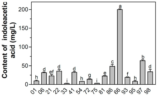
Of the 252 colonies screened for IAA production with plate assay, 16 showed distinctive color changes. IAA production of those isolates ranged from 3 to 200 mg/L (Figure 1). LX3, which showed the highest IAA production, was selected for further study.

Figure 1.
Screening of indoleacetic acid (IAA)-producing strains isolated from the maize rhizosphere soil collected from Shenyang Agricultural University, China, in which colony 66 is strain LX3. Error bars were calculated by standard deviations from triplicate runs for IAA production assay. Different letters on top of bars indicate significant differences (p < 0.05).
3.2. Phenotypic Properties
Isolate LX3 showed the maximum capability of producing IAA among the isolated strains from maize rhizosphere soil in the biochar-amended field. Isolate LX3 was a Gram-negative bacterium (Table 1). It was motile with peritrichous flagella and facultative anaerobic. Microscopic examination showed that bacterial cells were non-spore-forming rods with diameter of 0.6–1.0 μm and length of 1.2–3.0 μm. After strain LX3 was incubated on LB agar plates at 30 °C for 36 h, colonial morphology was white, circular, elevated, and smooth across a diameter of 1–3 mm.

Table 1.
Phenotypic characteristics of the newly isolated Enterobacter sp. LX3. Symbols: +, 90% or more strains were positive; −, 10% or less strains were positive.
3.3. Phenotypic Characteristics
Isolate LX3 grew well at 37 °C but could not hydrolyze corn oil and tributyrin (Table 1). A positive catalase test was achieved but a negative oxidase test was observed. Isolate LX3 gave positive results on enzyme activities including arginine dihydrolase, lysine decarboxylase and ornithine decarboxylase, but negative results on enzyme activities of β-galactosidase and phenylalanine deaminase. Glucose was fermented to produce both acid and gas at 30 °C but gas was not produced at 44.5 °C. Isolate LX3 was negative in testing hydrogen sulfide production but positive in testing indole production. Isolate LX3 also gave a positive result on Voges-Proskauer reaction but negative test in methyl red reaction. Gelatin was liquefied and esculin was hydrolyzed. Acid was produced from compounds: Amygdalin, arabinose, d-glucose, d-sorbitol, l-rhamnose and melibiose. No acid production was detected from compounds including d-mannitol, inositol and sucrose. Nitrate could be reduced into nitrite and tetrathionate could not be reduced. Positive alkaline reaction was detected in Simmons citrate broth.
Strain LX3 could utilize many carbohydrates including cellobiose, d-glucose, d-fructose, d-galactose, d-mannose, lactose, l-arabinose, l-rhamnose, maltose, melibiose, raffinose, sucrose, trehalose, d-mannitol, d-sorbitol and inositol as the sole carbon and energy source. A good growth was observed in the medium when yeast extract, peptone, tryptone, diammonium phosphate or ammonium sulfate were utilized as nitrogen sources. Strain LX3 could grow with citrate as sole carbon and energy source. It was not halophilic but able to grow in the medium containing 4% NaCl.
Isolate LX3 was capable of growing at pH range from 5.0 to 9.0, and optimum pH for cell growth was at pH 7.0. Cell growing temperature for strain LX3 was between 20 °C and 45 °C, and the maximal cell growth was observed at temperature of 30 °C.
3.4. Phylogenetic Analysis
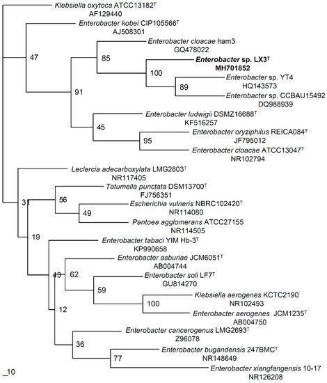
Sequencing of 16S rRNA gene gave a 1409-base sequence (accession MH701852, GenBank) for establishing phylogenetic position of strain LX3. Comparisons with all available sequences in GenBank database showed that strain LX3 had the most similarity in 16S rRNA gene sequence with genus Enterobacter species [23]. Calculation using MegAlign program in DNAStar program package indicated that sequence similarity was from 96.8% to 99.8% between strain LX3 and its closer relatives. The closest relatives of strain LX3 were E. cloacae ham3, Enterobacter sp. CCBAU15492 and Enterobacter sp. YT4 having 99.8%, 99.6% and 99.7% similarity in 16S rRNA gene sequence respectively. Strain LX3 also was close to other phylogenetic neighbors including E. asburiae, E. cancerogenus, E. ludwigii and Leclercia adecarboxylata, which has more than 98.7% similarity in the 16S rRNA gene sequence. A phylogenetic tree was constructed with neighbor-joining method and shown in Figure 2. It was clear to cluster strain LX3 into the clade of E. cloacae.

Figure 2.
Phylogenetic tree constructed based on 16S rRNA gene sequence comparisons between isolate LX3 and some close relatives. A percentage of 100 replications showed on branching points represented bootstrap value. Bar length indicates 1% sequence divergence.
3.5. Plant Growth Promotion
To examine potential role of strain LX3 in plant growth, isolate LX3 was evaluated with Chinese barley (Hordeum vulgare L.) for its plant growth promoting capability. Inoculation of barley plants with strain LX3 increased barley seedling growth when compared to control (Table 2). After inoculation for 10 days, an increase in dry weight of whole-plant relative to uninoculated control was 20.8%. Similarly, an increase in root length by 18.2% and in plant height of 21.6% was recorded. Chlorophyll production was increased by 8.1% when compared with the control.

Table 2.
Barley growth in the water with and without addition of culture fluid after 10 days.
4. Discussion
According to the description for the genus Enterobacter [24], these determinative phenotypic criteria were included: Gram-negative, motile by peritrichous flagella, facultative anaerobic rod, positive Voges-Proskauer reaction, negative test in methyl red reaction, no hydrolyzation of corn oil and tributyrin. Strain LX3 shared coincident key characteristics with genus Enterobacter (Table 1) [24]. Thus, strain LX3 was phenotypically classified in the genus Enterobacter.
Comparisons in 16S rRNA gene sequences between strain LX3 and its related genera also suggested that strain LX3 should be a member of the genus Enterobacter. Isolate LX3 showed more than 97.8% similarity to all species of genus Enterobacter in 16S rRNA gene sequences. These data coincided with a proposal that 93% or 95% similarity was used as a cut-off value to delineate genera [25,26]. As shown in the phylogenetic tree (Figure 2), strain LX3 has a close phylogenetic relationship with genus Enterobacter species from tree topology. It was clear to cluster strain LX3 into clade of E. cloacae that had a 99.8% similarity, in which Enterobacter sp. CCBAU15492 appears related to E. cloacae [27]. Those phylogenetic results proved that strain LX3 must be part of genus Enterobacter.
Strain LX3 coincided with the genus Enterobacter in phenotypic characteristics, whereas there were some notable differences in biochemical traits at species level (Table 3). Such discrepancies should be significant to distinguish strain LX3 from other members of genus Enterobacter. The data in Table 3 showed a distinction between strain LX3 and its nearest apparent phylogenetic neighbors of genus Enterobacter in a number of key characteristics, such as H2S production, indole production, urease, Voges-Proskauer reaction and enzyme activities including β-galactosidase, lysine decarboxylase and arginine dihydrolase. To distinguish strain LX3 from the most closely related E. cloacae, decisive characteristics were lysine decarboxylase, β-galactosidase, urease, indole production and acid production from both d-mannitol and sucrose. E. ludwigii can be distinguished from strain LX3 by urease test, indole production, lysine decarboxylase and β-galactosidase. Isolate LX3 also was differentiated from its closer neighbor Leclercia adecarboxylata in phenotypic properties, such as β-galactosidase, lysine decarboxylase, arginine dihydrolase and urease tests. Isolate LX3 showed positive lysine decarboxylase, but most of species in the genus Enterobacter were deficient in lysine decarboxylase production except for E. aerogenes. Whereas E. aerogenes showed difference from isolate LX3 in arginine dihydrolase activity, indole production and urease. Moreover, β-galactosidase could be detected in most of genus Enterobacter species, but not in strain LX3 and E. aerogenes. Both strain LX3 and E. tabaci were positive for indole production and Voges-Proskauer reaction, whereas E. tabaci was positive in H2S production and strain LX3 was not. Contrary characteristics occurred between strain LX3 and E. cancerogenus, such as β-Galactosidase, lysine decarboxylase, urease and indole production. Being different from most species in the genus Enterobacter, both strain LX3 and E. asburiae gave a positive result in urease test, in which discrepancy between strain LX3 and E. asburiae occurred in indole production and Voges-Proskauer reaction. As shown in Table 3, some differences between isolate LX3 and genus Enterobacter species also occurred in carbohydrate fermentation tests. Based on both phenotypic characteristics and phylogenetic analysis, isolate LX3 could be considered as a new species of the genus Enterobacter that is closely related with E. cloacae. A creation of new species was proposed in the genus Enterobacter despite needing more detailed taxonomic works to finish classification.

Table 3.
Key characteristics distinguishing strain LX3 from other Enterobacter species. Species: 1, Enterobacter sp. LX3; 2, Enterobacter bugandensis; 3, Enterobacter asburiae; 4, Enterobacter cancerogenus; 5, Enterobacter cloacae; 6, Enterobacter ludwigii; 7, Enterobacter oryziphilus; 8, Enterobacter tabaci; 9, Enterobacter kobei; 10, Enterobacter soli; 11, Enterobacter aerogenes; 12, Enterobacter xiangfangensis; 13, Leclercia adecarboxylata. Symbols: +, 90% or more strains were positive; −, 10% or less strains were positive; N, not be detected.
Some rhizobacteria can stimulate plant growth by phytohormone IAA synthesis, which showed differential roles in plant growth and development [3]. The maximal IAA production of 200 mg/L was detected in NB medium with l-tryptophan, which indicated that Enterobacter sp. LX3 was an active IAA-producer. Capability of strain LX3 promoting barley growth was determined by soil-free plant growth assays, although a number of plants are necessary for experimental replication. As previously confirmed that exogenous IAA could promote plant growth by enhancing formation of root hair and lateral roots [28], inoculation of barley plants with culture fluid of strain LX3 can stimulate barley growth (Table 2). Development in root system was promoted in root weight by 10.4% increase and in root length by 18.2% elongation. Enhancement in root development was one of the important indicators denoting beneficial effect of bacteria promoting plant growth, because rapid development in roots can help young seedlings obtain both water and nutrient from surrounding and increase their ability to anchor in soil [29]. The data suggested that plant height was increased by 21.6% and chlorophyll content in barley cells was increased by 8.1% when barley plants were inoculated with strain LX3. Results indicate that Enterobacter sp. LX3 is a potential biofertilizer for promoting barley growth.
5. Conclusions
One strain was obtained from maize rhizosphere in a biochar-amended field. It was a Gram-negative rod with peritrichous flagella and facultative anaerobic growth, which shared key characteristics with the genus Enterobacter. This isolate was identified as a new member of genus Enterobacter according to differential phenotypic characteristics combined with phylogenetic analysis, and designed as Enterobacter sp. LX3. Strain LX3 is an IAA-producer producing maximally 200 mg/L of IAA in NB medium. The root system and chlorophyll content were enhanced when isolate LX3 was used to inoculate barley plants.
Author Contributions
Conceptualization, X.L. and W.C.; methodology, Q.C., L.L. and W.T.; formal analysis, J.M. and X.D.; investigation, Q.C., L.L., W.T. and X.D.; resources, J.M.; data curation, L.L.; writing—original draft preparation, W.T.; writing—review and editing, X.L.; visualization, X.L.; supervision, W.C.
Funding
This research was funded by the National Key R&D Program of China (2017YFD0200800) and the National Natural Science Foundation of China (31771907).
Acknowledgments
This works was supported by Dalian Polytechnic University.
Conflicts of Interest
The authors declare no conflict of interest.
References
- Duca, D.; Lorv, J.; Patten, C.L.; Rose, D.; Glick, B.R. Indole-3-acetic acid in plant-microbe interactions. Antonie Van Leeuwenhoek 2014, 106, 85–125. [Google Scholar] [CrossRef] [PubMed]
- Tian, H.; De Smet, I.; Ding, Z. Shaping a root system: Regulating lateral versus primary root growth. Trends Plant Sci. 2014, 19, 426–431. [Google Scholar] [CrossRef] [PubMed]
- Egamberdieva, D.; Wirth, S.J.; Alqarawi, A.A.; Abd_Allah, E.F.; Hashem, A. Phytohormones and beneficial microbes: Essential components for plants to balance stress and fitness. Front. Microbiol. 2017, 8, 2104. [Google Scholar] [CrossRef] [PubMed]
- Ozdal, M.; Ozdal, O.G.; Sezen, A.; Algur, O.F.; Kurbanoglu, E.B. Continuous production of indole-3-acetic acid by immobilized cells of Arthrobacter agilis. 3 Biotech 2017, 7, 23. [Google Scholar] [CrossRef] [PubMed]
- Mukherjee, S.; Sen, S.K. Exploration of novel rhizospheric yeast isolate as fertilizing soil inoculant for improvement of maize cultivation. J. Sci. Food Agric. 2015, 95, 1491–1499. [Google Scholar] [CrossRef] [PubMed]
- Weselowski, B.; Nathoo, N.; Eastman, A.W.; MacDonald, J.; Yuan, Z.C. Isolation, identification and characterization of Paenibacillus polymyxa CR1 with potentials for biopesticide, biofertilization, biomass degradation and biofuel production. BMC Microbiol. 2016, 16, 244. [Google Scholar] [CrossRef] [PubMed]
- Liu, C.Y.; Zhang, F.; Zhang, D.J.; Srivastava, A.K.; Wu, Q.S.; Zou, Y.N. Mycorrhiza stimulates root-hair growth and IAA synthesis and transport in trifoliate orange under drought stress. Sci. Rep. 2018, 8, 1978. [Google Scholar] [CrossRef] [PubMed]
- Defez, R.; Andreozzi, A.; Bianco, C. The Overproduction of indole-3-acetic acid (IAA) in endophytes upregulates nitrogen fixation in both bacterial cultures and inoculated rice plants. Microb. Ecol. 2017, 74, 441–452. [Google Scholar] [CrossRef] [PubMed]
- Egamberdieva, D.; Wirth, S.; Behrendt, U.; Abd_Allah, E.F.; Berg, G. Biochar treatment resulted in a combined effect on soybean growth promotion and a shift in plant growth promoting rhizobacteria. Front. Microbiol. 2016, 7, 209. [Google Scholar] [CrossRef] [PubMed]
- King, E.O.; Ward, M.K.; Raney, D.E. Two simple media for the demonstration of phycocyanin and fluorescin. J. Lab. Clin. Med. 1954, 44, 301–307. [Google Scholar] [PubMed]
- Bric, J.M.; Bostock, R.M.; Silverstone, S.E. Rapid in situ assay for indoleacetic acid production by bacteria immobilized on a nitrocellulose membrane. Appl. Environ. Microbiol. 1991, 57, 535–538. [Google Scholar] [PubMed]
- Murray, R.G.E.; Doetsch, R.N.; Robinow, C.F. Determinative and cytological light microscopy. In Methods for General and Molecular Bacteriology; Gerhardt, P., Murray, R.G.E., Wood, W.A., Krieg, N.R., Eds.; American Society for Microbiology: Washington, DC, USA, 1994; pp. 21–41. [Google Scholar]
- Leifson, E. Staining, shape, and arrangement of bacterial flagella. J. Bcateriol. 1951, 62, 377–389. [Google Scholar]
- Smibert, R.M.; Krieg, N.R. Phenotypic characterization. In Methods for General and Molecular Bacteriology; Gerhardt, P., Murray, R.G.E., Wood, W.A., Krieg, N.R., Eds.; American Society for Microbiology: Washington, DC, USA, 1994; pp. 607–654. ISBN 1-55581-048-9. [Google Scholar]
- Moore, D.D.; Dowhan, D. Preparation and analysis of DNA. In Current Protocols in Molecular Biology; Ausubel, F.M., Brent, R., Kingston, R.E., Moore, D.D., Seidman, J.G., Smith, J.A., Struhl, R., Eds.; John Wiley & Sons: New York, NY, USA, 1997; Volume 1, pp. 241–245. ISBN 0-471-50338-X. [Google Scholar]
- Jin, Z.; Wang, C.; Li, X. Isolation and some properties of newly isolated oxalate-degrading Pandoraea sp. OXJ-11 from soil. J. App. Microbiol. 2007, 103, 1066–1073. [Google Scholar] [CrossRef] [PubMed]
- Johnson, M.; Zaretskaya, I.; Raytselis, Y.; Merezhuk, Y.; McGinnis, S.; Madden, T.L. NCBI BLAST: A better web interface. Nucl. Acids Res. 2008, 36, W5–W9. [Google Scholar] [CrossRef] [PubMed]
- Thompson, J.D.; Gibson, T.L.; Plewniak, F.; Jeanmougin, F.; Higgins, D.G. The CLUSTAL-X windows interface: Flexible strategies for multiple sequence alignment aided by quality analysis tools. Nucleic Acids Res. 1997, 25, 4876–4882. [Google Scholar] [CrossRef] [PubMed]
- Felsenstein, J. PHYLIP (Phylogeny Inference Package) Version 3.6; Department of Genome Sciences, University of Washington: Seattle, WA, USA, 2005. [Google Scholar]
- Gunning, T.; Cahill, D.M. A soil-free plant growth system to facilitate analysis of plant pathogen interactions in roots. J. Phytopathol. 2009, 157, 497–501. [Google Scholar] [CrossRef]
- Patten, C.L.; Glick, B.R. Role of Pseudomonas putida indoleacetic acid in development of the host plant root system. Appl. Environ. Microbiol. 2002, 68, 3795–3801. [Google Scholar] [CrossRef] [PubMed]
- Arnon, D.I. Copper enzymes in isolated chloroplasts. Polyphenoloxidases in Beta vulgaris. Plant Physiol. 1949, 24, 1–15. [Google Scholar] [CrossRef] [PubMed]
- Altschul, S.F.; Madden, T.L.; Schaffer, A.A.; Zhang, J.; Zhang, Z.; Miller, W.; Lipman, D.J. Gapped BLAST and PSI-BLAST: A new generation of protein database search programs. Nucleic Acids Res. 1997, 25, 3389–3402. [Google Scholar] [CrossRef] [PubMed]
- Grimont, P.A.D.; Grimont, F. Genus XII. Enterobacter. In Bergey’s Manual of Systematic Bacteriology; Brenner, D.J., Krieg, N.R., Staley, J.T., Eds.; Springer: Monroe, MI, USA, 2005; Volume 2, Part B; pp. 661–670. ISBN 978-0-387-24144-9. [Google Scholar]
- Fox, G.E.; Wisotzkey, J.D.; Jurtshuk, P. How close is close: 16S rRNA sequence identity may not be sufficient to guarantee species identity. Int. J. Syst. Bacteriol. 1992, 42, 166–170. [Google Scholar] [CrossRef] [PubMed]
- Wagner-Döbler, I.; Rheims, H.; Felske, A.; El-Ghezal, A.; Flade-Schröder, D.; Laatsch, H.; Lang, S.; Pukall, R.; Tindall, B.J. Oceanibulbus indolifex, gen. nov., sp. nov., a North Sea Alphaproteo-bacterium producing bioactive metabolites. Int. J. Syst. Evol. Microbiol. 2004, 54, 1177–1184. [Google Scholar] [CrossRef] [PubMed]
- Zhao, L.; Xu, Y.; Lai, X. Antagonistic endophytic bacteria associated with nodules of soybean (Glycine max L.) and plant growth-promoting properties. Braz. J. Microbiol. 2018, 49, 269–278. [Google Scholar] [CrossRef] [PubMed]
- Sun, P.F.; Fang, W.T.; Shin, L.Y.; Wei, J.Y.; Fu, S.F.; Chou, J.Y. Indole-3-acetic acid-producing yeasts in the phyllosphere of the carnivorous plant Drosera indica L. PLoS ONE 2014, 9, e114196. [Google Scholar] [CrossRef] [PubMed]
- Zhang, Y.; Liu, M.; Saiz, G.; Dannenmann, M.; Guo, L.; Tao, Y.; Shi, J.; Zuo, Q.; Butterbach-Bahl, K.; Li, G.; et al. Enhancement of root systems improves productivity and sustainability in water saving ground cover rice production system. Field Crops Res. 2017, 213, 186–193. [Google Scholar] [CrossRef]
© 2018 by the authors. Licensee MDPI, Basel, Switzerland. This article is an open access article distributed under the terms and conditions of the Creative Commons Attribution (CC BY) license (http://creativecommons.org/licenses/by/4.0/).