Abstract
Achieving room-temperature exciton–polariton condensation represents a frontier challenge in condensed matter physics and optoelectronics. However, its mainstream approach—distributed Bragg reflector (DBR) microcavities—faces widespread application challenges due to complex fabrication and high costs. Here, we report direct observation of the interaction between exciton and microcavity photons in Sn-doped CdS microsheet without extreme fabrication conditions. In situ PL and Raman mapping demonstrate the formation of superlattice structure. Using angle-resolved photoluminescence (ARPL) spectroscopy, we obtain Rabi splitting of polaritons up to 140 meV, and the exciton-like and photon-like components in low-polariton states at different cavity–exciton detuning were revealed at room temperature. Our work demonstrates the origin of optical microcavities and the light–matter coupling in CdS/CdS:SnS2 superlattice microwires.
1. Introduction
Exciton–polaritons, quasiparticles formed by the strong coupling of photons and excitons, have attracted significant attention due to their remarkable properties such as extremely light effective mass, long coherence length, and strong nonlinearity. [,,] Exciton–polaritons provide a promising platform for studying unique quantum phenomena, such as Bose–Einstein condensation (BEC), Fano resonance, superfluid, ultra-low threshold lasers, etc. [,,,,]. Experimental observations of exciton–polaritons have been achieved in various material systems, such as perovskites, III–V and II–VI semiconductor microlayers, or quantum well structures [,,,]. However, each of these systems faces distinct challenges: in perovskite materials, despite their high exciton binding energy, the nonlinear interactions of Frenkel excitons are typically weak, and most studies still rely on complex microcavity structures to achieve strong coupling [,]. In conventional III–V and II–VI semiconductors, the smaller exciton binding energy renders their polariton effects highly susceptible to thermal fluctuation annihilation at room temperature, limiting observations to cryogenic conditions. These limitations have driven researchers to actively explore novel micro/nanostructures combined with high-binding-energy exciton materials, aiming to realize strongly interacting exciton plasmons at room temperature without complex microcavities [,,].
CdS, a prototypical wurtzite-structured semiconductor, has garnered considerable attention in the study of exciton–photon interactions and lasing behavior due to its visible-range emission spectrum and direct bandgap of 2.42 eV [,,]. Wurtzite-type CdS belongs to the C6v point group, featuring a relatively stable hexagonal crystal lattice with tightly bonded, well-ordered atoms. However, due to the intrinsic inability of CdS to respond to photons below its excitation threshold energy, it cannot achieve light emission beyond the green spectrum [,,]. Indeed, bulk CdS exhibits a Rabi splitting value of 82 meV. However, Lambert K. van Vugt and colleagues achieved a substantially enhanced Rabi splitting of 200 meV in self-assembled CdS nanowire cavities at 77 K []. This work demonstrates that CdS provides a viable platform to investigate strong exciton–photon interactions. Nevertheless, the relatively weak exciton binding energy in CdS semiconductor materials confines observations of exciton–polaritons to cryogenic temperatures, rendering their detection at room temperature a formidable scientific challenge [,,,]. Rashaba et al. discovered that introducing appropriate defects can form a new quasiparticle—the defect-bound exciton. When this binding is weak, the oscillator strength of this bound state becomes significantly greater than that of the free exciton. This oscillator strength enhancement effect in localized states can effectively boost the efficiency of light–matter interactions []. When metal ions such as Sn are introduced into CdS nanowires as dopants, their amphiphilic nature simultaneously introduces donor and acceptor levels within the bandgap. This self-compensating effect, while limiting electrical mobility, enhances electron–phonon coupling. This enables the observation of strong oscillator strength and strong photon–exciton coupling []. Jacob B. Khurgin demonstrated that “superstrong coupling” states exceeding the CdS exciton binding energy (30 meV) can be achieved in Ag/CdS core–shell structures. When the silver nanosphere radius R0 = 10 nm, the maximum tensile splitting value reaches approximately 50 meV, and this splitting value increases with increasing radius R0 []. Li et al. successfully designed and fabricated a heterostructure composed of WS2–Si3N4 nanopore arrays and silver films. By ingeniously introducing defects through the heterostructure, three distinct resonance modes were formed, achieving a record-breaking large tensile splitting (318 meV) []. Sn(IV) doping in CdS nanowires disrupts the original lattice symmetry and induces lattice distortion, thereby creating favorable conditions for microcavity formation []. Furthermore, during Sn-doped CdS synthesis, multiple nanostructures emerge. This phenomenon primarily stems from tin acting as a catalyst during synthesis, guiding the oriented growth of nanowires through a vapor–liquid–solid mechanism. Specifically, liquid Sn captures vapor-phase precursors, forming a eutectic melt with CdS. Upon reaching supersaturation, the target material precipitates from droplets, yielding one-dimensional nanostructures. By controlling growth conditions such as heating rate, carrier gas flow rate, and weight ratio of the two precursors, different one-dimensional nanostructures can be produced, including CdS/CdS:SnS2 superlattice microwires [], comb-like CdS nanostructures [], core–shell CdS/CdO microwires [], and hollow CdS nanotubes []. These studies demonstrate that CdS nanostructures engineered through elemental doping strategies exhibit profound potential for exploring exciton–photon interactions [].
In this work, we report the direct observation of the interaction between excitons and microcavity photons in CdS/CdS:SnS2 superlattice microwires, achieved without relying on complex distributed Bragg reflector (DBR) technology. We demonstrate the strong exciton–photon coupling at room temperature in CdS/CdS:SnS2 superlattice microwires through angle-resolved spectroscopy. This robust coupling originates from interactions between excitons and photons confined within self-formed microcavity modes inherent to the microwire structure. Through analysis of experimental data via the coupled harmonic oscillator model, a Rabi splitting of 143 meV was obtained in CdS/CdS:SnS2 superlattice microwires, yielding Hopfield coefficients under different microcavity resonance conditions. As the detuning energy increases, the contribution of excitons (EX) to lower polaritons (LPs) gradually increases at small angles, while the contribution of photons (CM) to lower polaritons (LPs) gradually diminishes. Furthermore, as the mismatch grows, the polariton effect vanishes. These results demonstrate the origin of light–matter interactions in Fabry–Pérot microcavities within CdS/CdS:SnS2 superlattice microwires. They demonstrate that introducing Sn ions enhances the oscillator strength of excitons, enabling the observation of room-temperature exciton–polaritons and establishing a new architecture for studying exciton–polariton devices under ambient conditions.
2. Materials and Methods
2.1. Fabrication of the CdS/CdS:SnS2 Superlattice Microwires
Using chemical vapor deposition (CVD), CdS/CdS:SnS2 superlattice microwires were fabricated on 3.5 × 3.5 cm mica substrates in a tube furnace model SK2-2.5-13A. The precursor powders SnO2 (99.5%, Alfa Aesar, Heysham, UK) and CdS (99.999%, Alfa Aesar) were mixed and ground in a mortar at a mass ratio of 1:15. The porcelain boat containing the mixed powder was placed at the center of the single-zone tube furnace, while the mica substrate was positioned 15 cm downstream from the center. Prior to growth, the quartz tube was purged with high-purity Ar/H2 mixed gas (manufacturer: Praxair, Danbury, Connecticut) at a flow rate of 60 sccm for 1 h to purify the growth environment.
The growth process is summarized as follows: The temperature was first raised to 670 °C at a rate of 100 °C/min. At approximately 200 °C, SnO2 is reduced by hydrogen to form Sn vapor (SnO2 + 2H2 ↔ Sn + 2H2O). This vapor is transported by the carrier gas to the mica substrate surface, where it condenses to form Sn microwires. As the temperature continues to rise, CdS powder sublimates to form gaseous CdS, which gradually dissolves into the Sn droplets. Upon reaching supersaturation within the Sn droplets, CdS begins to epitaxially precipitate as micron-scale wires while encapsulating the Sn droplets. During this phase, the following reaction may occur within the tube: CdS + H2 ↔ Cd + H2S. The encapsulated Sn droplets gradually liquefy, refine, and ultimately melt away during the ongoing growth process. Subsequently, the system is heated at a rate of 20 °C/min from 670 °C to 870 °C and held at this temperature for 40 min. During this stage, the molten Sn liquid is segmented into multiple structures due to the increased size of the microwires and tensile effects, with continuous precipitation occurring at the growth front. At elevated temperatures, Sn reacts with H2S to form SnS (Sn + 2H2S ↔ SnS2 + H2). Finally, during the cooling process to room temperature, the molten CdS solidifies and crystallizes. This solidification process interacts with the periodic structure of SnS2, ultimately self-assembling into CdS/CdS:SnS2 superlattice microwires. Finally, the superlattice microwires are transferred onto a silicon wafer for characterization [].
2.2. Characterization
Self-assembled CdS/CdS:SnS2 superlattice microwires were characterized optically: X-ray diffraction (XRD, SMARTLAB, Hong Kong, China) investigated material elemental composition and lattice structure; Confocal fluorescence/Raman microscopy (WITEC alpha 300R, Ulm, Germany) characterized the optical properties of various Sn-doped CdS nanostructures. A 405 nm laser was used for micro-PL spectroscopy and PL imaging, while a 532 nm laser was employed for micro-PL spectroscopy, light-emitting imaging, Raman spectroscopy, and Raman imaging tests. An Angle-Resolved Spectrometer (ARMS, Shanghai Aidiop Technology Co., Ltd., Shanghai, China) was employed to investigate light–matter interactions within CdS/CdS:SnS2 superlattice microwires at the micro–nano scale. An excitation source of 405 nm continuous-wave (CW) GaN laser was used, with an angle range of 0° to 45°.
3. Results and Discussion
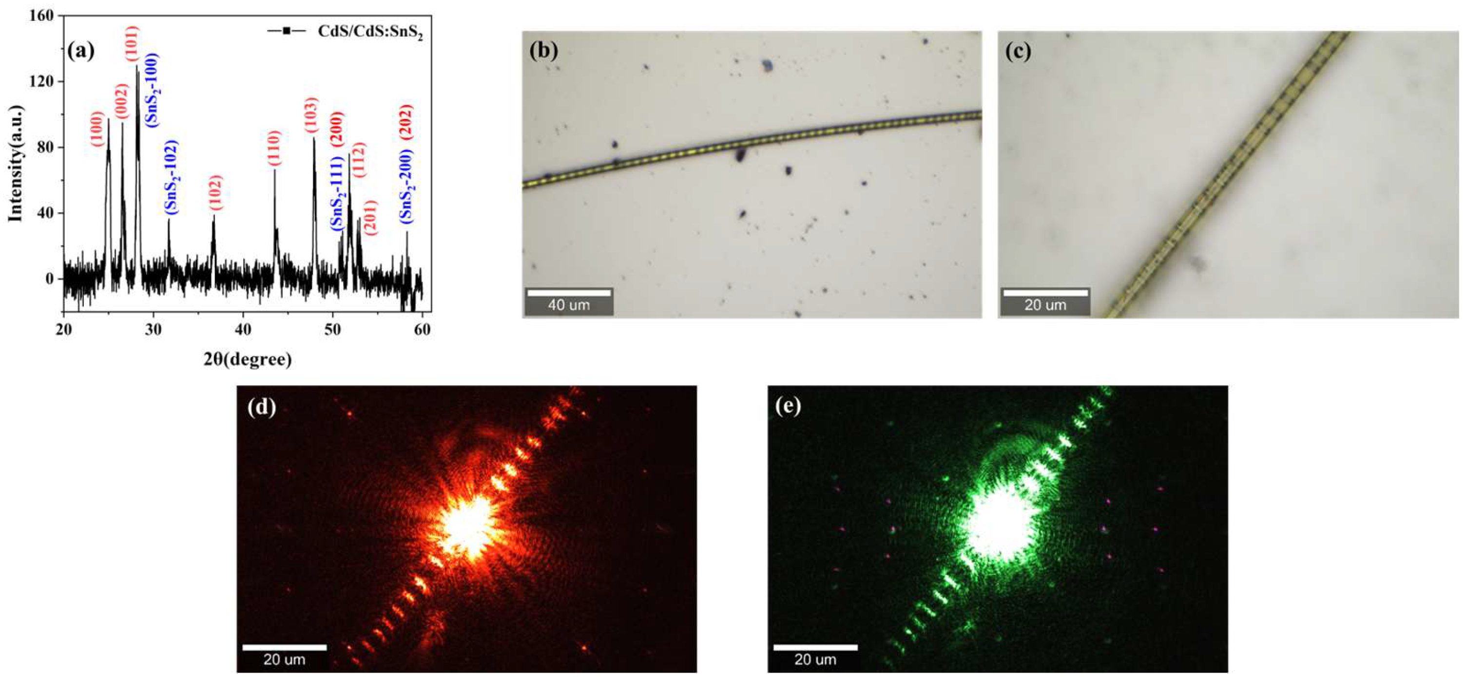
CdS/CdS: SnS2 superlattice microwires were fabricated using chemical vapor deposition (CVD). Figure 1a shows the XRD pattern of the CdS/CdS:SnS2 superlattice microwires. The pattern confirms the presence of two crystalline phases in the synthesized microwires: CdS (JCPDS No. 41-1049) and SnS2 (JCPDS No. 40-1467). No characteristic peaks from other solid impurities were detected. Figure 1b is a CCD image of a single nanowire, which shows that the lengths of the nanowire are greater by up to several hundreds of micrometers and the nanowire presents many periodic nodes. In addition, Figure 1d,e exhibit the typical micro-PL image of the single nanowire excited by the 633 nm laser and 532nm laser, respectively. The bright spot marks the laser excitation position and many periodic red or green dots are distributed on both sides of the excitation center, indicating a specific periodic emitting property. Among the nanowire, the emission dots are the CdS:SnS2 section and the segments of two dots are CdS. It can clearly be seen that the products are long microwires with regions of periodic and alternating indices of refraction, that is, microwire superlattices.
Figure 1.
XRD, CCD images, and dark-field luminescence images of the CdS/CdS:SnS2 superlattice microwires under laser excitation at different wavelengths. (a) XRD pattern of CdS/CdS:SnS2 superlattice microwires; (b) 10× and (c) 50× microscope CCD images of CdS/CdS:SnS2 superlattice microwire. Luminescence image of the CdS/CdS:SnS2 superlattice microwires under dark-field microscopy with continuous-wave laser excitation at (d) 633 nm and (e) 532 nm.
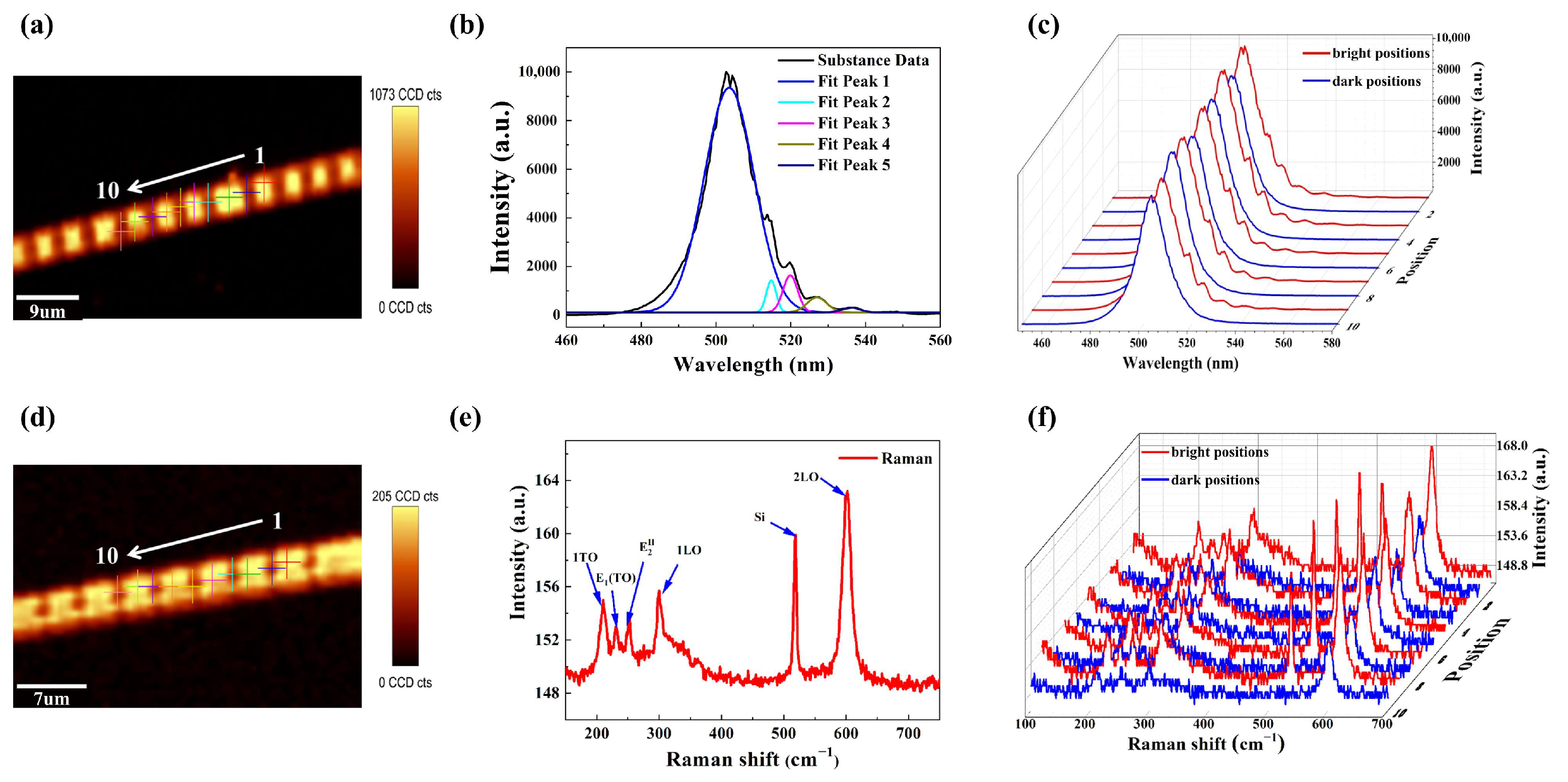
To further investigate the optical properties within the periodic structures, PL and Raman measurements were performed at different locations. Figure 2a presents the PL mapping image of CdS/CdS:SnS2 superlattice microwires, with ten distinct points selected across the structure for comparative spectral analysis at different locations. The dark-colored portion represents the CdS/SnS2 section, while the light-colored portion represents the CdS section. Figure 2b presents the PL spectrum from bright regions within the superlattice microwire, revealing several oscillatory peaks near the band edge. Figure 2c displays the PL spectrum extracted from positions 1 to 10 in Figure 2a, with red curves representing the spectrum selected from bright positions in the imaging and blue curves denoting those from dark positions. Notably, oscillatory peaks are observed exclusively in bright positions, while spectrum from dark positions lacks band-edge oscillations, demonstrating periodic luminescence distribution. In the photoluminescence spectra at different positions, periodic bright–dark variations occur along the microwire axis, with dark regions corresponding to CdS/SnS2 sections and bright regions to CdS sections. This indicates that photons undergo periodic propagation along the microwire axis due to the differing refractive indices of SnS2 and CdS, leading to the formation of a microwire superlattice structure. When laser light illuminates the sample, it excites numerous photons that propagate longitudinally along the microwire toward the CdS/SnS2 interface. Some photons are reflected, while others transmit and continue propagating. The reflected photons bounce back toward the opposite interface, where some are reflected again and others transmit through the interface. This repeated reflection and transmission creates oscillations within this small cavity. Therefore, in the superlattice microwire, the oscillation peak primarily originates from the reflection of photons at the SnS2 interface. Assuming the microwire contains ideal mirror facets forming a Fabry–Perot (F–P) cavity, the cavity length is given as follows:
where and represent the wavelengths of adjacent resonant peaks and denotes the refractive index. The calculated cavity length ranges from 3.36 μm to 5.12 μm, significantly shorter than the entire CdS/CdS:SnS2 superlattice microwires length yet comparable to the dimensions of bright and dark regions observed in PL spectrum. This suggests that such resonances likely originate from reflections at adjacent CdS/SnS2 interfaces within the microstructure.
Figure 2.
(a) PL mapping image with 10 positions selected for spectral analysis; (b) PL spectrum fitted by Gaussian curves; (c) PL spectrum corresponding to positions 1–10; (d) Raman mapping image with 10 positions selected for analysis; (e) Raman spectrum; (f) Raman spectrum from positions 1–10.
Figure 2d displays the Raman mapping image of CdS/CdS:SnS2 superlattice microwires, with ten distinct measurement points selected along the architecture for comparative spectral mapping at different locations. To further characterize the CdS/CdS:SnS2 superlattice microwires, we analyzed the Raman spectrum in the 200–800 cm−1 range, with the results shown in Figure 2e. Six distinct vibrational mode peaks of the CdS/CdS:SnS2 superlattice microwires are clearly observable in the spectrum: 209 cm−1, 228 cm−1, 250 cm−1, 302 cm−1, 520 cm−1, and 603 cm−1. These correspond to 209 cm−1, 228 cm−1, 250 cm−1, 302 cm−1, 520 cm−1, and 603 cm−1. Specifically, these include 209 cm−1 (CdS 1TO phonon), 228 cm−1 (CdS E1TO vibrational mode), 250 cm−1 (CdS higher-order mode), 302 cm−1 (CdS 1LO mode), and 603 cm−1 (CdS 2LO mode) [,]. Notably, the peak at 520 cm−1 does not belong to the CdS/CdS:SnS2 superlattice nanowire. Its appearance stems from the fact that the measured nanostructure was directly fabricated on a silicon substrate []. Figure 2f displays Raman spectrum corresponding to the ten positions marked in Figure 2d, with red curves designating the spectra from bright regions in the imaging and blue curves representing those from dark regions. The Raman spectra from bright regions exhibit greater intensity than those from dark regions. Therefore, the position-dependent PL spectrum indicates that the oscillatory peaks near the band edge in the superlattice originate from photon reflection at the CdS/SnS2 interface. Both the position-dependent PL spectrum and Raman spectrum further support the formation of the superlattice structure. Accordingly, the periodic distributions observed in PL mapping, Raman mapping, the PL spectrum, and the Raman spectrum provide indirect evidence for the formation of the microwire superlattices.
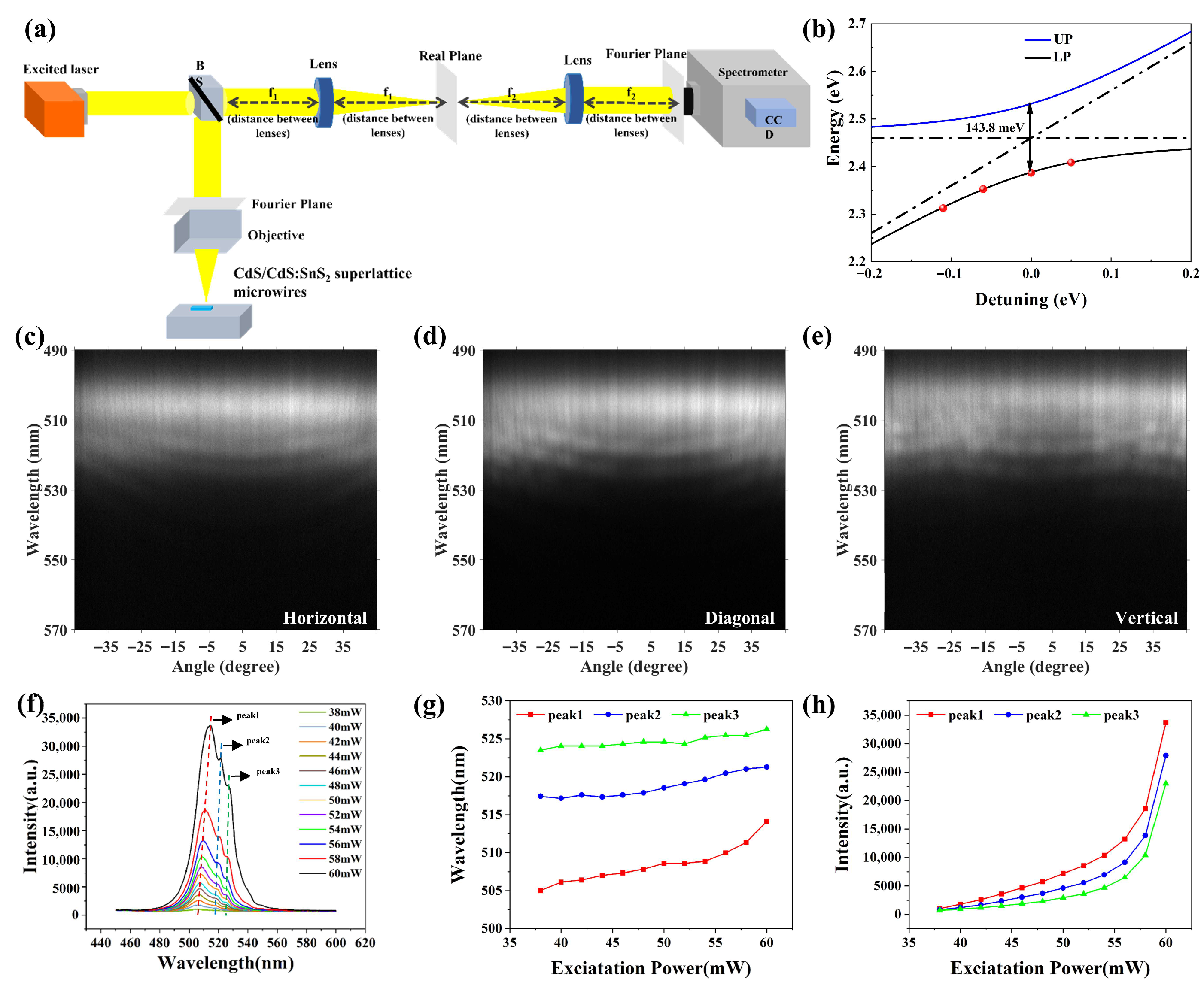
To identify the strong coupling regime of exciton–photon interaction, we comparatively study the ARPL spectra by ideal optical ARPL system with a 4-focal distance Fourier imaging configuration, as shown in Figure 3a. The experimental setup employs a 405 nm continuous-wave (CW) laser as the excitation source. The energy dispersion relations for the upper and lower polariton branches are derived from the angle-resolved photoluminescence (ARPL) spectra of CdS/CdS:SnS2 superlattice microwires using the coupled oscillator model, where the coupling strength is denoted as V and expressed as [,,,].
where gives the cavity photon dispersion, in which , is the initial cavity-exciton detuning.
Figure 3.
Exciton–polaritons in CdS/CdS:SnS2 superlattice microwires. (a) Schematic diagram of the optical path in an angular-resolved spectrometer. (b) Dispersion relation of exciton–polaritons in the CdS/CdS:SnS2 superlattice microwires. (c) ARPL spectrum of the CdS/CdS:SnS2 superlattice microwires parallel to the monochromator entrance slit. (d) ARPL spectrum of the CdS/CdS:SnS2 superlattice microwires at 45° to the monochromator entrance slit. (e) ARPL spectrum of the CdS/CdS:SnS2 superlattice microwires perpendicular to the monochromator entrance slit. (f) PL spectrum of CdS/CdS:SnS2 superlattice microwires under different excitation powers. (g) Dependence of PL intensity of peak1, peak2 and peak3 on excitation power. (h) Intensity shift related to the increase in the excitation power.
In optical microcavities, Rabi splitting represents the strength of exciton–photon coupling within the cavity. It corresponds to the energy difference between two new hybrid states (i.e., polaritons) formed after strong coupling between cavity photons and excitons—specifically, the minimum energy difference between the lower and upper polariton branches in the dispersion curve. Considering the Rabi splitting energy of this system, we can obtain the corresponding exciton–polariton energy–wavevector (E–K) dispersion curve at a detection angle of 0°, as shown in Figure 3b. Experimental data are indicated by red circles, corresponding to the lower-level exciton–polariton states obtained from the ARPL spectrum at 0° incidence. Theoretical calculations are represented by solid lines, where the blue curve depicts cavity photon dispersion and the black curve illustrates the fitted polariton dispersion under different cavity–exciton detuning conditions, revealing anti-crossing behavior. By performing a global fit of the experimental data for the lower polariton branch in the angle-resolved PL spectrum using the coupled resonator model, as shown in Figure 3b of the manuscript, the Rabi splitting of the proposed system is determined to be 148.3 meV. Massive Rabi splitting indicates the existence of strong light–matter interaction in the Sn-doped CdS microsheet. Figure 3c–e show the angle-resolved spectra of the microwire at different orientations relative to the spectrometer slit. Specifically, Figure 3c displays the angle-resolved spectrum when the CdS/CdS:SnS2 superlattice microwires are aligned parallel to the entrance slit of the monochromator. It can be observed that near the exciton energy, the curvature of the microcavity mode dispersion decreases progressively. Furthermore, the dispersion exhibits distinct repulsion-like behavior at large angles, with the dispersion curve becoming increasingly flattened. As the photon energy approaches the exciton energy, the spacing between the two adjacent curves progressively decreases which is a hallmark of exciton–polariton formation. These behaviors provide definitive evidence for the existence of strong coupling between the optical microcavity and the excitons, demonstrating that the dispersion curve of the microcavity mode is a continuous parabola. Figure 3d shows the angle-resolved spectrum when the CdS/CdS:SnS2 superlattice microwires are oriented at 45° relative to the entrance slit of the monochromator. In contrast to Figure 3c, the dispersion curve of the microcavity mode evolves into a parabolic-like shape with discontinuities. Figure 3e shows the angle-resolved spectrum when the CdS/CdS:SnS2 superlattice microwires are oriented perpendicular to the entrance slit of the monochromator. Between −45° to −25° and 25° to 45°, the dispersion is nearly linear at all angles within these ranges. In contrast, between −25° to 25°, the dispersion curve exhibits a discontinuous parabolic shape. Therefore, angle-resolved spectroscopy demonstrates the existence of strong exciton–photon coupling within the CdS/CdS:SnS2 superlattice microwires. Furthermore, the ARPL spectra acquired at different angles between the microwire and the monochromator entrance slit provide compelling evidence for anisotropic emission originating from the presence of SnS2.
Additionally, we performed excitation-power-dependent ARPL measurements. When the CdS/CdS:SnS2 superlattice microwire was oriented perpendicular to the spectrometer slit, power-dependent PL spectra were extracted from the ARPL mappings, as presented in Figure 3f. By combining the APPL spectra with Figure 3f, we identified three distinct emission peaks in the CdS/CdS:SnS2 superlattice microwire, with characteristic wavelengths at 505 nm, 517 nm, and 523 nm (positions marked by red, blue, and green vertical lines in the figure). According to the excitation-power-dependent PL spectrum, both the wavelengths and intensities of the three peaks underwent changes. The power-dependent wavelength shifts are presented in Figure 3g. As the excitation power increased, all three peaks exhibited a red shift, which is attributed to shifts in the conduction and valence bands induced by higher excitation power []. This red shift is a characteristic signature of excitons in semiconductor nanostructures under high excitation power, as exemplified by comparable spectral shifts observed in WSe2 nanobelt heterostructures and CdS nanobelts with increasing excitation power []. The three luminescence peaks exhibit similar emission behavior, with the first peak originating from intrinsic exciton emission and the other two arising from exciton–polariton emission. Meanwhile, with increasing power, the intensities of all three luminescence peaks rise, while the growth rate of peak 1 is significantly higher than those of the other two peaks. This occurs because Sn doping in the superlattice structure substantially enhances electron–phonon coupling, thereby providing compelling evidence for the existence of exciton–photon and exciton–carrier interactions within the system [].
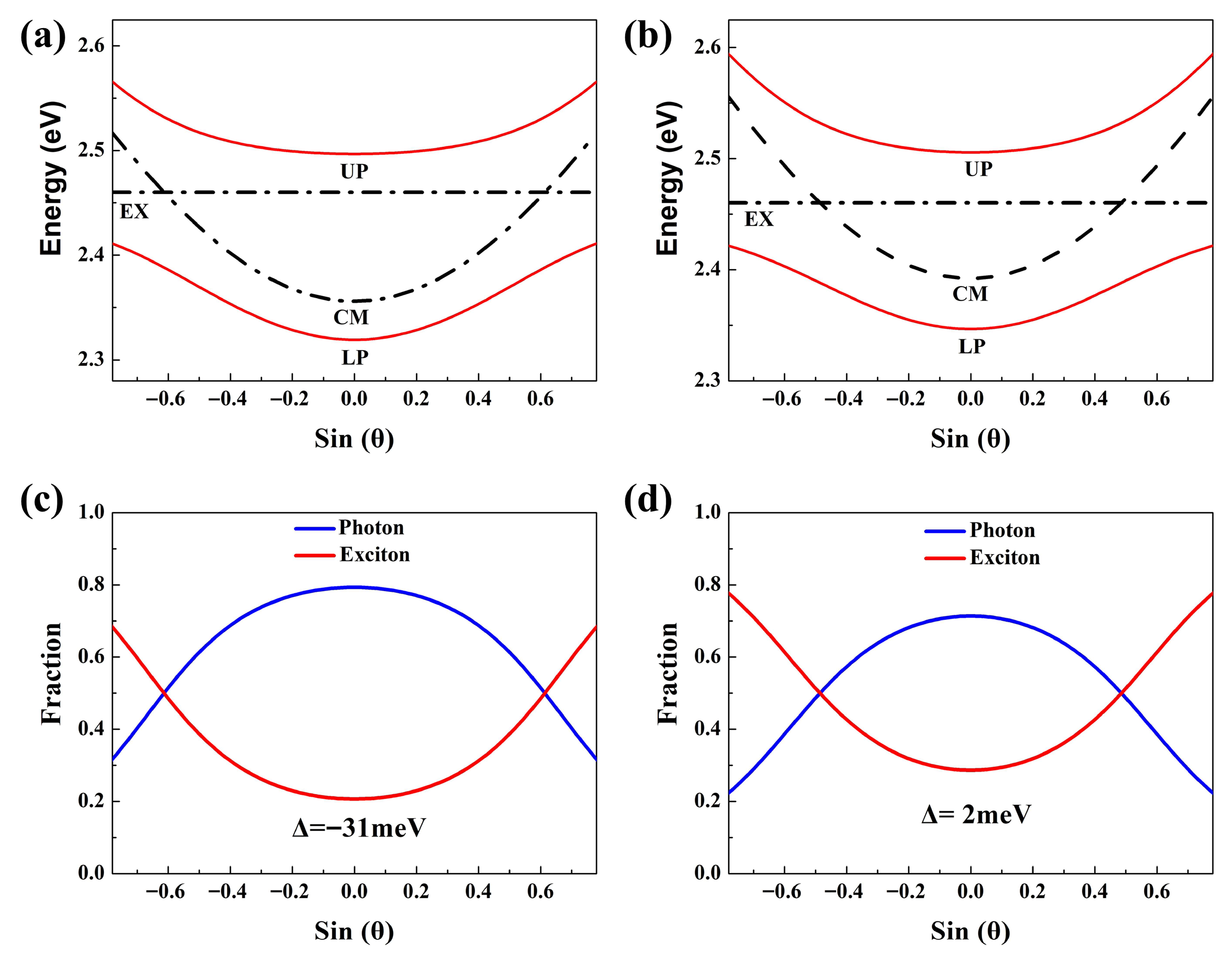
The properties of a polariton are primarily determined by its energy. When the polariton’s energy is closer to that of a photon, its properties are predominantly governed by the photon; when the polariton’s energy is closer to that of an exciton, its properties are predominantly governed by the exciton []. Since exciton–polaritons represent hybrid states of excitons and photons, the Hopfield coefficients obtained from fitting the lower polariton branch modes quantify the relative contributions of excitonic and photonic components within the polaritons []. Collectively, tuning the cavity–exciton detuning from positive to negative values transforms exciton-dominated exciton–polaritons into photon-dominated exciton–polariton states []. Based on theoretical fitting, we plot the single-mode polariton dispersion curves for CdS/CdS:SnS2 superlattice microwires with different hole excitation detunings of −31 meV and 2 meV, as shown in Figure 4a,b. Among them, the red curves represent theoretical fits for the upper and lower polariton branches, demonstrating how excitons and photons interact to form exciton–polaritons at varying detection angles. Here, the detuning () represents the energy difference between the cavity mode () energy and the exciton () energy () []. Respectively, the functional dependence of the Hopfield coefficients for the lower polariton branch on sin (θ) is presented in Figure 4c,d, where the cavity–exciton detuning energies are −31 meV and 2 meV. Concurrently, with increasing cavity–exciton detuning, the Hopfield coefficients reveal that the photonic fraction progressively dominates in the lower-branch exciton–polaritons at small angles. At a detuning of −31 meV, the lower polariton branch exhibits more photonic behavior at small angles, while at large angles, the exciton-to-photon ratio approaches 7:3. In comparison, for a detuning of 2 meV, the ratio approaches 4:1 at large angles. This demonstrates the angular and detuning dependencies of exciton–polaritons, wherein the present architecture provides a novel platform for investigating strong exciton–photon interactions.
Figure 4.
Analysis of exciton and photon fractions in CdS/CdS:SnS2 superlattice microwires. (a,b) Exciton–polariton dispersions for CdS/CdS:SnS2 superlattice microwires at cavity–exciton detunings of −31 meV and 2 meV, respectively. Red curves show theoretically fitted dispersions for the upper and lower polariton branches. Black dashed lines indicate the dispersions of the uncoupled exciton energy and cavity mode energy of the CdS/CdS:SnS2 superlattice microwires, respectively. (c,d) Hopfield coefficients corresponding to the photon fraction of the lower polariton branch cavity mode in (a,b), respectively, with the red and blue curves representing the exciton and photon components in the exciton–polariton state.
4. Conclusions
The CdS/CdS:SnS2 superlattice microwires intrinsically form an optical microcavity due to their structure, enabling the realization of exciton–polaritons at room temperature without requiring an external microcavity. Within the CdS/CdS:SnS2 superlattice microwires, photoluminescence spectra from distinct positions confirm that the optical microcavity in the superlattice structure originates from a F–P cavity mode formed by photon reflection at adjacent SnS2 interfaces. The behavior of exciton polaritons under varying cavity configurations was investigated using angle-resolved photoluminescence (ARPL) spectroscopy. By analyzing the experimental data with a coupled harmonic oscillator model, a Rabi splitting as large as 143.8 meV was obtained. The Hopfield coefficients within the exciton–polariton modes under different detuning values were discussed, revealing the variation process of the excitonic and photonic fractions in the exciton–polaritons. Anisotropic luminescence, induced by the presence of SnS2, was observed by measuring ARPL spectra at different angles between the CdS/CdS:SnS2 superlattice microwires and the spectrometer slit. Thus, CdS/CdS:SnS2 superlattice nanowires provide an excellent platform for studying strong light–matter interactions at the micro–nano scale. They serve as core components for developing next-generation low-power, high-speed integrated photonic chips, such as ultra-low-threshold nanolasers far exceeding the efficiency of conventional lasers, as well as ultrafast all-optical switches and logic gates based on polariton condensate currents. ultimately paving new pathways for room-temperature operation of on-chip quantum information processing and photonic quantum computing systems.
Author Contributions
Conceptualization, J.W. and Y.T.; methodology, C.J.; software, H.S.; validation, J.W., C.J. and H.S.; formal analysis, J.W.; investigation, J.W.; resources, J.W.; data curation, J.W.; writing—original draft preparation, J.W.; writing—review and editing, C.J.; visualization, C.J.; supervision, Y.T.; project administration, Y.T.; funding acquisition, Y.T. All authors have read and agreed to the published version of the manuscript.
Funding
This work was supported by the National Natural Science Foundation of China (62501541), Postdoctoral Fellowship Program of CPSF (No. GZC20241576), the Shanxi Province Basic Research Program (No. 202403021212341), the Open Project Program of Shanxi Key Laboratory of Advanced Semiconductor Optoelectronic Devices and Integrated Systems (No. 2024SZKF03), the National Natural Science Foundation of China (NSFC) (No. 62404207), and the Fundamental Research Program of Shanxi Province (No. 202303021212206 and No. 202403021212179).
Data Availability Statement
The original contributions presented in this study are included in the article. Further inquiries can be directed to the corresponding author.
Acknowledgments
One of the authors, Jia Wang, gratefully acknowledges the School of Semiconductor and Physics, North University of China, Taiyuan for providing instrumental facilities. All authors have read and agreed to the published version of the manuscript.
Conflicts of Interest
The authors declare no conflicts of interest.
References
- Deng, H.; Haug, H.; Yamamoto, Y. Exciton-polariton Bose-Einstein condensation. Rev. Mod. Phys. 2010, 82, 1489–1537. [Google Scholar] [CrossRef]
- Wang, T.; Zang, Z.; Gao, Y.; Lyu, C.; Gu, P.; Yao, Y.; Peng, K.; Watanabe, K.; Taniguchi, T.; Liu, X.; et al. Electrically Pumped Polarized Exciton-Polaritons in a Halide Perovskite Microcavity. Nano Lett. 2022, 22, 5175–5181. [Google Scholar] [CrossRef]
- Tumkur, T.; Zhu, G.; Noginov, M. Strong coupling of surface plasmon polaritons and ensembles of dye molecules. Opt. Express 2016, 24, 3921–3928. [Google Scholar] [CrossRef]
- Berghuis, A.M.; Castellanos, G.W.; Murai, S.; Pura, J.L.; Abujetas, D.R.; van Heijst, E.; Ramezani, M.; Sanchez-Gil, J.A.; Rivas, J.G. Room Temperature Exciton-Polariton Condensation in Silicon Metasurfaces Emerging from Bound States in the Continuum. Nano Lett. 2023, 23, 5603–5609. [Google Scholar] [CrossRef] [PubMed]
- Bhattacharya, P.; Xiao, B.; Das, A.; Bhowmick, S.; Heo, J. Solid State Electrically Injected Exciton-Polariton Laser. Phys. Rev. Lett. 2013, 110, 206403. [Google Scholar] [CrossRef] [PubMed]
- Byrnes, T.; Kim, N.Y.; Yamamoto, Y. Exciton-polariton condensates. Nat. Phys. 2014, 10, 803–813. [Google Scholar] [CrossRef]
- Hoffmann, D.K.; Singh, V.P.; Paintner, T.; Jaeger, M.; Limmer, W.; Mathey, L.; Hecker Denschlag, J. Second sound in the crossover from the Bose-Einstein condensate to the Bardeen-Cooper-Schrieffer superfluid. Nat. Commun. 2021, 12, 7074. [Google Scholar] [CrossRef]
- Huang, Y.; Su, R.; Wang, Y.; Zhu, C.; Feng, J.; Zhao, J.; Liu, Z.; Xiong, Q. A Fano Cavity-Photon Interface for Directional Suppression of Spectral Diffusion of a Single Perovskite Nanoplatelet. Nano Lett. 2022, 22, 8274–8280. [Google Scholar] [CrossRef]
- Larionova, Y.; Stolz, W.; Weiss, C.O. Optical bistability and spatial resonator solitons based on exciton-polariton nonlinearity. Opt. Lett. 2008, 33, 321–323. [Google Scholar] [CrossRef]
- Schweiner, F.; Ertl, J.; Main, J.; Wunner, G.; Uihlein, C. Exciton-polaritons in cuprous oxide: Theory and comparison with experiment. Phys. Rev. B 2017, 96, 245202. [Google Scholar] [CrossRef]
- Wu, X.; Zhang, S.; Song, J.; Deng, X.; Du, W.; Zeng, X.; Zhang, Y.; Zhang, Z.; Chen, Y.; Wang, Y.; et al. Exciton polariton condensation from bound states in the continuum at room temperature. Nat. Commun. 2024, 15, 3345. [Google Scholar] [CrossRef]
- Su, R.; Wang, J.; Zhao, J.; Xing, J.; Zhao, W.; Diederichs, C.; Liew, T.C.H.; Xiong, Q. Room temperature long-range coherent exciton polariton condensate flow in lead halide perovskites. Sci. Adv. 2018, 4, eaau0244. [Google Scholar] [CrossRef]
- Shang, Q.; Li, C.; Zhang, S.; Liang, Y.; Liu, Z.; Liu, X.; Zhang, Q. Enhanced optical absorption and slowed light of reduced-dimensional CsPbBr3 nanowire crystal by exciton–polariton. Nano Lett. 2020, 20, 1023–1032. [Google Scholar] [CrossRef] [PubMed]
- Du, W.; Zhang, S.; Zhang, Q.; Liu, X. Recent progress of strong exciton–photon coupling in lead halide perovskites. Adv. Mater. 2019, 31, 1804894. [Google Scholar] [CrossRef] [PubMed]
- Zaremba, M.; Kedziora, M.; Stanco, L.; Piskorski, K.; Kosiel, K.; Szerling, A.; Mazur, R.; Piecek, W.; Opala, A.; Sigurdsson, H.; et al. Optically Trapped Exciton-Polariton Condensates in a Perovskite Microcavity. Adv. Opt. Mater. 2025, 13, 2500304. [Google Scholar] [CrossRef]
- Kedziora, M.; Opala, A.; Mastria, R.; De Marco, L.; Krol, M.; Lempicka-Mirek, K.; Tyszka, K.; Ekielski, M.; Guziewicz, M.; Bogdanowicz, K.; et al. Predesigned perovskite crystal waveguides for room-temperature exciton-polariton condensation and edge lasing. Nat. Mater. 2024, 23, 1515–1522. [Google Scholar] [CrossRef] [PubMed]
- Gan, Y.; Shi, Y.; Ghosh, S.; Liu, H.; Xu, H.; Xiong, Q. Ultrafast neuromorphic computing driven by polariton nonlinearities. Elight 2025, 5, 9. [Google Scholar] [CrossRef]
- Sun, L.; Ren, M.-L.; Liu, W.; Agarwal, R. Resolving Parity and Order of Fabry-Perot Modes in Semiconductor Nanostructure Waveguides and Lasers: Young’s Interference Experiment Revisited. Nano Lett. 2014, 14, 6564–6571. [Google Scholar] [CrossRef]
- Tian, Y.; Yao, S.; Zou, B. Excitation-Power-Dependent Color Tuning in a Single Sn-Doped CdS Nanowire. Molecules 2024, 29, 5389. [Google Scholar] [CrossRef]
- Mi, Y.; Jin, B.; Zhao, L.; Chen, J.; Zhang, S.; Shi, J.; Zhong, Y.; Du, W.; Zhang, J.; Zhang, Q. High-quality hexagonal nonlayered CdS nanoplatelets for low-threshold whispering-gallery-mode lasing. Small 2019, 15, 1901364. [Google Scholar] [CrossRef]
- Wen, X.; Lu, Z.; Wang, G.-C.; Washington, M.A.; Lu, T.-M. Efficient and stable flexible Sb2Se3 thin film solar cells enabled by an epitaxial CdS buffer layer. Nano Energy 2021, 85, 106019. [Google Scholar] [CrossRef]
- Kennerud, K.L. Analysis of performance degradation in CdS solar cells. IEEE Trans. Aerosp. Electron. Syst. 2007, AES-5, 912–917. [Google Scholar] [CrossRef]
- Nizamoglu, S.; Mutlugun, E.; Akyuz, O.; Perkgoz, N.K.; Demir, H.V.; Liebscher, L.; Sapra, S.; Gaponik, N.; Eychmüller, A. White emitting CdS quantum dot nanoluminophores hybridized on near-ultraviolet LEDs for high-quality white light generation and tuning. New J. Phys. 2008, 10, 023026. [Google Scholar] [CrossRef]
- Van Vugt, L.K.; Piccione, B.; Cho, C.-H.; Nukala, P.; Agarwal, R. One-dimensional polaritons with size-tunable and enhanced coupling strengths in semiconductor nanowires. Proc. Natl. Acad. Sci. USA 2011, 108, 10050–10055. [Google Scholar] [CrossRef] [PubMed]
- Li, D.; Zhang, J.; Xiong, Q. Surface Depletion Induced Quantum Confinement in CdS Nanobelts. Acs Nano 2012, 6, 5283–5290. [Google Scholar] [CrossRef] [PubMed]
- Zheng, Q.; Zhou, W.; Peng, Y.; Yin, Y.; Zhong, M.; Zhao, Z.; Zhang, Q.; Tang, D.; Zeng, R.; Zou, B. Surface polarons and optical micro-cavity modulated broad range multi-mode emission of Te-doped CdS nanowires. Nanotechnology 2018, 29, 465709. [Google Scholar] [CrossRef] [PubMed]
- Huang, L.; Zhang, D.; Ge, C.; He, M.; Zeng, Z.; Wang, Y.; Liu, S.; Wang, X.; Pan, A. Enhancing circular polarization of photoluminescence of two-dimensional Ruddlesden-Popper perovskites by constructing van der Waals heterostructures. Appl. Phys. Lett. 2021, 119, 151101. [Google Scholar] [CrossRef]
- Liu, Y.; Jiang, Y.; Xu, Z.; Li, L.; Zhang, D.; Zheng, W.; Liang, D.; Zheng, B.; Liu, H.; Sun, X.; et al. Magnetic Doping Induced Strong Circularly Polarized Light Emission and Detection in 2D Layered Halide Perovskite. Adv. Opt. Mater. 2022, 10. [Google Scholar] [CrossRef]
- Rashba, E.; Gurgenishvili, G. Edge absorption theory in semiconductors. Sov. Phys. Solid State 1962, 4, 759. [Google Scholar]
- Zhang, M.; Wille, M.; Röder, R.; Heedt, S.; Huang, L.; Zhu, Z.; Geburt, S.; Grützmacher, D.; Schäpers, T.; Ronning, C. Amphoteric nature of Sn in CdS nanowires. Nano Lett. 2014, 14, 518–523. [Google Scholar] [CrossRef]
- Khurgin, J.B. Pliable polaritons: Wannier exciton-plasmon coupling in metal-semiconductor structures. Nanophotonics 2019, 8, 629–639. [Google Scholar] [CrossRef]
- Li, S.; Deng, F.; Huang, L.; Zhang, Y.; Zhou, L.; Lan, S. Greatly enhanced exciton-photon coupling in a WS2-Si3N4 nanohole array-Ag film heterostructure. Appl. Surf. Sci. 2025, 702, 163356. [Google Scholar] [CrossRef]
- Tian, Y.; Zhang, Y.; Yao, S.; Cheng, L.; Wen, H.; Guo, H.; Tang, J.; Liu, J.; Liu, X.; Zou, B. Room temperature exciton polariton condensation in high-quality Sn-doped CdS microsheet. Nano Res. 2025, 18, 94907525. [Google Scholar] [CrossRef]
- Dai, G.; Zou, B.; Wang, Z. Preparation and periodic emission of superlattice CdS/CdS:SnS2 microwires. J. Am. Chem. Soc. 2010, 132, 12174–12175. [Google Scholar] [CrossRef]
- Liu, R.; Li, Z.-A.; Zhang, C.; Wang, X.; Kamran, M.A.; Farle, M.; Zou, B. Single-step synthesis of monolithic comb-like CdS nanostructures with tunable waveguide properties. Nano Lett. 2013, 13, 2997–3001. [Google Scholar] [CrossRef]
- Zou, S.; Zhou, W.; Liu, R.; Zou, B. Cavity-enhanced microphotoluminescence in a core–shell n–p CdS/CdO micrometer wire and Its efficient surface photovoltage responses in the whole visible range. J. Phys. Chem. C 2017, 121, 14349–14358. [Google Scholar] [CrossRef]
- Zhang, L.; Zhang, Y.; Guo, Y.; Wang, Y.; Liu, R.; Chen, B.; Zhong, H.; Zou, B. Growth of CdS nanotubes and their strong optical microcavity effects. Nanoscale 2019, 11, 5325–5329. [Google Scholar] [CrossRef]
- Ma, W.; Lu, J.; Yang, Z.; Peng, D.; Li, F.; Peng, Y.; Chen, Q.; Sun, J.; Xi, J.; Pan, C. Crystal-orientation-related dynamic tuning of the lasing spectra of CdS nanobelts by piezoelectric polarization. ACS Nano 2019, 13, 5049–5057. [Google Scholar] [CrossRef]
- Xu, J.; Zhuang, X.; Guo, P.; Zhang, Q.; Ma, L.; Wang, X.; Zhu, X.; Pan, A. Dilute tin-doped CdS nanowires for low-loss optical waveguiding. J. Mater. Chem. C 2013, 1, 4391–4396. [Google Scholar] [CrossRef]
- Abdi, A.; Titova, L.V.; Smith, L.M.; Jackson, H.E.; Yarrison-Rice, J.M.; Lensch, J.L.; Lauhon, L.J. Resonant Raman scattering from CdS nanowires—Art. No. 043118. Appl. Phys. Lett. 2006, 88, 043118. [Google Scholar] [CrossRef]
- Khoo, K.H.; Zayak, A.T.; Kwak, H.; Chelikowsky, J.R. First-Principles Study of Confinement Effects on the Raman Spectra of Si Nanocrystals. Phys. Rev. Lett. 2010, 105, 115504. [Google Scholar] [CrossRef]
- Evans, T.J.S.; Schlaus, A.; Fu, Y.; Zhong, X.; Atallah, T.L.; Spencer, M.S.; Brus, L.E.; Jin, S.; Zhu, X.Y. Continuous-Wave Lasing in Cesium Lead Bromide Perovskite Nanowires. Adv. Opt. Mater. 2018, 6. [Google Scholar] [CrossRef]
- Wang, X.; Shoaib, M.; Wang, X.; Zhang, X.; He, M.; Luo, Z.; Zheng, W.; Li, H.; Yang, T.; Zhu, X.; et al. High-Quality In-Plane Aligned CsPbX3 Perovskite Nanowire Lasers with Composition-Dependent Strong Exciton—Photon Coupling. Acs Nano 2018, 12, 6170–6178. [Google Scholar] [CrossRef] [PubMed]
- Zhang, S.; Zhong, Y.; Yang, F.; Cao, Q.; Du, W.; Shi, J.; Liu, X. Cavity engineering of two-dimensional perovskites and inherent light-matter interaction. Photonics Res. 2020, 8, A72–A90. [Google Scholar] [CrossRef]
- Lv, Q.; Shen, X.; Li, X.; Liu, J.; Shan, Z.; Xiao, L.; Guo, P.; Ho, J.C. Laser-induced phase segregation of inorganic halide perovskite alloy nanowires for optical switch. Nano Res. 2025, 18, 94907119. [Google Scholar] [CrossRef]
- Yang, X.; Wu, R.; Zheng, B.; Luo, Z.; You, W.; Liu, H.; Li, L.; Zhang, Y.; Tan, Q.; Liang, D.; et al. A Waveguide-Integrated Two-Dimensional Light-Emitting Diode Based on p-Type WSe2/n-Type CdS Nanoribbon Heterojunction. Acs Nano 2022, 16, 4371–4378. [Google Scholar] [CrossRef]
- Cao, B.; Tian, Y.; Wen, H.F.; Guo, H.; Wu, X.; Li, L.; Zhang, Z.; Liu, L.; Zhu, Q.; Tang, J. Recent progress on fabrication, spectroscopy properties, and device applications in Sn-doped CdS micro-nano structures. J. Semicond. 2024, 45, 091101. [Google Scholar] [CrossRef]
- Ouyang, C.; Li, Y.; Fu, X.; Zeng, Z.; Huang, L.; Liu, S.; Wang, X.; Gao, T.; Pan, A. Room temperature exciton-polaritons in high-quality 2D Ruddlesden–Popper perovskites (BA)2(MA)n−1PbnI3n+1 (n = 3, 4). Appl. Phys. Lett. 2020, 117, 221107. [Google Scholar] [CrossRef]
- Han, Q.; Wang, J.; Lu, J.; Sun, L.; Lyu, F.; Wang, H.; Chen, Z.; Wang, Z. Transition between exciton-polariton and coherent photonic lasing in all-inorganic perovskite microcuboid. ACS Photonics 2020, 7, 454–462. [Google Scholar] [CrossRef]
Disclaimer/Publisher’s Note: The statements, opinions and data contained in all publications are solely those of the individual author(s) and contributor(s) and not of MDPI and/or the editor(s). MDPI and/or the editor(s) disclaim responsibility for any injury to people or property resulting from any ideas, methods, instructions or products referred to in the content. |
© 2025 by the authors. Licensee MDPI, Basel, Switzerland. This article is an open access article distributed under the terms and conditions of the Creative Commons Attribution (CC BY) license (https://creativecommons.org/licenses/by/4.0/).