Article

Effect of White Light-Emitting Diode Illuminants Recommended by the CIE on Colors of Dental Ceramic Materials

by
Javier Ruiz-López
1,2,
Manuel Melgosa
1,
Razvan Ghinea
1,2,3,
Maria Tejada-Casado
1,2,
Ioana-Sofia Pop-Ciutrila
4 and
María M. Pérez
1,2,*
1
Department of Optics, Faculty of Science, Campus Fuentenueva, Edificio Mecenas, s/n., University of Granada, 18071 Granada, Spain
2
Instituto de Investigación Biosanitaria ibs.GRANADA, 18011 Granada, Spain
3
Department of Physics, Faculty of Sciences, University of Craiova, 13 AI Cuza Street, 200585 Craiova, Romania
4
Department of Conservative Dentistry and Endodontics, Faculty of Dental Medicine, Iuliu Hatieganu University of Medicine and Pharmacy, 33 Motilor Street, 400001 Cluj-Napoca, Romania
*
Author to whom correspondence should be addressed.
Appl. Sci. 2023, 13(3), 1518; https://doi.org/10.3390/app13031518
Submission received: 12 December 2022 / Revised: 17 January 2023 / Accepted: 19 January 2023 / Published: 24 January 2023

Abstract

:
The aim of this study was to evaluate color inconstancy of dental ceramics under the white light-emitting diode illuminants recently proposed by the CIE. From spectral reflectance factors of 18 dental ceramics (VST, NSP, and IEC; shades A1, A2, A3, A3.5, B2, and C2), the corresponding colors under illuminant D65 and CIE 1931 Standard Colorimetric Observer were computed for all samples, using the chromatic adaptation transform CIECAT16. CIEDE2000 color differences between dental ceramics illuminated by CIE D65 standard illuminant and different white LED illuminants were calculated. Perceptibility and acceptability thresholds (PT00 and AP00) in dental ceramics were used to analyze color changes. Color gamut size was within the same range for all illuminants and for all ceramics, since MCDM computed values were 4.1–4.4 for VST, 4.0–4.2 for NSP, and 4.3–4.6 for IEC. For all ceramics and shades, the color inconstancies were higher than 50:50% PT00 and, in general, lower than 50:50% AT00. The effect of CIE-proposed LED illuminants on dental ceramic is perceptible and slightly lower than on natural teeth. Dental clinicians should consider these lighting effects on the visual appearance of dental ceramics.

1. Introduction

The color appearance of teeth needs to be matched and reproduced by restorative dental materials to achieve acceptable aesthetic conditions. The ISO/TR 2864:2016 Dentistry—Guidance on Colour Measurement [1] describes visual and instrumental methods for assessment of subjects related to color shade and interconvertibility of monochromatic and polychromatic dental tissues and/or dental materials. Bearing this in mind, the characteristic of the light source (or illuminant) used to illuminate a given sample for a color measurement must be specified. The official illuminants used in colorimetry have been published by the Commission Internationale de l’Éclairage (CIE) (see Abbreviations list in supplementary materials) [2]. This published set of illuminants includes spectral data approaching daylights and various incandescent, fluorescent, discharge, and LED lamps. Among all the descriptors of an illuminant, in particular, the spectral power distribution (SPD) and correlated color temperature (CCT) [2] are of utter importance for the experimental setup when evaluating color appearance of teeth and dental materials. A light source which represents noon daylight (CCT of 6500K) with a color rendering index (CRI) of 90 or greater is recommended when visual color assessments are made in dentistry [1]. In this context, it is important to point out that the color difference exhibited by a specimen across different viewing conditions is known as color inconstancy [3].
The CIE has been responsible for introducing the main color systems, illumination/detection geometries and color difference (ΔE), and color appearance concepts used in color science [4]. When considering CIELAB color space, the CIELAB color difference Δ E a b * has been the standard parameter employed by many users to measure total color difference. However, the CIE [2] recommend the use of the CIEDE2000 color difference formula ( Δ E 00 ) [5] because it provides a better fit than the CIELAB formula for visual color differences managed in many different industrial applications, including dentistry [6,7,8,9].
In dentistry, visual judgments are the most frequently used method for evaluating color. Thus, adequate knowledge of the limits of the perception of colors in dentistry is crucial for understanding color differences in clinical dentistry [10]. Just perceptible refers to the lowest color difference that can be detected by an average human observer with normal color vision. A color difference noticeable by 50% of the observers corresponds to a 50:50% perceptibility threshold (PT). Similarly, the difference in color that is considered as acceptable for 50% of observers corresponds to a 50:50% acceptability threshold (AT) [11]. The visual color thresholds are of extreme importance in interpreting color differences in clinical dentistry and dental research [12,13]. Several studies have established the value of color discrimination thresholds in dentistry [13] and, in particular, Ghinea et al. [14] determined the values of CIEDE2000 color difference thresholds in dental ceramics (PT00 = 1.25 and AT00 = 2.23).
General lighting is currently undergoing a revolutionary shift towards solid state devices, in particular light emitting diode (LED)-based technologies, as a replacement for classic fluorescent lamps. LEDs save energy and provide the same or better quality of illumination and visual comfort than classic fluorescent lamps. Researchers [15] investigated the perceived quality of LED sources in psychophysical experiments, considering color difference assessment and color appearance estimation. The results show that LEDs outperformed conventional fluorescent lamp-based simulators for the CIE illuminant D50. Furthermore, colorimetric measures revealed that LED simulators can achieve the desired quality according to the relevant ISO and CIE standards [16]. Another study [17] showed that an optimized LED spectrum illumination based on human perception of color differences significantly improved the visibility of tissue texture compared to illuminants such as CIE D65.
In order to provide an approximate representation of the available range of white LED sources, the CIE proposed a series of 9 specific LED illuminants [2]: LEDS B1 to B5 illuminants (typical phosphor-converted blue LEDs with different CCTs); LED BH1 illuminant (of mixing phosphor-converted blue and red LEDs); LED RGB1 illuminant (mixture of red, green, and blue LEDs); and LEDs V1 and V2 (traditional phosphor-converted violet LEDs with modified CCTs) [2]. Currently, B1 to B5 LEDs are the most frequently used. The SPDs of the 9 LED illuminants proposed were obtained from experimental measurements of a total of 1298 commercial white LED conventional sources [18,19].
The effect that these nine CIE white illuminants have on visual appearance has been evaluated for cosmetic applications [20] and museum lighting [21]. In dentistry, a recent study [22] evaluated the color inconstancy of natural teeth measured under white light-emitting diode illuminants. The authors concluded that the CIE-proposed LED illuminants produced perceptible and no acceptable color and whiteness differences on the evaluated teeth when compared with CIE standard illuminants D65 and A. Nevertheless, to the best of our knowledge, there are no studies that have evaluated color inconstancy for dental ceramics when illuminated with the newly proposed CIE LED illuminants relative to their color as illuminated with the CIE-recommended standard illuminant. Therefore, the main purpose of this study was to evaluate the color changes of dental ceramics based on CIE LED illuminants relative to the CIE D65 standard illuminant recommended for dentistry, considering color difference thresholds in dental ceramics.

2. Materials and Methods

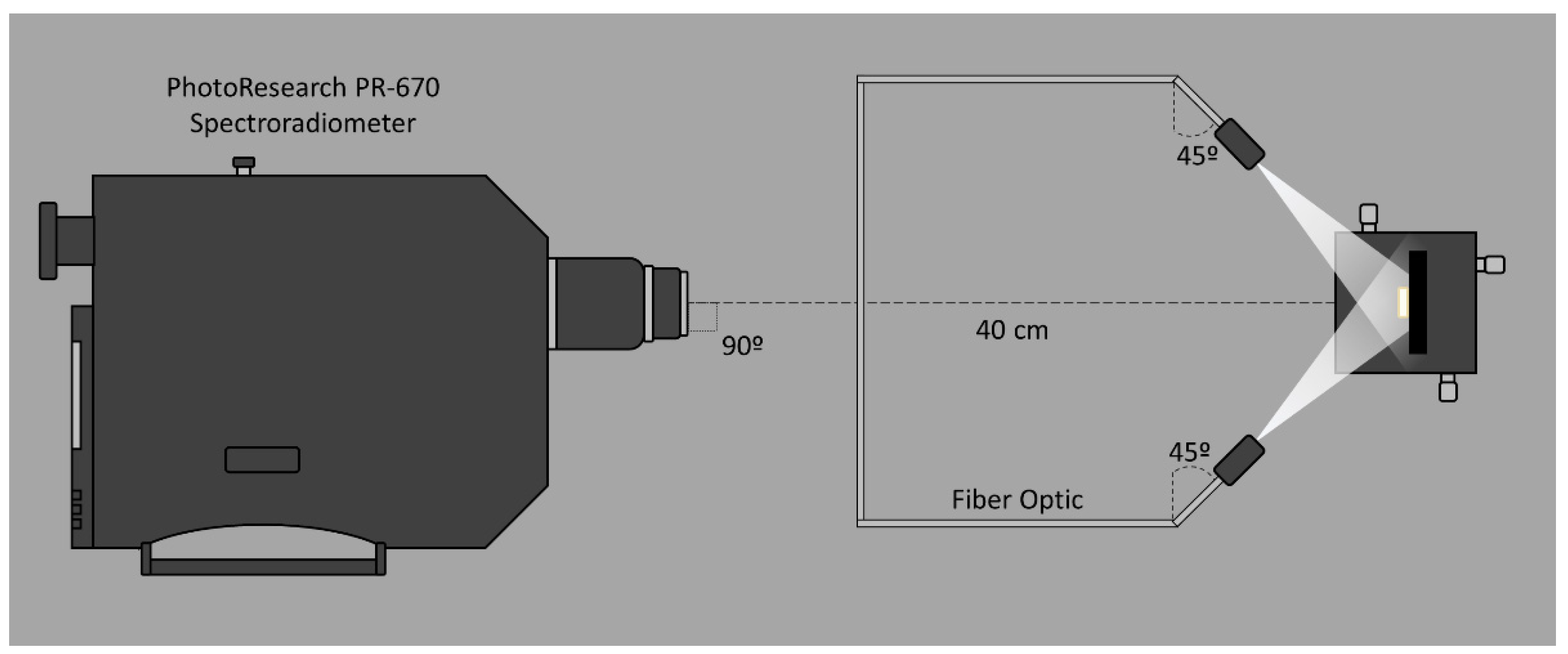
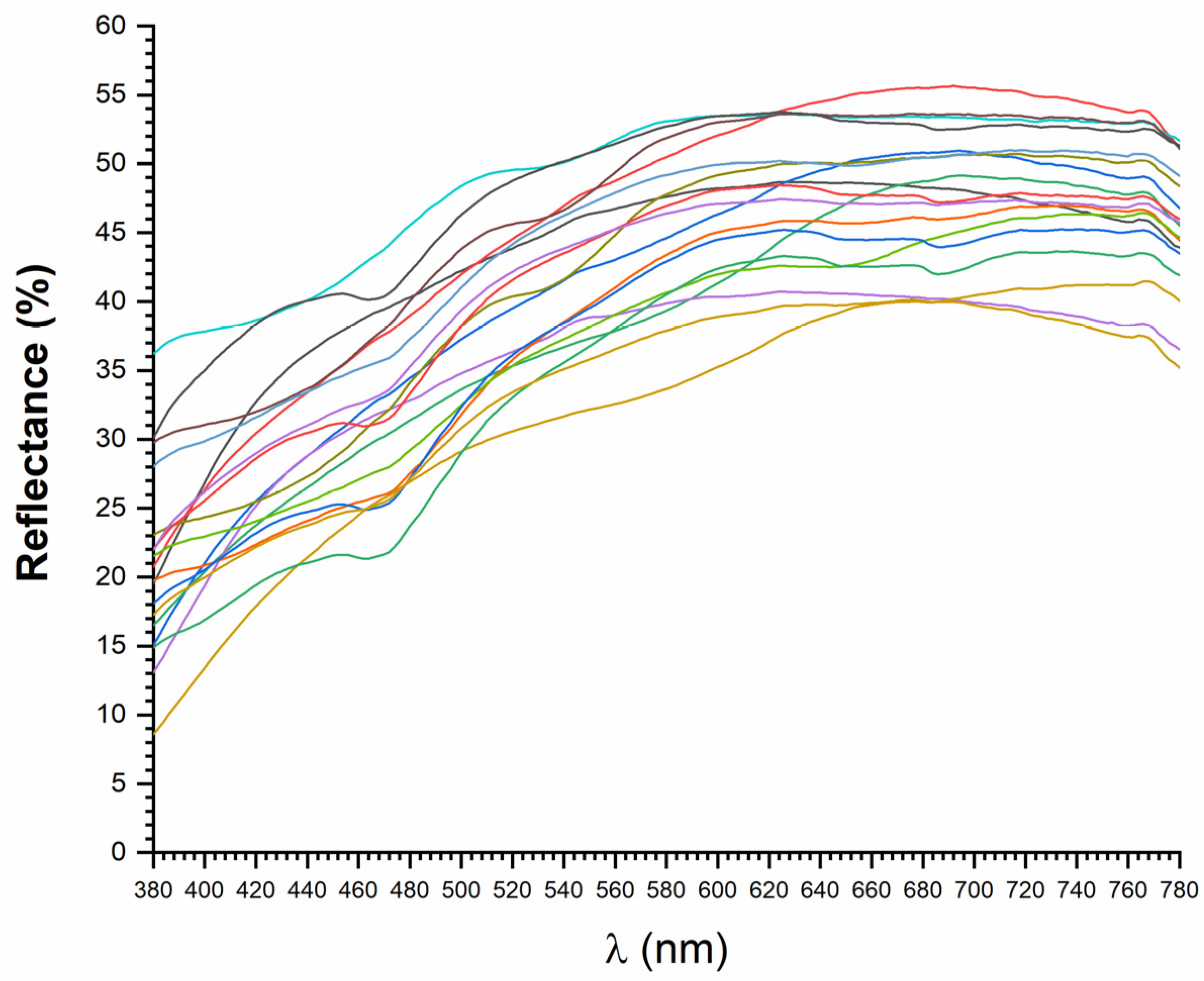
2.1. Samples and Illuminants

The spectral reflectance factors of 3 ceramic restorative materials of different classifications (Table 1) were measured using a non-contact measuring system. The device was composed of a spectroradiometer (PR 670-Photo Research, Chatsworth, CA, USA) and a double-fiber optic light cable (Oriol Research, Newport Corporation, CA, USA) coupled to a Xe-Arc light source (Oriol Research, Newport Corporation, CA, USA). Measurements (380–780 nm in steps of 2 nm) were performed using the CIE illuminating/measuring geometry 45°:0° and a spectrally calibrated reflectance standard (SRS-3, PhotoResearch, CA, USA) in a dark room, where the specimens were placed 40 cm away from the spectroradiometer over a black 50 × 50 mm ceramic tile background (L* = 3.1, a* = 0.7 and b* = 2.4) (Ceram, Stafford, UK) (Figure 1). For each specimen, 3 samples (2.0 mm-thick) were prepared and 3 repeated measurements without replacement were performed for each sample, and the results were averaged into a single value. Figure 2 shows the mean measured spectral reflectance factors of the 18 ceramic specimens evaluated in this study. An experienced investigator performed all spectral reflectance factor measurements of dental ceramics.
Measured spectral reflectance factors (Figure 2) were converted into CIE tristimulus values (Xi, Yi, Zi (i = 1…18)) using the CIE 1931 2° Standard Colorimetric Observer and D65 standard illuminant as well as the 9 CIE white LED illuminants (Figure 3). According to a previous study [22], corresponding colors were computed using a chromatic adaptation transform (CAT) [3], that is, different tristimulus values are obtained from colors with the same visual appearance as the initial ones. This was undertaken in order to make meaningful comparisons, since a common viewing condition was achieved. Then, from these corresponding colors, a conventional color difference formula could be used to obtain the ‘color inconstancy’ index [3].
The CAT known as CIECAT16 [23,24,25] was used to calculate the corresponding colors for the CIE 1931 Standard Colorimetric Observer as illuminated with D65 standard illuminant. This was achieved using the CIE tristimulus values (Xi, Yi, Zi) of the 18 dental ceramics calculated using D65 standard illuminant and the 9 CIE LEDs illuminants (in all cases using the CIE 1931 2° Standard Colorimetric Observer), assuming an adaptation luminance LA = 64 cd/m2 (equal to a photopic illuminance of 1000 lx), a degree of adaptation D = 1, and an average surround (F = 1). Since the corresponding colors were computed under the aforementioned conditions, an adequate comparison of the CIE L*, a*, b* chromaticity coordinates of each sample computed under different illuminants was possible, since a common reference viewing condition was achieved while maintaining the different visual color appearances of all dental ceramics studied under each illuminant.

2.2. Data Processing
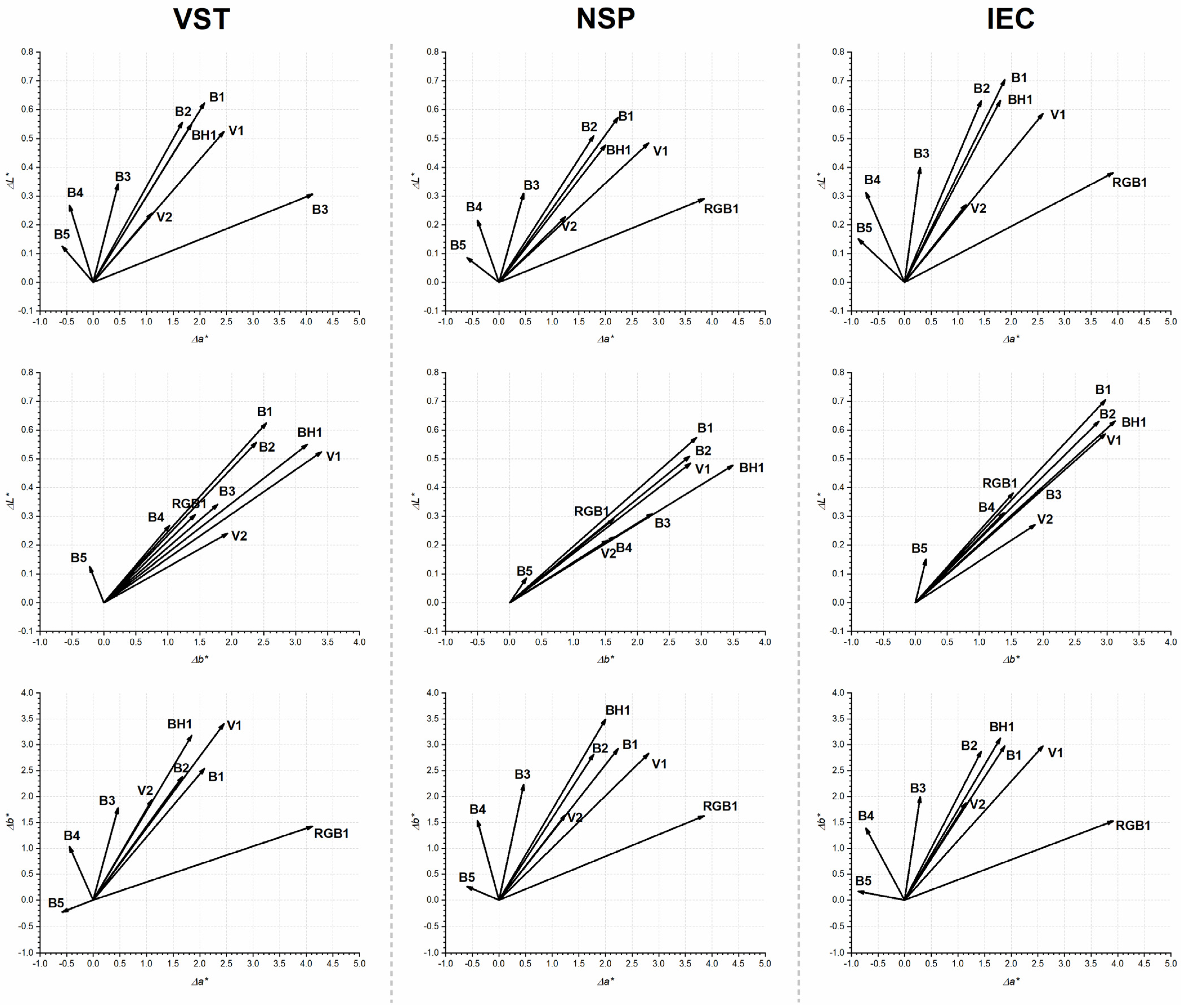
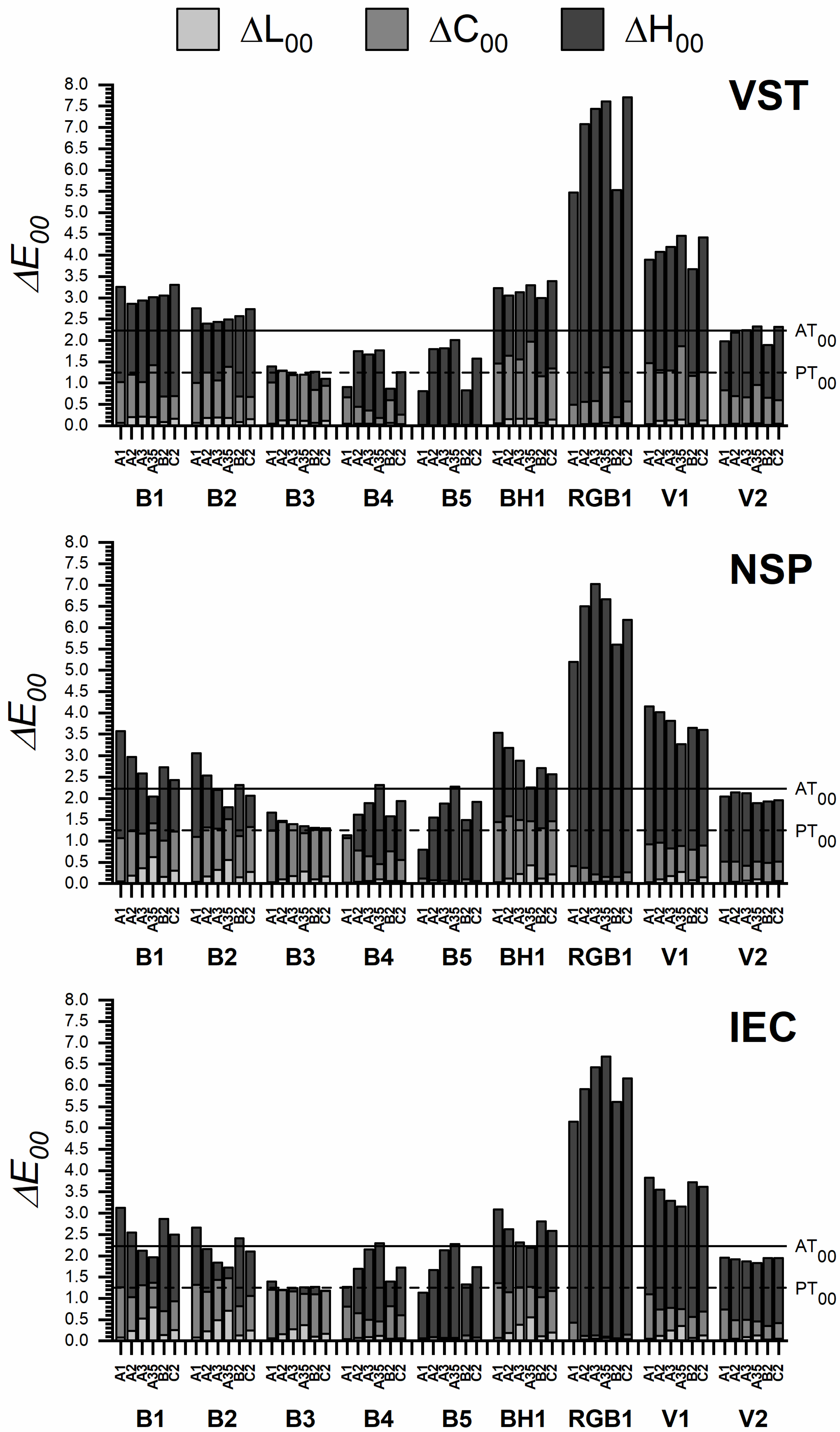
The CIELAB color gamut of the dental ceramics studied can be described by using the average values of the CIELAB chromaticity coordinates L * ¯ , a * ¯ ,   b * ¯ of all samples with their standard deviations (SD) using the range of each CIELAB chromaticity coordinates or by using the mean color difference from the mean (MCDM) [26]. In this study, the MCDM was used:
MCDM = i = 1 N L i * L * ¯ a i * a * ¯ b i * b * ¯ 1 / 2 N
CIEDE2000 (∆𝐸00) color-difference formula [27] was used to assess color inconstancy:
Δ E 00 = Δ L k L S L 2 + Δ C k C S C 2 + Δ H k H S H 2 + R T Δ C k C S C Δ H k H S H 1 2
where k L S L , k C S C , and k H S H are correction terms used for weighting the metric differences to the CIEDE2000 components. Parametric factors ( k L , k C and k H ) were set to 1 in this study (i.e., we used CIEDE2000(1:1:1)).
The 50:50% perceptibility (PT) and acceptability (AT) color thresholds for dental ceramics described in previous literature [14] (PT00 = 1.25 [95%CI 0.69–2.22] and AT00 = 2.23 [95%CI 1.55–3.00] were used to interpret the findings of the study.
In addition, total color differences in CIEDE2000 units ( Δ E 00 ) can also be described in terms of corresponding differences of the 3 main perceptual attributes of color: lightness, chroma, and hue (ΔL00, ΔC00 and ΔH00, respectively) [28]. These 3 attributes can be presented in percentage and calculated as: % Δ a t t r i b u t e = 100 Δ a t t r i b u t e / Δ E 00 2 and therefore, from this definition, it follows that:
% Δ L 00 + % Δ C 00 + % Δ H 00 = 100

3. Results

Table 2, Table 3 and Table 4 show, for each shade and each illuminant, the average CIELAB coordinates for the three measured samples with their corresponding standard deviations (in brackets). The three ceramics studied showed similar color gamut sizes for all illuminants, as reflected by the narrow range of the MCDM: 4.1–4.4 for VST (Table 2), 4.0–4.2 for NSP (Table 3), and 4.3–4.6 for IEC (Table 4).
Figure 4 shows, as an example, the average color shifts (or color inconstancies) in terms of the relative displacements of the centers of gravity of the color gamuts with respect to the D65 illuminant, differentiating the a *   b * ,   a *   L * , and b *   L * planes for shade A1 (similar results can be found for the remaining shades). In terms of the D65 illuminant color shifts, it can be observed that (1) all LED illuminants studied produced a* b* shifts in several different directions of the CIELAB diagram; (2) in general, shifts in b* were larger than those in a*; and (3) the L* coordinate showed the lowest shifts for the nine LED illuminants, producing in all cases increases below 0.8 units.
Figure 5 shows total color differences (color inconstancies) between chromaticity coordinates obtained with each LED illuminant and chromaticity coordinates obtained with the D65 illuminant. Differences in lightness, chroma, and hue components of the total CIEDE2000 color difference are shown. It can be noted that in general for all illuminants, differences in hue made the greatest contribution to the total color difference with respect to illuminant D65 (Figure 5), in particular for LED B5 and LED RGB1, independently of the dental ceramic analyzed. However, in the case of LED B3 for all ceramics and LED B2 for IEC ceramic, chroma differences for all shades analyzed were mainly found. Lightness differences were almost negligible with respect to D65, due to the fact that all the illuminants tested had Y = 100 [22].
Most interesting, for all tested ceramics and shades, the CIEDE2000 color differences representative of color inconstancies were higher than 50:50% PT00 and, in general, lower than 50:50% AT00, only for LEDs B2, B3, B4, and V2.

4. Discussion

The goal of any dental restoration is to create a natural appearance that is highly aesthetic while remaining functional [29]. Among all available current dental restorative materials, dental ceramics have been widely used for dental restorations due to their exceptional aesthetics, durability, and mechanical properties [30].
Perceived color of objects depends initially on three main factors [3]: the physical characteristics of the objects, the spectral properties of the illuminant, and the sensitivity of the photoreceptors in the human eye. Thus, color inconstancy generated by a change in the light that illuminates a dental tissue or a dental material can be found. It is therefore of great interest to understand the visual effects caused by an illuminant change on dental materials, and, in particular, dental ceramics.
A key issue in restorative dentistry is the improvement of the relationship between what it is observed and what it is computed with instrumental color measurements. In fact, whenever possible, instrumental and visual color matching methods should be used jointly for dental color assessment, as they complement each other and can lead towards better aesthetic outcomes. However, different studies [7,31] have reported that the differences between visual and instrumental shade matching are still substantial and not homogeneous for shade distribution found in instrumental examination. The color appearance models (CAMs), which make use of CATs as their main components [3], are primarily aimed at providing viewing-condition-specific methods for the transformation of tristimulus values X, Y, Z to or from perceptual attribute correlates. It should be noted that the CIELAB color space is considered as the first color appearance model. CIELAB provides equations to obtain the three main attributes of color perceived by the human eye (L*, C* and H*) from instrumentally measured X, Y, Z values, using only the X, Y, Z values of the ‘reference white’ X 0 ,     Y 0   , Z 0   to perform the CAT. In dentistry, CIELAB color space and its associated color coordinates L * , a * , b * are widely used [4,32,33,34]. The values of these coordinates are strongly affected by the illuminant used in their computations, and changes in lighting alter the perceived color of most materials, as has been proven in natural teeth [22].
Color differences in dentistry are usually evaluated using the CIEDE2000 total color-difference formula ( Δ E 00 ) [2,27] based on the CIELAB color space. Δ E 00 incorporates specific corrections for the non-uniformity of the CIELAB color space and is currently recommended by CIE and ISO/TR 28642:2016 [1,2]. The actual aesthetic success of dental restorations depends on the visual perception by the observers rather than the numerical data obtained from instrumental measurements of specimens. Specific visual thresholds for color discrimination in dentistry have already been proposed in literature [13], and are useful for industrial quality control. According to the latest guidance on color measurements in dentistry published by the International Organization for Standardization ISO/TR 28642:2016 [1], color variation should be assessed based on comparisons with 50:50 % thresholds.
From Figure 5, the size of color changes registered for each LED illuminant when referred to the CIE standard illuminant D65 fall within the following ranges: 0.8 (shade A1, LED B5) to 7.7 (shade C2, LED RGB1) CIEDE2000 units for VTS ceramic; 0.8 (shade A1, LED B5) to 7.0 (shade A3, LED RGB1) CIEDE2000 units for NSP ceramic; and 1.1 (shade A1 LED B5) to 6.7 (shade A3.5, LED RGB1) CIEDE2000 units for IEC ceramic. It should be noted that LED B5 is, among the new LED standard illuminants proposed by the CIE, the closest one to D65 standard illuminant in terms of CCT and xy-chromaticity coordinates [3,18], which could justify the low values of color inconstancy found for this LED illuminant. The ranges found are similar to those reported in a previous work [22] for color inconstancy in human teeth (1.4–8.2 CIEDE2000 units). In addition, CIEDE2000 color inconstancy in teeth [22] under the LED RGB1 showed the highest values, and B3, B4, and B5 the lowest, similar to what was found for dental ceramics in this study. Furthermore, as reported in CIEDE2000 regarding color inconstancy in teeth [22], color changes were mainly due to hue changes in LEDs RGB1, B4, and B5, and chroma changes in LED B3. Thus, our results showed similar trends of color inconstancy (when changing from D65 to CIE white LED illuminants) in human teeth and dental ceramics. Nevertheless, it should be noted that in the case of the studied dental ceramics, the size of color inconstancy produced by the CIE white LEDs is lower than in human teeth (0.8–7.0 versus 1.4–8.2 CIEDE2000 units). Therefore, the effect of LED illuminants on dental ceramic has a lower magnitude than on natural teeth.
According to the literature, the 50:50% perceptibility/acceptability color difference thresholds for dental ceramics can be considered as 1.25/2.23 CIEDE2000 units, respectively [14]. Therefore, it can be stated that, in general, for all shades and dental ceramics studied, the color produced perceptible color change from D65 to a white LED illuminant in most cases and unacceptable color change in some cases (e.g., LED RGB1). Consequently, it can be considered that the evaluated shade may have a high color inconstancy in comparison with the perceptible threshold value of a shade under two different illuminants. In particular, RGB1, V1, and BH1 LED illuminants produced the highest color inconstancy with respect to the recommended illuminant in dentistry (CIE D65). This result was also reported in natural teeth, although in this study [22] other values of 50:50% visual thresholds (0.8/1.8) [9] were used to evaluate the effect of the white LED illuminants on color of teeth. On the other hand, the results found in the present study (Figure 5) also show that if a proper selection of the LED illuminants is made (e.g., LED B3, B4 and B5), a visually acceptable color inconstancy (with respect to the recommend illuminant CIE D65) can be achieved. In summary, these findings imply that when a change in illumination from D65 to white LEDs is produced, the color shift of the materials analyzed will be perceptible to an average human observer with normal color vision. In some cases (e.g., change from D65 to LED RGB1), such color changes will be clearly unacceptable to an average human observer.
Lastly, our results show that, by using LED illuminants in dental ceramics, different color shifts, mainly for C* and h (Figure 5), may be achieved. These results prove that the use of the CIE-proposed white LEDs [2] have the potential to affect the color appearance of a dental ceramic material in various ways. Given that different LED sources are generally available on the market, our presented results using different prototypes of white LEDs are consistent with what is currently available [18]. With respect to aesthetic dentistry, choosing LEDs that generate maximum or minimum color shifts in relation to outdoor equivalent illumination (D65) is plausible.

5. Conclusions

Within the limitations of the present study, it can be stated that the visual overall appearance of the studied dental ceramics is affected by the LED illuminants proposed by the CIE. Specifically, the color shift of the materials analyzed will be perceptible and in some cases even unacceptable to an average human observer with normal color vision.
Dental ceramics showed similar trends of color inconstancy to those found for in vivo human teeth. Nevertheless, it should be noted that the effect of these LED illuminants on dental ceramics has a lower magnitude than that found in natural teeth.
The visual color appearance of a dental ceramic changes according to the selected white LEDs used, which is interesting in the context of aesthetic dentistry. Knowledge of these effects may enable dental clinicians to select white LEDs that generate minimum color shifts relative to the CIE D65 standard illuminant. Therefore, it is important that dentists be aware of the influence of lighting on the visual appearance of dental ceramics in aesthetic dental restorations. Future studies with real subjects and in clinical conditions should be performed, where natural teeth and adjacent dental ceramics can be evaluated together.

Supplementary Materials

The following supporting information can be downloaded at: https://www.mdpi.com/article/10.3390/app13031518/s1, Table S1: Abbreviations list.

Author Contributions

Conceptualization, writing—original draft, supervision, M.M. and M.M.P.; methodology, investigation, J.R.-L., M.M., R.G., M.T.-C. and I.-S.P.-C.; formal analysis, writing—review and editing, J.R.-L., M.M. and M.M.P.; funding acquisition, M.M.P. and M.M. All authors have read and agreed to the published version of the manuscript.

Funding

This research was funded by FEDER/Junta de Andalucía-Consejería de Transformación Económica, Industria, Conocimiento y Universidades, Spain, grant number A-FQM-30-UGR20, Ministerio de Ciencia, Innovación y Universidades, Spain, research project PGC2018-101904-A-100, and Junta de Andalucía, Spain, grant number P20-00200.

Institutional Review Board Statement

Not applicable.

Informed Consent Statement

Not applicable.

Data Availability Statement

Not applicable.

Acknowledgments

The authors acknowledge funding support from the Junta de Andalucía, Spain (A-FQM-30-UGR20 and P20-00200), Ministerio de Ciencia, Innovación y Universidades, Spain (PGC2018-101904-A-100).

Conflicts of Interest

The authors declare no conflict of interest.

References

  1. ISO/TR28642:2016; International Organization for Standardization—Technical Report(E): Dentistry—Guidance on Color Measurements. ISO: Geneva, Switzerland, 2016.
  2. The International Commission on Illumination. CIE 015:2018 Colorimetry, 4th ed.; The International Commission on Illumination: Vienna, Austria, 2018. [Google Scholar]
  3. Fairchild, M.D. Color Appearance Models, 3rd ed.; Wiley: Chichester, UK, 2013. [Google Scholar]
  4. Della Bona, A. Color and Appearance in Dentistry, 1st ed.; Springer: Cham, Switzerland, 2020. [Google Scholar]
  5. Luo, M.R.; Cui, G.; Rigg, B. The Development of the CIE 2000 Colour-Difference Formula: CIEDE2000. Color Res. Appl. 2001, 26, 340–350. [Google Scholar] [CrossRef]
  6. Gómez-Polo, C.; Muñoz, M.P.; Lorenzo Luengo, M.C.; Vicente, P.; Galindo, P.; Martín Casado, A.M. Comparison of the CIELab and CIEDE2000 Color Difference Formulas. J. Prosthet. Dent. 2016, 115, 65–70. [Google Scholar] [CrossRef]
  7. Pecho, O.E.; Ghinea, R.; Alessandretti, R.; Pérez, M.M.; della Bona, A. Visual and Instrumental Shade Matching Using CIELAB and CIEDE2000 Color Difference Formulas. Dent. Mater. 2016, 32, 82–92. [Google Scholar] [CrossRef]
  8. Pérez, M.M.; Saleh, A.; Yebra, A.; Pulgar, R. Study of the Variation between CIELAB ΔE* and CIEDE2000 Color-Differences of Resin Composites. Dent. Mater. J. 2007, 26, 21–28. [Google Scholar] [CrossRef] [Green Version]
  9. Paravina, R.D.; Ghinea, R.; Herrera, L.J.; Bona, A.D.; Igiel, C.; Linninger, M.; Sakai, M.; Takahashi, H.; Tashkandi, E.; del Mar Perez, M. Color Difference Thresholds in Dentistry. J. Esthet. Restor. Dent. 2015, 27, S1–S9. [Google Scholar] [CrossRef]
  10. Pérez, M.M.; Pecho, O.E.; Ghinea, R.; Pulgar, R.; Della Bona, A. Recent Advances in Color and Whiteness Evaluations in Dentistry. Curr. Dent. 2019, 1, 23–29. [Google Scholar] [CrossRef]
  11. Melgosa, M.; Hita, E.; Romero, J.; del Barco, L.J. Color-Discrimination Thresholds Translated from the CIE (x, y, Y) Space to the CIE 1976 (L*,A*,B*). Color. Res. Appl. 1994, 19, 10–18. [Google Scholar]
  12. Chu, S.J.; Trushkowsky, R.D.; Paravina, R.D. Dental Color Matching Instruments and Systems. Review of Clinical and Research Aspects. J. Dent. 2010, 38, e2–e16. [Google Scholar] [CrossRef]
  13. Paravina, R.D.; Pérez, M.M.; Ghinea, R. Acceptability and Perceptibility Thresholds in Dentistry: A Comprehensive Review of Clinical and Research Applications. J. Esthet. Restor. Dent. 2019, 31, 103–112. [Google Scholar] [CrossRef]
  14. Ghinea, R.; Pérez, M.M.; Herrera, L.J.; Rivas, M.J.; Yebra, A.; Paravina, R.D. Color Difference Thresholds in Dental Ceramics. J. Dent. 2010, 38, e57–e64. [Google Scholar] [CrossRef]
  15. Gu, H.T.; Pointer, M.R.; Liu, X.Y.; Ronnier Luo, M. Quantifying the Suitability of CIE D50 and a Simulators Based on LED Light Sources. Color. Res. Appl. 2017, 42, 408–418. [Google Scholar] [CrossRef]
  16. ISO 3664:2008; International Organization for Standardization: Viewing Conditions—Graphic Technology and Photography. ISO: Geneva, Switzerland, 2008.
  17. Wang, H.; Cuijpers, R.H.; Vogels, I.M.L.C.; Ronnier Luo, M.; Heynderickx, I.; Zheng, Z. Optimising the Illumination Spectrum for Tissue Texture Visibility. Light. Res. Technol. 2018, 50, 757–777. [Google Scholar] [CrossRef]
  18. Jost, S.; Ngo, M.; Ferrero, A.; Poikonen, T.; Pulli, T.; Thorseth, A.; Blattner, P. Determination of Illuminants Representing Typical White Light Emitting Diodes Sources. In Proceedings of the CIE Midterm Meeting 2017, Jeju Island, Republic of Korea, 21–28 October 2017; pp. 427–432. [Google Scholar]
  19. Kokka, A.; Poikonen, T.; Blattner, P.; Jost, S.; Ferrero, A.; Pulli, T.; Ngo, M.; Thorseth, A.; Gerloff, T.; Dekker, P.; et al. Development of White LED Illuminants for Colorimetry and Recommendation of White LED Reference Spectrum for Photometry. Metrologia 2018, 55, 526. [Google Scholar] [CrossRef]
  20. Melgosa, M.; Richard, N.; Fernández-Maloigne, C.; Xiao, K.; de Clermont-Gallerande, H.; Jost-Boissard, S.; Okajima, K. Colour Differences in Caucasian and Oriental Women’s Faces Illuminated by White Light-Emitting Diode Sources. Int. J. Cosmet. Sci. 2018, 40, 244–255. [Google Scholar] [CrossRef]
  21. Martínez-Domingo, M.Á.; Melgosa, M.; Okajima, K.; Medina, V.J.; Collado-Montero, F.J. Spectral Image Processing for Museum Lighting Using CIE LED Illuminants. Sensors 2019, 19, 5400. [Google Scholar] [CrossRef] [Green Version]
  22. Melgosa, M.; Ruiz-López, J.; Li, C.; García, P.A.; della Bona, A.; Pérez, M.M. Color Inconstancy of Natural Teeth Measured under White Light-Emitting Diode Illuminants. Dent. Mater. 2020, 36, 1680–1690. [Google Scholar] [CrossRef]
  23. Li, C.; Li, Z.; Wang, Z.; Xu, Y.; Luo, M.R.; Cui, G.; Melgosa, M.; Brill, M.H.; Pointer, M. Comprehensive Color Solutions: CAM16, CAT16, and CAM16-UCS. Color Res. Appl. 2017, 42, 703–718. [Google Scholar] [CrossRef]
  24. Li, C.; Xu, Y.; Wang, Z.; Luo, M.R.; Cui, G.; Melgosa, M.; Brill, M.H.; Pointer, M. Comparing Two-Step and One-Step Chromatic Adaptation Transforms Using the CAT16 Model. Color Res. Appl. 2018, 43, 633–642. [Google Scholar] [CrossRef]
  25. The International Commission on Illumination. CIE 248:2022 The CIE Colour Appearance Model for Colour Management Systems: CIECAM16; The International Commission on Illumination: Vienna, Austria, 2022. [Google Scholar]
  26. Berns, R. Billmeyer and Saltzman’s Principles of Color Technology, 3rd ed.; Wiley: New York, NY, USA, 2000. [Google Scholar]
  27. ISO/CIE 11664-6:2022; International Organization for Standardization: Colorimetry—Part 6: CIEDE2000 Colour-Difference Formula. ISO: Vienna, Austria, 2014.
  28. Nobbs, J.H. A Lightness, Chroma and Hue Splitting Approach to CIEDE2000 Colour Differences. Adv. Colours Sci. Technol. 2002, 5, 46–53. [Google Scholar]
  29. Da Costa, J.; Fox, P.; Ferracane, J. Comparison of Various Resin Composite Shades and Layering Technique with a Shade Guide. J. Esthet. Restor. Dent. 2010, 22, 114–124. [Google Scholar] [CrossRef]
  30. Bajraktarova-Valjakova, E.; Korunoska-Stevkovska, V.; Kapusevska, B.; Gigovski, N.; Bajraktarova-Misevska, C.; Grozdanov, A. Contemporary Dental Ceramic Materials, A Review: Chemical Composition, Physical and Mechanical Properties, Indications for Use. Open Access Maced. J. Med. Sci. 2018, 6, 1742. [Google Scholar] [CrossRef] [PubMed] [Green Version]
  31. Brandt, J.; Nelson, S.; Lauer, H.C.; von Hehn, U.; Brandt, S. In Vivo Study for Tooth Colour Determination—Visual versus Digital. Clin. Oral Investig. 2017, 21, 2863–2871. [Google Scholar] [CrossRef] [PubMed]
  32. Ruiz-López, J.; Pulgar, R.; Lucena, C.; Pelaez-Cruz, P.; Cardona, J.C.; Perez, M.M.; Ghinea, R. Impact of Short-Term Dental Dehydration on in-Vivo Dental Color and Whiteness. J. Dent. 2021, 105, 103560. [Google Scholar] [CrossRef] [PubMed]
  33. Ruiz-López, J.; Perez, M.M.; Lucena, C.; Pulgar, R.; López-Toruño, A.; Tejada-Casado, M.; Ghinea, R. Visual and Instrumental Coverage Error of Two Dental Shade Guides: An in Vivo Study. Clin. Oral Investig. 2022, 26, 5961–5968. [Google Scholar] [CrossRef]
  34. Ruiz-López, J.; Espinar, C.; Lucena, C.; de la Cruz Cardona, J.; Pulgar, R.; Pérez, M.M. Effect of Thickness on Color and Translucency of a Multi-Color Polymer-Infiltrated Ceramic-Network Material. J. Esthet. Restor. Dent. 2022. [Google Scholar] [CrossRef]
Figure 1. Experimental device configuration used for color measurements.
Applsci 13 01518 g001
Figure 2. Average spectral reflectance factors on black background of the 18 ceramic materials studied in this study.
Applsci 13 01518 g002
Figure 3. Normalized (Y = 100) spectral power distributions of the 9 white CIE LED illuminants and CIE D65 illuminant [2].
Applsci 13 01518 g003
Figure 4. Projections of the color shifts produced by each one of the nine LED illuminants on the a *   b * ,   a *   L * , and b *   L * planes, using as common origin the coordinates under illuminant D65 for shade A1. Results for the three dental ceramics analyzed are distinguished (left: VST; center: NSP; right: IEC).
Applsci 13 01518 g004
Figure 5. Color inconstancy for each shade and ceramic material when the illuminant changes from D65 standard illuminant to each of the LED illuminants considered. (First: VST; second: NSP; third: IEC). Perceptibility (PT00)–(dashed line) and acceptability (AT00)–(solid line) color threshold values are indicated.
Applsci 13 01518 g005
Table 1. Characteristics and information on the ceramic materials included in this study. All resin-based composite data were provided by manufacturers.
MaterialManufacturerClassificationShades
Vita Suprinity
Translucent
(VST)
Vita Zahnfabrik
(Bad Säckingen, Germany)
ZrO2 lithium silicate glass ceramicA1
A2
A3
A3.5
B2
C2
Noritake Super
Porcelain EX-3
(NSP)
Kuraray Noritake Dental
(Tokyo, Japan)
Conventional feldspathic ceramic
IPS-Empress CAD
Low Translucency
(IEC)
Ivoclar Vivadent
(Schaan, Liechtenstein)
Leucite-reinforced glass-ceramic
Table 2. Mean CIELAB color coordinate values and standard deviations (in parentheses) for the VST ceramic shades under D65 and nine LED illuminants (corresponding colors under D65 illuminant and CIE 1931 standard colorimetric observer).
VST D65LED B1LED B2LED B3LED B4LED B5LED BH1LED RGB1LED V1LED V2
A1 L * 73.5 (0.2)74.1 (0.2)74.1 (0.2)73.8 (0.2)73.8 (0.2)73.6 (0.2)74.1 (0.2)73.8 (0.2)74.0 (0.2)73.7 (0.2)
a *   −0.7 (0.0)1.4 (0.0)1.0 (0.0)−0.2 (0.0)−1.1 (0.0)−1.3 (0.0)1.1 (0.0)3.4 (0.1)1.8 (0.0)0.4 (0.0)
b *   9.8 (0.2)12.4 (0.2)12.2 (0.2)11.6 (0.2)10.9 (0.2)9.6 (0.2)13.0 (0.2)11.3 (0.2)13.2 (0.2)11.8 (0.2)
A2 L * 74.8 (0.7)75.8 (0.7)75.7 (0.7)75.3 (0.7)75.2 (0.7)74.9 (0.7)75.7 (0.7)75.5 (0.7)75.7 (0.7)75.2 (0.7)
a *   0.6 (0.1)2.6 (0.1)2.2 (0.1)0.7 (0.1)−0.7 (0.2)−0.9 (0.2)2.6 (0.1)6.6 (0.0)3.8 (0.1)2.2 (0.1)
b *   14.9 (0.7)17.4 (0.6)17.4 (0.6)17.0 (0.7)16.3 (0.7)14.9 (0.7)18.3 (0.7)15.8 (0.5)18.0 (0.7)16.7 (0.7)
A3 L * 71.3 (0.2)72.3 (0.2)72.2 (0.2)71.8 (0.2)71.6 (0.2)71.4 (0.2)72.2 (0.2)72.0 (0.2)72.2 (0.2)71.7 (0.2)
a *   0.7 (0.2)3.0 (0.1)2.5 (0.1)0.9 (0.1)−0.5 (0.1)−0.7 (0.1)3.0 (0.2)7.1 (0.3)4.1 (0.2)2.4 (0.2)
b *   15.1 (0.5)17.4 (0.5)17.4 (0.5)17.1 (0.5)16.4 (0.5)15.1 (0.5)18.4 (0.5)15.8 (0.4)18.2 (0.5)16.9 (0.5)
A3.5 L * 68.0 (0.3)69.0 (0.3)68.8 (0.3)68.4 (0.3)68.2 (0.3)68.0 (0.3)68.9 (0.3)68.9 (0.3)69.0 (0.3)68.4 (0.3)
a *   2.0 (0.0)4.3 (0.0)3.8 (0.0)2.1 (0.0)0.7 (0.0)0.4 (0.0)4.4 (0.0)8.7 (0.0)5.7 (0.0)3.8 (0.0)
b *   13.4 (0.2)15.8 (0.1)15.7 (0.2)15.2 (0.2)14.5 (0.2)13.2 (0.2)16.7 (0.2)14.7 (0.1)16.6 (0.2)15.2 (0.2)
B2 L * 68.2 (0.7)68.9 (0.7)68.8 (0.7)68.6 (0.7)68.5 (0.7)68.4 (0.7)68.8 (0.7)68.5 (0.7)68.8 (0.7)68.5 (0.7)
a *   −1.1 (0.1)1.0 (0.1)0.6 (0.1)−0.6 (0.1)−1.5 (0.1)−1.7 (0.1)0.7 (0.1)3.1 (0.0)1.3 (0.1)0.0 (0.1)
b *   11.0 (0.5)13.1 (0.4)13.0 (0.4)12.6 (0.5)11.9 (0.5)10.7 (0.5)13.9 (0.4)11.8 (0.3)14.1 (0.5)12.8 (0.5)
C2 L * 63.8 (0.2)64.7 (0.2)64.6 (0.2)64.2 (0.2)64.0 (0.2)63.8 (0.2)64.6 (0.2)64.6 (0.2)64.7 (0.2)64.2 (0.2)
a *   0.5 (0.2)3.1 (0.2)2.6 (0.2)0.9 (0.2)−0.4 (0.2)−0.8 (0.2)3.0 (0.2)6.9 (0.3)4.0 (0.3)2.2 (0.2)
b *   14.1 (0.1)15.8 (0.1)15.8 (0.1)15.7 (0.1)15.0 (0.1)13.8 (0.1)17.1 (0.1)14.6 (0.1)17.1 (0.1)15.7 (0.1)
Table 3. Mean CIELAB color coordinate values and standard deviations (in parentheses) for the NSP ceramic shades under D65 and nine LED illuminants (corresponding colors under D65 illuminant and CIE 1931 standard colorimetric observer).
NSP D65LED B1LED B2LED B3LED B4LED B5LED BH1LED RGB1LED V1LED V2
A1 L * 76.7 (0.6)77.3 (0.6)77.2 (0.6)77.0 (0.6)76.9 (0.6)76.8 (0.6)77.2 (0.6)77.0 (0.6)77.2 (0.6)77.0 (0.6)
a *   −1.1 (0.1)1.1 (0.0)0.7 (0.0)−0.7 (0.1)−1.5 (0.1)−1.7 (0.1)0.9 (0.1)2.7 (0.1)1.7 (0.0)0.1 (0.1)
b *   9.9 (0.5)12.8 (0.5)12.7 (0.5)12.1 (0.5)11.4 (0.5)10.1 (0.5)13.4 (0.5)11.5 (0.4)12.7 (0.5)11.5 (0.5)
A2 L * 75.2 (0.2)76.2 (0.1)76.1 (0.1)75.7 (0.1)75.6 (0.1)75.3 (0.2)76.1 (0.1)75.7 (0.1)76.1 (0.1)75.6 (0.1)
a *   0.1 (0.0)2.1 (0.0)1.7 (0.0)0.3 (0.0)−0.9 (0.0)−1.2 (0.0)2.2 (0.0)5.5 (0.1)3.2 (0.0)1.7 (0.0)
b *   14.7 (0.2)17.5 (0.2)17.5 (0.2)17.2 (0.2)16.5 (0.2)15.2 (0.2)18.3 (0.2)15.5 (0.2)17.4 (0.2)16.3 (0.2)
A3 L * 72.2 (0.1)73.5 (0.1)73.3 (0.1)72.9 (0.1)72.7 (0.1)72.4 (0.1)73.3 (0.1)72.9 (0.1)73.3 (0.1)72.7 (0.1)
a *   0.6 (0.0)2.5 (0.0)2.1 (0.0)0.6 (0.0)−0.8 (0.0)−1.0 (0.0)2.7 (0.0)6.9 (0.1)4.0 (0.0)2.4 (0.0)
b *   18.2 (0.3)20.7 (0.3)20.8 (0.3)20.7 (0.3)20.2 (0.4)18.9 (0.4)21.6 (0.3)18.2 (0.3)20.6 (0.3)19.6 (0.3)
A3.5 L * 69.3 (0.3)70.7 (0.3)70.6 (0.3)70.1 (0.3)69.9 (0.3)69.6 (0.3)70.6 (0.3)70.1 (0.3)70.5 (0.3)69.9 (0.3)
a *   1.0 (0.1)2.3 (0.1)1.9 (0.1)0.6 (0.1)−1.0 (0.1)−1.1 (0.1)2.5 (0.1)7.3 (0.2)4.0 (0.1)2.7 (0.1)
b *   20.2 (0.2)22.6 (0.2)22.7 (0.2)22.4 (0.2)22.0 (0.2)20.8 (0.2)23.1 (0.2)19.9 (0.2)22.5 (0.2)21.7 (0.2)
B2 L * 74.0 (0.1)74.9 (0.1)74.8 (0.1)74.5 (0.1)74.4 (0.1)74.2 (0.1)74.8 (0.1)74.4 (0.1)74.7 (0.1)74.3 (0.1)
a *   −1.0 (0.0)0.8 (0.0)0.3 (0.0)−0.9 (0.0)−2.1 (0.0)−2.2 (0.0)0.6 (0.0)3.5 (0.1)1.7 (0.0)0.4 (0.0)
b *   14.2 (0.3)16.8 (0.3)16.8 (0.3)16.3 (0.3)15.8 (0.3)14.5 (0.3)17.3 (0.3)14.9 (0.2)16.8 (0.3)15.8 (0.3)
C2 L * 68.1 (0.5)69.2 (0.5)69.1 (0.5)68.7 (0.5)68.6 (0.5)68.3 (0.5)69.1 (0.5)68.7 (0.5)69.0 (0.5)68.5 (0.5)
a *   0.2 (0.0)1.8 (0.0)1.4 (0.0)0.1 (0.0)−1.2 (0.1)−1.3 (0.1)1.8 (0.0)5.5 (0.0)3.1 (0.0)1.7 (0.0)
b *   15.8 (0.3)18.3 (0.2)18.3 (0.2)17.9 (0.2)17.4 (0.3)16.2 (0.3)18.8 (0.2)16.2 (0.2)18.2 (0.2)17.3 (0.2)
Table 4. Mean CIELAB color coordinate values and standard deviations (in parentheses) for the IEC ceramic shades under D65 and nine LED illuminants (corresponding colors under D65 illuminant and CIE 1931 standard colorimetric observer).
IEC D65LED B1LED B2LED B3LED B4LED B5LED BH1LED RGB1LED V1LED V2
A1 L * 76.4 (0.1)77.1 (0.1)77.1 (0.1)76.8 (0.1)76.8 (0.1)76.6 (0.1)77.1 (0.1)76.8 (0.1)77.0 (0.1)76.7 (0.1)
a *   −0.8 (0.0)1.1 (0.0)0.7 (0.0)−0.5 (0.0)−1.5 (0.0)−1.6 (0.0)1.0 (0.0)3.1 (0.1)1.8 (0.0)0.4 (0.0)
b *   11.1 (0.1)14.1 (0.1)14.0 (0.1)13.1 (0.1)12.5 (0.1)11.3 (0.1)14.2 (0.1)12.6 (0.1)14.1 (0.1)13.0 (0.1)
A2 L * 72.3 (0.8)73.4 (0.8)73.3 (0.8)72.9 (0.8)72.8 (0.8)72.5 (0.8)73.3 (0.8)72.9 (0.8)73.2 (0.8)72.7 (0.8)
a *   −0.7 (0.0)1.0 (0.0)0.6 (0.0)−0.6 (0.0)−1.9 (0.0)−2.1 (0.0)1.0 (0.0)4.3 (0.1)2.1 (0.0)0.7 (0.0)
b *   16.2 (0.3)18.7 (0.2)18.8 (0.2)18.2 (0.3)17.8 (0.3)16.6 (0.3)19.0 (0.2)16.5 (0.2)18.7 (0.2)17.8 (0.3)
A3 L * 69.1(0.4)70.5 (0.4)70.3 (0.4)69.9 (0.4)69.7 (0.4)69.4 (0.4)70.3 (0.4)69.9 (0.4)70.3 (0.4)69.7 (0.4)
a *   0.5 (0.0)1.8 (0.0)1.4 (0.0)0.2 (0.0)−1.3 (0.0)−1.5 (0.0)2.1 (0.0)6.3 (0.1)3.4 (0.0)2.1 (0.0)
b *   19.7 (0.3)22.1 (0.3)22.2 (0.3)21.7 (0.3)21.4 (0.4)20.3 (0.4)22.3 (0.3)19.4 (0.2)21.9 (0.3)21.2 (0.3)
A3.5 L *   67.2 (0.6)68.7 (0.6)68.6 (0.6)68.0 (0.6)67.8 (0.6)67.5 (0.6)68.6 (0.6)68.0 (0.6)68.5 (0.6)67.8 (0.6)
a *   1.0 (0.1)2.2 (0.1)1.8 (0.1)0.6 (0.1)−1.1 (0.1)−1.3 (0.1)2.6 (0.1)7.5 (0.2)4.0 (0.1)2.7 (0.1)
b *   22.6 (0.6)24.7 (0.5)24.9 (0.5)24.6 (0.6)24.4 (0.6)23.2 (0.6)25.0 (0.5)21.6 (0.4)24.5 (0.5)24.0 (0.5)
B2 L * 72.4 (0.4)73.2 (0.4)73.1 (0.4)72.8 (0.4)72.7 (0.4)72.5 (0.4)73.1 (0.4)72.8 (0.4)73.0 (0.4)72.7 (0.4)
a *   −1.6 (0.0)0.4 (0.0)−0.1 (0.0)−1.4 (0.0)−2.6 (0.0)−2.7(0.0)0.1 (0.0)2.9 (0.1)1.2 (0.0)−0.2 (0.0)
b *   14.7 (0.4)17.0 (0.4)17.0 (0.4)16.6 (0.4)16.2 (0.4)15.0 (0.4)17.6 (0.4)15.1 (0.3)17.1 (0.4)16.2 (0.4)
C2 L * 66.3 (0.4)67.3 (0.4)67.2 (0.4)66.8 (0.4)66.7 (0.4)66.5(0.4)67.2 (0.4)66.9 (0.4)67.1 (0.4)66.7 (0.4)
a *   −0.5 (0.0)1.2 (0.0)0.8 (0.0)−0.5 (0.0)−1.7 (0.0)−1.9 (0.0)1.2 (0.0)4.6 (0.0)2.4 (0.0)1.0 (0.0)
b *   15.8 (0.2)18.1 (0.1)18.1 (0.2)17.8 (0.2)17.4 (0.2)16.2 (0.2)18.6 (0.2)16.0 (0.1)18.1 (0.2)17.3 (0.2)
Disclaimer/Publisher’s Note: The statements, opinions and data contained in all publications are solely those of the individual author(s) and contributor(s) and not of MDPI and/or the editor(s). MDPI and/or the editor(s) disclaim responsibility for any injury to people or property resulting from any ideas, methods, instructions or products referred to in the content.

