Article

Chemical Composition and Valorization of Broccoli Leaf By-Products (Brassica oleracea L. Variety: Italica) to Ameliorate Reno-Hepatic Toxicity Induced by Gentamicin in Rats

by
Hany Ezzat Khalil
1,2,*,†,
Miada F. Abdelwahab
2,†,
Promise Madu Emeka
1,
Lorina I. Badger-Emeka
3,
Sara Mohamed Naguib Abdel Hafez
4,
Khalid A. AlYahya
5,
Al-Shaimaa F. Ahmed
6,
Aliaa F. Anter
6,
Nada M. Abdel-Wahab
2,
Katsuyoshi Matsunami
7 and
Alyaa Hatem Ibrahim Selim
8
1
Department of Pharmaceutical Sciences, College of Clinical Pharmacy, King Faisal University, Al-Ahsa 31982, Saudi Arabia
2
Department of Pharmacognosy, Faculty of Pharmacy, Minia University, Minia 61519, Egypt
3
Department of Biomedical Sciences, College of Medicine, King Faisal University, Al-Ahsa 31982, Saudi Arabia
4
Department of Histology and Cell Biology, Faculty of Medicine, Minia University, Minia 61519, Egypt
5
Department of Surgery, College of Medicine, King Faisal University, Al-Ahsa 36363, Saudi Arabia
6
Department of Pharmacology and Toxicology, Faculty of Pharmacy, Minia University, Minia 61519, Egypt
7
Department of Pharmacognosy, Graduate School of Biomedical & Health Sciences, Hiroshima University, 1-2-3 Kasumi, Minami-ku, Hiroshima 734-8553, Japan
8
Department of Pharmacognosy, Faculty of Pharmacy, Sohag University, Sohag 82524, Egypt
*
Author to whom correspondence should be addressed.
Those authors have equally contributed to this work.
Appl. Sci. 2022, 12(14), 6903; https://doi.org/10.3390/app12146903
Submission received: 6 June 2022 / Revised: 4 July 2022 / Accepted: 4 July 2022 / Published: 7 July 2022

Abstract

:
Broccoli (Brassica oleracea) is reported to possess antioxidant activity that could potentially prevent oxidative damage to tissues caused by many diseases. In the present study, we investigated the preventive effect of broccoli leaf by-product extract (BL) on gentamicin-induced renal and hepatic injury by measuring tissue antioxidant activities and morphological apoptotic changes. Broccoli leaf was thoroughly extracted with 70% methanol to yield the total methanol extract (TME). The total phenolic content (TPC) was determined. Thirty male rats were divided into five groups (six animals/group). Group I received phosphate-buffered saline orally, while group II was treated with gentamicin (100 mg/kg i.p. intraperitoneal) for ten days. Group III and group IV animals were given BL (200 mg/kg and 400 mg/kg, respectively) plus gentamicin treatment. Group V received L-cysteine (1 mmole/kg) plus gentamicin. Antioxidant and biochemical parameters, such as transaminase (AST), alanine transaminase (ALT) and alkaline phosphatase (ALP), creatinine, and urea, and mRNA expression levels of tumor necrosis factor alpha (TNF-α), interleukin-1β (IL-1β), and nuclear factor kappa B (NFkB) were determined in various groups, along with the quantification of inflammatory and apoptotic cells in hepatic and renal tissues. Malondialdehyde (MDA), glutathione (GSH), and superoxide dismutase (SOD) levels were determined in liver and renal samples. Histopathological studies of the liver and kidneys were also carried out. The TME was subjected to various and repeated techniques of chromatography to yield caffeic acid, gallic acid, and methyl gallate. The TPC was 6.47 mg Gallic Acid Equivalent/g of dry extract. Gentamicin increased the levels of serum AST, ALT, ALP, creatinine, and urea. The MDA and GSH contents and theactivity levels of the antioxidant enzyme SOD decreased in liver and kidney samples with gentamicin administration. BL administration dose-dependently prevented the alteration in biochemical parameters and was supported by low levels of tubular and glomerular injuries induced by gentamicin. This study valorizes the potential of BL as a preventive candidate in cases of gentamicin-induced liver and kidney toxicity and recommends further clinical studies using BL to validate its utilization for human consumption and as a source of phenolics for nutraceutical and pharmaceutical purposes.

1. Introduction

Gentamicin (GTN) is an antibiotic that is widely used to treat severe Gram-negative bacterial infections. This is because of its unique bactericidal and post-antibiotic characteristics coupled with low levels of resistance [1]. However, its clinical use is associated with severe renal injury, imposing a therapeutic limitation [2,3]. The literature has shown that about 30% of patients treated for more than seven days with GTN showed signs of renal injury [4]. GTN causes oxidative stress, which taxes the nephrons because of the consequent induced inflammation, enhancing the damage [5]; this action culminates in turning on the apoptotic pathway [1]. Abundant evidence shows that oxidative stress induces the production of reactive oxygen species (ROS), which are critical in acute renal injury [6]. This is because ROS, in their interactions, mediate apoptosis by inducing the apoptotic signaling pathway [7]. Several studies have demonstrated the molecular mechanism by which GTN induce renal injury. This is seen to include renal tubular damage leading to necrosis and glomerular congestion, subsequently causing renal dysfunction [8]. Furthermore, reports show that GTN increases serum liver enzymes and induces histological lesions, reflecting its hepatotoxic effects. The pathogenesis of GTN-induced liver toxicity was proposed to be mainly due to inflammation and oxidative stress [9,10]. Numerous strategies and agents have been employed in an attempt to attenuate the renal toxicity caused by GTN, with limited success [11,12,13]. These agents, if scientifically proven to be efficacious and without side effects, could be clinically important in enhancing the safety of GTN. Therefore, reviving the use of this important antibiotic will be of great benefit, particularly on the heels of the increasing antimicrobial resistance and the lack of development of new antibiotics [4]. From this background, and based on the premise that many plants demonstrate hepatoprotective and nephroprotective potential through the prevention of oxidative stress [14,15,16], we were encouraged to investigate the protective role of broccoli against GTN-induced nephrotoxicity and hepatotoxicity in rats as a model for drug-induced toxicities. Broccoli (Brassica oleracea L. variety italica) (B. oleracea) is reported to have a high content of bioactive phytochemicals, such as phenolic compounds, glucosinolates, vitamin C, and mineral nutrients. Additionally, previous studies showed that broccoli extract can exert beneficial effects against chronic diseases of the cardiovascular system and cancer [17]. In addition, numerous reports showed that broccoli prevents oxidative stress, which is implicated in many diseases [18]. Broccoli florets, consumed commonly for their nutritional benefits, have received more attention from researchers. However, the leaves, which have been used as a source of fiber, are now reported to have a much higher content of antioxidants compared to the florets [17]. Thus, the present study aimed to extract, isolate, and identify the contents of broccoli leaf by-product extract (BL) and then to examine the potential benefits of BL against GTN-induced renal and hepatic injury, whereby markers of oxidative stress including, malondialdehyde (MDA), glutathione (GSH), and superoxide dismutase (SOD), were determined in liver and renal samples. Aspartate transaminase (AST), alanine transaminase (ALT), alkaline phosphatase (ALP), creatinine, and urea were assessed. Additionally, to investigate its influence on inflammatory pathways, the mRNA expression levels of tumor necrosis factor alpha (TNF-α), interleukin-1β (IL-1β), and nuclear factor kappa B (NFkB) were evaluated in liver and kidney tissues.

2. Results

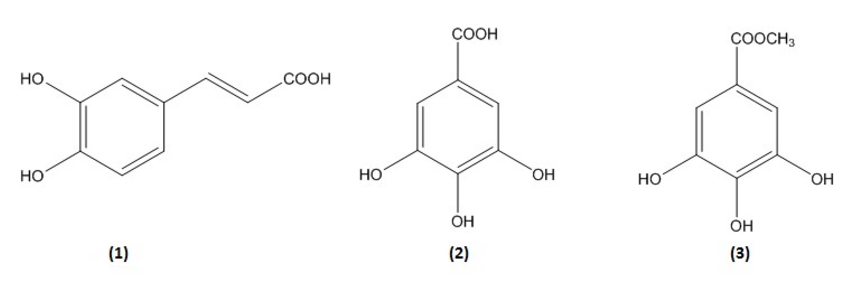
2.1. Isolation and Identification of Secondary Metabolites

The total methanol extract (TME) of freeze-dried B. oleracea (26 g) was subjected to various and repeated techniques of chromatography to yield three pure compounds: caffeic acid (1) [19,20], gallic acid (2) [21], and methyl gallate (3) [22] (Figure 1). The structures were elucidated via the inspection of 1D-and 2D-NMR spectroscopic data, including 1H, 13C, DEPT, HMQC, and HMBC (Supplementary Materials). The results were compared with those available in the literature.
Caffeic acid (1): 1H NMR (DMSO-d6, 400 MHz) δ: 7.43 (d, J= 15.9 Hz, H-7), 7.04 (1H, s, H-2), 6.96 (1H, d, J = 7.9 Hz, H-6), 6.76 (1H, d, J = 7.9 Hz, H-5), 6.18 (1H, d, J = 15.8 Hz, H-8); 13C NMR (DMSO-d6, 100 MHz) δ: 125.66 (C-1), 115.71 (C-2), 145.5 (C-3), 148.08 (C-4), 115.08 (C-5), 121.13 (C-6), 144.55 (C-7), 114.58 (C-8), 167.88 (C-9).
Gallic acid (2): 1H NMR (400 MHz, DMSO-d6): 6.94 (2H, s, H-2, H-6); 13C NMR (100 MHz, DMSO-d6): δ 120.21 (C-1), 108.7 (C-2, C-6), 137.94 (C-4), 145.35 (C-3, C-5), 167.46 (C-7).
Methyl gallate (3): 1H NMR (400 MHz, DMSO-d6): δ 6.96 (2H, s, H-2, H-6), 3.75 (3H, s, COOCH3); 13C NMR (100 MHz, DMSO-d6): δ 119.28 (C-1), 108.48 (C-2, C-6), 138.37 (C-4), 145.53 (C-3, C-5), 166.30 (C-7), 51.54 (CH3).

2.2. Determination of the Total Phenolic Content (TPC)

The TPC in TME was calculated using the regression equation: y = 0.169x − 0.0053, R² = 0.9578. The quantity of phenolic constituents was expressed as the equivalent milligrams of standard gallic acid per gram of dried plant extract (mg GAE/g). The TPC was 6.47 mg GAE/g of dry extract.

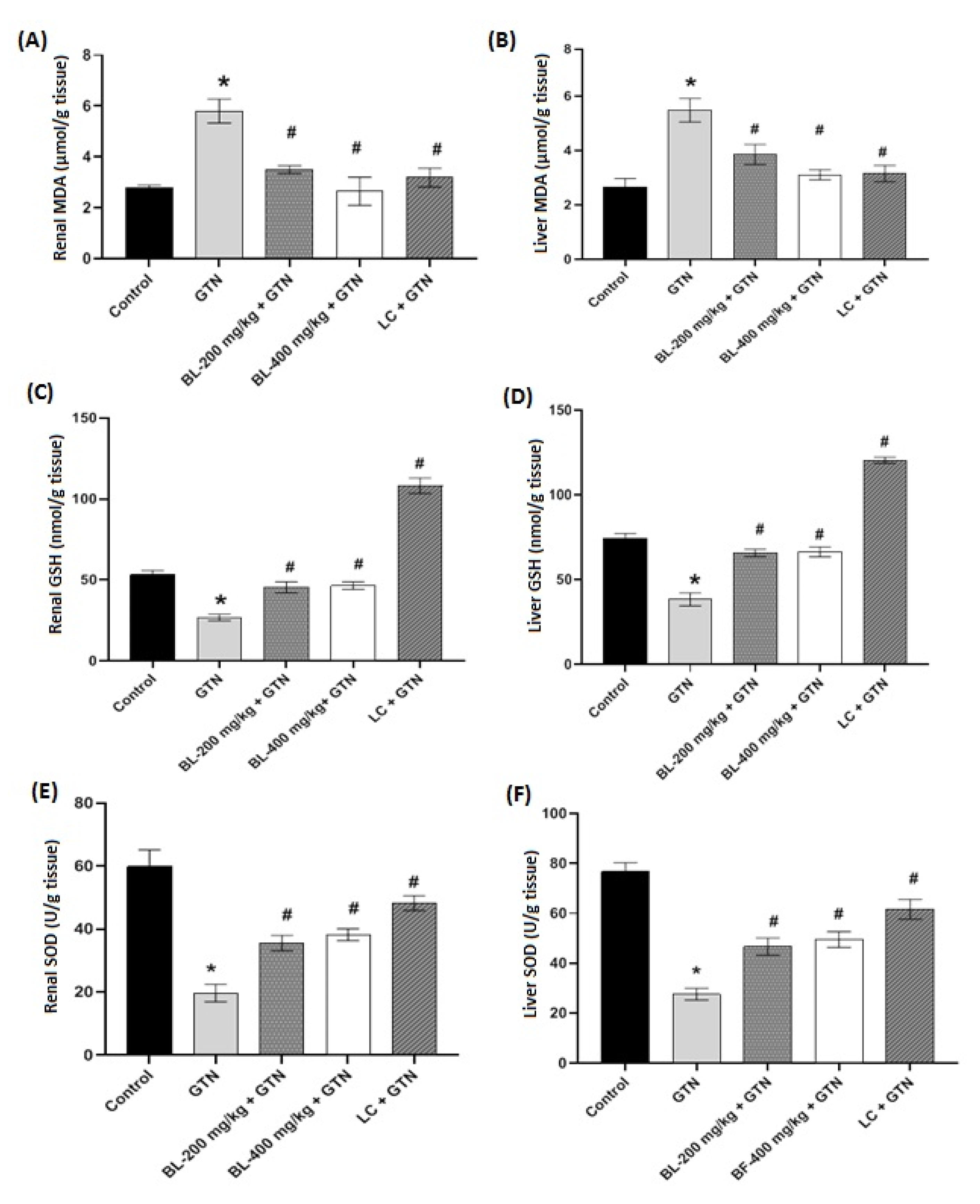
2.3. Effect of Broccoli Leaf Extract on Oxidative Stress Markers

There was a significant difference (p < 0.01) between the renal MDA levels of the control and GTN-treated mice, indicating that GTN produced a profound increase. However, considering broccoli leaf extract (BL) plus GTN-treated groups, including L-cysteine (LC) + GTN, all significantly (p < 0.001) reduced MDA levels were produced by GTN. In addition, there was no difference between the BL+GTN and LC+GTN treatments compared to the control. Here, BL 400 mg/kg + GTN brought the MDA level to a similar value as that in the control group (Figure 2A). On the other hand, the MDA levels in liver tissue samples exhibited similar trends as the renal MDA levels, as shown in Figure 2B. However, the treatment groups BL 400 mg/kg + GTN and LC + GTN resulted in similar reductions compared to GTN alone. The reduction in MDA indicates an alleviation of tissue lipid peroxidation by BL. Figure 2C presents the renal GSH content, and it shows that the GTN treatment significantly (p < 0.001) reduced the renal tissue GSH content. Although the BL (200 and 400 mg/kg) + GTN groups exhibited significant (p < 0.01) increases in the GSH content, they were lower compared to the control. The LC+GTN treatment produced a highly significant (p < 0.001) increase in the GSH content, confirming that it is a precursor of GSH. The liver tissue GSH contents showed similar profiles between GTN induction and treatment with BL. Comparably, treatment with L-cysteine also increased the GSH content much more than the control (Figure 2D). The renal and liver tissue superoxide dismutase (SOD) activity levels are presented in Figure 2E,F, respectively. The results show that there was a significant (p < 0.001) reduction in the activity of renal tissue SOD with the treatment of GTN compared to the control. However, all other treatment groups significantly increased the activity levels of both renal and liver tissue SOD compared to GTN treatment. In both cases, the LC + GTN treatment showed a better profile in increasing the SOD activity levels. Although the BL (200 and 400 mg/kg) + GTN treatment groups produced significant increases compared to GTN alone, they did not increase the SOD activity close to that in the control group.

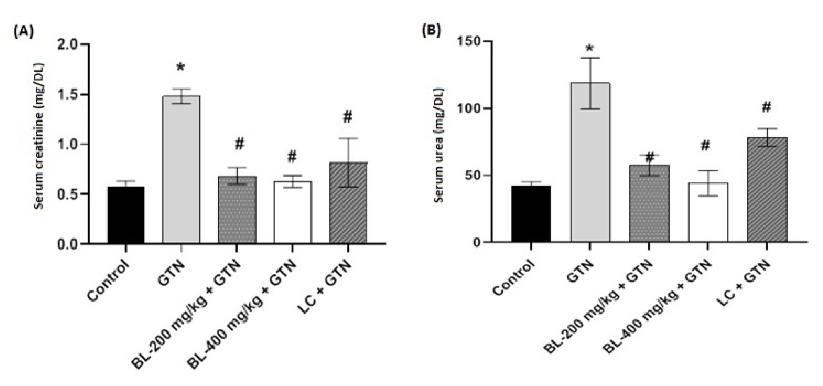
2.4. Effects of Broccoli Leaf Extract on Serum Creatinine and Urea

The serum creatinine and urea levels were evaluated, and the results are presented in Figure 3A,B. Our results showed that GTN significantly increased (p < 0.001) the level of serum creatinine. Treatments with BL (200 and 400 mg/kg) after the administration of GTN decreased the serum creatinine levels, similar to the control group, in a significant manner (p < 0.01). Overall, all treatment groups after GTN induction showed markedly reduced serum creatinine levels (Figure 3A). In the case of serum urea, treatments with BL (200 and 400 mg/kg) after the administration of GTN significantly decreased (p < 0.001) its level. The effects of BL produced levels that were very similar to the control, supporting the antioxidant activity potential of the extracts (Figure 3B).

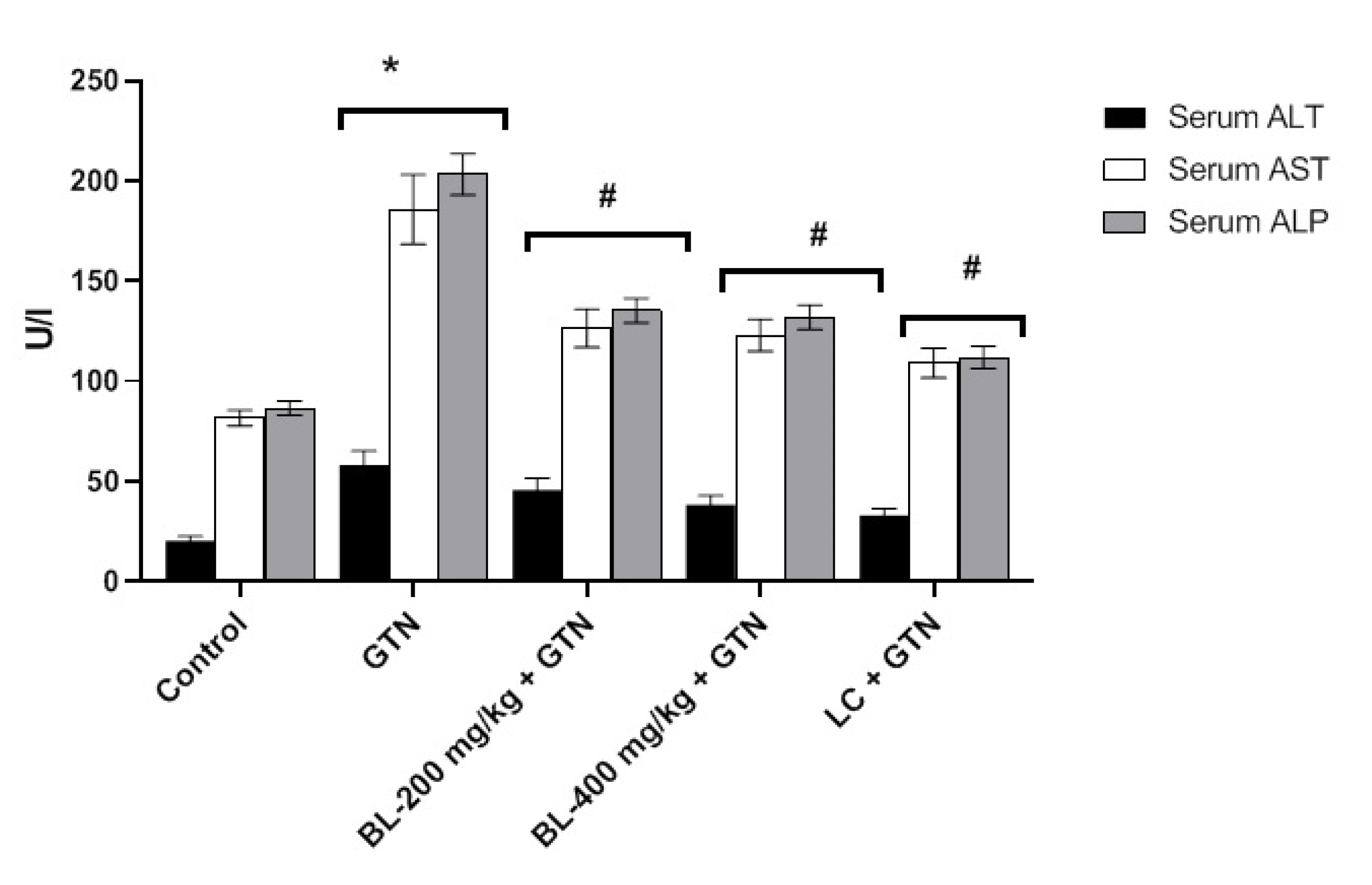
2.5. Effect of Broccoli Leaf Extract on Liver Enzymes

Figure 4 shows the levels of the liver enzymes AS, ALT, and ALP under the GTN, BL (200 and 400 mg/kg), and LC treatments after the administration of GTN. The results indicated that GTN increased the levels of ALT, AST, and ALP compared to the control. Although the treatments with BL and LC in the presence of GTN produced a significant difference (p < 0.01) compared to GTN alone, the control enzyme levels were lower in all groups.

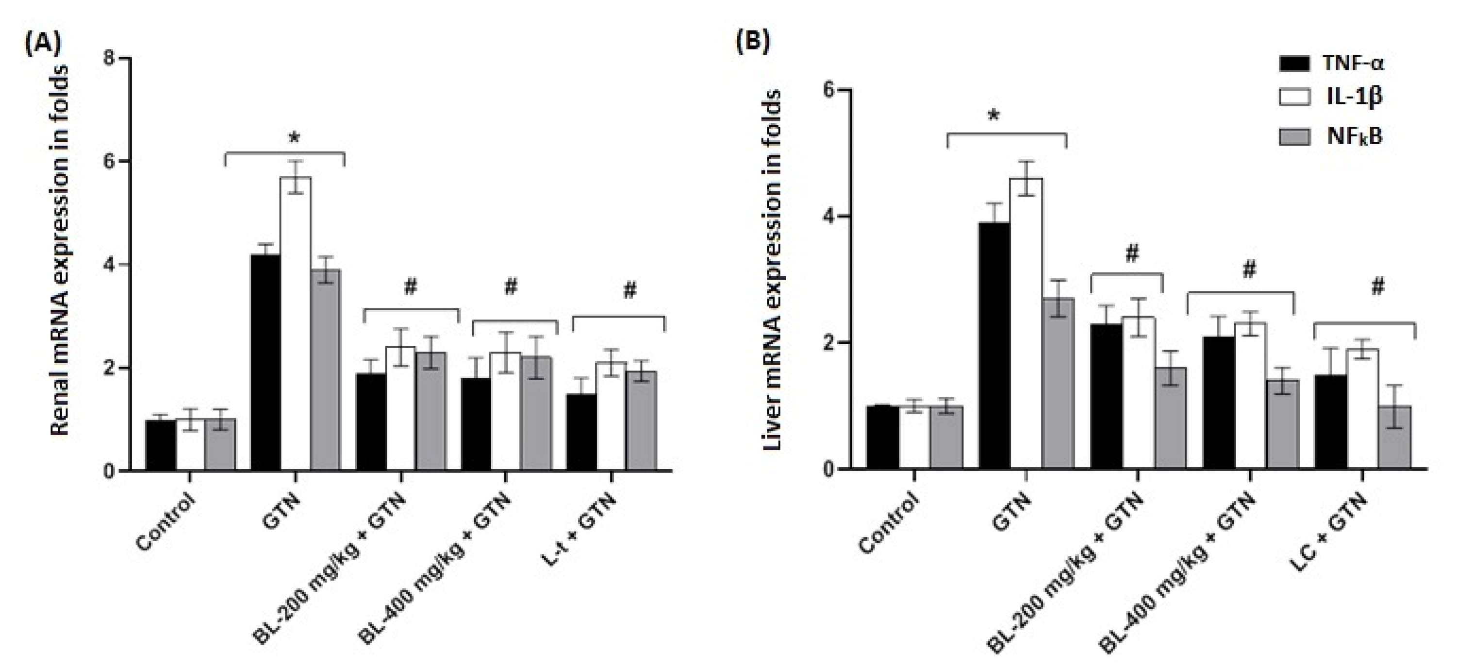
2.6. Effects of Broccoli Leaf Extract on Renal and Liver mRNA Expression Levels (TNF-α, IL-1β, and NFkB)

The renal and liver mRNA expression levels of TNF-α, IL-1β, and NFkB were evaluated, and the results are presented in Figure 5A,B. Figure 5A shows that the renal mRNA expression levels of TNF-α, IL-1β, and NFkB were elevated significantly (p < 0.001) in the GTN-treated group compared to the control and showed significant decreases in treatments with BL (200 and 400 mg/kg) + GTN. In Figure 5B, the liver mRNA expression levels of TNF-α, IL-1β, and NFkB showed a similar pattern to that of the renal mRNA cytokine expression levels. However, the results also indicated that IL-1β expression was higher in all GTN treatment groups compared to the control for both the renal and liver mRNA expression levels. Overall, the results indicated that treatments with BL (200 and 400 mg/kg) after GTN induction also appeared to reduce the mRNA expression levels of TNF-α, IL-1β, and NFkB in a similar fashion in both tissue types.

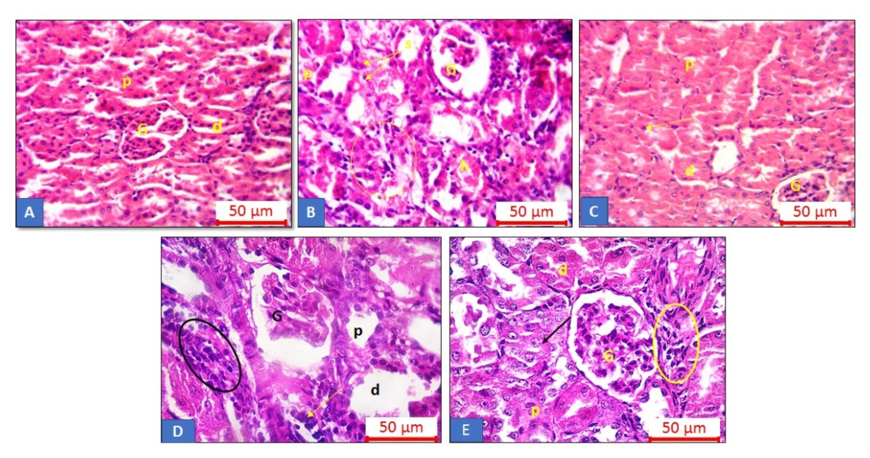
2.7. Histopathological Analysis of Renal Tissue

The control group showed normal lobular organization of the renal cortex. Glomerular capillary tufts occupying the renal glomeruli were clearly seen. The proximal convoluted tubules (PCTs) had a narrow lumen and a highly acidophilic cytoplasm. The distal convoluted tubules (DCTs) had a wider lumen and less acidophilic cytoplasm with vesicular nuclei. The GTN treatment group displayed a disturbed lobular architecture with inflammatory cell infiltration. Atrophic glomeruli with dilated PCTs and DCTs were noticed among the sections. The more numerous apoptotic cells lining the tubules and intra-tubular debris with a hyalin cast were also detected in this group. The BL 200 mg/kg + GTN group showed amelioration of the renal histological structure but with a less disturbed lobular architecture, while dilated tubules with few apoptotic cells and inflammatory cells were frequently seen among the sections. The BL 400 mg/kg + GTN group showed amelioration of the renal histological structure but with focal areas of dilated tubules and inflammatory cell infiltration (Figure 6). Meanwhile, the LC group exhibited restoration of the normal architecture; the renal tissue approached the normal structure, but a few apoptotic cells were still seen.

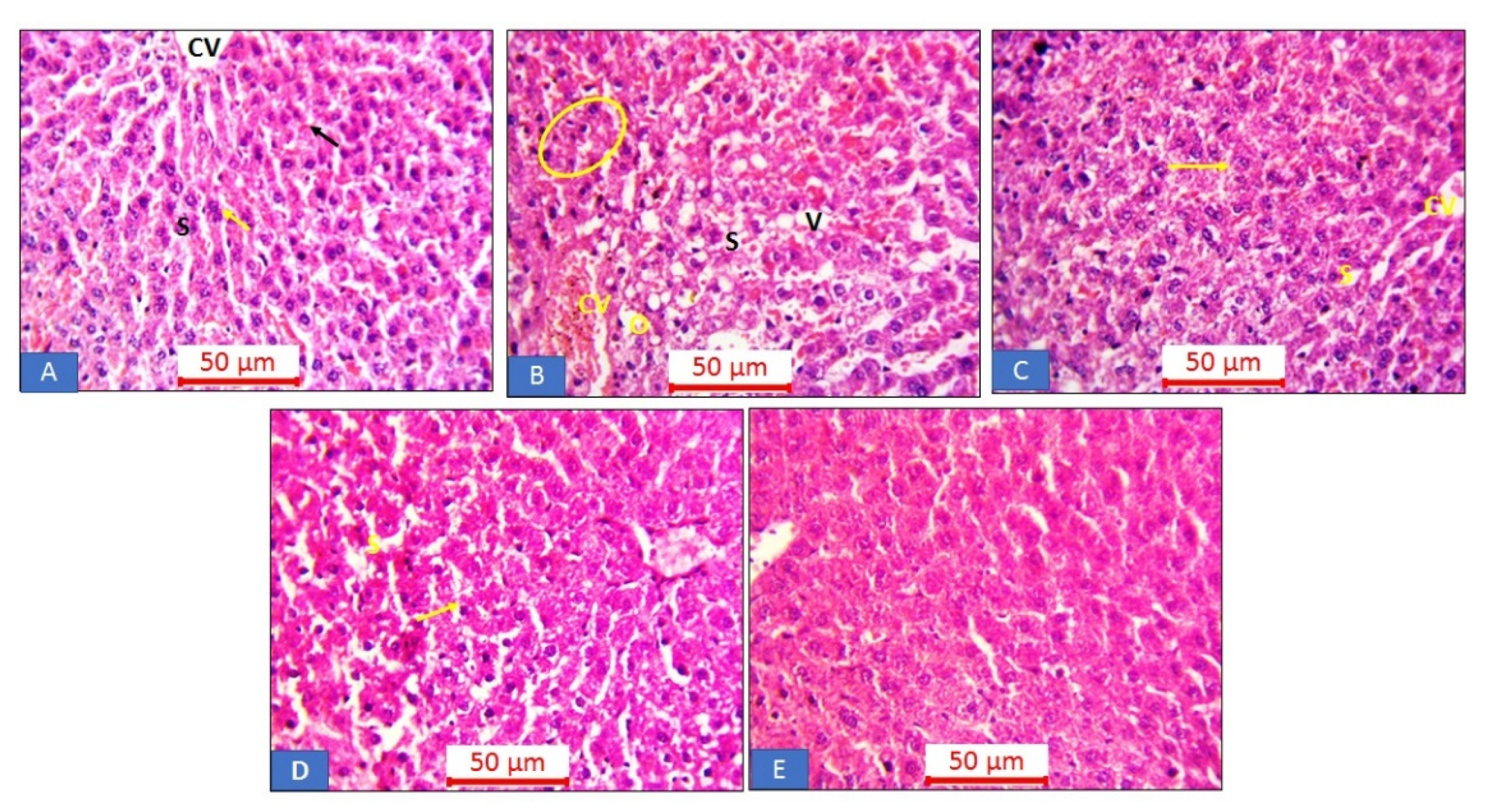
2.8. Histopathological Analysis of Liver Tissue

The control group showed normal hepatic architecture with a central vein and cords of hepatocytes with a granular cytoplasm and large rounded vesicular nuclei, separated by the blood sinusoids. Binucleated hepatocytes were seen. On the other hand, the GTN group showed a disturbed hepatic architecture. Dilated central veins and blood sinusoids were observed. Edema and inflammatory cell infiltrations were frequently seen among the sections. The BL 200 mg/kg + GTN group showed slight dilatation of blood sinusoids with a few apoptotic cells, while the BL 400 mg/kg + GTN group displayed hepatic tissue nearly approaching the normal structure. Furthermore, the LC group exhibited a more or less normal structure, central vein, blood sinusoids, and hepatocytes (Figure 7).

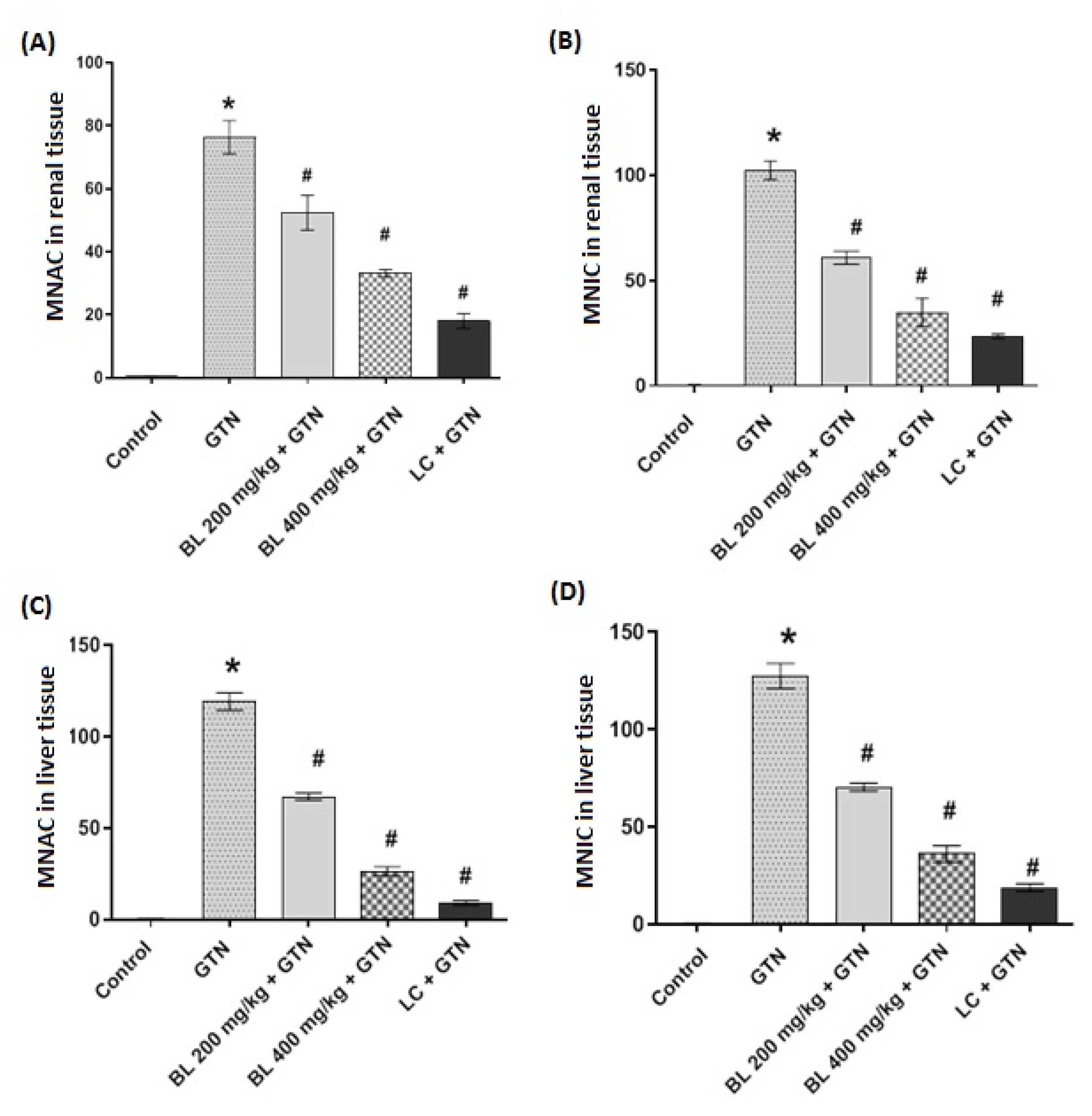
2.9. Morphometric Analysis of Renal and Hepatic Tissues

Figure 8A–D show the morphometric results of the renal and hepatic sections, indicating the mean number of inflammatory cells and the mean number of apoptotic cells. The results of the morphometric analysis showed that GTN significantly increased (p < 0.01) the numbers of apoptotic and inflammatory cells in both the renal and liver tissues. As shown, BL 200 mg/kg + GTN reduced these numbers significantly (p < 0.01). However, BL 400 mg/kg + GTN showed a better profile in reducing both apoptotic and inflammatory cell changes induced by GTN in both the renal and liver tissues. The overall results correlate with the antioxidant and anti-inflammatory activities observed for other groups.

3. Discussion

The present study evaluated the antioxidant, anti-inflammatory, and anti-apoptotic potential of broccoli leaf extract (BL (200 and 400 mg/kg) + GTN) in GTN-induced renal and liver toxicity. GTN is an aminoglycoside antibiotic employed for the treatment of serious Gram-negative bacterial infections with a well-documented nephrotoxic and hepatotoxic adverse effect profile [23,24]. The documented evidence shows that GTN can induce oxidative stress and apoptosis in both the kidneys and liver [25]. Our results hereby confirm that GTN induces oxidative adverse effects by decreasing the antioxidant status and increasing the expression levels of proinflammatory cytokines. Therefore, in mitigating GTN’s adverse effects, the present study investigated the potential of broccoli leaf extract. Broccoli is usually consumed as a fresh vegetable, and its antioxidant activity is ranked second among ten common vegetables [26,27]. Chemically, many reports have shown the presence of various constituents in BL [28,29], and based on that the current study demonstrated the isolation and identification of three major phenolic compounds in the TME of BL. Based on 1H, 13C, DEPT, HMQC, and HMBC spectral data, the three molecules were identified as caffeic acid, gallic acid, and methyl gallate. The current findings regarding the TPC align with the reported values [17,30]. The higher TPC was also noticed through the abundance of the abovementioned phenolic compounds from the TME. The presence of such phenolic acids and their potential to ameliorate GTN-induced oxidative stress [31] provoked us to evaluate the effects of the leaf by-product against GTN-induced toxicity. Biologically, the effect of the broccoli leaf extract was assessed against GTN-induced oxidative stress in rat kidneys and liver. Water extracts of broccoli were found to possess profound antioxidant activity according to the report by Jang et al. [32]. Our results showed that BL (200 and 400 mg/kg) treatments significantly reduced the lipid peroxidation (MDA) induced by GTN in both the kidney and liver tissues of experimental rats. In addition, the renal and liver GSH and SOD activity levels were markedly increased by the treatments with BL (200 and 400 mg/kg) extract in the presence of GTN exposure. The aforementioned observation in the present study is similar to that in the studies by Raeeszadeh et al. [33] and Lei et al. [34]. They indicated in their reports that broccoli extracts alleviated drug-induced oxidative stress in experimental animals. The documented evidence shows that GTN increases the serum levels of creatinine and urea [35]. The results of the present study showed that BL (200 and 400 mg/kg) treatments reduced the influence of GTN in both serum creatinine and serum urea. In this regard, we observed that the broccoli leaf extract showed better activity compared to the LC treatment. Regarding liver enzymes (ALT, AST, and ALP), GTN significantly elevated the activity levels of these enzymes. However, as shown in the results, the treatment with BL (200 and 400 mg/kg) significantly reduced the levels of these enzymes. Hence, from this observation, it does appear that the broccoli extract has the potential to alleviate GTN-induced liver damage. Interestingly, our results appear to agree with a similar report documented by Raeeszadah et al. [33]. They examined the mitigating potential of BL in arsenic-induced rat poisoning. ALT is an indicator of necrosis; therefore, elevated ALT and AST levels, according to a previous finding [36], indicate acute and chronic hepatic necrosis. Inflammatory cytokines, such as TNF-α and IL-1β, and signaling pathway activation, such as for NFkB, are elevated by GTN according to previous report and could lead to necrosis and apoptosis. The findings of the present study are in total agreement with the authors, who reported that the induction of inflammatory cytokines (TNF-α and IL-1β) follows the activation of NFkB [37]. Therefore, the activation of TNF-α by GTN leads to the subsequent activation of other proinflammatory pathways, which include NFkB. In effect, the observed oxidative and inflammatory stress in both the kidneys and liver in this study could be related to the production of these inflammatory cytokines and the activation of their pathways. Our results show that the BL (200 and 400 mg/kg) treatments reduced the mRNA expression levels of TNF-α, IL-1β, and NFkB in both the kidney and liver tissues. Therefore, the findings suggest that BL could possess both anti-inflammatory and antioxidant activities. Our results are similar to those in previous documented studies that reported that broccoli extract inhibited the production of proinflammatory cytokines and furthermore suppressed the NFkB signaling pathway [38]. Parallel histological examination studies revealed that GTN treatment caused inflammation, necrosis, and apoptosis of both the kidneys and liver in rats [39,40]. The histological pictures showed a dilated central vein, blood sinusoids, edema, and inflammatory cell infiltration. Therefore, increasing the antioxidant status might be an important strategy in preventing organ damage due to GTN administration. Our results revealed that the treatments with BL (200 and 400 mg/kg) ameliorated the histological renal and liver structures and reduced inflammatory cell infiltration. The analysis of the extent of apoptotic and inflammatory activities showed that the GTN treatment induced increased levels of inflammatory cells as well as apoptosis in both the renal and liver tissues. Conversely, the treatments with BL (200 and 400 mg/kg) decreased the inflammatory and apoptotic activities induced by GTN. The present results clearly indicate that inflammation and apoptosis triggered by GTN could be attenuated with BL treatment.

4. Materials and Methods

4.1. General Procedures and Chemicals

NMR spectra were obtained using an Avance 400 NMR spectrometer (1H-NMR: 400 MHz and 13C-NMR: 100 MHz, Bruker, Switzerland) and a freeze dryer (Millrock technology, New York, NY, USA). Silica gel column chromatography (SCC) was carried out on silica gel 60 (Sigma–Aldrich, Darmstadt, Germany) and Diaion-HP-20 (Sigma–Aldrich, Darmstadt, Germany). Pre-coated silica gel 60 F254 plates (Sigma–Aldrich, Darmstadt, Germany) were used for thin-layer chromatography (PTLC). The visualization was performed using 10% vanillin–sulfuric acid in ethanol with a hotplate (150 °C). Assay kits for MDA, SOD, and GSH were procured from Sigma–Aldrich, (St. Louis, MO, USA). ALT, AST, and ALP kits were also obtained (Quimica Clinica Aplicada S.A, Amposta, Spain). All other chemicals were of analytical grade.

4.2. Plant Material

Broccoli (Brassica oleracea L. variety: italica) leaves were collected during October 2021 from a local market in Al-hasa, Eastern Province, Saudi Arabia. The middle part of the midrib was removed. The plant was kindly identified by Eng. Mamdouh Shokry, Director of El-Zohria Botanical Garden, Giza, Egypt. A voucher specimen (BOi-Oct-2021) of the leaves was deposited in the Department of Pharmaceutical Sciences, College of Clinical Pharmacy, King Faisal University, Al-Ahsa, Saudi Arabia.

4.3. Extraction and Isolation of Secondary Metabolites

Coarse freeze-dried powdered leaves of B. oleracea (1 kg) were thoroughly extracted three times with 15 L of 70% methanol for one week at 25 °C with regular stirring and storage in darkness. The resulting extracts were evaporated to dryness using a rotary evaporator over a temperature range of 40–45 °C to yield a dark green extract weighing 89.2 g that was labeled as the total methanol extract (TME). The TME (70 g) was defatted using n-hexane, whereby the TME was suspended in 500 mL of deionized water partitioned with n-hexane (5 times each with 3 L) [41]. The resulting n-hexane fractions were concentrated to dryness to yield 20 g. The remaining mother liquor (50 g) was subjected to column chromatography using Diaion HP-20 (500 g) as the stationary phase and was then successively eluted with water to remove sugars and carbohydrates followed by washing with 30%, 50%, and 100% methanol to yield the following fractions: water (21 g), 30% methanol (named BOI, 6.9 g), 50% methanol (named BOII, 8.3 g), and 100% methanol (named BOIII, 13.8 g) (4 g). With the guidance of TLC patterns, the BOII fraction (8.3 g) was subjected to SCC (500 g, using 5 L of chloroform–methanol–water (ratio of 15:6:1) as the mobile phase, followed by repeated PTLC to yield the pure compounds (compound (1) (12 mg), compound (2) (26 mg), and compound (3) (14 mg)).

4.4. Determination of the Total Phenolic Content (TPC)

TPC was evaluated using the Folin–Ciocalteu index protocol described by Hany et al., with modifications [42]. A stock solution with a concentration of 1 mg/mL of TME was established in methanol. The Folin–Ciocalteu reagent (0.5 mL) and 6 mL of double-distilled water were sequentially added to 0.1 mL of stock solution of each fraction. Later, 1.5 mL of a 20% sodium carbonate solution was added to obtain a final volume of 10 mL. The reaction was completed over 2 h at 25 °C. Then, the absorbance levels of different mixtures were measured at 760 nm. A calibration curve of gallic acid (standard) was prepared using serial dilutions (0.1, 0.2, 0.3, 0.4, and 0.5 mg/mL in distilled water).

4.5. Animals

Thirty adult male Wistar rats weighing 220–250 g (10–12 weeks) were obtained from Nahda University, Beni Suef (NUB) animal care facility. The animals were housed under standard laboratory conditions and were maintained on a 12-h light/dark cycle to allow acclimatization for 2 weeks. The animals were allowed free access to food and water and were maintained at temperatures of 25–27 °C. The study protocol was approved by the Commission on the Ethics of Scientific Research, Faculty of Pharmacy, Minia University, registration number ES17/2020. The animals were then randomly assigned to five treatment groups, consisting of six rats per group.

4.6. Experimental Protocol

The rats were divided into 5 (five) treatment groups of 6 rats per group. The animals were treated for 10 days. Group I, the untreated control, was given 0.5 mL/kg phosphate-buffered saline (PBS) solution. The other groups were group II (GTN 100 mg/kg), group III (BL 200 mg/kg + GTN), group IV (BL-400 mg/kg + GTN), and group V (LC 1 mmole/kg + GTN). The experimental animals were treated daily for 10 days. GTN was administered via i.p, while broccoli extracts and L-cysteine were given orally using an oral tube. At the end of the study, the rats were euthanized by cervical dislocation under ether anesthesia. The ether was administered using the “open-drop” method with an ether-impregnated cotton ball in a bell jar for induction, followed by inhalation via a simple face cone. As stipulated by the institutional guidelines for the use of animals, we used about five drops, corresponding to 3–4 mL. The blood was collected via cardiac puncture. Then, the rats were euthanized via decapitation. The kidneys and liver were carefully dissected. The tissue samples were thoroughly rinsed in phosphate-buffered saline (PBS) to remove excess blood before being stored at −85 °C for analysis. The collected samples were used to estimate biochemical markers of antioxidants such as MDA, GSH, and SOD. Additionally, the mRNA expression levels of TNF-α, IL-1β, and NFkB were investigated.

4.7. Biochemical Analysis

The kidney and liver samples were harvested and then washed in saline using an ice bath. These samples were homogenized in phosphate-buffered saline in a ratio of 1:10 (w/v) homogenates and then centrifuged at 13,000× g (4 °C) for 30 min; the supernatant was then collected to determine the levels of MDA, SOD, and GSH. The levels of MDA, as a marker for lipid peroxidation, were determined using the thiobarbituric-acid-reactive substances method by determining the levels of thiobarbituric-acid-reactive substances, as earlier described by Emeka et al. [43]. A phosphoric acid solution (1%) and a measured volume of thiobarbituric acid were added to each sample homogenate, which was then incubated at 95 °C for 1 h. The concentration of the subsequent thiobarbituric-acid-reactive substances was then measured spectrophotometrically at 535 nm. SOD assays were performed according to the method described by Weydert and Cullen [44]. Xanthine–xanthine oxidase was used to generate O2 radicals, and the subsequent nitro blue tetrazolium (NBT) reduction was used as an indicator of O2 radical production. As SOD competes with NBT for the generated O2 radicals, the percent reduction in NBT was calculated from the amount of SOD present. The GSH levels were determined by following the manufacturer’s protocol, which can be described briefly as a colorimetric reaction of 5,5′-dithiobis(2-nitrobenzoic acid). Kidney and liver GSH contents were measured at 412 nm using a standard curve plot.

4.8. Serum Analysis of ALT, AST, ALP, Urea, and Creatinine Levels

Blood samples were harvested and centrifuged at 2000× g for 15 min using a refrigerated centrifuge to obtain serum samples from the cells. Subsequently, the ALT, AST, ALP, urea, and creatinine levels were determined in the serum using routine colorimetric methods on a Roche modular auto-analyzer (Roche modular auto-analyzer, Tokyo, Japan) [45].

4.9. mRNA Extraction and Quantification

Fresh portions of kidney and liver samples were dissected and washed with ice-cold PBS, and then the cell-free supernatant was removed. The total RNA was extracted using the modified Trizol (Thermo Fisher, San Jose, CA, USA) method. The extracted RNA was quantified using a NanoDrop system, and 300 ng/target was used for the cDNA preparation against inflammatory mRNA markers using the MQ basic cDNA synthesis kit (Molequle-on, Takara, Kusatsu, Shiga, Japan) [46]. The forward and reverse primers for TNF-α, IL-1β, NFkB, and GAPDH are shown in Table 1. The expression levels of the cDNA products were quantified via quantitative real-time RT-PCR (qRT-PCR) using the SYBR master mix method. An applied biosystems Vii7A PCR system was used, and the mRNA was quantified using the ΔΔcT method. The house keeping target GAPDH was used as an internal control for the normalization of mRNA expression.

4.10. Kidney and Liver Histopathological Examinations

Kidney tissue samples from each rat were harvested; then, the specimens were fixed in Blouin’s Solution, dehydrated in ascending degrees of alcohol, cleared with xylene, rapidly embedded in paraffin wax, and sectioned (5 µm thickness). The sections were stained with H&E [46]. The prepared slides were then examined under light microscopy (Olympus CX23LEDRFS1, Olympus, Tokyo, Japan) to study the histopathological changes.

4.11. Morphometric Analysis of Renal and Hepatic Tissues

A morphometric estimation using Leica QWin 500 image analysis software (Leica Microsystems, Wetzlar, Germany) was performed for evaluation with power ×400. These parameters were detected in 10 non-overlapping sections from each slide from each rat to calculate the mean number of inflammatory cells and the mean number of apoptotic cells [47].

4.12. Statistical Analysis

The results obtained here are expressed as means ± SD and were analyzed using Graph Pad Prism software version 8.2 (San Diego, CA, USA). Comparisons between the control and treatment groups were performed using one-way analysis of variance, and the differences between the groups were measured using Tukey’s multiple comparisons test. Statistical significance was taken as p < 0.01.

5. Conclusions

From the foregoing, this study has demonstrated the high value of the TPC and the isolation and identification of three pure phenolic compounds in the TME of broccoli leaf extract, namely, caffeic acid, gallic acid, and methyl gallate. The results showed that the broccoli leaf extract treatment abolished GTN-mediated lipid peroxidation by reducing MDA formation, as well as restoring GSH and SOD levels in both renal and liver tissues. Moreover, in both tissues, serum creatinine and urea levels were returned to normal with reduced liver enzyme levels elevated by GTN administration. The histological examinations also revealed ameliorated renal and liver tissues following treatment with different concentrations of broccoli leaf extract in a dose-dependent manner. The inflammatory and apoptotic processes induced by GTN were mitigated, as evidenced by the reduction in the mRNA expression levels of TNF-α, IL-1β, and NFkB, indicating that the broccoli extract has high potential for restoring normal cell function in the presence of GTN-induced oxidative stress and is able to prevent both inflammatory and apoptotic cellular damage. Based on these findings, we recommend that future clinical studies utilize broccoli agro-waste leaf for human consumption and as a source of phenolics for nutraceutical and pharmaceutical purposes.

Supplementary Materials

The following are available online at https://www.mdpi.com/article/10.3390/app12146903/s1: Supplementary S. The 1D- and 2D-NMR spectroscopic data for caffeic acid (1), gallic acid (2), and methyl gallate (3).

Author Contributions

H.E.K., conceptualization, funding acquisition, methodology, resources, writing—review and editing, writing—original draft preparation, supervision; M.F.A., methodology, writing—review and editing, writing—original draft preparation; P.M.E., methodology, writing—review and editing, writing—original draft preparation; L.I.B.-E., methodology, writing—review and editing, writing—original draft preparation; S.M.N.A.H., methodology, writing—original draft preparation; K.A.A., methodology, writing—review and editing; N.M.A.-W., methodology; A.-S.F.A., methodology; A.F.A., methodology; K.M., conceptualization, writing—review and editing; A.H.I.S., methodology, writing—review and editing, writing—original draft preparation. All authors have read and agreed to the published version of the manuscript.

Funding

This research was funded by the Deanship of Scientific Research (DSR), King Faisal University, Saudi Arabia (grant number 1811002).

Institutional Review Board Statement

The study was conducted in accordance with the Declaration of Helsinki, and approved by the Commission on the Ethics of Scientific Research, Faculty of Pharmacy, Minia University, registration number ES17/2020.

Informed Consent Statement

Not applicable.

Data Availability Statement

The data presented in this study are available on request from the corresponding author.

Acknowledgments

The authors thank the Deanship of Scientific Research (DSR), King Faisal University, Saudi Arabia, for funding this research group project (grant number 1811002) and the College of Clinical Pharmacy, King Faisal University.

Conflicts of Interest

The authors declare no conflict of interest.

References

  1. Negrette-Guzmán, M.; Huerta-Yepez, S.; Medina-Campos, O.N.; Zatarain-Barrón, Z.L.; Hernández-Pando, R.; Torres, I.; Tapia, E.; Pedraza-Chaverri, J. Sulforaphane attenuates gentamicin-induced nephrotoxicity: Role of mitochondrial protection. Evid. Based Complement. Alternat. Med. 2013, 2013, 135314. [Google Scholar] [CrossRef] [PubMed] [Green Version]
  2. Lopez-Novoa, J.M.; Quiros, Y.; Vicente, L.; Morales, A.I.; Lopez-Hernandez, F.J. New insights into the mechanism of aminoglycoside nephrotoxicity: An integrative point of view. Kidney Int. 2011, 79, 33–45. [Google Scholar] [CrossRef] [PubMed] [Green Version]
  3. Udupa, V.; Prakash, V. Gentamicin induced acute renal damage and its evaluation using urinary biomarkers in rats. Toxicol. Rep. 2019, 6, 91–99. [Google Scholar] [CrossRef] [PubMed]
  4. Ali, B.H.; Al Za’abi, M.; Blunden, G.; Nemmar, A. Experimental gentamicin nephrotoxicity and agents that modify it: A mini-review of recent research. Basic Clin. Pharmacol. Toxicol. 2011, 109, 225–232. [Google Scholar] [CrossRef]
  5. Bledsoe, G.; Shen, B.; Yao, Y.-Y.; Hagiwara, M.; Mizell, B.; Teuton, M.; Grass, D.; Chao, L.; Chao, J. Role of tissue kallikrein in prevention and recovery of gentamicin-induced renal injury. Toxicol. Sci. 2008, 102, 433–443. [Google Scholar] [CrossRef] [Green Version]
  6. Koyner, J.L.; Ali, R.S.; Murray, P.T. Antioxidants. Nephron Exp. Nephrol. 2008, 109, e109–e117. [Google Scholar] [CrossRef]
  7. Juan, S.-H.; Chen, C.-H.; Hsu, Y.-H.; Hou, C.-C.; Chen, T.-H.; Lin, H.; Chu, Y.-L.; Sue, Y.-M. Tetramethylpyrazine protects rat renal tubular cell apoptosis induced by gentamicin. Nephrol. Dial. Transplant. 2007, 22, 732–739. [Google Scholar] [CrossRef] [Green Version]
  8. Abouzed, T.K.; Sherif, E.A.E.; Barakat, M.E.S.; Sadek, K.M.; Aldhahrani, A.; Nasr, N.E.; Eldomany, E.; Khailo, K.; Dorghamm, D.A. Assessment of gentamicin and cisplatin-induced kidney damage mediated via necrotic and apoptosis genes in albino rats. BMC Vet. Res. 2021, 17, 350. [Google Scholar] [CrossRef]
  9. Ali, F.E.; Hassanein, E.H.; Bakr, A.G.; El-Shoura, E.A.; El-Gamal, D.A.; Mahmoud, A.R.; Abd-Elhamid, T.H. Ursodeoxycholic acid abrogates gentamicin-induced hepatotoxicity in rats: Role of NF-κB-p65/TNF-α, Bax/Bcl-xl/Caspase-3, and eNOS/iNOS pathways. Life Sci. 2020, 254, 117760. [Google Scholar] [CrossRef]
  10. Arjinajarn, P.; Chueakula, N.; Pongchaidecha, A.; Jaikumkao, K.; Chatsudthipong, V.; Mahatheeranont, S.; Norkaew, O.; Chattipakorn, N.; Lungkaphin, A. Anthocyanin-rich Riceberry bran extract attenuates gentamicin-induced hepatotoxicity by reducing oxidative stress, inflammation and apoptosis in rats. Biomed. Pharmacother. 2017, 92, 412–420. [Google Scholar] [CrossRef]
  11. Ali, B. Agents ameliorating or augmenting experimental gentamicin nephrotoxicity: Some recent research. Food Chem. Toxicol. 2003, 41, 1447–1452. [Google Scholar] [CrossRef]
  12. Cekmen, M.; Otunctemur, A.; Ozbek, E.; Cakir, S.S.; Dursun, M.; Polat, E.C.; Somay, A.; Ozbay, N. Pomegranate extract attenuates gentamicin-induced nephrotoxicity in rats by reducing oxidative stress. Ren. Fail. 2013, 35, 268–274. [Google Scholar] [CrossRef] [Green Version]
  13. Nagai, J.; Takano, M. Molecular aspects of renal handling of aminoglycosides and strategies for preventing the nephrotoxicity. Drug Metab. Pharmacokinet. 2004, 19, 159–170. [Google Scholar] [CrossRef]
  14. Hashish, E.A.; Elgaml, S.A. Hepatoprotective and nephroprotective effect of curcumin against copper toxicity in rats. Indian J. Clin. Biochem. 2016, 31, 270–277. [Google Scholar] [CrossRef] [Green Version]
  15. Yarijani, Z.M.; Najafi, H.; Shackebaei, D.; Madani, S.H.; Modarresi, M.; Jassemi, S.V. Amelioration of renal and hepatic function, oxidative stress, inflammation and histopathologic damages by Malva sylvestris extract in gentamicin induced renal toxicity. Biomed. Pharmacother. 2019, 112, 108635. [Google Scholar] [CrossRef]
  16. Tlili, N.; Feriani, A.; Saadoui, E.; Nasri, N.; Khaldi, A. Capparis spinosa leaves extract: Source of bioantioxidants with nephroprotective and hepatoprotective effects. Biomed. Pharmacother. 2017, 87, 171–179. [Google Scholar] [CrossRef]
  17. Hwang, J.-H.; Lim, S.-B. Antioxidant and anticancer activities of broccoli by-products from different cultivars and maturity stages at harvest. Prev. Nutr. Food Sci. 2015, 20, 8. [Google Scholar] [CrossRef] [Green Version]
  18. Borowski, J.; Szajdek, A.; Borowska, E.J.; Ciska, E.; Zieliński, H. Content of selected bioactive components and antioxidant properties of broccoli (Brassica oleracea L.). Eur. Food Res. Technol. 2008, 226, 459–465. [Google Scholar] [CrossRef]
  19. Liu, Q.; Yu, J.; Liao, X.; Zhang, P.; Chen, X. One-step separation of antioxidant compounds from Erythrina variegata by high speed counter-current chromatography. J. Chromatogr. Sci. 2015, 53, 730–735. [Google Scholar] [CrossRef] [Green Version]
  20. Ibrahim, H.-I.M.; Darrag, H.M.; Alhajhoj, M.R.; Khalil, H.E. Biomolecule from Trigonella stellata from Saudi Flora to Suppress Osteoporosis via Osteostromal Regulations. Plants 2020, 9, 1610. [Google Scholar] [CrossRef]
  21. Park, J.-C.; Hwang, Y.-H.; Choi, D.-R.; Jung, D.-Y.; Park, J.-G.; Hur, J.-M.; Kim, S.-J.; Kim, S.-N.; Kim, M.-S. A triterpenoid glucoside and phenolic compounds from Rosa davurica. Nat. Prod. Sci. 2003, 9, 31–33. [Google Scholar]
  22. Chaudhuri, D.; Ghate, N.B.; Singh, S.S.; Mandal, N. Methyl gallate isolated from Spondias pinnata exhibits anticancer activity against human glioblastoma by induction of apoptosis and sustained extracellular signal-regulated kinase 1/2 activation. Pharmacogn. Mag. 2015, 11, 269. [Google Scholar]
  23. Banday, A.A.; Farooq, N.; Priyamvada, S.; Yusufi, A.N.; Khan, F. Time dependent effects of gentamicin on the enzymes of carbohydrate metabolism, brush border membrane and oxidative stress in rat kidney tissues. Life Sci. 2008, 82, 450–459. [Google Scholar] [CrossRef]
  24. Bulboacă, A.E.; Porfire, A.S.; Rus, V.; Nicula, C.A.; Bulboacă, C.A.; Bolboacă, S.D. Protective Effect of Liposomal Epigallocatechin-Gallate in Experimental Gentamicin-Induced Hepatotoxicity. Antioxidants 2022, 11, 412. [Google Scholar] [CrossRef]
  25. Babaeenezhad, E.; Nouryazdan, N.; Nasri, M.; Ahmadvand, H.; Sarabi, M.M. Cinnamic acid ameliorate gentamicin-induced liver dysfunctions and nephrotoxicity in rats through induction of antioxidant activities. Heliyon 2021, 7, e07465. [Google Scholar] [CrossRef]
  26. Kaur, C.; Kumar, K.; Anil, D.; Kapoor, H. Variations in antioxidant activity in broccoli (Brassica oleracea L.) cultivars. J. Food Biochem. 2007, 31, 621–638. [Google Scholar] [CrossRef]
  27. Sun, T.; Powers, J.R.; Tang, J. Evaluation of the antioxidant activity of asparagus, broccoli and their juices. Food Chem. 2007, 105, 101–106. [Google Scholar] [CrossRef]
  28. Li, Z.; Lee, H.W.; Liang, X.; Liang, D.; Wang, Q.; Huang, D.; Ong, C.N. Profiling of phenolic compounds and antioxidant activity of 12 cruciferous vegetables. Molecules 2018, 23, 1139. [Google Scholar] [CrossRef] [PubMed] [Green Version]
  29. Gupta, S.; Burman, S.; Nair, A.B.; Chauhan, S.; Sircar, D.; Roy, P.; Dhanwat, M.; Lahiri, D.; Mehta, D.; Das, R. Brassica oleracea Extracts Prevent Hyperglycemia in Type 2 Diabetes Mellitus. Prev. Nutr. Food Sci. 2022, 27, 50. [Google Scholar] [CrossRef] [PubMed]
  30. Liu, M.; Zhang, L.; Ser, S.L.; Cumming, J.R.; Ku, K.-M. Comparative Phytonutrient Analysis of Broccoli By-Products: The Potentials for Broccoli By-Product Utilization. Molecules 2018, 23, 900. [Google Scholar] [CrossRef] [PubMed] [Green Version]
  31. Ghaznavi, H.; Fatemi, I.; Kalantari, H.; Hosseini Tabatabaei, S.M.T.; Mehrabani, M.; Gholamine, B.; Kalantar, M.; Mehrzadi, S.; Goudarzi, M. Ameliorative effects of gallic acid on gentamicin-induced nephrotoxicity in rats. J. Asian Nat. Prod. Res. 2018, 20, 1182–1193. [Google Scholar] [CrossRef]
  32. Jang, H.W.; Moon, J.-K.; Shibamoto, T. Analysis and antioxidant activity of extracts from broccoli (Brassica oleracea L.) sprouts. J. Agric. Food Chem. 2015, 63, 1169–1174. [Google Scholar] [CrossRef]
  33. Raeeszadeh, M.; Karimi, P.; Khademi, N.; Mortazavi, P. The Effect of Broccoli Extract in Arsenic-Induced Experimental Poisoning on the Hematological, Biochemical, and Electrophoretic Parameters of the Liver and Kidney of Rats. Evid. Based Complement. Alternat. Med. 2022, 2022, 3509706. [Google Scholar] [CrossRef]
  34. Lei, P.; Zhao, W.; Pang, B.; Yang, X.; Li, B.-L.; Ren, M.; Shan, Y.-J. Broccoli sprout extract alleviates alcohol-induced oxidative stress and endoplasmic reticulum stress in C57BL/6 mice. J. Agric. Food Chem. 2018, 66, 5574–5580. [Google Scholar] [CrossRef]
  35. Al-Kuraishy, H.M.; Al-Gareeb, A.I.; Al-Nami, M.S. Irbesartan attenuates gentamicin-induced nephrotoxicity in rats through modulation of oxidative stress and endogenous antioxidant capacity. Int. J. Prev. Med. 2020, 11, 16. [Google Scholar]
  36. Roy, S.; Bhattacharya, S. Arsenic-induced histopathology and synthesis of stress proteins in liver and kidney of Channa punctatus. Ecotoxicol. Environ. Saf. 2006, 65, 218–229. [Google Scholar] [CrossRef]
  37. Jaikumkao, K.; Pongchaidecha, A.; Thongnak, L.-o.; Wanchai, K.; Arjinajarn, P.; Chatsudthipong, V.; Chattipakorn, N.; Lungkaphin, A. Amelioration of renal inflammation, endoplasmic reticulum stress and apoptosis underlies the protective effect of low dosage of atorvastatin in gentamicin-induced nephrotoxicity. PLoS ONE 2016, 11, e0164528. [Google Scholar]
  38. Bessler, H.; Djaldetti, M. Broccoli and human health: Immunomodulatory effect of sulforaphane in a model of colon cancer. Int. J. Food Sci. Nutr. 2018, 69, 946–953. [Google Scholar] [CrossRef]
  39. Kalkan, Y.; Kapakin, K.A.T.; Kara, A.; Atabay, T.; Karadeniz, A.; Simsek, N.; Karakus, E.; Can, I.; Yildirim, S.; Ozkanlar, S. Protective effect of Panax ginseng against serum biochemical changes and apoptosis in kidney of rats treated with gentamicin sulphate. J. Mol. Histol. 2012, 43, 603–613. [Google Scholar] [CrossRef]
  40. Ezema, A.S.; Ihedioha, T.E.; Num-Adom, S.M.; Ihedioha, J.I. Alterations in serum activity of hepatocellular enzymes, levels of liver function markers, and liver histology of dogs given high (nephrotoxic) doses of gentamicin. Comp. Clin. Path. 2022, 31, 115–122. [Google Scholar] [CrossRef]
  41. Khalil, H.E.; Ibrahim, H.-I.M.; Ahmed, E.A.; Emeka, P.M.; Alhaider, I.A. Orientin, a Bio-Flavonoid from Trigonella hamosa L., Regulates COX-2/PGE-2 in A549 Cell Lines via miR-26b and miR-146a. Pharmaceuticals 2022, 15, 154. [Google Scholar] [CrossRef]
  42. Khalil, H.E.; Al Ahmed, A. Phytochemical Analysis and Free Radical Scavenging Activity of Carthamus oxyacantha growing in Saudi Arabia: A Comparative Study. Int. J. Pharm. Sci. Rev. Res. 2017, 45, 51–55. [Google Scholar]
  43. Emeka, P.M.; Rasool, S.T.; Morsy, M.A.; Islam, M.I.H.; Chohan, M.S. Protective effects of lutein against vancomycin-induced acute renal injury in mice via upregulation of peroxisome proliferator-activated receptor gamma/nuclear factor erythroid 2-related factor 2 and inhibition nuclear factor-kappaB/caspase 3. Korean J. Physiol. Pharmacol. 2021, 25, 321–331. [Google Scholar] [CrossRef]
  44. Weydert, C.J.; Cullen, J.J. Measurement of superoxide dismutase, catalase and glutathione peroxidase in cultured cells and tissue. Nat. Protoc. 2010, 5, 51–66. [Google Scholar] [CrossRef] [Green Version]
  45. Chatterjee, A.; Khanra, R.; Chattopadhyay, M.; Ghosh, S.; Sahu, R.; Nandi, G.; Maji, H.S.; Chakraborty, P. Pharmacological studies of rhizomes of extract of Cyperus tegetum, emphasized on anticancer, anti-inflammatory and analgesic activity. J. Ethnopharmacol. 2022, 289, 115035. [Google Scholar] [CrossRef]
  46. Khalil, H.E.; Abdelwahab, M.F.; Emeka, P.M.; Badger-Emeka, L.I.; Thirugnanasambantham, K.; Ibrahim, H.-I.M.; Naguib, S.M.; Matsunami, K.; Abdel-Wahab, N.M. Ameliorative Effect of Ocimum forskolei Benth on Diabetic, Apoptotic, and Adipogenic Biomarkers of Diabetic Rats and 3T3-L1 Fibroblasts Assisted by In Silico Approach. Molecules 2022, 27, 2800. [Google Scholar] [CrossRef]
  47. Beshay, O.N.; Ewees, M.G.; Abdel-Bakky, M.S.; Hafez, S.M.N.A.; Abdelrehim, A.B.; Bayoumi, A.M. Resveratrol reduces gentamicin-induced EMT in the kidney via inhibition of reactive oxygen species and involving TGF-β/Smad pathway. Life Sci. 2020, 258, 118178. [Google Scholar] [CrossRef]
Figure 1. Structures of pure isolated constituents from TME of B. oleracea. Caffeic acid (1), Gallic acid (2), and Methyl gallate (3).
Applsci 12 06903 g001
Figure 2. The effects of (GTN), (GTN + BL (200 and 400 mg/kg)), and (LC + GTN) on antioxidant enzyme activities after treatment. MDA in renal tissue (A) and liver tissue (B). GSH in renal tissue (C) and liver tissue (D). SOD in renal tissue (E) and liver tissue (F). Results represent the mean ± SD. * Shows a significant difference (p < 0.01) compared to the control; # represents a significant difference between GTN and the other treatment groups. Broccoli leaf extract (BL), malondialdehyde (MDA), glutathione (GSH), superoxide dismutase (SOD), L-cysteine (LC) Gentamicin (GTN).
Applsci 12 06903 g002
Figure 3. The effects of (GTN), (GTN + BL (200 and 400 mg/kg)), and (LC + GTN) on serum creatinine (A) and serum urea (B). Results presented as the mean ± SD. * Shows a significant difference (p < 0.01) compared to the control; # represents a significant difference between GTN and other treatment groups. Broccoli leaf extract (BL), L-cysteine (LC), Gentamicin (GTN).
Applsci 12 06903 g003
Figure 4. The levels of liver enzymes ALT, AST, and ALP treated with (GTN), (BL (200 and 400 mg/kg) + GTN), and (LC + GTN). Results represent the mean ± SD. * Shows a significant difference (p < 0.01) compared to the control; # represents a significant difference between GTN and other treatment groups. Broccoli leaf extract (BL), L-cysteine (LC), Gentamicin (GTN), aspartate transaminase (AST), alanine transaminase (ALT), and alkaline phosphatase (ALP).
Applsci 12 06903 g004
Figure 5. mRNA expressions of TNF-α, IL-1β, and NFkB in renal (A) liver (B) tissues after treatment with (GTN), (BL (200 and 400 mg/kg) + GTN), and (LC+GTN). Results represent the mean ± SD. * Shows a significant difference (p < 0.001) compared to the control; # represents a significant difference between GTN and the other treatment groups. Broccoli leaf extract (BL), L-cysteine (LC), Gentamicin (GTN), tumor necrosis factor alpha (TNF-α), interleukin-1β (IL-1β), and nuclear factor kappa B (NFkB).
Applsci 12 06903 g005
Figure 6. Photomicrographs of renal tissue. Control group (A): showing normal organization of the renal cortex with normal glomerular capillary tufts (g). PCTs (p) have a narrow lumen and a highly acidophilic cytoplasm, while DCTs (d) have a wider lumen and less acidophilic cytoplasm with vesicular nuclei (arrow). GTN group (B): showing disturbed lobular architecture with inflammatory cells (circle) with atrophic glomerulus (g), dilated PCTs (P), DCTs (d), and numerous apoptotic cells (arrows) lining the tubules. Intratubular debris (s) with hyalin cast (h) was also present. LC group (C): showing restoration of normal architecture approaching the normal structure but still with a few apoptotic cells (arrow). (BL 200 mg/kg) group (D): showing amelioration of the renal histological structure but with less disturbed lobular architecture with dilated tubules (p,d) with few apoptotic (arrow) and inflammatory (circle) cells. (BL 400 mg/kg) group (E) showing amelioration of the renal histological structure but with focal areas of dilated tubules (d) and inflammatory cell infiltration (circle). Broccoli leaf extract (BL), L-cysteine (LC), Gentamicin (GTN), proximal convoluted tubules (PCTs), and distal convoluted tubules (DCTs).
Applsci 12 06903 g006
Figure 7. Photomicrographs of liver tissue. Control group (A): showing normal hepatic architecture with a central vein (CV) and cords of hepatocytes (arrow) with a granular cytoplasm and large rounded vesicular nuclei and separated by the blood sinusoids (S). Binucleated hepatocytes (green arrow) are also seen. GTN group (B): showing disturbed normal hepatic architecture. Notice the dilated central vein (CV) and blood sinusoids (S). Oedema (O) and inflammatory cell infiltration (circle) could be seen with vacuolated hepatocytes (V). LC group (C): showing a fairly normal structure; central vein (CV), blood sinusoids (S), and hepatocytes (arrow). (BL 200 mg/kg) group (D): showing slight dilatation of blood sinusoids (S) with a few apoptotic cells (arrow). (BL 400 mg/kg) group (E) showing hepatic tissue nearly approaching the normal structure. Broccoli leaf extract (BL), L-cysteine (LC), and Gentamicin (GTN).
Applsci 12 06903 g007
Figure 8. Histogram showing the morphometric results of the renal and liver sections indicating MNAC in renal tissue (A), MNIC in renal tissue (B), MNAC in liver tissue (C), and MNIC in liver tissue (D). Data represent the mean ± SD (n = 6). * represents a significant difference (p < 0.05) from the control group, while # shows a significant difference from the GTN treatment group. Broccoli leaf extract (BL), L-cysteine (LC), Gentamicin (GTN), mean number of apoptotic cells (MNAC), and mean number of inflammatory cells (MNIC).
Applsci 12 06903 g008
Table 1. Real-time PCR primer details.
Primer NameForward SequenceReverse SequenceProduct Size
TNF-αCTCTTCTGCCTGCTGCACTTTGATGGGCTACAGGCTTGTCACTC188
IL-1β GAAATTCCTGATCCAGACAAAAACATCACTTCAATGGCCTCTGTGTAG194
NFkBTGGACCTTCCAGGATGAGGACAGTTCATCTCGGAGCCTGTAGTG201
GAPDH GCAAGGATACTGAGAGCAAGAGGGATGGAATTGTGAGGGAGATG204
Publisher’s Note: MDPI stays neutral with regard to jurisdictional claims in published maps and institutional affiliations.

