Abstract
The purpose of this multicenter randomized controlled trial was to compare the clinical, radiographic, and patient-centered outcomes of early loaded mandibular overdentures deliberately placed on two or three implants. The outcomes were: implant and prosthesis success and survival rates; biological and technical complications; marginal bone loss; patient satisfaction; and periodontal parameters. The results showed no differences between the groups in any of the outcomes analyzed. With the limitations of the present study, and looking at long-term follow-up, the gold standard of prosthetic rehabilitations with attachments, in agreement with the scientific community, should prefer two non-splinted implants.
1. Introduction
Today completely edentulous patients with atrophic mandible or maxilla more frequently ask for fixed rehabilitations. Nevertheless, overdenture with implants retentions represent one of the best solutions to achieve an optimal masticatory and phonetic function and to satisfy the higher esthetic request. According to McGill consensus statement, implant-retained overdentures (IRO) have become a standard option for the prosthetic treatment of the edentulous jaws, both with immediate and the delayed loading protocols []. The stable anchoring of implant overdenture contributes importantly to these successful results. Two- to four-implant–retained mandibular overdentures have been proven to be a successful treatment option for edentulous patients, allowing sufficient retention and support [,]. Placement of three or more implants should increases retention and constitutes an angular relationship instead of a straight-line relationship. Despite that, there is a lack of studies evaluating the ideal number of implants to retaining an overdenture [].
A recent meta-analysis demonstrated no statistically significant differences between splinted and unsplinted attachment systems with regard to marginal bone loss, complications, and implant survival rate []. For the latter, unsplinted implants should be considered the gold standard. Several attachment systems have been developed [,]. Among these, retentive anchor with titanium matrix and locator may be a better choice from a financial point of view, taking into consideration the initial low cost of the components and also the reduced number of complications. In a long term retrospective analysis, implant overdenture showed high implant and prosthetic survival rates, low complications, high patient satisfaction, and good biological parameters. Within these, low-profile attachments showed lower number of complications [].
The purpose of this randomized controlled trial, is to compare implant and prosthetic success and survival rates, biological and technical complications, marginal bone loss, patient satisfaction, and peri-implant tissue health between two or three non-splinted implant-supported overdentures. The hypothesis was that there were no differences between the groups.
2. Materials and Methods
This study was structured as a multicenter randomized controlled trial with parallel groups. Patients, in the present study, presented with a completely edentulous mandible, required overdenture prosthetic rehabilitation with implants.
They were enrolled and treated in seven public and private centers in Europe between December 2017 and November 2018. This study was conducted in accordance with the principles outlined in the 1964 Declaration of Helsinki for Biomedical Research Involving Human Subjects, as amended in 2013, and received ethical approval from the coordinating center located in Albania (protocol number 4/2018). Patients were duly informed about the nature of the study. A written informed consent form for surgical and prosthetic procedures, as well as for the use of clinical and radiological data, was obtained for each patient. This research was registered on Clinical trial.gov (NCT03640910), and the manuscript was written according to CONSORT guidelines.
At the time of enrollment, healthy individuals, aged 24 years or older, with a completely edentulous mandible or deficient dentition in the mandible, scheduled to receive an implant-supported overdenture again were considered.
Exclusion criteria were general contraindications to oral surgery, pregnancy, or lactation, intravenous bisphosphonate therapy, alcohol or drug abuse, heavy smoking (≥20 cigarettes/day), radiation therapy to the head or neck region within the past five years, parafunctional activity, untreated periodontitis, psychiatric therapy, or unrealistic expectations, immunosuppressed or immunocompromised, lack of opposing teeth/occluding dentition in the area intended for implant placement, acute infection in the area intended for bone augmentation and implant placement, poor oral hygiene and motivation, patients participating in other studies, and allergy or adverse reaction to restorative materials.
For initial screening and evaluation, preoperative photographs, orthopanoramic radiographs, and periodontal status were obtained.
Severely compromised tooth elements were extracted three months prior to implant placement and finalization of the new temporary complete removable denture.
On the day of surgery, a single dose of an antibiotic (2 g amoxicillin or 600 mg clindamycin or 500 mg azithromycin or clarithromycin if allergic to penicillin) was administered 1 h before implant placement.
Immediately before surgery, participants rinsed with 0.2% chlorhexidine mouthwash for 1 min. Local anesthesia prescribed by the surgeon was administered. The flapless procedure or a minimally invasive mucoperiosteal flap was lifted. Then, patients were randomly assigned to receive two (control group) or three (test group) non-splinted implants.
Implants were placed in the interforaminal region of the mandible according to a one-stage approach. Any brand of implants that provide OT-Equator OT attachments (Rhein83, Bologna, Italy) was placed according to the manufacturer suggestions, in order to achieve an insertion torque of at least 35 N cm. The implant lengths were dictated by the preoperative radiographs. Jaw bone quality was rated during the dental implant surgery, by the tactile resistance during drilling.
After surgery, the patients were instructed to avoid brushing and trauma at the surgical site. A post-surgical cold and soft diet was recommended. Smokers were recommended to avoid smoking for 2 weeks postoperatively, and oral hygiene instructions were given (Chlorhexidine 0.12% rinses 3 times/day). Analgesics (600 mg of ibuprofen or other) were prescribed as needed. Sutures (if present) were removed after 10 days.
The prosthetic procedures began eight weeks after implant placement. All the patients received reliable, fully extended impressions of both jaws taken using a replica of the patient’s removable denture, rebased with polysulfide-based or polyether material. The master cast was poured with low expansion, class IV gypsum. Master cast and antagonists were mounted in a semi adjustable articulator using the actual functional occlusion of the patient. Then, a diagnostic prosthetic setup was made and tried-in the patient’s mouth, in which the functional and aesthetic parameters were checked. A definitive metal reinforced, complete removable denture was delivered in both groups within four weeks after second surgery.
After tissue healing was complete, low-profile OT Equator attachments (Rhein83) were screwed onto the implants using the OT Equator Square Screwdriver (Rhein83), with a torque range of 22–25 N cm, as specified by the manufacturer. The heights of the attachments ranged from 0.5 to 7.0 mm, depending on the height of the transition zone of each implant, which was easily measured using the colored Tissue Height Measurer Gauge (Rhein83) after removal of the healing screw. Next, spaces to accept the steel cage of the female housing were prepared in the mounting surface of the removable complete mandibular prosthesis, including small holes between the created space and the surface of the prosthesis to allow resin to escape. Silicone protective discs (Rhein83) were placed over the OT Equator attachments. Extra-soft retentive copings (yellow, 600 g) were initially inserted into the female steel housing, attached to the OT Equator, and finally secured to the prosthesis using self-curing acrylic resin while the patient held the prosthesis in occlusion, in a direct manner. When polymerization was complete, the prosthesis was removed and the silicone discs were removed. Excess acrylic was finished with laboratory burs, and the prosthesis was polished with laboratory grommets. One month after delivery of the prosthesis, the yellow retentive plugs were replaced with a stronger-grip type (pink, 1200 g).
In both groups, the occlusion was developed to provide a lingualized occlusion with balanced contacts during function, avoiding any premature contact. However, when the opposing arch was a removable complete denture, the over-jet had to be left deliberately wide, two to five mm to avoid interference during function. Instructions were given to patients and recall visits were scheduled for occlusal adjustments and oral hygiene quality control every six months and, for retentive cap replacement, every year.
Outcome measures were: implant and prosthetic success and survival rate; biological and technical complications; marginal bone loss; and patient satisfaction (Oral Health Impact Profile, OHIP-22).
- -
- Implant failure was considered if it exhibited mobility, assessed by tapping or swinging the implant head with the metal handles of two instruments, progressive marginal bone loss or infection, and any mechanical complication that rendered the implant unusable, although still mechanically stable in bone.
- -
- A prosthesis was considered a failure if it presented evidence of reprocessing except for accepted maintenance (includes patrice/matrix activation/repair/replacement, with a limit of two patrice or matrix replacements in the first year and five replacements in five years, and one relining of the base of the overdenture in five years).
- -
- Complications: Any biological (pain, swelling, suppuration, etc.) and/or mechanical (attachment loosening, fracture of the denture’s base and/or fracture or detachment of the teeth) complications were evaluated.
- -
- The marginal bone levels were evaluated using digital or conventional intraoral periapical radiographs taken with the parallelism technique by means of the Rinn centering device, at implant placement, at loading (baseline), and one year after loading. Radiographs were accepted or rejected for evaluation based on the clarity of the implant wires. All legible radiographs were uploaded in jpeg format to an image analysis software package (ImageJ; National Institutes of Health, http://imagej.nih.gov/ij, accessed on 27 August 2021) that was calibrated using the known length or diameter of the dental implants and displayed on a 24 in LCD screen (iMac, Apple, Cuppertino, CA, USA) and evaluated under standardized conditions (ISO 12646:2004). The marginal bone levels were determined from linear measurements performed by two independent (semi-blinded) trained examiners on each periapical radiograph, from the mesial and distal margin of the implant neck to the most coronal point where the bone appeared to be in contact with the implant.
- -
- Quality of life was assessed by the Oral Health Impact Profile (OHIP-19; Allen and Locker 2002) questionnaire, which was completed by the participants. The questionnaire consisted of seven subscales FL = Functional limitation, P1 = Physical pain, P2 = Psychological discomfort, D1 = Physical disability, D2 = Psychological disability, D3 = Social disability, H = Handicap, with two to three questions each. Participants chose from five possible responses for each question as follows: 4 = very often; 3 = fairly often; 2 = occasionally; 1 = hardly ever; 0 = never/do not know. Lower OHIP total scores were suggestive of improvement in oral health-related quality of life. The questionnaire was administered before treatment and one month and one year after definitive prosthesis delivery by a blinded examiner.
- -
- Bleeding index and plaque index were evaluated at four sites around each implant-abutment interface at baseline and at the one-year after loading examination with a dedicate periodontal probe.
Statistical Analysis
Statistical analysis was developed in order to find differences between groups. A priori sample size calculation was performed on-line (https://clincalc.com/stats/samplesize.aspx, accessed on accessed on 27 August 2021) basing of a preliminary report [], given: alpha 0.05 beta 0.2; and power 0.80. The total sample size was 44 patients for each group. Twenty centers were involved with six patients each. Of these, three patients to be treated with two implants, and same number of patients to be treated with three implants. Data was planned to be collected at 1, 3, and 5 years after loading.
Data were recorded in a spreadsheet (Numbers for Mac OS X). A statistician with expertise in dentistry analyzed the data using the same software. Descriptive analysis was performed for numeric parameters using mean ± standard deviation with confidence interval (95% CI). Differences in the proportion for dichotomous outcomes (patients with implant failures, prosthesis failures, and complications) were compared using the Fisher’s exact. Differences of means at patient level for continuous outcomes (OHIP, marginal bone loss, BoP. and PI) were compared by independent sample t tests. All statistical analyses were performed at patient level and conducted at a 0.05 level of significance.
3. Results
Thirty-seven patients were screened for eligibility, but only 34 participants were consecutively enrolled in the trial by the seven participating centers. Three patients refused to participate. Each center was supposed to enroll six patients (three patients in each group), but five centers out of seven did not enroll all the patients. In particular, two centers recruited six patients; two centers recruited five patients; two centers recruited four patients; and only one center recruited three patients. Finally, 14 patients were randomized in the test group (42 implants) and 20 patients were randomized in the control group (40 implants). No patients dropped out after randomization at the one-year examination. The main baseline patients’ and implants’ characteristics of the 34 patients that were actually randomized are presented in Table 1.

Table 1.
Main patients and implants characteristics.
There were no significant baseline imbalances between the two groups.
3.1. Implant and Prosthesis Failures
At the one-year follow-up, two implants failed in the test group, one at each center, while no implants were lost in the control group. The difference was not statistically significant (p = 0.4941). Both patients lost the middle implant before loading (failed osseointegration). The implants were replaced 3 months later with no other complications/failures. In the meantime, the patient wore the prosthesis attached to the other two implants/attachments. At the one-year follow-up, no prostheses failed in both groups (p = 1.0).
3.2. Complications
At the one-year follow-up, three complications were experienced in the control group, while, only one complication was experienced in the test group. In the control groups, three complications were experienced at two centers. All of these complications were early loss of retention of the caps (first month). At the center two and seven, one patient each showed an early loss of retention of the caps (first two weeks). The yellow retentive caps were replaced chairside with a stronger type. In the control group, one patient showed an early loss of retention of the middle cap that was treated for the control group. Comparison of complications were not statistically significant (0.6272). Comparison of mean marginal bone loss, OHIP, mean BI, and PI are reported in Table 2. There were no statistically significant differences between groups in any of the tested secondary outcomes.
Table 2.
Comparison of MBL, OHIP, BI, and PI between groups.
4. Discussion
The present randomized controlled trial was aimed to compare implant and prosthetic success and survival rate, biologic and technical complications, marginal bone loss, patients’ satisfaction, and peri-implant tissue health between three (Figure 1) or two (Figure 2) unsplinted, implant-retained, overdentures. The null hypothesis that there are no differences between groups can be accepted.
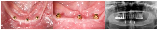
Figure 1.
Explanatory case (test group).
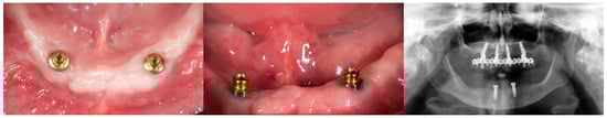
Figure 2.
Explanatory case (control group).
Looking at the maxilla, the ideal number of implants required to retain an overdenture is yet to be clarified. However, the greater trend is to place at least four implants, splinted or not, in order to ensure a higher survival rate of implants, regarding stress distribution []. In the mandible, Pisani et al. [], in an in vitro study on 3D finite element models of mandibular overdentures, demonstrated that the two-implant-retained overdentures exhibited lower stresses than the single- implant-retained overdentures. A similar study was conducted by Liu et al. [] concluding that placing a third implant placed between the classical two did not eliminate denture rotation around the fulcrum line. In addition, no strain concentration was found in cortical bone around the middle implant. These results are in agreement with another in vitro research revealing that the bone was insensitive to the number of implants or attachment type. After this observation, the authors recommended to use two implants in the canine region. The results of the present research are in agreement with the aforementioned studies, confirming that there are no benefits to placing one more implant, at least one year after loading.
Looking into the future, a possible benefit to place three implants could be to remake the implant-retained overdenture with an implant-supported restoration, both fixed or hybrid (fixed/removable). The so called “Exit Strategy” allows to move from a removable to fixed denture, limiting the biologic and economic impact for the patients. In this way, the OT Equator attachments can work both as an attachment system and as an intermediate abutment for fixed restorations [,,,].
The main limitation of the present study was the small sample size and the kind of implant used, which may influence the primary and secondary outcome variables.
This happened most likely due to the COVID-19 pandemic, which poses as a life-threatening risk to elderly patients, particularly when affected by co-morbidities and/or fragilities []. This allowed to enroll a minor number of patients compared to the exerted sample. Nevertheless, although the number of patients was unbalanced, a total of 34 patients were treated up to one year after loading, with no unbalancing between patients’ and implants’ characteristics. Most of the researches aimed to find the optimal number of implants to retain an overdenture were conducted in vitro. On the contrary, this is one of the few in vivo, randomized, and controlled trials.
5. Conclusions
There are no differences in all the investigated outcomes. Waiting for further studies, it is possible to conclude that adjunctive implant besides the two to retain an implant overdenture are not needed.
Author Contributions
M.T.: conceptualization, investigation, original draft preparation, methodology; M.M.: investigation; R.S.: investigation; E.F.: investigation; A.C.: investigation; E.X.: investigation; S.M.L.: investigation; S.M.: writing—review and editing, data curation; F.M.C.: investigation; R.R.y.B.: supervision; M.C. and G.C.: supervision, funding acquisition. All authors have read and agreed to the published version of the manuscript.
Funding
This research received no external funding.
Institutional Review Board Statement
The study was conducted according to the guidelines of the Declaration of Helsinki, and approved by the Institutional Review Board of the Aldent University, Tirana, Albania (protocol code 4/2018, 2 April 2018).
Informed Consent Statement
Informed consent was obtained from all subjects involved in the study.
Data Availability Statement
Data is available on request to corresponding author.
Conflicts of Interest
The authors declare no conflict of interest.
References
- Feine, J.S.; Carlsson, G.E.; Awad, M.A.; Chehade, A.; Duncan, W.; Gizani, S.; Head, T.; Lund, J.P.; MacEntee, M.; Mericske-Stern, R.; et al. The McGill Consensus Statement on Overdentures. Int. J. Prosthodont. 2002, 15, 413–414. [Google Scholar] [PubMed]
- Batenburg, R.H.; Meijer, H.J.; Raghoebar, G.M.; Vissink, A. Treatment concept for mandibular overdentures supported by endosseous implants: A literature review. Int. J. Oral Maxillofac. Implant. 1998, 13, 539–545. [Google Scholar]
- Batenburg, R.H.; Raghoebar, G.M.; Van Oort, R.P.; Heijdenrijk, K.; Boering, G. Mandibular overdentures supported by two or four endosteal implants: A prospective, comparative study. Int. J. Oral Maxillofac. Surg. 1998, 27, 435–439. [Google Scholar] [CrossRef]
- Leão, R.S.; Moraes, S.L.D.; Vasconcelos, B.C.E.; Lemos, C.A.A.; Pellizzer, E.P. Splinted and unsplinted overdenture attachment systems: A systematic review and meta-analysis. J. Oral Rehabil. 2018, 45, 647–656. [Google Scholar] [CrossRef] [PubMed]
- Ortensi, L.; Martinolli, M.; Borromeo, C.; Ceruso, F.M.; Gargari, M.; Xhanari, E.; Tallarico, M. Effectiveness of Ball Attachment Systems in Implant Retained- and Supported-Overdentures: A Three- to Five-Year Retrospective Examination. Dent. J. 2019, 7, 84. [Google Scholar] [CrossRef] [PubMed] [Green Version]
- Tallarico, M.; Ortensi, L.; Martinolli, M.; Casucci, A.; Ferrari, E.; Malaguti, G.; Montanari, M.; Scrascia, R.; Vaccaro, G.; Venezia, P.; et al. Multicenter Retrospective Analysis of Implant Overdentures Delivered with Different Design and Attachment Systems: Results Between One and 17 Years of Follow-Up. Dent. J. 2018, 6, 71. [Google Scholar] [CrossRef] [PubMed] [Green Version]
- Di Francesco, F.; De Marco, G.; Carnevale, U.A.G.; Lanza, M.; Lanza, A. The number of implants required to support a maxillary overdenture: A systematic review and meta-analysis. J. Prosthodont. Res. 2019, 63, 15–24. [Google Scholar] [CrossRef] [PubMed]
- Pisani, M.X.; Presotto, A.G.C.; Mesquita, M.F.; Barão, V.A.R.; Kemmoku, D.T.; Del Bel Cury, A.A. Biomechanical behavior of 2-implant– and single-implant–retained mandibular overdentures with conventional or mini implants. J. Prosthet. Dent. 2018, 120, 421–430. [Google Scholar] [CrossRef] [PubMed]
- Liu, J.; Pan, S.; Dong, J.; Mo, Z.; Fan, Y.; Feng, H. Influence of implant number on the biomechanical behaviour of mandibular implant-retained/supported overdentures: A three-dimensional finite element analysis. J. Dent. 2013, 41, 241–249. [Google Scholar] [CrossRef] [PubMed]
- Tallarico, M.; Cervino, G.; Scrascia, R.; Uccioli, U.; Lumbau, A.I.; Meloni, S.M. Minimally Invasive Treatment of Edentulous Maxillae with Overdenture Fully Supported by a Cad/Cam Titanium Bar with a Low-Profile Attachment Screwed on Four or Six Implants: A Case Series. Prosthesis 2020, 2, 53–64. [Google Scholar] [CrossRef]
- Cicciù, M.; Cervino, G.; Milone, D.; Risitano, G. FEM Investigation of the Stress Distribution over Mandibular Bone Due to Screwed Overdenture Positioned on Dental Implants. Materials 2018, 11, 1512. [Google Scholar] [CrossRef] [PubMed] [Green Version]
- Acampora, R.; Montanari, M.; Scrascia, R.; Ferrari, E.; Pasi, M.; Cervino, G.; Meloni, S.M.; Lumbau, A.I.; Erta, X.; Koshovari, A.; et al. 1-Year Evaluation of OT Bridge Abutments for Immediately Loaded Maxillary Fixed Restorations: A Multi-center Study. Eur. J. Dent. 2021, 15, 290–294. [Google Scholar] [PubMed]
- Scrascia, R.; Cicciù, M.; Manco, C.; Miccoli, A.; Cervino, G. Angled Screwdriver Solutions and Low-Profile Attachments in Full Arch Rehabilitation with Divergent Implants. Appl. Sci. 2021, 11, 1122. [Google Scholar] [CrossRef]
- Tallarico, M.; Cicciù, M.; Lumbau, A.I.; Meloni, S.M. Coronavirus Disease 2019 Coexistence in the Daily Practice. Eur. J. Dent. 2020, 14 (Suppl. 01), S171–S176. [Google Scholar] [CrossRef] [PubMed]
Publisher’s Note: MDPI stays neutral with regard to jurisdictional claims in published maps and institutional affiliations. |
© 2021 by the authors. Licensee MDPI, Basel, Switzerland. This article is an open access article distributed under the terms and conditions of the Creative Commons Attribution (CC BY) license (https://creativecommons.org/licenses/by/4.0/).