Article

Field Prevalence and Pathological Features of Edwardsiella tarda Infection in Farmed American Bullfrogs (Aquarana catesbeiana)

1
College of Veterinary Medicine, South China Agricultural University, Guangzhou 510642, China
2
Zhaoqing Branch Center of Guangdong Laboratory for Lingnan Modern Agricultural Science and Technology, Zhaoqing 526238, China
3
Zhaoqing Dahuanong Biology Medicine Co., Ltd., Zhaoqing 526238, China
*
Authors to whom correspondence should be addressed.
These authors contributed equally to this work.
Animals 2025, 15(17), 2487; https://doi.org/10.3390/ani15172487
Submission received: 19 July 2025 / Revised: 20 August 2025 / Accepted: 20 August 2025 / Published: 25 August 2025
(This article belongs to the Section Veterinary Clinical Studies)

Simple Summary

Bullfrogs are now widely farmed in China, yet hidden illnesses can quietly erode profits and animal welfare. In this study, we looked for the bacterium Edwardsiella tarda in apparently healthy bullfrogs from four farms in Guangdong Province. Laboratory tests showed that between two and three out of every five frogs carried the germ in their intestines, usually in low numbers. The strains we isolated already withstand some older medicines such as penicillin and tetracycline, though newer and more powerful antibiotics still work. When healthy frogs were exposed to the bacterium in the laboratory, very large doses killed them quickly, but much smaller doses were enough to cause long-lasting gut inflammation that slows growth. Because infected frogs often show no clear signs of disease, the pathogen can spread unnoticed in ponds and may become harder to treat if antibiotics are used incorrectly. Regular health checks, clean-water management and careful selection of medicines will help farmers keep their stock healthy, protect farm income, and reduce the risk that this organism will threaten other animals or people.

Abstract

Edwardsiella tarda is a zoonotic facultative intracellular bacterium whose impact on farm-raised amphibians is still poorly defined. We recovered seven strains from American bullfrogs (Aquarana catesbeiana) on four farms in Guangdong, China, and combined field surveillance with molecular and pathological investigations. Phylogenetic analysis of 16S rRNA and rpoB sequences confirmed species identity. Quantitative PCR of 192 apparently healthy frogs revealed intestinal carriage at every farm, with prevalence ranging from 39 to 77 percent and bacterial loads of 105–106 CFU/mL, indicating widespread subclinical colonisation. Virulence profiling demonstrated a conserved core gene set (gadB, mukF, citC, fimA, ompA) and accessory variation confined to the flagellar gene fliC. The strains resisted trimethoprim, ampicillin, and tetracyclines, yet remained susceptible to third generation cephalosporins, carbapenems, and most aminoglycosides. Infection trials showed that although very high inocula caused acute fatalities, an inoculum of 108 CFU/mL was sufficient to induce persistent enteritis characterised by suppressed tight junction proteins, elevated cytokine expression, and marked intestinal damage. These findings demonstrate that E. tarda circulates silently in bullfrog culture, carries an amphibian adapted virulence profile and still responds to key antimicrobials, providing a baseline for risk assessment, surveillance, and targeted control in amphibian aquaculture.

1. Introduction

Edwardsiella tarda is a Gram-negative, facultatively anaerobic, non-capsulated bacterium belonging to the order Enterobacterales and is recognised as a facultative intracellular pathogen [1,2,3]. Its host spectrum is broad, encompassing fish, amphibians, birds, mammals, and even humans [4,5]. In fish such as flounder and seahorse, E. tarda infection is chiefly characterised by enteritis accompanied by anorexia and intestinal fluid accumulation [6,7]. Reported avian cases, mostly involving ichthyophagous species, include septicaemia in crested ibis [8] and renal masses in common loons [9]. Consumption of raw or under-cooked fish contaminated with E. tarda can likewise lead to human fulminant septicaemia and become life-threatening [10], although human infection more commonly presents as self-limiting gastroenteritis that may progress to chronic diarrhoea [2,11]. Isolation of E. tarda has also been documented in pigs, puppies, and bullfrogs, yet the pathogenicity of these isolates has not been examined in detail [12,13,14].
The American bullfrog (Aquarana catesbeiana), native to North America, has become one of the fastest-growing aquaculture commodities in China. Driven by high economic returns, producers frequently adopt high-density stocking, which promotes water eutrophication and makes bacterial diseases difficult to control [15,16]. At present, meningitis-like disease caused by Elizabethkingia miricola is widespread [17,18], and red-leg syndrome (acute septicaemia) associated with Aeromonas, Streptococcus, and other bacteria attracts considerable attention because of the resulting mass mortalities [16,19]. In contrast, certain chronic conditions lack evident clinical signs and are therefore easily overlooked; surveys indicate that farmers primarily rely on sensory cues for routine monitoring [20]. Intensified farming increases animals’ susceptibility to chronic diseases and facilitates subclinical transmission of infectious agents [21,22]. Although such diseases may not cause immediate mortality, they markedly reduce production performance and profitability, while subclinically infected animals may continuously shed pathogens into the environment, greatly complicating control measures and amplifying the risk of spread [23,24].
Recently, we conducted an epidemiological survey across several bullfrog farms in Guangdong Province, China. During ongoing epidemiological surveillance, we identified multiple bullfrog populations that showed reduced feed intake but few overt clinical signs. The present study isolates and identifies the aetiological agent, delineates its epidemiological characteristics and disease progression, and thus aims to fill a knowledge gap concerning chronic infections in bullfrog culture while providing data useful for farm management.

2. Materials and Methods

2.1. Epidemiological Survey

Between 2022 and 2025, during a broad epidemiological survey, we identified a disease characterised by low mortality, minimal external symptoms, and reduced feed intake in farmed bullfrogs from Guangzhou, Zhaoqing, Huizhou, and Yunfu. For these asymptomatic cases, we typically collected 53 frogs from at least three randomly selected ponds per farm. Among them, 48 individuals were examined microscopically for parasites, and pooled liver, spleen, kidney, and intestinal tissues (50 mg total) were subjected to DNA extraction. The remaining DNA was stored at −20 °C for subsequent molecular analyses. The other five frogs were used for necropsy and bacterial isolation, and based on the bacterial isolation results, the DNA samples from the initial 48 individuals were reanalysed.

2.2. Experimental Animals

Healthy bullfrogs (20 ± 2 g) were obtained from a farm in Zhaoqing with no known history of disease. The frogs were maintained in a controlled laboratory environment at 25 ± 1 °C, with regular feeding and daily water replacement. All bullfrogs were humanely euthanised by cervical dislocation either prior to tissue collection or at the end of the experiments.

2.3. Histopathology

Liver and intestinal tissues were collected from both naturally infected bullfrogs (n = 5) and healthy individuals (n = 5). In the experimental infection model, intestinal samples were obtained from five frogs at each point (1, 3, 5, 9, and 14 days post-infection), with five uninfected frogs used as controls. All tissue samples were immersed in 4% paraformaldehyde for 96 h to ensure adequate fixation. Following fixation, the samples were dehydrated, cleared, and embedded in paraffin blocks using routine histological procedures. Sections were cut at a thickness of 3 to 5 μm, then stained with Mayer’s hematoxylin and eosin. Stained slides were examined under a light microscope (Eclipse Ci, Nikon, Tokyo, Japan) for histopathological assessment.

2.4. Bacterial Isolation and Identification

To isolate pathogenic bacteria, bullfrogs were dissected under sterile conditions, and samples were obtained from visibly affected liver, spleen, kidney, and intestinal tissues. The samples were inoculated onto brain heart infusion (BHI) agar and cultured at 30 °C for 18 h. Dominant bacterial colonies were selected and purified through repeated streaking. Genomic DNA was extracted from these purified isolates. Species identification was performed by amplifying and sequencing both the 16S rRNA and rpoB genes, following primer sets and PCR protocols previously established in our laboratory [25]. Sequencing was conducted by Tianyi Huayu Gene Technology Co., Ltd., Suzhou, China, and the resulting sequences were aligned with reference data in the NCBI database for preliminary classification. All bacterial strains were preserved at −80 °C for downstream analyses. One or two representative isolates from each site (designated E1, E2, E4, E5, E7, E8, and E9) were used for phylogenetic reconstruction using MEGA 7 software.

2.5. Quantitative Detection of FV3 and E. tarda in Field Samples

A total of 192 DNA samples preserved at −20 °C, as described in Section 2.1, were thawed on ice and subjected to qPCR detection and quantification of FV3 and E. tarda using previously published methods [26,27]. The target genes were the major capsid protein gene for FV3 and the gyrB gene for E. tarda. Primers and probe used in this assay are listed in Table 1. Samples with a cycle threshold value below 35 were considered positive. Based on the standard curves provided in the cited literature and the dilution factor introduced during DNA extraction, the viral or bacterial load in each sample was calculated (expressed as copies/μL for FV3 and CFU/μL for E. tarda). All reactions were performed in triplicate.

2.6. Antimicrobial Susceptibility Testing

Antimicrobial susceptibility of seven E. tarda isolates from bullfrogs was evaluated using the standard agar disc diffusion method according to the CLSI M100-Ed34 Performance Standards for Antimicrobial Susceptibility Testing [28]. A total of 18 antibiotics were tested, including Cefotaxime, Chloramphenicol, Streptomycin, Gentamicin, Imipenem, Tetracycline, and eleven additional agents to be supplemented. All antibiotic discs were obtained from Hangzhou Microbial Reagent Co., Ltd., Hangzhou, China. Escherichia coli ATCC 25,922 was used as the quality control strain and yielded inhibition zone diameters within the CLSI-recommended ranges. Based on the diameter of inhibition zones, isolates were classified as susceptible, intermediate, or resistant.

2.7. Detection of Virulence Genes

To determine virulence gene profiles, all E. tarda isolates were examined for eight genes previously reported as pathogenicity determinants in this species [29,30,31,32]. The target genes were fliC (flagellin gene), ompA (outer membrane protein A gene), fimA (fimbrial adhesin-like protein gene), gadB (glutamate decarboxylase gene), katB (catalase gene), citC (citrate lyase ligase gene), mukF (chromosome partitioning protein MukF gene), and eseD (type III secretion system effector protein D gene). PCR amplification followed protocols established in our earlier work. Products were resolved on 1 percent agarose gels and visualised with a gel imaging system (Bio-Rad, Berkeley, CA, USA). A clear band of the expected size was recorded as positive (“+”), whereas the absence of a band was recorded as negative (“−”).

2.8. Infection Model

2.8.1. Challenge Experiment

E. tarda Strain E2 was cultured overnight at 28 °C in brain heart infusion broth. The resulting stationary-phase cells were serially diluted with sterile 0.65% physiological saline to prepare four inocula containing 1.2 × 10⁹, 1.2 × 10⁸, 1.2 × 10⁷, and 1.2 × 10⁶ CFU/mL, respectively. Groups of ten healthy bullfrogs were each given 0.2 mL of one dilution by intraperitoneal injection; the control group received an equivalent volume of sterile saline. All frogs were held in laboratory tanks at 25 ± 1 °C, and clinical signs together with mortality were recorded daily for fourteen consecutive days.

2.8.2. Disease Progression Analysis

Fifty clinically healthy bullfrogs were allocated to the infection group and injected intraperitoneally with 200 µL of an E. tarda E2 suspension (1.2 × 10⁸ CFU/mL). A negative-control group of ten frogs received 200 µL of sterile physiological saline by intraperitoneal injection. At 1, 3, 5, 9, and 14 days post-injection, five frogs were randomly selected from each group. From every frog two intestinal samples were taken: one fragment was fixed in 4% paraformaldehyde for histopathology (Section 2.3), and the second fragment was pooled with those from the other four frogs sampled on the same day for gene-expression analysis (Section 2.9). All animals were maintained throughout the trial under the husbandry conditions described previously.

2.9. Intestinal Gene Expression

To investigate the impact of E. tarda infection on intestinal barrier integrity and mucosal inflammation in bullfrogs, we assessed the expression of key genes associated with epithelial tight junctions (Claudin-7, Occludin, and ZO-2) and pro-inflammatory signalling (TNF, IL-8, and IL-1β). These genes were selected based on their established roles in maintaining epithelial homeostasis and mediating acute immune responses in mucosal tissues [19,33]. The amphibian 18S rRNA gene was used as an internal reference [34]. Relative transcript levels were calculated using the 2−ΔΔCT method, with the day-0 control group serving as the calibrator.
Table 1. The information of primers used in this study.
Target GenePrimer Sequence (5′-3′)Reference
IL-1βF: CAGTGATTAGACACCCAGGAACG[19]
R: GACCTTCATTTCAGTGGAGCATTC
IL-8F: GCTTGTGCCACCCTTACCCTC[19]
R: GCTCTACCCAAGAAGCAGAAGGA
TNFF: CACAGCACCCAGGAGCAACTXM_040323112.1
R: GGGATGATGTGGAAGTGGAGC
ZO-2F: TGAGGATGGACATTTTGACCGTAG[33]
R: CATAGCCTCGGTCTGGACTGGAT
Claudin-7F: GCTCTCCATTGTTCTCGGGGT[33]
R: CCCAGCAGAAAGATGAAGCCTC
OccludinF: GCTCTCCACCTGGCATCATCA[33]
R: GCAAAACCCATTCCCATCTGAG
RNA 18SF: CGTTGATTAAGTCCCTGCCCTT[34]
R: GCCGATCCGAGGACCTCACTA
16S rRNAF: AGAGTTTGATCCTGGTCAGAACGAACGCT[25]
R: TACGGCTACCTTGTTACGACTTCACCCC
gadBF: ATTTGGATTCCCGCTTTGGT[31]
R: GCACGACGCCGATGGTGTTC
mukFF: TTGCTGGCTATCGCTACCCT[31]
R: AACTCATCGCCGCCCTCTTC
citCF: TTTCCGTTTGTGAATCAGGTC[31]
R: AATGTTTCGGCATAGCGTTG
fimAF: CTGTGAGTGGTCAGGCAAGC[31]
R: TAACCGTGTTGGCGTAAGAGC
katBF: CTTAGCCATCAGCCCTTCC[31]
R: GCGAGTGCCGTAGTCCTT
EseDF: TTCAGGGTGGTCAGTATCTCGAY643478.1
R: TCAACAGACGCAGCAAAGC
OmpAF: ACCCGTCTGGACTATCAGTATGTGQ259743.1
R: GCGGCTGAGTAACTTCTTCTTT
FliCF: CGCTGATGGCACAGAATAACAP040123.1
R: TCAGATAGGGCTTTGTCCAG
rpoBF: GCAGTGAAAGARTTCTTTGGTTC[25]
R: GTTGCATGTTNGNACCCA
Et-gyrBF: TGGCGACACCGAGCAGA[26]
R: ACAAACGCCTTAATCCCACC
FV3-MCPF: GACGAGAGACAGGCCATGAG[27]
R: GGAAGGGTGTGTGACGTTCT
Probe: ACCTCCTGATCCA

2.10. PCR and Quantitative PCR

Primers and probes used in this study are listed in Table 1. Conventional PCR was performed to amplify the 16S rRNA and rpoB genes for bacterial identification (Section 2.4), as well as eight virulence genes described in Section 2.7. All reaction mixtures and thermal cycling conditions followed the protocol reported by Lin et al., 2023 [35].
Quantitative PCR assays for the detection of E. tarda and FV3 in field samples were carried out using the reagent systems and thermal cycling conditions described in previous studies [26,27]. Each reaction was performed in triplicate.
For intestinal gene expression analysis, total RNA was extracted from pooled intestinal samples collected at different time points (Section 2.8.2), reverse-transcribed to cDNA, and subjected to qPCR using the reagent system and thermal profile described by Lin et al., 2024 [19]. All reactions were performed in triplicate.

2.11. Statistical Analysis

Data are presented as the mean ± standard deviation from three independent trials. Prior to hypothesis testing, normal distribution was confirmed with the Kolmogorov–Smirnov test and equality of variances with the Levene test; both criteria were satisfied for every variable. Group differences were therefore evaluated with a single-factor analysis of variance. All statistical procedures were performed in SPSS version 26 (IBM, Armonk, NY, USA), and figures were prepared with GraphPad Prism version 10.4.1 (GraphPad Software, San Diego, CA, USA). Significance thresholds were defined as follows: ns for p > 0.05, * for p < 0.05, ** for p < 0.01 and *** for p < 0.001.

3. Results

3.1. Clinical Features

Gross findings are summarised in Figure 1. Diseased bullfrogs showed inflamed and oedematous intestines with dilated capillaries and occasional intussusception (Figure 1A). A subset of animals exhibited enlarged, pale livers (Figure 1B). Histologically, compared with healthy controls (Figure 2A,C), infected intestines displayed markedly shortened and disorganised villi, focal epithelial loss, pronounced submucosal oedema, and dense inflammatory cell infiltration (Figure 2B). Liver sections from the same animals revealed disrupted hepatic cords, hepatocellular swelling, and sinusoidal congestion (Figure 2D). These combined gross and microscopic lesions indicate that E. tarda infection compromises the intestinal barrier and initiates a systemic inflammatory response.

3.2. Molecular Identification

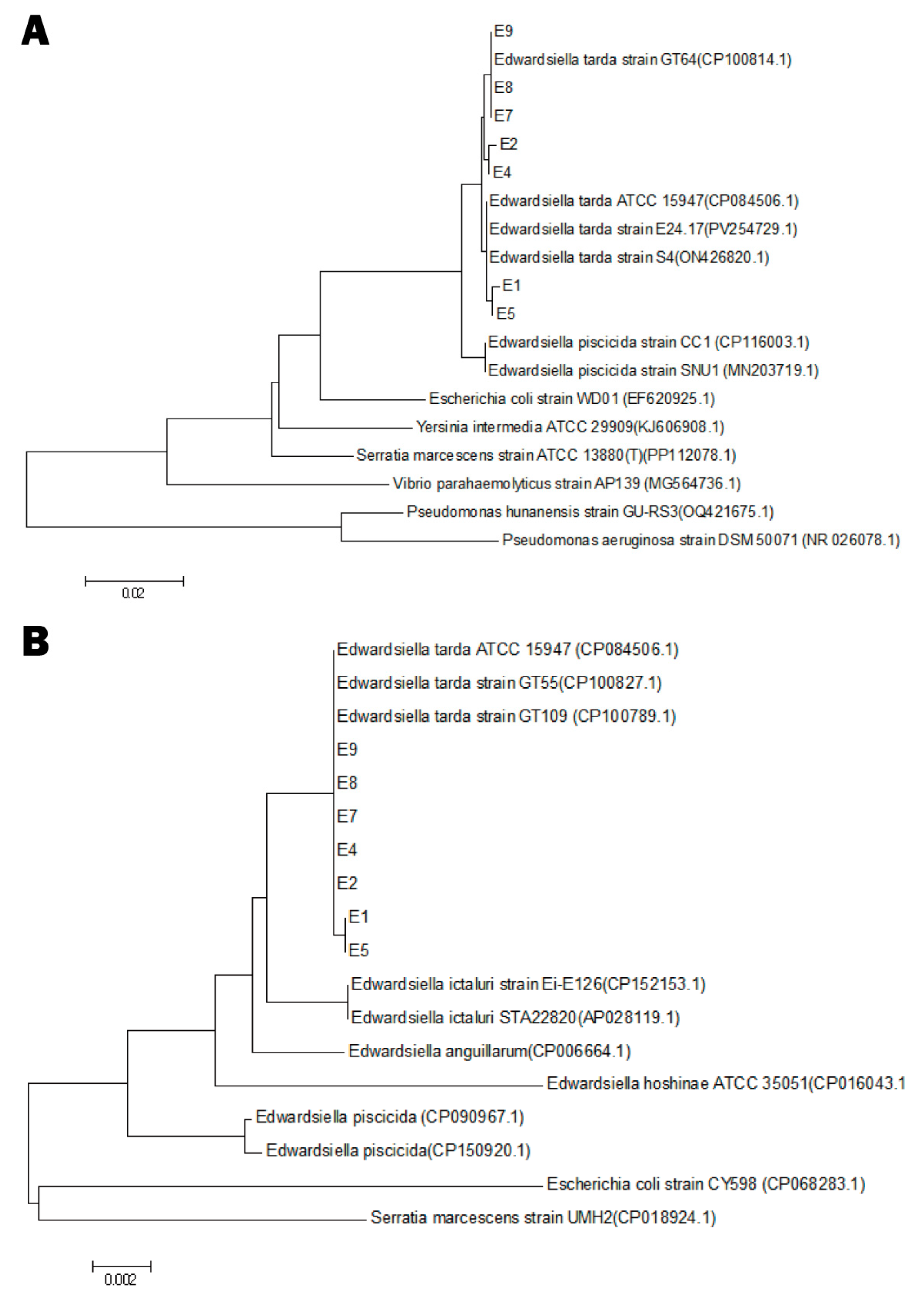
No parasites were detected in any of the sampled frogs, and FV3-specific qPCR was negative across all DNA extracts. A single bacterial morphotype was consistently recovered from the bacteriological cultures of liver, spleen, kidney, or intestinal tissues of sampled frogs. After purification the colonies were circular, smooth, white, and 0.5–1.0 mm in diameter, with no obvious haemolytic activity was observed on 5% sheep-blood agar (Figure S1). Sequences of 16S rRNA and rpoB from seven representative isolates (E1, E2, E4, E5, E7, E8, E9) have been deposited in GenBank (accession numbers: PV915438–PV915444 for 16S rRNA; PV935302–PV935308 for rpoB). BLAST analysis (performed using the NCBI online tool https://blast.ncbi.nlm.nih.gov/Blast.cgi, accessed in June 2025) showed > 99% identity to reference strains of E. tarda. Neighbour-joining phylogenies based on both loci grouped all seven isolates into a single, strongly supported E. tarda clade (Figure 3).

3.3. Prevalence and Bacterial Load

Using a previously published quantitative PCR method specific for E. tarda, all forty-eight DNA samples from each farm were re-examined. The detection rates in Guangzhou, Zhaoqing, Huizhou, and Yunfu were 45.8%, 39.6%, 77.1%, and 66.7%, respectively, indicating a high prevalence of the pathogen (Figure 4A). For positive specimens, most bacterial loads ranged from 102 to 103 CFU/µL (equivalent to 105 to 106 CFU/mL), reflecting generally low levels of colonisation across the four farms (Figure 4B).

3.4. Virulence Gene Profiling

PCR screening (Table 2) demonstrated that all seven E. tarda isolates (E1, E2, E4, E5, E7, E8, E9) possessed the core virulence genes gadB, mukF, citC, fimA, and ompA. Among the accessory loci examined, variation was confined to the flagellar gene fliC, which was detected in isolates E2 and E4 but absent from the other five strains. The type III secretion effector gene eseD and the oxidative stress gene katB were not amplified from any isolate. These findings indicate that the isolates share conserved metabolic and adhesion gene complements, and that flagellar motility potential is the only variable trait detected in the present study.

3.5. Antimicrobial Susceptibility Patterns

The antibiotic resistance of seven representative E. tarda strains isolated from four different regions of Guangdong Province was tested using the paper disc diffusion method, as shown in Table 3. The strains were generally resistant to trimethoprim, ampicillin, and tetracyclines. They were mostly susceptible to cephalosporins, fluoroquinolones, and carbapenems, although some strains (E1, E5) were resistant to ciprofloxacin. The aminoglycoside antibiotics showed variability: most strains were susceptible to amikacin and gentamicin, but exhibited intermediate resistance to kanamycin, with only strain E7 being susceptible. Overall, the prevalent strains exhibited multiple drug resistance, with certain regional differences observed among the strains.

3.6. Challenge Experiment

To confirm the pathogenicity of E. tarda in bullfrogs, strain E2 was administered at four inoculum levels. As shown in Figure 5, the highest dose of 1.2 × 10⁹ CFU/mL produced a cumulative mortality of 90% within fourteen days, with the peak loss recorded on day 4. At 1.2 × 10⁸ CFU/mL, only sporadic deaths occurred during the first week; the mortality peak shifted to the second week, and overall lethality declined. Survival after fourteen days reached 80–90% in groups inoculated with 1.2 × 10⁷ or 1.2 × 10⁶ CFU/mL. Bacteria re-isolated from the intestines of dead frogs were sequenced and confirmed as E. tarda. No deaths or lesions were observed in the saline control group. All remaining frogs were humanely euthanised at the end of the trial.

3.7. Disease Progression

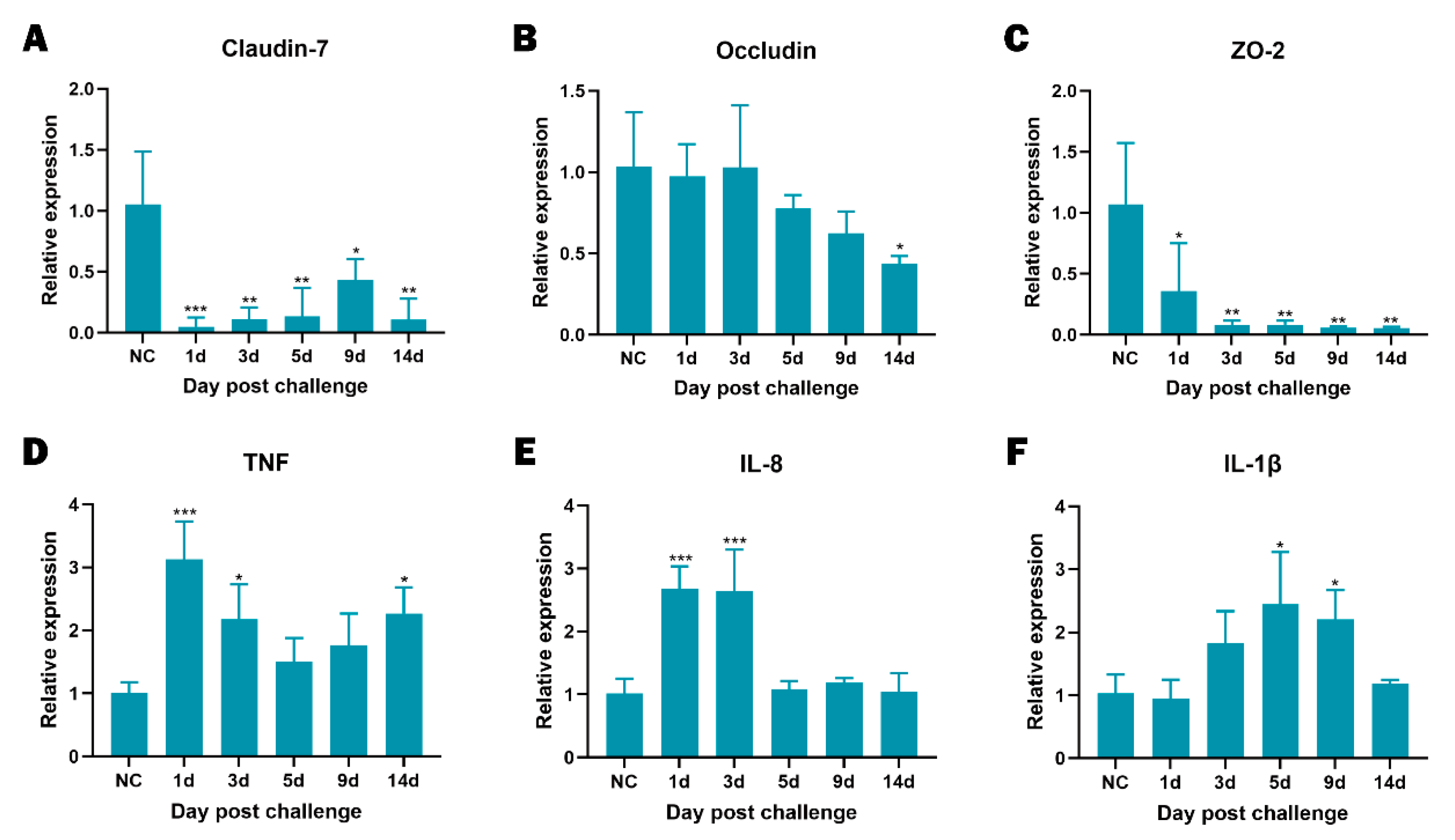
To clarify the course of infection we combined sequential histology with quantitative PCR. Figure 6 shows that intestinal lesions progressed from mild epithelial degeneration with modest villus shortening on days 1 to 3, to pronounced submucosal oedema, vascular congestion, and focal haemorrhage on days 5 to 9, and culminated on day 14 in deep ulceration with partial loss of the intestinal wall. In concordance with these structural changes, expression of the tight-junction genes Claudin-7, Occludin, and ZO-2 was already significantly lower than in healthy controls as early as day 1 post-infection and remained depressed thereafter (Figure 7A–C), indicating rapid and persistent barrier disruption. The pro-inflammatory transcripts TNF, IL-8, and IL-1β peaked between days 3 and 5 (Figure 7D–F). By day 14, IL-8 and IL-1β had returned to baseline, whereas TNF remained significantly elevated (p < 0.05). Collectively, the histological and molecular data demonstrate that E. tarda infection causes an early loss of epithelial integrity, provokes an acute cytokine surge, and leads to sustained tissue injury despite partial attenuation of the inflammatory response.

4. Discussion

E. tarda infection is a recognised threat in fin-fish aquaculture, yet its pathogenic behaviour in farmed amphibians has received little systematic scrutiny. Through longitudinal surveillance across multiple farms, we show that E. tarda is widely endemic in Chinese bullfrog production, usually circulating as low-level, subclinical infection. By coupling field surveys with laboratory analyses we characterised the circulating isolates’ virulence repertoires, antimicrobial-resistance profiles, and disease-course features, thereby providing the first comprehensive baseline dataset for E. tarda infection in bullfrogs. These data address a critical knowledge gap for disease risk assessment and management in this rapidly expanding sector.
Field sampling revealed that most frogs harbour E. tarda at modest bacterial loads, a condition that nevertheless depresses growth and compromises immune competence through persistent intestinal inflammation. This chronic, largely silent presentation contrasts sharply with the fulminant septicaemia typically provoked by other bacterial agents of bullfrogs, such as Aeromonas, Acinetobacter, Streptococcus, and Elizabethkingia, which precipitate sudden mass mortalities [16,19,35,36]. Consistent with reports that E. tarda can sustain prolonged stimulation of host defences [37], our challenge model demonstrated that pathogenesis remains dose-dependent: when frogs received an inoculum of 1 × 10⁹ CFU/mL, cumulative mortality approached 90%, mirroring the acute septicaemic outcomes documented in infected fish species [38]. Although human cases rarely progress beyond self-limiting gastroenteritis, once bacteraemia is established the clinical course accelerates and case-fatality rates become alarmingly high [39]. Early recognition of subtle disease cues and an appreciation of the ensuing time-course are therefore indispensable for effective outbreak prevention and control in amphibian culture.
Infected hosts consistently developed progressive enteritis driven by a combination of direct bacterial invasion and dysregulated immune signalling. Experimental infection markedly elevated IL-1β and IFN-γ and thereby sustained mucosal inflammation that compromised intestinal barrier integrity [40]. Previous work indicates that Edwardsiella species can impair Th17-cell function, destabilise host–microbiota metabolic crosstalk and selectively enrich Proteobacteria, amplifying epithelial injury [41]. Parallel studies in teleosts have shown activation of the MyD88/IRAK4/TAK1 cascade with over-expression of IKKβ and IL-1β, culminating in necrotic lesions of the intestine and liver [42], while loss of goblet cells and tight-junction integrity further increases intestinal permeability, locking the tissue into an inflammation–damage loop [40,43]. Human infections, though usually self-contained, may similarly masquerade as inflammatory-bowel-disease-like pathology during protracted inflammation [11]. Our bullfrog model reproduced these hallmarks: IL-1β and IL-8 transcripts rose sharply then gradually declined as cytokine stores were exhausted, whereas the tight-junction proteins Claudin-7, ZO-2, and Occludin remained significantly down-regulated throughout infection. Histopathological examination corroborated the molecular findings, showing pronounced inflammatory-cell infiltration and progressive structural damage of the intestinal tissue. Collectively, the data underline a mechanistic convergence that spans fish, amphibian, and human hosts.
Differences in virulence-factor repertoires can translate into distinct pathogenic profiles among circulating strains. All bullfrog isolates in this study harbour gadB, mukF, citC, fimA, and OmpA, genes previously shown to enhance intracellular and acid-stress survival [44,45], accelerate proliferation [46], reinforce intestinal colonisation [47], and promote adhesion, invasion, and immune evasion [30]. Intriguingly, whereas fish-derived E. tarda rely on katB to withstand H2O2 and phagocyte-mediated killing [48,49], this gene was absent from every bullfrog isolate examined. Amphibian macrophages depend more on CSF1/IL34-regulated antioxidant pathways [50,51], and their oxidative-stress response varies across metamorphic stages [52,53]; teleosts, in contrast, employ a dual Keap1a/Keap1b system and robust katG up-regulation to counter oxidative pressure [54,55]. Such host-specific immune landscapes may relax the selective pressure for katB retention in amphibian-adapted strains. Alternatively, bullfrog isolates could compensate via redundant antioxidant systems, such as alkyl-hydroperoxide reductase (Ahp) or other peroxidases [56,57]. These hypotheses warrant further experimental validation.
An analogous pattern of host adaptation is evident in antimicrobial susceptibility. All bullfrog isolates were resistant to trimethoprim, ampicillin, and tetracyclines, which matches the widespread detection of trimethoprim and tetracycline residues in aquaculture ponds and sediments across South China [58,59] and corresponds to the continued environmental presence of blaTEM and related β lactamase genes in livestock and municipal wastewater despite the official prohibition of penicillin in fish farming since 2002 [60,61]. In sharp contrast to the rapidly rising resistance to cephalosporins and fluoroquinolones that has been reported for other aquaculture pathogens [62,63], our E. tarda isolates remained fully susceptible to third-generation cephalosporins, carbapenems and, apart from two strains, ciprofloxacin. This susceptibility may reflect limited direct exposure of E. tarda in bullfrog systems to these higher level agents, or an incomplete acquisition of transferable resistance determinants such as qnr or extended spectrum β lactamase genes that have already spread through bacterial communities in the Pearl River basin and other Chinese waters [64,65]. Sustained genomic surveillance that combines resistance genotyping with farm level audits of antimicrobial use is therefore critical for preventing the convergence of high virulence and broad drug resistance.
These interpretations are constrained by the limited number of isolates and farms included, which may not capture the full diversity of E. tarda in bullfrog production. In addition, the study did not include molecular analysis of resistance genes, which limits the resolution of resistance mechanisms. Future genomic studies are needed to explore the relationship between phenotypic resistance and resistance genes.

5. Conclusions

This study provides the first systematic evidence of subclinical E. tarda infection in farmed bullfrogs, integrating field detection, histopathology, virulence profiling, and antimicrobial resistance assessment. The results highlight a conserved core of virulence genes adapted to amphibian hosts and a distinct resistance pattern marked by reduced susceptibility to commonly used antibiotics but preserved sensitivity to critical drugs. These findings offer a valuable reference for disease monitoring and control in amphibian aquaculture and underscore the importance of continued surveillance and responsible antimicrobial stewardship to safeguard animal health and production sustainability.

Supplementary Materials

The following supporting information can be downloaded at https://www.mdpi.com/article/10.3390/ani15172487/s1; Figure S1: Colony morphology and haemolytic activity of Edwardsiella tarda isolates on 5% sheep blood agar.

Author Contributions

Conceptualization, methodology, investigation, writing—original draft preparation, Y.Y.; conceptualization, writing—original draft preparation, writing—review and editing, Y.H.; formal analysis, visualisation, investigation, F.L.; investigation, formal analysis, Z.C.; writing—original draft preparation, writing—review and editing, visualisation, H.L.; funding acquisition, resources, writing—review and editing, R.C. All authors have read and agreed to the published version of the manuscript.

Funding

This study was supported by the Special Project for Research and Development in Key areas of Guangdong Province (CN) (2022B1111040001), and the independent project of Zhaoqing Branch Center of Guangdong Laboratory for Lingnan Modern Agricultural Science and Technology (P20211154-0102-03).

Institutional Review Board Statement

The study was conducted in accordance with the Declaration of Helsinki and approved by the Ethics Committee of the Animal Experiments Committee of Zhaoqing Da Huanong Biopharmaceutical Co., Ltd. (LL-G-20250430-01).

Informed Consent Statement

Not applicable.

Data Availability Statement

The original contributions presented in this study are included in the article. Further inquiries can be directed to the corresponding author.

Conflicts of Interest

Author Ruiai Chen was employed by Zhaoqing Dahuanong Biology Medicine Co., Ltd. The remaining authors declare that the research was conducted in the absence of any commercial or financial relationships that could be construed as a potential conflict of interest.

Abbreviations

The following abbreviations are used in this manuscript:
E. tardaEdwardsiella tarda
FV3Frog Virus 3
rpoBRNA Polymerase Subunit B
16S rRNA16S Ribosomal RNA
CFUColony-Forming Unit
BHIBrain Heart Infusion
CLSIClinical and Laboratory Standards Institute
AhpAlkyl-Hydroperoxide Reductase
ILInterleukin
TNFTumour Necrosis Factor
ZO-2Zonula Occludens-2

References

  1. Fujita, M.; Ito, H.; Oshida, J.; Kobayashi, D. Fulminant Edwardsiella tarda bacteremia following near-drowning episode in a patient without cirrhosis: A case report. J. Infect. Chemother. 2025, 31, 102591. [Google Scholar] [CrossRef]
  2. Leung, K.Y.; Wang, Q.; Zheng, X.; Zhuang, M.; Yang, Z.; Shao, S.; Achmon, Y.; Siame, B.A. Versatile lifestyles of Edwardsiella: Free-living, pathogen, and core bacterium of the aquatic resistome. Virulence 2022, 13, 5–18. [Google Scholar] [CrossRef]
  3. Hara, C.; Tanaka, T.; Nishiwada, S.; Kirihataya, Y.; Yoshimura, A. Acute cholecystitis with sepsis due to Edwardsiella tarda: A case report. Surg. Case Rep. 2023, 9, 184. [Google Scholar] [CrossRef] [PubMed]
  4. Peng, L.T.; Li, D.L.; Yang, D.X.; Peng, B. Taurine promotes Oreochromis niloticus survival against Edwardsiella tarda infection. Fish Shellfish Immunol. 2022, 129, 137–144. [Google Scholar] [CrossRef] [PubMed]
  5. Li, M.; Wu, M.; Sun, Y.; Sun, L. Edwardsiella tarda TraT is an anti-complement factor and a cellular infection promoter. Commun. Biol. 2022, 5, 637. [Google Scholar] [CrossRef] [PubMed]
  6. Park, S.B.; Aoki, T.; Jung, T.S. Pathogenesis of and strategies for preventing Edwardsiella tarda infection in fish. Vet. Res. 2012, 43, 67. [Google Scholar] [CrossRef]
  7. Wang, X.; Wang, F.; Chen, G.; Yang, B.; Chen, J.; Fang, Y.; Wang, K.; Hou, Y. Edwardsiella tarda induces enteritis in farmed seahorses (Hippocampus erectus): An experimental model and its evaluation. Fish Shellfish Immunol. 2020, 98, 391–400. [Google Scholar] [CrossRef]
  8. Xie, H.; Li, W.; Zhang, R.; Li, H.; Zhang, Y.; Guo, R.; Zhang, J.; Li, J.; Qing, B.; Duan, W.; et al. Edwardsiella tarda causing septicemia in a wild crested ibis (Nipponia nippon). J. Wildl. Dis. 2024, 60, 232–235. [Google Scholar] [CrossRef]
  9. Lewis, N.L.; Ware, C.; Griffin, M.J. Edwardsiella tarda isolated from a kidney mass in a common loon (Gavia immer). J. Wildl. Dis. 2023, 59, 804–806. [Google Scholar] [CrossRef]
  10. Zhou, Y.; Ren, D.; Li, Y.; Gui, S. A case of septic shock caused by drug-resistant Edwardsiella tarda and literature review. BMC Infect. Dis. 2025, 25, 393. [Google Scholar] [CrossRef]
  11. Chida, R.; Iio, K.; Kaizuka, H.; Hirai, Y.; Ishida, Y.; Yamanaka, G. Chronic diarrhea associated with Edwardsiella tarda gastroenteritis: A case report and literature review. Pediatr. Infect. Dis. J. 2022, 41, e440–e442. [Google Scholar] [CrossRef] [PubMed]
  12. Owens, D.R.; Nelson, S.L.; Addison, J.B. Isolation of Edwardsiella tarda from swine. Appl. Microbiol. 1974, 27, 703–705. [Google Scholar] [CrossRef] [PubMed]
  13. van Assche, J. Edwardsiella tarda infection in a puppy with possible parvovirus infection. Vet. Rec. 1991, 129, 475–476. [Google Scholar] [CrossRef]
  14. Mauel, M.J.; Miller, D.L.; Frazier, K.S.; Hines, M.E., II. Bacterial pathogens isolated from cultured bullfrogs (Rana catesbeiana). J. Vet. Diagn. Investig. 2002, 14, 431–433. [Google Scholar] [CrossRef]
  15. Zhang, C.X.; Huang, K.K.; Wang, L.; Song, K.; Zhang, L.; Li, P. Apparent digestibility coefficients and amino-acid availability of common protein ingredients in the diets of bullfrog (Rana/Lithobates catesbeiana). Aquaculture 2015, 437, 38–45. [Google Scholar] [CrossRef]
  16. Di, Z.H.; Zheng, J.P.; Zhang, H.W.; Wang, X.; Guo, G.Y.; Lin, Y.Q.; Han, Q.; Li, R.S.; Yang, N.; Fan, L.X.; et al. Natural outbreaks and molecular characteristics of Streptococcus agalactiae infection in farmed American bullfrog (Rana catesbeiana). Aquaculture 2022, 551, 737885. [Google Scholar] [CrossRef]
  17. An, R.; Hou, G.; Sun, X.; Wang, L.; Zhang, C.; Han, Y.; Li, Y.; Wu, T.; Shi, Q.; Zhu, Z.; et al. Outbreaks of Elizabethkingia miricola caused fatal meningitis-like disease in cultured bullfrogs. Transbound. Emerg. Dis. 2024, 2024, 4733320. [Google Scholar] [CrossRef]
  18. Wei, D.; Cheng, Y.; Xiao, S.; Liao, W.; Yu, Q.; Han, S.; Huang, S.; Shi, J.; Xie, Z.; Li, P. Natural occurrences and characterization of Elizabethkingia miricola infection in cultured bullfrogs (Rana catesbeiana). Front. Cell. Infect. Microbiol. 2023, 13, 1094050. [Google Scholar] [CrossRef]
  19. Lin, H.; Zeng, G.; Yu, Y.; Li, H.; Chen, K.; Qin, Z.; Jiang, B.; Li, W.; Su, Y.; Li, L.; et al. Acute septicemia and diagnostic evaluation of Aeromonas veronii infection in American bullfrogs (Aquarana catesbeiana). Aquaculture 2023, 740, 349. [Google Scholar] [CrossRef]
  20. Manoli, C.; Di Bianco, S.; Sigwalt, A.; Defois, J.; Dufay-Lefort, A.C.; Gambara, T.; Gabriac, M.S.; Leblanc Maridor, M.; Duvauchelle Waché, A. Informational resources used by farmers with ruminants and monogastrics for animal health monitoring: Importance of sensory indicators. Animal 2024, 18, 101053. [Google Scholar] [CrossRef]
  21. Semangoen, T.; Chotigawin, R.; Sangnim, T.; Chailerd, N.; Yodkeeree, S.; Pahasup-Anan, T.; Huanbutta, K. Antimicrobial efficacy of Mangosteen (Garcinia mangostana) peel extracts in airborne microbial control within livestock farming environments. Microb. Pathog. 2025, 204, 107618. [Google Scholar] [CrossRef] [PubMed]
  22. Go, Y.Y.; Ho, J.H.P.; Tam, K.W.S.; Kamali, M.; Zhang, Y.; Lau, C.C.Y.; Li, S.H.; Wilson, M.T.; Guo, Z.; Li, R.; et al. Investigation of the first African swine fever outbreak in a domestic pig farm in Hong Kong. Transbound. Emerg. Dis. 2023, 2023, 1720474. [Google Scholar] [CrossRef] [PubMed]
  23. Af Sandeberg, A.; Båge, R.; Nyman, A.K.; Agenäs, S.; Hansson, H. Linking animal health measures in dairy cows to farm-level economic outcomes: A systematic literature mapping. Animal 2023, 17, 100971. [Google Scholar] [CrossRef] [PubMed]
  24. Park, H.E.; Park, J.S.; Park, H.T.; Shin, J.I.; Kim, K.M.; Park, S.R.; Choi, J.G.; Jung, M.; Kang, H.L.; Baik, S.C.; et al. Fetuin as a potential serum biomarker to detect subclinical shedder of bovine paratuberculosis. Microb. Pathog. 2022, 169, 105675. [Google Scholar] [CrossRef]
  25. Liu, C.; Chang, Q.; Zhang, D.F.; Li, K.B.; Wang, F.; Lin, M.H.; Shi, C.B.; Jiang, L.; Wang, Q.; Bergmann, S.M. Aeromonas shuberti as a cause of multi-organ necrosis in internal organs of Nile tilapia (Oreochromis niloticus). J. Fish Dis. 2018, 41, 1529–1538. [Google Scholar] [CrossRef]
  26. Rong, X.; Liao, M.; Zhang, Z.; Wang, Y.; Liu, Z.; Li, B.; Wang, L.; Chen, G. Development of an SYBR Green I real-time PCR assay for detection of Edwardsiella tarda and its application. J. Fish. China 2013, 37, 1829–1838. (In Chinese) [Google Scholar] [CrossRef]
  27. Li, H.; Liu, X.; Sun, J.; Lin, H.; Chen, K.; Hu, Y.; Jiang, M.; Jiang, B.; Li, W.; Su, Y.; et al. Frog virus 3: Prevalence survey and quantitative analysis in American bullfrogs using TaqMan MGB probe real-time fluorescence quantitative PCR. Aquaculture 2025, 605, 742527. [Google Scholar] [CrossRef]
  28. CLSI. Performance Standards for Antimicrobial Susceptibility Testing, 34th ed.; CLSI: Wayne, PA, USA, 2024. [Google Scholar]
  29. Sun, X.M.; Yoshida, A.; Toutani, F.; Shimizu, T.; Oda, T.; Osatomi, K. Cloning, DNA sequence, and expression of flagellins from high- and low-virulence strains of Edwardsiella tarda and their macrophage-stimulating activities. Microb. Pathog. 2023, 176, 105993. [Google Scholar] [CrossRef]
  30. Zhou, Q.; Hu, Y.; You, Y.; Gao, Y.; Wang, X.; Qin, L. Functional analysis of OmpA and its contribution to pathogenesis of Edwardsiella tarda. Microb. Pathog. 2024, 193, 106760. [Google Scholar] [CrossRef]
  31. Wang, X.; Yan, M.; Wang, Q.; Ding, L.; Li, F. Identification of Edwardsiella tarda isolated from duck and virulence-genes detection. Afr. J. Microbiol. Res. 2012, 6, 4970–4975. [Google Scholar]
  32. Wang, B.; Mo, Z.L.; Xiao, P.; Li, J.; Zou, Y.X.; Hao, B.; Li, G.Y. EseD, a putative T3SS translocon component of Edwardsiella tarda, contributes to virulence in fish and is a candidate for vaccine development. Mar. Biotechnol. 2010, 12, 678–685. [Google Scholar] [CrossRef] [PubMed]
  33. Chen, K.; Lin, H.; Sun, J.; Li, H.; Ma, J.; Qin, Z.; Jiang, B.; Li, W.; Wang, Q.; Su, Y.; et al. Archidendron clypearia extract: Modulating intestinal inflammation and enhancing disease resistance in aquacultured American bullfrogs (Aquarana catesbeiana). Aquac. Rep. 2025, 42, 102744. [Google Scholar] [CrossRef]
  34. Huang, X.; Feng, Y.; Tang, H.; Xiong, G.; Li, L.; Yang, Y.; Wang, K.; Ouyang, P.; Geng, Y.; Chen, D.; et al. Candidate animal disease model of Elizabethkingia spp. infection in humans, based on the systematic pathology and oxidative damage caused by E. miricola in Pelophylax nigromaculatus. Oxidative Med. Cell. Longev. 2019, 2019, 6407524. [Google Scholar] [CrossRef] [PubMed]
  35. Lin, H.; Ma, J.; Sun, J.; Qin, Z.; Jiang, B.; Li, W.; Wang, Q.; Su, Y.; Lin, L.; Liu, C. Identification and characterization of Klebsiella pneumoniae from farmed American bullfrogs (Rana catesbeiana). Microbiol. Spectr. 2023, 11, e0357922. [Google Scholar] [CrossRef]
  36. Lin, H.; Sun, J.; Li, H.; Chen, K.; Qin, Z.; Jiang, B.; Li, W.; Wang, Q.; Su, Y.; Lin, L.; et al. Enhanced virulence of Acinetobacter johnsonii at low temperatures induces acute immune response and systemic infection in American bullfrogs (Aquarana catesbeiana). Vet. Microbiol. 2025, 302, 110404. [Google Scholar] [CrossRef]
  37. Wang, F.; Xiong, N.X.; Ou, J.; Zhong, Z.R.; Xie, Q.; Huang, J.F.; Li, K.X.; Huang, M.Z.; Fang, Z.X.; Kuang, X.Y.; et al. Immunometabolic interplay in Edwardsiella tarda-infected crucian carp (Carassius auratus) and in vitro identification of the antimicrobial activity of apolipoprotein D. Int. J. Biol. Macromol. 2024, 278, 134898. [Google Scholar] [CrossRef]
  38. Pirarat, N.; Maita, M.; Endo, M.; Katagiri, T. Lymphoid apoptosis in Edwardsiella tarda septicemia in tilapia (Oreochromis niloticus). Fish Shellfish Immunol. 2007, 22, 608–616. [Google Scholar] [CrossRef]
  39. Tonosaki, K.; Yonenaga, K.; Mikami, T.; Mizuno, T.; Oyama, S. Acute cholecystitis, sepsis, and disseminated intravascular coagulation caused by Edwardsiella tarda in an elderly woman. Tokai J. Exp. Clin. Med. 2021, 46, 51–53. [Google Scholar]
  40. Ding, L.; Wu, X.; Lin, J.; Zhang, J.; Shi, H.; Hong, M.; Fang, Z. Butylparaben disordered intestinal homeostasis in Chinese, striped-necked turtles (Mauremys sinensis). Ecotoxicol. Environ. Saf. 2023, 262, 115193. [Google Scholar] [CrossRef]
  41. Yang, J.; Wang, Q.; Wang, Z.; Zhang, Y.; Liu, Q.; Yang, D. Edwardsiella piscicida infection-induced tryptophan-kynurenine pathway impairs Th17 cells to drive intestinal inflammation in teleost. Fish Shellfish Immunol. 2025, 163, 110425. [Google Scholar] [CrossRef]
  42. Wang, C.; Chen, Q.; Tang, M.; Wei, T.; Zou, J. Effects of TLR2/4 signalling pathway in western mosquitofish (Gambusia affinis) after Edwardsiella tarda infection. J. Fish Dis. 2023, 46, 299–307. [Google Scholar] [CrossRef]
  43. Macura, B.; Kiecka, A.; Szczepanik, M. Intestinal permeability disturbances: Causes, diseases and therapy. Clin. Exp. Med. 2024, 24, 232. [Google Scholar] [CrossRef]
  44. Rai, R.; Mathew, B.J.; Chourasia, R.; Singh, A.K.; Chaurasiya, S.K. Glutamate decarboxylase confers acid tolerance and enhances survival of mycobacteria within macrophages. J. Biol. Chem. 2025, 301, 108338. [Google Scholar] [CrossRef]
  45. Saint Martin, C.; Caccia, N.; Darsonval, M.; Gregoire, M.; Combeau, A.; Jubelin, G.; Dubois-Brissonnet, F.; Leroy, S.; Briandet, R.; Desvaux, M. Spatially localised expression of the glutamate decarboxylase gadB in Escherichia coli O157:H7 microcolonies in hydrogel matrices. npj Sci. Food 2023, 7, 55. [Google Scholar] [CrossRef]
  46. Seba, M.; Boccard, F.; Duigou, S. Activity of MukBEF for chromosome management in E. coli and its inhibition by MatP. eLife 2024, 12, RP91185. [Google Scholar] [CrossRef] [PubMed]
  47. Kostiuk, B.; Becker, M.E.; Churaman, C.N.; Black, J.J.; Payne, S.M.; Pukatzki, S.; Koestler, B.J. Vibrio cholerae alkalizes its environment via citrate metabolism to inhibit enteric growth in vitro. Microbiol. Spectr. 2023, 11, e0491722. [Google Scholar] [CrossRef] [PubMed]
  48. Srinivasa Rao, P.S.; Yamada, Y.; Leung, K.Y. A major catalase (KatB) required for resistance to H2O2 and phagocyte-mediated killing in Edwardsiella tarda. Microbiology 2003, 149, 2635–2644. [Google Scholar] [CrossRef] [PubMed]
  49. Xiao, J.; Chen, T.; Wang, Q.; Zhang, Y. Comparative roles of catalases KatB and KatG in fitness and pathogenesis of fish pathogen Edwardsiella tarda. Lett. Appl. Microbiol. 2012, 54, 425–432. [Google Scholar] [CrossRef]
  50. Hossainey, M.R.H.; Hauser, K.A.; Garvey, C.N.; Kalia, N.; Garvey, J.M.; Grayfer, L. A perspective into the relationships between amphibian (Xenopus laevis) myeloid cell subsets. Philos. Trans. R. Soc. B 2023, 378, 20220124. [Google Scholar] [CrossRef]
  51. Yaparla, A.; Popovic, M.; Hauser, K.A.; Rollins-Smith, L.A.; Grayfer, L. Amphibian (Xenopus laevis) macrophage subsets vary in their responses to the chytrid fungus Batrachochytrium dendrobatidis. J. Fungi 2025, 11, 311. [Google Scholar] [CrossRef]
  52. Martin, C.; Capilla-Lasheras, P.; Monaghan, P.; Burraco, P. Impact of chemical pollution across major life transitions: Meta-analysis on oxidative stress in amphibians. Proc. Biol. Sci. 2024, 291, 20241536. [Google Scholar] [CrossRef]
  53. Su, R.; Chen, F.; Zhang, X.; Qin, Y.; Zhang, Y.; Zhang, W. Immune-defence adaptation of Strauchbufo raddei population in heavy-metal-polluted area: Developmental and environmental perspectives. J. Environ. Manag. 2024, 371, 123126. [Google Scholar] [CrossRef]
  54. Bian, L.; Nguyen, V.T.; Tamaoki, J.; Endo, Y.; Dong, G.; Sato, A.; Kobayashi, M. Genetic hyperactivation of Nrf2 causes larval lethality in Keap1a- and Keap1b-double-knockout zebrafish. Redox Biol. 2023, 62, 102673. [Google Scholar] [CrossRef]
  55. Liu, Y.; Zhang, J.; Zhao, H.; Zhong, F.; Li, J.; Zhao, L. VBNC Cronobacter sakazakii survives in macrophages by resisting oxidative stress and evading recognition. BMC Microbiol. 2024, 24, 458. [Google Scholar] [CrossRef]
  56. Si, M.; Hu, M.; Yang, M.; Peng, Z.; Li, D.; Zhao, Y. Characterization of oxidative-stress-induced cgahp, an alkyl hydroperoxide reductase gene, from Corynebacterium glutamicum. Biotechnol. Lett. 2023, 45, 1309–1326. [Google Scholar] [CrossRef]
  57. Xu, X.J.; Cui, R.; Liu, Y.Y.; Liu, W.R.; Wang, Z.L.; Li, C.M.; Ju, Y.X. Regulation of alkyl hydroperoxidase D by AhpD-R in the antioxidant system of Pseudomonas aeruginosa. Biochem. Biophys. Res. Commun. 2025, 763, 151797. [Google Scholar] [CrossRef]
  58. Gu, Y.G.; Gao, Y.P.; Jiang, S.J.; Jordan, R.W.; Yang, Y.F. Ecotoxicological risk of antibiotics and their mixtures to aquatic biota assessed with the DGT technique in sediments. Ecotoxicology 2023, 32, 536–543. [Google Scholar] [CrossRef] [PubMed]
  59. Ren, X.; Qin, Y.; Zhang, Y.; Xie, J.; Diao, X.; Altaf, M.M. Regional distribution differences of antibiotics in a tropical marine aquaculture area: Insights into antibiotic management and risk assessment. Sci. Total Environ. 2024, 954, 176391. [Google Scholar] [CrossRef] [PubMed]
  60. Sarıçam İnce, S.; Akan, M. Distribution of antimicrobial resistance and virulence markers in chicken-origin Proteus mirabilis isolates. Acta Vet. Hung. 2024, 72, 11–20. [Google Scholar] [CrossRef] [PubMed]
  61. Salazar, C.; Giménez, M.; Riera, N.; Parada, A.; Puig, J.; Galiana, A.; Grill, F.; Vieytes, M.; Mason, C.E.; Antelo, V.; et al. Human microbiota drives hospital-associated antimicrobial resistance dissemination in the urban environment and mirrors patient case rates. Microbiome 2022, 10, 208. [Google Scholar] [CrossRef]
  62. Oliver, C.; Céspedes, C.; Santibañez, N.; Ruiz, P.; Romero, A. Sub-inhibitory concentrations of florfenicol increase biofilm formation of Piscirickettsia salmonis. J. Fish Dis. 2023, 46, 591–596. [Google Scholar] [CrossRef]
  63. Colon, V.A.; Lugsomya, K.; Lam, H.K.; Wahl, L.C.; Parkes, R.S.V.; Cormack, C.A.; Horlbog, J.A.; Stevens, M.; Stephan, R.; Magouras, I. Serotype diversity and antimicrobial resistance profile of Salmonella enterica isolates from freshwater turtles sold for human consumption in wet markets in Hong Kong. Front. Vet. Sci. 2022, 9, 912693. [Google Scholar] [CrossRef]
  64. Zhang, Y.; Li, J.; Wu, T.; Ma, K.; Cheng, Z.; Yi, Q.; Dai, Y.; Wang, B.; Chen, Y.; Wang, B.; et al. Characteristics of antibiotic resistance genes and microbial-community distribution in Wanfeng Lake, upper Pearl River, China. Environ. Sci. Pollut. Res. 2023, 30, 83214–83230. [Google Scholar] [CrossRef]
  65. Fu, P.; Zhai, J.; Yang, X.; Gao, J.; Ren, Z.; Guo, B.; Qi, P. Distribution and influencing factors of antibiotic resistance genes in two mussel species along the coasts of the East China Sea and the Yellow Sea. J. Hazard. Mater. 2025, 488, 137399. [Google Scholar] [CrossRef]
Figure 1. Gross lesions in naturally infected bullfrogs. (A) Intestine showing diffuse inflammation, pronounced oedema, dilated serosal capillaries, and an occasion of intussusception. (B) Liver from an affected frog with marked enlargement and a pale, haemorrhagic appearance indicative of blood loss.
Animals 15 02487 g001
Figure 2. Histopathological alterations in intestine and liver. (A) Intestine of a healthy control frog displaying tall, regularly arranged villi and an intact epithelial lining. (B) Intestine of an infected frog with villus shortening, disorganisation, focal epithelial loss, pronounced submucosal oedema, and dense inflammatory infiltration. (C) Liver of a healthy control frog showing well-aligned hepatic cords and clear sinusoids. (D) Liver of an infected frog exhibiting disrupted hepatic cords, hepatocellular swelling, and sinusoidal congestion.
Animals 15 02487 g002
Figure 3. Phylogenetic analysis of seven bullfrog-derived E. tarda isolates. (A) Neighbour-joining tree inferred from partial 16S rRNA sequences of isolates E1, E2, E4, E5, E7, E8, E9 together and reference strains. (B) Neighbour-joining tree based on the corresponding rpoB fragments from the same isolates and reference taxa. Both trees were generated in MEGA 7, with bootstrap values calculated from 1000 replicates. Scale bars indicate substitutions per nucleotide position.
Animals 15 02487 g003
Figure 4. Spatial distribution and bacterial load of E. tarda in naturally infected bullfrogs. (A) Map of Guangdong Province indicating the locations of the four surveyed farms and the corresponding detection rates of E. tarda in each farm (n = 48 frogs per site). (B) Scatter plot showing bacterial loads (log10 CFU/µL) for all 48 frogs sampled at each site; horizontal lines represent the median for each farm.
Animals 15 02487 g004
Figure 5. Survival of bullfrogs challenged with E. tarda strain E2.
Animals 15 02487 g005
Figure 6. Histological progression of bullfrog intestine after E. tarda infection. Haematoxylin and eosin micrographs are presented, from left to right, for the healthy control and for samples collected at 1, 3, 5, 9, and 14 days post-infection. The control tissue shows intact villi with no inflammatory infiltrate. Mild epithelial degeneration and slight villus shortening are visible at days 1 and 3. By day 5, pronounced submucosal oedema and vascular congestion are evident, becoming more severe with focal haemorrhage and dense leucocyte infiltration at day 9. Deep ulceration and segmental loss of the intestinal wall are observed at day 14. Scale bar, 100 µm.
Animals 15 02487 g006
Figure 7. Temporal expression of intestinal genes in bullfrogs after E. tarda infection. Relative mRNA levels of (A) Claudin-7, (B) Occludin, (C) ZO-2, (D) TNF, (E) IL-8, and (F) IL-1β were determined by quantitative PCR at 0, 1, 3, 5, 9, and 14 days post-infection. Differences among time points were evaluated by one-way ANOVA. Significance relative to the day-0 control (NC) is indicated as follows: *, p < 0.05; **, p < 0.01; ***, p < 0.001.
Animals 15 02487 g007
Table 2. Virulence gene profiling of 7 E. tarda strains.
StrainsVirulence Factor Genes
gadBmukFcitCfimAkatBeseDompAfliC
E1+++++
E2++++++
E4++++++
E5+++++
E7+++++
E8+++++
E9+++++
Note: “+”, positive; “−”, negative.
Table 3. Antibiotic susceptibility patterns of 7 E. tarda strains to 18 antimicrobial agents.
ClassificationsAntibioticsContent (µg)Criteria (mm)Isolation Sites/Isolated Strains/Inhibition Zone (mm)
HuizhouZhaoqingGuangzhouYunfuATCC 25922 (CLSI Range)
RISDDSE1E5E2E4E7E8E9
CephalosporinsCeftazidim30≤1718–20-≥2125 S25 S22 S21 S27 S27 S28 S27 (25–32)
Cefepime 30≤18-19–24≥2526 S24 SDD28 S28 S28 S28 S30 S36 (31–37)
FluoroquinolonesCiprofloxacin5≤2122–25-≥2616 R18 R28 S28 S29 S29 S30 S29 (29–38)
Ofloxacin5≤1213–15-≥1622 S20 S21 S22 S23 S20 S22 S30 (29–33)
ChloramphenicolChloramphenicol30≤1213–17-≥1813 I16 I15 I16 I18 S16 I16 I25 (21–27)
AminoglycosidesStreptomycin10≤1112–14 ≥1518 S14 I18 S18 S16 S18 S16 S18 (12–20)
Amikacin30≤1617–19-≥2025 S21 S24 S22 S23 S22 S26 S21 (19–26)
Gentamicin10≤1415–17 ≥1824 S22 S24 S24 S24 S26 S22 S22 (19–26)
Kanamycin30≤1314–17-≥1814 I14 I16 I15 I18 S17 I17 I18 (17–25)
PenicillinAmpicillin10≤1314–16-≥170 R0 R0 R0 R0 R0 R0 R17 (15–22)
CarbapenemImipenem10≤1920–22-≥2326 S30 S28 S28 S30 S28 S28 S28 (26–32)
TetracyclineTetracycline30≤1112–14-≥1510 R15 S8 R8 R18 S22 S18 S23 (18–25)
Doxycycline30≤1011–13-≥1410 R11 I10 R12 I10 R10 R11 R21 (18–24)
Minocycline30≤1213–15-≥167 R10 R9 R9 R8 R9 R10 R20 (19–25)
DHFR inhibitorTrimethoprim5≤1011–15-≥160 R0 R0 R0 R0 R0 R0 R22 (21–28)
Note: S, susceptible. I, intermediate. R, resistant. SDD, Susceptible-Dose Dependent. DHFR inhibitor, Dihydrofolate reductase inhibitor.
Disclaimer/Publisher’s Note: The statements, opinions and data contained in all publications are solely those of the individual author(s) and contributor(s) and not of MDPI and/or the editor(s). MDPI and/or the editor(s) disclaim responsibility for any injury to people or property resulting from any ideas, methods, instructions or products referred to in the content.