Next Issue
Volume 8, June
Previous Issue
Volume 7, December


Epigenomes, Volume 8, Issue 1 (March 2024) – 11 articles

Cover Story (view full-size image): 
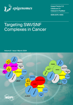
SWI/SNF enzymes are heterogeneous multi-subunit complexes that harness ATP hydrolysis to remodel chromatin, facilitating crucial cellular processes such as transcription, DNA replication, and repair. Within mammalian cells, distinct sub-complexes, namely cBAF, ncBAF, and PBAF, exhibit diverse subunit compositions and perform distinct genomic functions. Dysregulation of the SWI/SNF complex and its sub-complexes is a prominent feature of many human cancers, rendering SWI/SNF subunits appealing targets for therapeutic intervention. Current approaches in cancer therapeutics focus on pharmacological agents engineered to selectively bind and disrupt the activities of SWI/SNF complexes or specific sub-complexes, offering promising avenues for cancer treatment. View this paper
  • Issues are regarded as officially published after their release is announced to the table of contents alert mailing list.
  • You may sign up for e-mail alerts to receive table of contents of newly released issues.
  • PDF is the official format for papers published in both, html and pdf forms. To view the papers in pdf format, click on the "PDF Full-text" link, and use the free Adobe Reader to open them.
Order results
Result details
Select all
Export citation of selected articles as:
13 pages, 3569 KiB  
Article
Opposite and Differently Altered Postmortem Changes in H3 and H3K9me3 Patterns in the Rat Frontal Cortex and Hippocampus
by Karolina Dulka, Noémi Lajkó, Kálmán Nacsa and Karoly Gulya
Epigenomes 2024, 8(1), 11; https://doi.org/10.3390/epigenomes8010011 - 18 Mar 2024
Viewed by 1026
Abstract
Temporal and spatial epigenetic modifications in the brain occur during ontogenetic development, pathophysiological disorders, and aging. When epigenetic marks, such as histone methylations, in brain autopsies or biopsy samples are studied, it is critical to understand their postmortem/surgical stability. For this study, the [...] Read more.
Temporal and spatial epigenetic modifications in the brain occur during ontogenetic development, pathophysiological disorders, and aging. When epigenetic marks, such as histone methylations, in brain autopsies or biopsy samples are studied, it is critical to understand their postmortem/surgical stability. For this study, the frontal cortex and hippocampus of adult rats were removed immediately (controls) or after a postmortem delay of 15, 30, 60, 90, 120, or 150 min. The patterns of unmodified H3 and its trimethylated form H3K9me3 were analyzed in frozen samples for Western blot analysis and in formalin-fixed tissues embedded in paraffin for confocal microscopy. We found that both the unmodified H3 and H3K9me3 showed time-dependent but opposite changes and were altered differently in the frontal cortex and hippocampus with respect to postmortem delay. In the frontal cortex, the H3K9me3 marks increased approximately 450% with a slow parallel 20% decrease in the unmodified H3 histones after 150 min. In the hippocampus, the change was opposite, since H3K9me3 marks decreased steadily by approximately 65% after 150 min with a concomitant rapid increase of 20–25% in H3 histones at the same time. Confocal microscopy located H3K9me3 marks in the heterochromatic regions of the nuclei of all major cell types in the control brains: oligodendrocytes, astrocytes, neurons, and microglia. Therefore, epigenetic marks could be affected differently by postmortem delay in different parts of the brain. Full article
(This article belongs to the Collection Feature Papers in Epigenomes)
Show Figures

Figure 1

16 pages, 7042 KiB  
Article
Exploring Transcriptional Regulation of Beta Cell SASP by Brd4-Associated Proteins and Cell Cycle Control Protein p21
by Jasmine Manji, Jasmine Pipella, Gabriel Brawerman and Peter J. Thompson
Epigenomes 2024, 8(1), 10; https://doi.org/10.3390/epigenomes8010010 - 06 Mar 2024
Viewed by 1131
Abstract
Type 1 diabetes (T1D) is a metabolic disease resulting from progressive autoimmune destruction of insulin-producing pancreatic beta cells. Although the majority of beta cells are lost in T1D, a small subset undergoes senescence, a stress response involving growth arrest, DNA damage response, and [...] Read more.
Type 1 diabetes (T1D) is a metabolic disease resulting from progressive autoimmune destruction of insulin-producing pancreatic beta cells. Although the majority of beta cells are lost in T1D, a small subset undergoes senescence, a stress response involving growth arrest, DNA damage response, and activation of a senescence-associated secretory phenotype (SASP). SASP in beta cells of the nonobese diabetic (NOD) mouse model of T1D and primary human islets is regulated at the level of transcription by bromodomain extra-terminal (BET) proteins, but the mechanisms remain unclear. To explore how SASP is transcriptionally regulated in beta cells, we used the NOD beta cell line NIT-1 to model beta cell SASP and identified binding partners of BET protein Brd4 and explored the role of the cyclin-dependent kinase inhibitor p21. Brd4 interacted with a variety of proteins in senescent NIT-1 cells including subunits of the Ino80 chromatin remodeling complex, which was expressed in beta cells during T1D progression in NOD mice and in human beta cells of control, autoantibody-positive, and T1D donors as determined from single-cell RNA-seq data. RNAi knockdown of p21 during senescence in NIT-1 cells did not significantly impact viability or SASP. Taken together, these results suggest that Brd4 interacts with several protein partners during senescence in NIT-1 cells, some of which may play roles in SASP gene activation and that p21 is dispensable for the SASP in this beta cell model. Full article
(This article belongs to the Collection Epigenetic Mechanisms in Diabetes Research)
Show Figures

Figure 1

11 pages, 1307 KiB  
Review
Dynamics and Epigenetics of the Epidermal Differentiation Complex
by Wiesława Leśniak
Epigenomes 2024, 8(1), 9; https://doi.org/10.3390/epigenomes8010009 - 29 Feb 2024
Viewed by 1065
Abstract
Epidermis is the outer skin layer built of specialized cells called keratinocytes. Keratinocytes undergo a unique differentiation process, also known as cornification, during which their gene expression pattern, morphology and other properties change remarkably to the effect that the terminally differentiated, cornified cells [...] Read more.
Epidermis is the outer skin layer built of specialized cells called keratinocytes. Keratinocytes undergo a unique differentiation process, also known as cornification, during which their gene expression pattern, morphology and other properties change remarkably to the effect that the terminally differentiated, cornified cells can form a physical barrier, which separates the underlying tissues from the environment. Many genes encoding proteins that are important for epidermal barrier formation are located in a gene cluster called epidermal differentiation complex (EDC). Recent data provided valuable information on the dynamics of the EDC locus and the network of interactions between EDC gene promoters, enhancers and other regions, during keratinocytes differentiation. These data, together with results concerning changes in epigenetic modifications, provide a valuable insight into the mode of regulation of EDC gene expression. Full article
Show Figures

Figure 1

20 pages, 2263 KiB  
Review
A Comparative Analysis of Mouse Imprinted and Random X-Chromosome Inactivation
by Rebecca M. Malcore and Sundeep Kalantry
Epigenomes 2024, 8(1), 8; https://doi.org/10.3390/epigenomes8010008 - 10 Feb 2024
Viewed by 1332
Abstract
The mammalian sexes are distinguished by the X and Y chromosomes. Whereas males harbor one X and one Y chromosome, females harbor two X chromosomes. To equalize X-linked gene expression between the sexes, therian mammals have evolved X-chromosome inactivation as a dosage compensation [...] Read more.
The mammalian sexes are distinguished by the X and Y chromosomes. Whereas males harbor one X and one Y chromosome, females harbor two X chromosomes. To equalize X-linked gene expression between the sexes, therian mammals have evolved X-chromosome inactivation as a dosage compensation mechanism. During X-inactivation, most genes on one of the two X chromosomes in females are transcriptionally silenced, thus equalizing X-linked gene expression between the sexes. Two forms of X-inactivation characterize eutherian mammals, imprinted and random. Imprinted X-inactivation is defined by the exclusive inactivation of the paternal X chromosome in all cells, whereas random X-inactivation results in the silencing of genes on either the paternal or maternal X chromosome in individual cells. Both forms of X-inactivation have been studied intensively in the mouse model system, which undergoes both imprinted and random X-inactivation early in embryonic development. Stable imprinted and random X-inactivation requires the induction of the Xist long non-coding RNA. Following its induction, Xist RNA recruits proteins and complexes that silence genes on the inactive-X. In this review, we present a current understanding of the mechanisms of Xist RNA induction, and, separately, the establishment and maintenance of gene silencing on the inactive-X by Xist RNA during imprinted and random X-inactivation. Full article
(This article belongs to the Special Issue X-Chromosome Inactivation)
Show Figures

Figure 1

22 pages, 1039 KiB  
Review
Targeting SWI/SNF Complexes in Cancer: Pharmacological Approaches and Implications
by Megan R. Dreier, Jasmine Walia and Ivana L. de la Serna
Epigenomes 2024, 8(1), 7; https://doi.org/10.3390/epigenomes8010007 - 04 Feb 2024
Viewed by 2059
Abstract
SWI/SNF enzymes are heterogeneous multi-subunit complexes that utilize the energy from ATP hydrolysis to remodel chromatin structure, facilitating transcription, DNA replication, and repair. In mammalian cells, distinct sub-complexes, including cBAF, ncBAF, and PBAF exhibit varying subunit compositions and have different genomic functions. Alterations [...] Read more.
SWI/SNF enzymes are heterogeneous multi-subunit complexes that utilize the energy from ATP hydrolysis to remodel chromatin structure, facilitating transcription, DNA replication, and repair. In mammalian cells, distinct sub-complexes, including cBAF, ncBAF, and PBAF exhibit varying subunit compositions and have different genomic functions. Alterations in the SWI/SNF complex and sub-complex functions are a prominent feature in cancer, making them attractive targets for therapeutic intervention. Current strategies in cancer therapeutics involve the use of pharmacological agents designed to bind and disrupt the activity of SWI/SNF complexes or specific sub-complexes. Inhibitors targeting the catalytic subunits, SMARCA4/2, and small molecules binding SWI/SNF bromodomains are the primary approaches for suppressing SWI/SNF function. Proteolysis-targeting chimeras (PROTACs) were generated by the covalent linkage of the bromodomain or ATPase-binding ligand to an E3 ligase-binding moiety. This engineered connection promotes the degradation of specific SWI/SNF subunits, enhancing and extending the impact of this pharmacological intervention in some cases. Extensive preclinical studies have underscored the therapeutic potential of these drugs across diverse cancer types. Encouragingly, some of these agents have progressed from preclinical research to clinical trials, indicating a promising stride toward the development of effective cancer therapeutics targeting SWI/SNF complex and sub-complex functions. Full article
Show Figures

Figure 1

22 pages, 1006 KiB  
Review
Orchestrating Asymmetric Expression: Mechanisms behind Xist Regulation
by Samuel Jesus Luchsinger-Morcelle, Joost Gribnau and Hegias Mira-Bontenbal
Epigenomes 2024, 8(1), 6; https://doi.org/10.3390/epigenomes8010006 - 01 Feb 2024
Viewed by 1738
Abstract
Compensation for the gene dosage disequilibrium between sex chromosomes in mammals is achieved in female cells by repressing one of its X chromosomes through a process called X chromosome inactivation (XCI), exemplifying the control of gene expression by epigenetic mechanisms. A critical player [...] Read more.
Compensation for the gene dosage disequilibrium between sex chromosomes in mammals is achieved in female cells by repressing one of its X chromosomes through a process called X chromosome inactivation (XCI), exemplifying the control of gene expression by epigenetic mechanisms. A critical player in this mechanism is Xist, a long, non-coding RNA upregulated from a single X chromosome during early embryonic development in female cells. Over the past few decades, many factors involved at different levels in the regulation of Xist have been discovered. In this review, we hierarchically describe and analyze the different layers of Xist regulation operating concurrently and intricately interacting with each other to achieve asymmetric and monoallelic upregulation of Xist in murine female cells. We categorize these into five different classes: DNA elements, transcription factors, other regulatory proteins, long non-coding RNAs, and the chromatin and topological landscape surrounding Xist. Full article
(This article belongs to the Special Issue X-Chromosome Inactivation)
Show Figures

Figure 1

21 pages, 7681 KiB  
Article
Angio-Long Noncoding RNA MALAT1 (rs3200401) and MIAT (rs1061540) Gene Variants in Ovarian Cancer
by Manal S. Fawzy, Afaf T. Ibrahiem, Dalia Mohammad Osman, Amany I. Almars, Maali Subhi Alshammari, Layan Tariq Almazyad, Noof Daif Allah Almatrafi, Renad Tariq Almazyad and Eman A. Toraih
Epigenomes 2024, 8(1), 5; https://doi.org/10.3390/epigenomes8010005 - 29 Jan 2024
Viewed by 1467
Abstract
The genotyping of long non-coding RNA (lncRNA)-related single-nucleotide polymorphisms (SNPs) could be associated with cancer risk and/or progression. This study aimed to analyze the angiogenesis-related lncRNAs MALAT1 (rs3200401) and MIAT (rs1061540) variants in patients with ovarian cancer (OC) using “Real-Time allelic discrimination polymerase [...] Read more.
The genotyping of long non-coding RNA (lncRNA)-related single-nucleotide polymorphisms (SNPs) could be associated with cancer risk and/or progression. This study aimed to analyze the angiogenesis-related lncRNAs MALAT1 (rs3200401) and MIAT (rs1061540) variants in patients with ovarian cancer (OC) using “Real-Time allelic discrimination polymerase chain reaction” in 182 formalin-fixed paraffin-embedded (FFPE) samples of benign, borderline, and primary malignant ovarian tissues. Differences in the genotype frequencies between low-grade ovarian epithelial tumors (benign/borderline) and malignant tumors and between high-grade malignant epithelial tumors and malignant epithelial tumors other than high-grade serous carcinomas were compared. Odds ratios (ORs)/95% confidence intervals were calculated as measures of the association strength. Additionally, associations of the genotypes with the available pathological data were analyzed. The heterozygosity of MALAT1 rs3200401 was the most common genotype (47.8%), followed by C/C (36.3%). Comparing the study groups, no significant differences were observed regarding this variant. In contrast, the malignant epithelial tumors had a higher frequency of the MIAT rs1061540 C/C genotype compared to the low-grade epithelial tumor cohorts (56.7% vs. 37.6, p = 0.031). The same genotype was significantly higher in high-grade serous carcinoma than its counterparts (69.4% vs. 43.8%, p = 0.038). Multivariate Cox regression analysis showed that the age at diagnosis was significantly associated with the risk of OC development. In contrast, the MIAT T/T genotype was associated with a low risk of malignant epithelial tumors under the homozygote comparison model (OR = 0.37 (0.16–0.83), p = 0.017). Also, MIAT T allele carriers were less likely to develop high-grade serous carcinoma under heterozygote (CT vs. CC; OR = 0.33 (0.12–0.88), p = 0.027) and homozygote (TT vs. CC; OR = 0.26 (0.07–0.90), p = 0.034) comparison models. In conclusion, our data provide novel evidence for a potential association between the lncRNA MIAT rs1061540 and the malignant condition of ovarian cancer, suggesting the involvement of such lncRNAs in OC development. Full article
Show Figures

Figure 1

26 pages, 3568 KiB  
Article
Epigenetics of Genes Preferentially Expressed in Dissimilar Cell Populations: Myoblasts and Cerebellum
by Melanie Ehrlich, Kenneth C. Ehrlich, Michelle Lacey, Carl Baribault, Sagnik Sen, Pierre-Olivier Estève and Sriharsa Pradhan
Epigenomes 2024, 8(1), 4; https://doi.org/10.3390/epigenomes8010004 - 26 Jan 2024
Viewed by 1793
Abstract
While studying myoblast methylomes and transcriptomes, we found that CDH15 had a remarkable preference for expression in both myoblasts and cerebellum. To understand how widespread such a relationship was and its epigenetic and biological correlates, we systematically looked for genes with similar transcription [...] Read more.
While studying myoblast methylomes and transcriptomes, we found that CDH15 had a remarkable preference for expression in both myoblasts and cerebellum. To understand how widespread such a relationship was and its epigenetic and biological correlates, we systematically looked for genes with similar transcription profiles and analyzed their DNA methylation and chromatin state and accessibility profiles in many different cell populations. Twenty genes were expressed preferentially in myoblasts and cerebellum (Myob/Cbl genes). Some shared DNA hypo- or hypermethylated regions in myoblasts and cerebellum. Particularly striking was ZNF556, whose promoter is hypomethylated in expressing cells but highly methylated in the many cell populations that do not express the gene. In reporter gene assays, we demonstrated that its promoter’s activity is methylation sensitive. The atypical epigenetics of ZNF556 may have originated from its promoter’s hypomethylation and selective activation in sperm progenitors and oocytes. Five of the Myob/Cbl genes (KCNJ12, ST8SIA5, ZIC1, VAX2, and EN2) have much higher RNA levels in cerebellum than in myoblasts and displayed myoblast-specific hypermethylation upstream and/or downstream of their promoters that may downmodulate expression. Differential DNA methylation was associated with alternative promoter usage for Myob/Cbl genes MCF2L, DOK7, CNPY1, and ANK1. Myob/Cbl genes PAX3, LBX1, ZNF556, ZIC1, EN2, and VAX2 encode sequence-specific transcription factors, which likely help drive the myoblast and cerebellum specificity of other Myob/Cbl genes. This study extends our understanding of epigenetic/transcription associations related to differentiation and may help elucidate relationships between epigenetic signatures and muscular dystrophies or cerebellar-linked neuropathologies. Full article
(This article belongs to the Collection Feature Papers in Epigenomes)
Show Figures

Figure 1

21 pages, 1039 KiB  
Article
Sex-Specific Associations between Prenatal Exposure to Di(2-ethylhexyl) Phthalate, Epigenetic Age Acceleration, and Susceptibility to Early Childhood Upper Respiratory Infections
by Sarah M. Merrill, Nicole Letourneau, Gerald F. Giesbrecht, Karlie Edwards, Julia L. MacIsaac, Jonathan W. Martin, Amy M. MacDonald, David W. Kinniburgh, Michael S. Kobor, Deborah Dewey, Gillian England-Mason and The APrON Study Team
Epigenomes 2024, 8(1), 3; https://doi.org/10.3390/epigenomes8010003 - 26 Jan 2024
Viewed by 1791
Abstract
Di(2-ethylhexyl) phthalate (DEHP) is a common plasticizer that can affect immune system development and susceptibility to infection. Aging processes (measured as epigenetic age acceleration (EAA)) may mediate the immune-related effects of prenatal exposure to DEHP. This study’s objective was to examine associations between [...] Read more.
Di(2-ethylhexyl) phthalate (DEHP) is a common plasticizer that can affect immune system development and susceptibility to infection. Aging processes (measured as epigenetic age acceleration (EAA)) may mediate the immune-related effects of prenatal exposure to DEHP. This study’s objective was to examine associations between prenatal DEHP exposure, EAA at three months of age, and the number of upper respiratory infections (URIs) from 12 to 18 months of age using a sample of 69 maternal–child pairs from a Canadian pregnancy cohort. Blood DNA methylation data were generated using the Infinium HumanMethylation450 BeadChip; EAA was estimated using Horvath’s pan-tissue clock. Robust regressions examined overall and sex-specific associations. Higher prenatal DEHP exposure (B = 6.52, 95% CI = 1.22, 11.81) and increased EAA (B = 2.98, 95% CI = 1.64, 4.32) independently predicted more URIs. In sex-specific analyses, some similar effects were noted for boys, and EAA mediated the association between prenatal DEHP exposure and URIs. In girls, higher prenatal DEHP exposure was associated with decreased EAA, and no mediation was noted. Higher prenatal DEHP exposure may be associated with increased susceptibility to early childhood URIs, particularly in boys, and aging biomarkers such as EAA may be a biological mechanism. Larger cohort studies examining the potential developmental immunotoxicity of phthalates are needed. Full article
(This article belongs to the Collection Feature Papers in Epigenomes)
Show Figures

Graphical abstract

19 pages, 740 KiB  
Review
The Genomic Shock Hypothesis: Genetic and Epigenetic Alterations of Transposable Elements after Interspecific Hybridization in Plants
by Carlos de Tomás and Carlos M. Vicient
Epigenomes 2024, 8(1), 2; https://doi.org/10.3390/epigenomes8010002 - 27 Dec 2023
Viewed by 1758
Abstract
Transposable elements (TEs) are major components of plant genomes with the ability to change their position in the genome or to create new copies of themselves in other positions in the genome. These can cause gene disruption and large-scale genomic alterations, including inversions, [...] Read more.
Transposable elements (TEs) are major components of plant genomes with the ability to change their position in the genome or to create new copies of themselves in other positions in the genome. These can cause gene disruption and large-scale genomic alterations, including inversions, deletions, and duplications. Host organisms have evolved a set of mechanisms to suppress TE activity and counter the threat that they pose to genome integrity. These includes the epigenetic silencing of TEs mediated by a process of RNA-directed DNA methylation (RdDM). In most cases, the silencing machinery is very efficient for the vast majority of TEs. However, there are specific circumstances in which TEs can evade such silencing mechanisms, for example, a variety of biotic and abiotic stresses or in vitro culture. Hybridization is also proposed as an inductor of TE proliferation. In fact, the discoverer of the transposons, Barbara McClintock, first hypothesized that interspecific hybridization provides a “genomic shock” that inhibits the TE control mechanisms leading to the mobilization of TEs. However, the studies carried out on this topic have yielded diverse results, showing in some cases a total absence of mobilization or being limited to only some TE families. Here, we review the current knowledge about the impact of interspecific hybridization on TEs in plants and the possible implications of changes in the epigenetic mechanisms. Full article
(This article belongs to the Collection Epigenetic Control in Plants)
Show Figures

Figure 1

31 pages, 1212 KiB  
Review
Inheritance of Stress Responses via Small Non-Coding RNAs in Invertebrates and Mammals
by Maria C. Ow and Sarah E. Hall
Epigenomes 2024, 8(1), 1; https://doi.org/10.3390/epigenomes8010001 - 19 Dec 2023
Cited by 1 | Viewed by 1700
Abstract
While reports on the generational inheritance of a parental response to stress have been widely reported in animals, the molecular mechanisms behind this phenomenon have only recently emerged. The booming interest in epigenetic inheritance has been facilitated in part by the discovery that [...] Read more.
While reports on the generational inheritance of a parental response to stress have been widely reported in animals, the molecular mechanisms behind this phenomenon have only recently emerged. The booming interest in epigenetic inheritance has been facilitated in part by the discovery that small non-coding RNAs are one of its principal conduits. Discovered 30 years ago in the Caenorhabditis elegans nematode, these small molecules have since cemented their critical roles in regulating virtually all aspects of eukaryotic development. Here, we provide an overview on the current understanding of epigenetic inheritance in animals, including mice and C. elegans, as it pertains to stresses such as temperature, nutritional, and pathogenic encounters. We focus on C. elegans to address the mechanistic complexity of how small RNAs target their cohort mRNAs to effect gene expression and how they govern the propagation or termination of generational perdurance in epigenetic inheritance. Presently, while a great amount has been learned regarding the heritability of gene expression states, many more questions remain unanswered and warrant further investigation. Full article
(This article belongs to the Special Issue Transgenerational Epigenetic Inheritance)
Show Figures

Figure 1

Previous Issue
Next Issue
Back to TopTop