

Cerebral Aneurysms and Arteriovenous Malformation: Preliminary Experience with the Use of Near-Infrared Fluorescence Imaging Applied to Endoscopy

 , , , , , ,
Abstract
1. Introduction
2. Materials and Methods
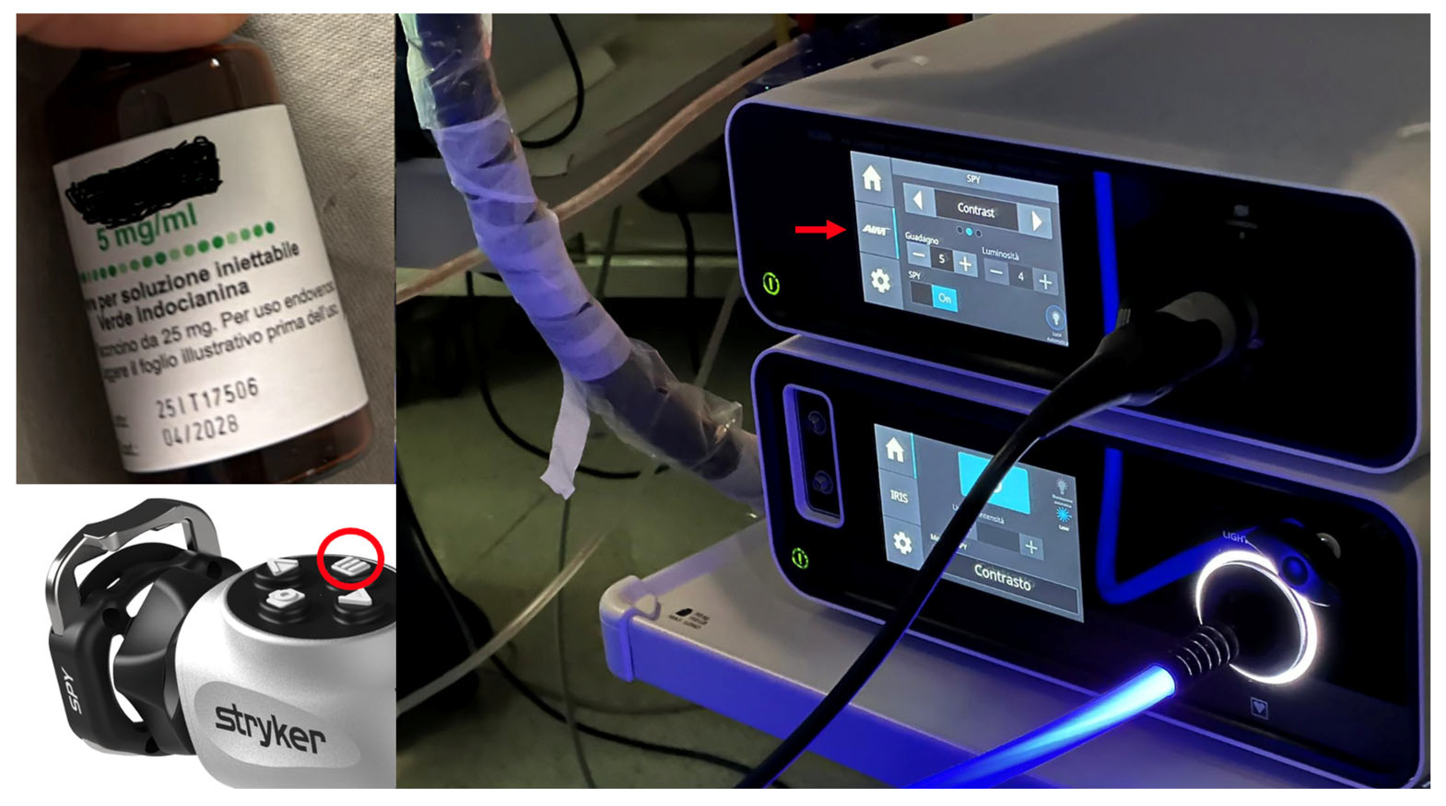
2.1. Study Cohort and Surgical Equipment
2.2. Preparation Protocol
2.3. Use of the Endoscope with Fluorescence Visualization and IntraOperative Evaluation
3. Results
3.1. Patients Overview
- (1)
- A 57-year-old male patient with a middle cerebral artery aneurysm (M2 segment) with a diameter of 1.2 cm, neurologically intact. The patient underwent clipping of the aneurysm neck.
- (2)
- A 73-year-old female patient with temporal AVM originating from the left middle cerebral artery and discharge into the ipsilateral transverse sinus, with a 3 cm nidus. The AVM manifested itself with an episode of fluent aphasia. The patient underwent preoperative embolization and microsurgical removal of the nidus (see Figure 1).
- (3)
- A 46-year-old female patient with an aneurysm of the anterior communicating artery with a diameter of 1 cm, neurologically intact. The patient underwent clipping of the aneurysm neck.
- (4)
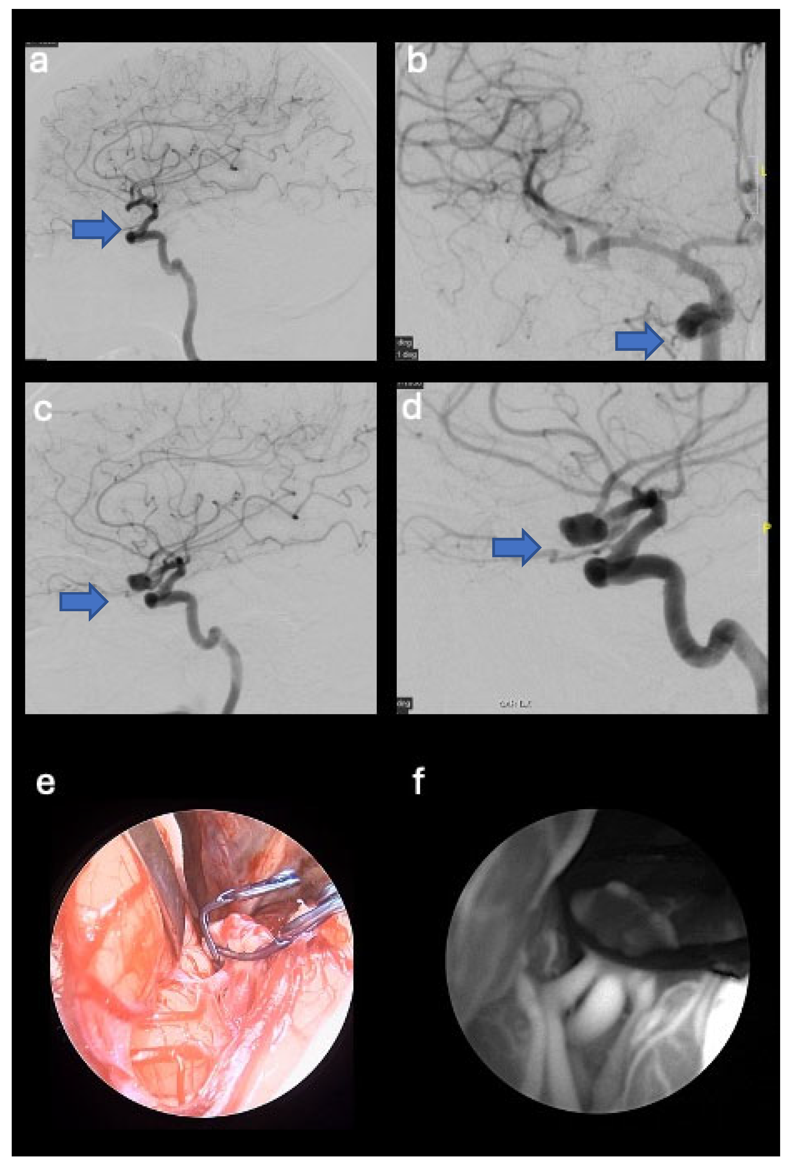
- A 44-year-old female patient with a right middle cerebral artery aneurysm (M1-M2 union) with a diameter of 1 cm, a history of headaches. The patient underwent clipping of the aneurysm neck (see Figure 2).
- (5)
- A 31-year-old male patient with frontal AVM originating from the right anterior cerebral artery and discharge into the sagittal sinus, with a 2.5 cm nidus. The patient presented with weakness in the left upper limb and underwent microsurgical removal of the nidus.

3.2. Illustrative Cases
3.2.1. Case Number 2
3.2.2. CASE Number 4
4. Discussion
5. Conclusions
Author Contributions
Funding
Institutional Review Board Statement
Informed Consent Statement
Data Availability Statement
Conflicts of Interest
References
- De Notaris, M.; Sacco, M.; Corrivetti, F.; Dallan, I.; Cavallo, L.M.; Somma, T.; Parbonetti, G.; Colamaria, A.; Solari, D. Indocyanine Green Endoscopy for Pituitary Adenomas with Parasellar Extension: Results from a Preliminary Case Series. World Neurosurg. 2022, 166, e692–e702. [Google Scholar] [CrossRef] [PubMed]
- Srinivasan, V.M.; Shlobin, N.A.; Karahalios, K.; Scherschinski, L.; Rahmani, R.; Graffeo, C.S.; Burkhardt, J.K.; Chaurasia, B.; Catapano, J.S.; Labib, M.A.; et al. Adoption of Advanced Microneurosurgical Technologies: An International Survey. World Neurosurg. 2022, 157, e473–e483. [Google Scholar] [CrossRef] [PubMed]
- Vega-Moreno, D.A.; Janković, D.; Azouz, H.; Nakipuria, M.; Kato, Y. Dual Microscope Indocyanine Green Video Angiography and Endoscopic Review to Treat Intracranial Aneurysm: A Review of the Literature Regarding a Case. Asian J. Neurosurg. 2023, 18, 701–707. [Google Scholar] [CrossRef] [PubMed] [PubMed Central]
- Muto, J.; Mine, Y.; Nishiyama, Y.; Murayama, K.; Hayakawa, M.; Hasegawa, M.; Lee, J.K.Y.; Hirose, Y. Intraoperative Real-Time Near-Infrared Image-Guided Endoscopic Endonasal Surgery for Pituitary Tumors. World Neurosurg. 2023, 175, e218–e229. [Google Scholar] [CrossRef] [PubMed]
- Ferlendis, L.; Veiceschi, P.; Capelli, S.; Agresta, G.; Leocata, A.; Pozzi, F.; Locatelli, D. Ultrahigh-Definition-3-Dimensional Exoscope-Assisted Clipping of a Right Middle Cerebral Artery Unruptured Aneurysm with Indocyanine Green Video Angiography: Operative Video. World Neurosurg. 2023, 179, 102–103. [Google Scholar] [CrossRef] [PubMed]
- Inokuchi, G.; Mine, M.; Tamagawa, K.; Tatehara, S.; Yui, M.; Uozumi, Y.; Fujita, Y.; Nakai, T.; Nibu, K.I. Indocyanine green fluorescence visualizes landmark arteries for endoscopic sinus and skull base surgery. Am. J. Otolaryngol. 2024, 45, 104343. [Google Scholar] [CrossRef] [PubMed]
- Della Puppa, A.; Rossetto, M.; Volpin, F.; Rustemi, O.; Grego, A.; Gerardi, A.; Ortolan, R.; Causin, F.; Munari, M.; Scienza, R. Microsurgical Clipping of Intracranial Aneurysms Assisted by Neurophysiological Monitoring, Microvascular Flow Probe, and ICG-VA: Outcomes and Intraoperative Data on a Multimodal Strategy. World Neurosurg. 2018, 113, e336–e344. [Google Scholar] [CrossRef] [PubMed]
- Mansour, A.; Endo, T.; Inoue, T.; Sato, K.; Endo, H.; Fujimura, M.; Tominaga, T. Clipping of an anterior spinal artery aneurysm using an endoscopic fluorescence imaging system for craniocervical junction epidural arteriovenous fistula: Technical note. J. Neurosurg. Spine 2019, 31, 279–284. [Google Scholar] [CrossRef] [PubMed]
- Devgan, Y.; Mayilvaganan, S.; Mishra, A.; Chand, G.; Agarwal, G.; Agarwal, A. Comparison of indocyanine green angiography vs. intraoperative parathyroid hormone in early prediction of risk of post-thyroidectomy hypocalcemia: A prospective cohort study. Ann. Med. Surg. 2024, 86, 678–688. [Google Scholar] [CrossRef] [PubMed] [PubMed Central]
- Teng, C.W.; Huang, V.; Arguelles, G.R.; Zhou, C.; Cho, S.S.; Harmsen, S.; Lee, J.Y.K. Applications of indocyanine green in brain tumor surgery: Review of clinical evidence and emerging technologies. Neurosurg. Focus 2021, 50, E4. [Google Scholar] [CrossRef] [PubMed]
- Ferroli, P.; Acerbi, F.; Albanese, E.; Tringali, G.; Broggi, M.; Franzini, A.; Broggi, G. Application of intraoperative indocyanine green angiography for CNS tumors: Results on the first 100 cases. In Intraoperative Imaging; Acta Neurochirurgica Supplementum (Volume 109); Springer: Berlin/Heidelberg, Germany, 2011; pp. 251–257. [Google Scholar] [CrossRef] [PubMed]
- Cho, S.S.; Salinas, R.; Lee, J.Y.K. Indocyanine-Green for Fluorescence-Guided Surgery of Brain Tumors: Evidence, Techniques, and Practical Experience. Front. Surg. 2019, 6, 11. [Google Scholar] [CrossRef] [PubMed] [PubMed Central]
- Yannuzzi, L.A. Indocyanine green angiography: A perspective on use in the clinical setting. Am. J. Ophthalmol. 2011, 151, 745–751.e1. [Google Scholar] [CrossRef] [PubMed]
- Lau, C.T.; Au, D.M.; Wong, K.K.Y. Application of indocyanine green in pediatric surgery. Pediatr. Surg. Int. 2019, 35, 1035–1041. [Google Scholar] [CrossRef] [PubMed]
- Raabe, A.; Beck, J.; Gerlach, R.; Zimmermann, M.; Seifert, V. Near-infrared indocyanine green video angiography: A new method for intraoperative assessment of vascular flow. Neurosurgery 2003, 52, 132–139; discussion 139. [Google Scholar] [CrossRef] [PubMed]
- Jeon, J.W.; Cho, S.S.; Nag, S.; Buch, L.; Pierce, J.; Su, Y.S.; Adappa, N.D.; Palmer, J.N.; Newman, J.G.; Singhal, S.; et al. Near-Infrared Optical Contrast of Skull Base Tumors During Endoscopic Endonasal Surgery. Oper Neurosurg 2019, 17, 32–42. [Google Scholar] [CrossRef] [PubMed] [PubMed Central]
- Della Puppa, A.; Rustemi, O.; Scienza, R. The “ICG Entrapment Sign” in Cerebral Aneurysm Surgery Assisted by Indocyanine Green Videoangiography. World Neurosurg. 2017, 97, 287–291. [Google Scholar] [CrossRef] [PubMed]
- Balamurugan, S.; Agrawal, A.; Kato, Y.; Sano, H. Intra operative indocyanine green video-angiography in cerebrovascular surgery: An overview with review of literature. Asian J. Neurosurg. 2011, 6, 88–93. [Google Scholar] [CrossRef] [PubMed] [PubMed Central]
- Chibbaro, S.; Tacconi, L. Extracranial-intracranial bypass for the treatment of cavernous sinus aneurysms. J. Clin. Neurosci. 2006, 13, 1001–1005. [Google Scholar] [CrossRef] [PubMed]
- Takagi, Y.; Samamura, K.; Hashimoto, N. Intra operative Near-infrared Indocyanine green video angiography performed with surgical microscope—Applications in Cerebrovascular surgery. Eur. Neurol. Rev. 2008, 3, 66–68. [Google Scholar] [CrossRef]
- Mery, F.J.; Amin-Hanjani, S.; Charbel, F.T. Is an angiographically obliterated aneurysm always secure? Neurosurgery 2008, 62, 979–982; discussion 982. [Google Scholar] [CrossRef] [PubMed]
- Della Puppa, A.; Rustemi, O.; Rossetto, M.; Gioffrè, G.; Munari, M.; Charbel, F.T.; Scienza, R. The “squeezing maneuver” in microsurgical clipping of intracranial aneurysms assisted by indocyanine green videoangiography. Neurosurgery 2014, 10 (Suppl. S2), 208–212; discussion 212–213. [Google Scholar] [CrossRef] [PubMed]
- Zitek, H.; Hejcl, A.; Sadeh, M.; Charbel, F.T.; Sames, M. Occipital artery to vertebral artery bypass for treatment of bilateral vertebral artery occlusion with QMRA as an adjunct to diagnostic assessment. Acta Neurochir 2024, 166, 203. [Google Scholar] [CrossRef] [PubMed] [PubMed Central]
- Acerbi, F.; Prada, F.; Vetrano, I.G.; Falco, J.; Faragò, G.; Ferroli, P.; DiMeco, F. Indocyanine Green and Contrast-Enhanced Ultrasound Videoangiography: A Synergistic Approach for Real-Time Verification of Distal Revascularization and Aneurysm Occlusion in a Complex Distal Middle Cerebral Artery Aneurysm. World Neurosurg. 2019, 125, 277–284. [Google Scholar] [CrossRef] [PubMed]
- Hashimoto, K.; Kinouchi, H.; Yoshioka, H.; Kanemaru, K.; Ogiwara, M.; Yagi, T.; Wakai, T.; Fukumoto, Y. Efficacy of Endoscopic Fluorescein Video Angiography in Aneurysm Surgery-Novel and Innovative Assessment of Vascular Blood Flow in the Dead Angles of the Microscope. Oper Neurosurg 2017, 13, 471–481. [Google Scholar] [CrossRef] [PubMed]
- Catapano, G.; Sgulò, F.; Laleva, L.; Columbano, L.; Dallan, I.; de Notaris, M. Multimodal use of indocyanine green endoscopy in neurosurgery: A single-center experience and review of the literature. Neurosurg. Rev. 2018, 41, 985–998. [Google Scholar] [CrossRef] [PubMed] [PubMed Central]
- Wong, A.K.; Wong, R.H. Keyhole clipping of a low-lying basilar apex aneurysm without posterior clinoidectomy utilizing endoscopic indocyanine green video angiography. Surg. Neurol. Int. 2020, 11, 31. [Google Scholar] [CrossRef] [PubMed] [PubMed Central]
- Bruneau, M.; Appelboom, G.; Rynkowski, M.; Van Cutsem, N.; Mine, B.; De Witte, O. Endoscope-integrated ICG technology: First application during intracranial aneurysm surgery. Neurosurg. Rev. 2013, 36, 77–84. [Google Scholar] [CrossRef]
- De Oliveira, J.G.; Beck, J.; Seifert, V.; Teixeira, M.J.; Raabe, A. Assessment of flow in perforating arteries during intracranial aneurysm surgery using intraoperative near-infrared indocyanine green videoangiography. Neurosurgery 2008, 62 (Suppl. S3), SHC1300–SHC1310. [Google Scholar] [CrossRef]
- Fischer, G.; Rediker, J.; Oertel, J. Endoscope-versus microscope-integrated near-infrared indocyanine green videoangiography in aneurysm surgery. J. Neurosurg. 2018, 131, 1413–1422. [Google Scholar] [CrossRef]
- Kalavakonda, C.; Sekhar, L.N.; Ramachandran, P.; Hechl, P. Endoscope-assisted microsurgery for intracranial aneurysms. Neurosurgery 2002, 51, 1119–1126. [Google Scholar] [CrossRef]
- Mielke, D.; Malinova, V.; Rohde, V. Comparison of intraoperative microscopic and endoscopic ICG angiography in aneurysm surgery. Neurosurgery 2014, 10 (Suppl. S3), 418–425. [Google Scholar] [CrossRef] [PubMed]
- Nishiyama, Y.; Kinouchi, H.; Senbokuya, N.; Kato, T.; Kanemaru, K.; Yoshioka, H.; Horikoshi, T. Endoscopic indocyanine green video angiography in aneurysm surgery: An innovative method for intraoperative assessment of blood flow in vasculature hidden from microscopic view. J. Neurosurg. 2012, 117, 302–308. [Google Scholar] [CrossRef] [PubMed]
- Washington, C.W.; Zipfel, G.J.; Chicoine, M.R.; Derdeyn, C.P.; Rich, K.M.; Moran, C.J.; Cross, D.T.; Dacey, R.G. Comparing indocyanine green videoangiography to the gold standard of intraoperative digital subtraction angiography used in aneurysm surgery. J. Neurosurg. 2013, 118, 420–427. [Google Scholar] [CrossRef] [PubMed]
- Oda, J.; Kato, Y.; Chen, S.F.; Sodhiya, P.; Watabe, T.; Imizu, S.; Sano, H.; Hirose, Y. Intraoperative near-infrared indocyanine green-videoangiog-raphy (ICG-VA) and graphic analysis of fluorescence intensity in cerebral aneurysm surgery. J. Clin. Neurosci. 2011, 18, 1097–1100. [Google Scholar] [CrossRef] [PubMed]
- Chen, D.Y.; Xu, C.S.; Fu, K.; Ma, Y.H.; Zhang, T.B.; Zou, Y.C.; Chen, J.C. Application of neuroendoscopy combined with fluorescence angiography in anterior circulation aneurysm clipping. Zhonghua Yi Xue Za Zhi 2021, 101, 254–258. (In Chinese) [Google Scholar] [CrossRef] [PubMed]

| Age | Sex | Clinical Data | CV Disease | Neurosurgical Procedure | Postoperative Outcome | |
|---|---|---|---|---|---|---|
| 1 | 57 | M | Neurologically intact | Left middle cerebral artery aneurysm (M2 segment) with a diameter of 1.2 cm | Clipping of the aneurysm neck | Neurologically intact; total exclusion of the aneurysm. No bleeding events. |
| 2 | 73 | F | Episode of fluent aphasia | Left temporal AVM originating from the left middle cerebral artery and venous drainage into the ipsilateral transverse sinus (3 cm nidus) | Preoperative embolization and the microsurgical removal of the nidus | Complete removal of the nidus and transient fluent aphasia lasting three months; no bleeding events. |
| 3 | 46 | F | Neurologically intact | Anterior communicating artery aneurysm with a diameter of 1 cm | Clipping of the aneurysm neck | Neurologically intact, total exclusion of the aneurysm, and no bleeding events. |
| 4 | 44 | F | Headache | Right middle cerebral artery aneurysm (M1-M2 union) with a diameter of 1.1 cm | Clipping of the aneurysm neck | Neurologically intact, total exclusion of the aneurysm, and no bleeding events. |
| 5 | 31 | M | Weakness in the left upper limb | Right frontal AVM originating from the right anterior cerebral artery and venous drainage into the sagittal sinus (2.5 cm nidus) | Microsurgical removal of the nidus | Complete removal of the nidus and partial recovery of weakness. No bleeding events. |
Disclaimer/Publisher’s Note: The statements, opinions and data contained in all publications are solely those of the individual author(s) and contributor(s) and not of MDPI and/or the editor(s). MDPI and/or the editor(s) disclaim responsibility for any injury to people or property resulting from any ideas, methods, instructions or products referred to in the content. |
© 2024 by the authors. Licensee MDPI, Basel, Switzerland. This article is an open access article distributed under the terms and conditions of the Creative Commons Attribution (CC BY) license (https://creativecommons.org/licenses/by/4.0/).
Share and Cite
Aiudi, D.; Iacoangeli, A.; Mattioli, A.; Raggi, A.; Dobran, M.; Polonara, G.; Gigli, R.; Iacoangeli, M.; Gladi, M. Cerebral Aneurysms and Arteriovenous Malformation: Preliminary Experience with the Use of Near-Infrared Fluorescence Imaging Applied to Endoscopy. J. Pers. Med. 2024, 14, 1117. https://doi.org/10.3390/jpm14121117
Aiudi D, Iacoangeli A, Mattioli A, Raggi A, Dobran M, Polonara G, Gigli R, Iacoangeli M, Gladi M. Cerebral Aneurysms and Arteriovenous Malformation: Preliminary Experience with the Use of Near-Infrared Fluorescence Imaging Applied to Endoscopy. Journal of Personalized Medicine. 2024; 14(12):1117. https://doi.org/10.3390/jpm14121117
Chicago/Turabian StyleAiudi, Denis, Alessio Iacoangeli, Andrea Mattioli, Alessio Raggi, Mauro Dobran, Gabriele Polonara, Riccardo Gigli, Maurizio Iacoangeli, and Maurizio Gladi. 2024. "Cerebral Aneurysms and Arteriovenous Malformation: Preliminary Experience with the Use of Near-Infrared Fluorescence Imaging Applied to Endoscopy" Journal of Personalized Medicine 14, no. 12: 1117. https://doi.org/10.3390/jpm14121117
APA StyleAiudi, D., Iacoangeli, A., Mattioli, A., Raggi, A., Dobran, M., Polonara, G., Gigli, R., Iacoangeli, M., & Gladi, M. (2024). Cerebral Aneurysms and Arteriovenous Malformation: Preliminary Experience with the Use of Near-Infrared Fluorescence Imaging Applied to Endoscopy. Journal of Personalized Medicine, 14(12), 1117. https://doi.org/10.3390/jpm14121117