Abstract
Background: Macular hole (MH) can severely impair central vision. Although it can be treated with vitrectomy surgery, avoiding recurrence and improving visual acuity are still priorities to be addressed. This study aims to reveal the trends and hotspots about MH. Methods: The Web of Science Core Collection (WOSCC) was used to perform a bibliometric analysis investigating trends of MH research from 2002 to 2021. We evaluated the details of associated regions, institutions, authors, and journals. To construct and overlay network visualizations, VOSviewer software was used. Results: In total, 1518 publications were collected. Our analysis showed that MH research is becoming increasingly relevant, with Japan achieving the largest number of publications (291), largest number of citations (7745 in total), and highest h-index value (48). Retina published the most publications on this topic, totaling more than the next two journals combined. An analysis of keyword co-occurrence was evaluated, highlighting several novel keywords of interest, such as flap technique, transplantation, epiretinal proliferation (EP), foveal microstructure, and retinal sensitivity. Conclusions: Details on MH research were uncovered by comprehensively analyzing the global trends and hotspots over the past two decades, presenting valuable information for future MH research. Japan, the USA, and China hold leading positions in research on this topic. Amendable surgical methods are a potential focus for improving prognosis.
1. Introduction
The vitreomacular interface (VMI) includes vitreomacular adhesion, vitreomacular traction, and potentially macular hole (MH), arising from tissue defects that may harm the retinal internal limiting membrane (ILM) or even the photoreceptor (PR) layer [1]. The impact of MH varies widely, making it a retinal disease with a broad spectrum consisting of different subtypes, each with its own defined pathogenesis, morphology, and specific therapeutic option [2].
According to the pathogenesis, MH has been conveniently divided into two types, idiopathic MH and secondary MH. The latter is attributed to but not limited to pathologic conditions [3] such as trauma [4], high myopia [5], uveitis, macular telangiectasia [6], macular schisis [5], idiopathic retinal vasoproliferative tumor [7], proliferative diabetic retinopathy (PDR) [8], and various retinal pathologies. MH may lead to vision loss, central dark spots and metamorphopsia, which can occur suddenly or gradually [9,10]. Reports of its prevalence vary widely, from 0.02% to 0.33%, in publications [9].
In the past 20 years, a lot of MH research has been published, so exploring the hotspots and trends will better guide future research. While bibliometrics can extract quantitative data through statistical analysis of scholarly publications to measure their impact [11], none are available for the topic of MH to our knowledge. Therefore, this study aims to explore published research through a bibliometric approach to informatively visualize the current MH landscape. Our study objectively assesses publication numbers, country/region contributions, co-authorship, individual productivity, and participating journals in the MH field. These were achieved through co-citation analysis and keyword co-occurrence analysis [12].
2. Materials and Methods
2.1. Search Methods
Science Citation Index Expanded in the Web of Science Core Collection (WOSCC) was used to search for publications included in this study. All the literature results included were published from 2002 to 2021 and were searched for on 12 October 2022. The keyword used was “Title = macular hole”, and 2466 results from the literature were identified. In total, 1514 articles and 77 reviews were included. Figure 1 depicts the selection process.
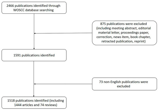
Figure 1.
Flowchart of the search strategy of our study.
2.2. Data Collection and Bibliometric Analysis
Bibliographic information, such as author, research institution, country/region, keywords, and citations were downloaded from WOSCC databases as TXT. Network visualizations and overlay visualizations were then used for input, when data are analyzed using VOSviewer version 1.6.18 (Supported by the Centre for Science and Technology Studies of Leiden University). Microsoft Excel 2019 was used to plot graphs. Relative research interest (RRI) was calculated using the number of MH publications divided by all publications in all fields within a year. H-index was obtained from the WOSCC, for it is useful in quantifying a researcher’s output. The prediction model was f(x) = ax3 + bx2 + cx + d (x means the year and f(x) means the cumulative publications in a certain year), which was calculated using Microsoft Excel 2019.
In the network visualization, each node with a label represents an element, such as countries/regions. Occurrence frequencies such as publications are visualized using the size of nodes. Collaborative relationships between two nodes are reflected by links, with thickness and length depicting the strength of connection and relevance, respectively. Different clusters of nodes are codified using colors. In the overlay visualizations, all nodes were color-coded, highlighting the average time of appearance, written as average appearing year (AAY). Yellow means more recent, while blue means earlier appearance.
2.3. Research Ethics
All data in this article were searched and downloaded from a public database (WOSCC). No human subjects or animals were involved, so no ethical review was required for this study.
3. Results
3.1. Countries/Regions
From 2002 to 2021, Japan contributed the most publications (291, 19.17%), followed by the USA (281, 18.51%), and then China (181, 11.92%) (Figure 2A). China in particular saw a substantial increase in MH publications. Moreover, RRI ranging from 0.003% to 0.005% indicated that the global interest has been high over the past two decades (Figure 2B).
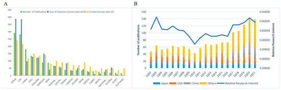
Figure 2.
Contributions of different countries/regions. (A) Top 20 countries/regions in MH publications. Green bar means the number of publications, blue bar means the sum of citations in total (actual value multiplied by 0.05), and yellow bar means the h-index (actual value multiplied by 5). (B) The proportion and RRI of Japan, USA, China and others in the MH field from 2002 to 2021. The left axis means the number of publications, and the right axis means the RRI. RRI: relative research interest.
The minimum number of documents of a country/region was set as five, and 33 countries reached that threshold out of 62. Then, we analyzed the co-authorship of the 33 countries and regions (Supplementary Figure S1); this analysis returned seven clusters. The red cluster, consisting of eight countries/regions, Egypt, India, Italy, Portugal, Saudi Arabia, Spain, Sweden, and Turkey, was the largest. We also color-coded the keywords by the AAY (Supplementary Figure S2). The yellow nodes, such as for China, India, and Italy, were the ones that published more papers recently.
3.2. Citations and H-Index
WOS citation reports showed that all publications related to MH have been cited 30,862 times since 2002 (16,714 citations without self-citations; 74 h-index). Based on the number of publications, the number of citations, and the h-index of each country/region, Japan (291 publications; 7745 citations in total; 48 h-index), the USA (281 publications; 7680 citations in total; 43 h-index), and China (181 publications; 1959 citations in total; 23 h-index) were the three most influential countries/regions in MH research. Of the top 10 authors, Kampik A accumulated the most citations (1712 citations in total; 1684 without self-citations; 17 h-index). Among the top 10 organizations, the University of Munich contributed the most citations (1910 citations in total; 1848 without self-citations; 18 h-index) (Table 1).

Table 1.
The top 10 productive countries/regions, organizations, and authors from 2002 to 2021.
3.3. Publication Trends for the Future
The number of MH publications has continued to increase in the past two decades, and the growth curves demonstrated a significant correlation between year and cumulative publications (Figure 3). Japan and the USA held a steady and fast rate of growth (Figure 3B,C). Furthermore, the prediction model showed that China saw a much faster rate of growth since 2015 and may reach approximately 350 cumulative publications in 2025 (Figure 3D).
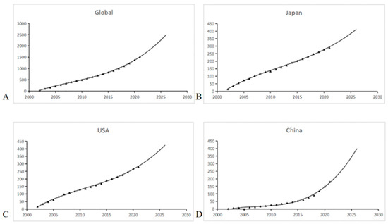
Figure 3.
The growth and prediction curves for publications. (A) Global; (B) Japan; (C) the USA; (D) China.
3.4. Research Organizations
The 1518 publications were written by 1405 organizations. The top 10 most productive organizations for MH research published 297 publications, accounting for 19.58% of the total publications (Table 1). The most productive organizations for MH research were the University of London (44 publications), followed by University College London (35 publications), and Moorfields Eye Hospital NHS Foundation Trust (34 publications). The co-authorship analysis network reflected the cooperation relationship among these institutions (Supplementary Figure S3).
3.5. Source Journals
The 1518 publications were submitted to 108 journals. Table 2 lists the top 10 active journals publishing MH research. American journals represented nearly half of all the publications included in the top 10 journals. Retina published the most publications (274, 18.05%), with the American Journal of Ophthalmology (141, 9.29%) in second place and Graefes Archive for Clinical and Experimental Ophthalmology (112, 7.38%) in third place.
Table 2.
Top 10 source journals on MH research from 2002 to 2021.
3.6. Co-Authorship
A total of 4676 authors published the 1518 retrieved publications. Table 1 lists the top 10 authors. In VOSviewer, the minimum number of publications published by an author was set as five. Of the 5284 authors, 183 authors were above that threshold. The co-authorship network, including 102 authors, was divided into 10 clusters, which are represented by different colors (Figure 4).
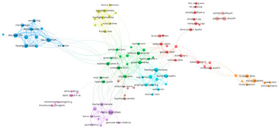
Figure 4.
The co-authorship network. Each node with a label represents an author. The size of nodes represents the number of publications. The links between two nodes reflect the collaborative relationships between the two authors. The color of nodes means different clusters the node belongs to.
The top 10 papers were displayed in Table 3; the most cited paper was published in Ophthalmology, named The International Vitreomacular Traction Study Group Classification of Vitreomacular Adhesion, Traction, and Macular Hole. The first author was Duker, Jay S.
Table 3.
Top 10 most highly cited publications ranked by number of citations.
3.7. Co-Cited Authors and References
Gass J (716 co-citations) achieved the top place, followed by Michalewska Z (618 co-citations) and Kelly N (453 co-citations), among the top 10 most co-cited authors.
The minimum number of citations of a cited reference was set as 20. Of the 8988 cited references, 380 co-cited references met the threshold and were selected for analysis. References with a high similarity were found to be in seven clusters, colored in red, green, blue, yellow, orange, purple, and cyan (Supplementary Figure S4).
3.8. Keywords Co-Occurrence
The minimum co-occurrence of a keyword was set as 25. Of the 2129 keywords used in MH research, 84 met the threshold and were selected for analysis. Based on this network, the keywords with a high similarity were clustered. The four main clusters are color-coded in red, yellow, green, blue, and purple (Figure 5A). Keywords were also colored according to the AAY (Figure 5B). Those colored yellow, such as flap technique, transplantation, epiretinal proliferation, foveal microstructure, and retinal sensitivity, were the ones that appeared most recently.
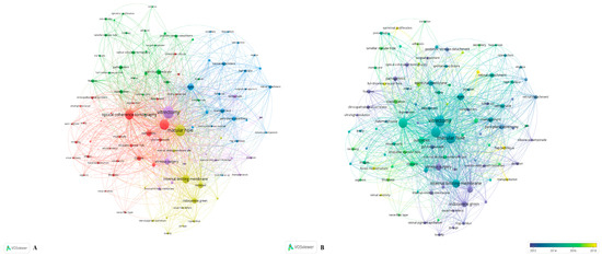
Figure 5.
Keywords analysis by VOSviewer. (A) Network visualization of keywords in the field of MH research. Each color represents a single cluster, and the keywords with high similarity were clustered. (B) Keywords were color-coded by AAY. Yellow means more recently, and blue means earlier. AAY: average appearing year.
4. Discussion
4.1. Global Trends
Visualization of the bibliometric information was used to present a better overview of the current research status, trends, and hotspots on MH research from 2002 to 2021. In total, 1518 manuscripts were screened.
According to Supplementary Figure S1, the size of the nodes represented the publications of each country/region. The lines reflected the collaborative relationships between two nodes (the more frequent collaboration was, the thicker the connecting lines are). Although Japan had the most publications, most citations, and highest h-index, the USA engaged in the highest number of collaborations with other countries/regions. This may be attributed to the USA being the first country to perform surgical techniques for treating MH. Kelly and Wendel in the USA wrote a seminal report on the description of vitrectomy surgery for MH in 1991 [13]. In 1997, Eckardt et al. reported that ILM peeling was effective to prevent the recurrence of MH [14]. The success of closure rates has also improved with the continuous improvement of the operation [14,15,16,17,18,19,20,21,22,23,24].
The large number of surgeries performed and the development of ophthalmology in the USA have enabled many clinical studies of MH to be performed. This has contributed to the USA’s great influence in the field of MH research. Intriguingly, there were tight collaborative relationships between the top five countries/regions, while Japan and China cooperated less with others.
In total, 4676 authors from 1405 research organizations in 62 countries/regions have published publications on the topic, showing that MH research has garnered widespread interest worldwide. As shown in Supplementary Figure S3, the co-occurrence analysis network reflects the collaborative relationships between these institutions. In this network, there was extensive cooperation among organizations without geographical restrictions, while there were quite a few institutions nodes that were not linked to any other nodes. More academic cooperation and exchange between countries/regions and organizations are expected in the future.
Retina, the American Journal of Ophthalmology, and Graefes Archive for Clinical and Experimental were the top three journals with the highest number of publications in the field of MH research. Retina presented the highest number of publications, more than the second- and third-highest numbers of publications combined. This could be attributed to Retina being a classic journal, focused on the introduction of new surgical techniques. The main treatment for MH is surgery, which falls within the focus of Retina. In conclusion, the three above-mentioned journals were the three most influential journals in the field of MH research. In addition, among the top 10 most sourced journals, there were six American journals. That is to say, American journals were the leading journals on MH research by quantity.
4.2. Research Hotspots
The top five keywords were vitrectomy, macular hole, surgery, optical coherence tomography (OCT), and ILM. Pars plana vitrectomy (PPV) combined with ILM peeling was considered the standard procedure in the treatment of MH [25,26]. Some also suggested using gas tamponade to promote the closure of LMH [27,28]. MH formation typically evolves through four stages, which were first described by Gass [1,29]. OCT is the gold standard for MH diagnosis. In Figure 5B, yellow symbolizes more recently while blue represents earlier. Attention has been paid not only to the restoration of retinal structure but also to the restoration of visual function. Researchers are starting to focus on some novel keywords, such as flap technique, transplantation, epiretinal proliferation (EP), foveal microstructure, and retinal sensitivity.
EP is mainly composed of non-contractile GFAP-positive gliotic tissue derived from Müller cells [30]. It was first described by Witkin et al. in 2006 on an ultrahigh-resolution OCT [31]. Thus, EP could be considered a form of chronic severe gliosis in the retina. Some studies suggested that EP was a preoperative risk factor for adverse surgical outcomes [32,33,34] and could be used as a biomarker for poorer MH surgical outcomes. However, Lai et al. [35] found that preservation of EP during lamellar MH surgery was vital and could contribute to successful surgery. Therefore, EP could be one of the future hotspots in MH research. These results remind surgeons to pay close attention to EP, as it may provide a strategic approach that maximizes favorable outcomes.
However, even after the anatomical closure of MH, some patients do not experience significant improvement in visual quality. Research addressing this issue may contain keywords such as foveal microstructure and retinal sensitivity, which both place focus on the consistency of structure and function. Associations between EZ parameters and visual function have been established for several retinal diseases [36,37,38]. Foveal microstructure observations are useful for evaluating visual function.
Transplantation is another novel keyword in recent years. Although vitreous surgery combined with ILM peeling yielded a closure rate of MH as high as 90% [39,40], the risk of surgery failure might be increased, and the MH closure rate is reduced when meeting with a large MH. Aiming at a refractory larger MH, other surgical treatments should be sought, such as re-vitrectomy with extended ILM peeling [41], autologous free ILM flap transplantation [42], or transplantation of the lens capsule [43]. What do we do when an insufficient ILM or lens capsule is left? The Grewal team, thus, proposed “autologous neurosensory retinal free flap transplantation”, in 2019 [44,45]. This surgery technique has achieved a high success rate concerning outcomes that can withstand the length of time [46]. It opens up a new perspective on treatment, presenting alternatives to better cover the hole and achieve effective closure rates, such as amniotic membrane.
However, some limitations should be noted. Firstly, the bibliometrics software could not distinguish the real author contribution among complicated partnerships, which requires researchers to read the original literature themselves. Secondly, papers not recorded in WOSCC were not included, thus limiting the comprehensiveness of our study. Moreover, though our analysis was objectively completed by software, how best to interpret these results will have inherent subjective bias by the individuals involved.
5. Conclusions
Our study examined MH research to date and comprehensively analyzed the trends of the past two decades. In particular, Japan, the USA, and China held the leading positions in MH research. Amendable surgical methods are a potential focus for improving prognosis. With the help of information visualization analysis, clinicians are able to have a view of the current status and trends of this research field, grasp hotspots in MH research, make better clinical decisions, and predict and guide the future direction of MH research.
Supplementary Materials
The following supporting information can be downloaded at: https://www.mdpi.com/article/10.3390/jpm13010075/s1. Supplementary Figure S1: Network visualization of co-authorship of 33 countries and regions in the field of MH research; Supplementary Figure S2: The co-authorship map of 33 countries and regions were color-coded by AAY; Supplementary Figure S3: The co-authorship analysis network of institutions by VOSviewer; Supplementary Figure S4: The analysis of the co-cited references.
Author Contributions
Conceptualization, all authors; investigation, Y.S. (Yiyang Shu) and Y.S. (Yuting Shao); resources, Y.W.; writing—review and editing, Y.B., Y.S. (Yiyang Shu), Y.S. (Yuting Shao) and Y.W.; supervision and funding acquisition, Y.B. All authors have read and agreed to the published version of the manuscript.
Funding
Supported by grants from the National Natural Science Foundation of China (No. 82070920) and the Project of Shanghai Science and Technology (No. 201409006500).
Institutional Review Board Statement
Not applicable.
Informed Consent Statement
Not applicable.
Data Availability Statement
Not applicable.
Conflicts of Interest
The authors declare no conflict of interest.
References
- Gass, J.D. Idiopathic senile macular hole. Its early stages and pathogenesis. Arch. Ophthalmol. 1988, 106, 629–639. [Google Scholar] [CrossRef] [PubMed]
- Premi, E.; Donati, S.; Azzi, L.; Porta, G.; Metrangolo, C.; Fontanel, L.; Morescalchi, F.; Azzolini, C. Macular Holes: Main Clinical Presentations, Diagnosis, and Therapies. J. Ophthalmol. 2022, 2022, 2270861. [Google Scholar] [CrossRef] [PubMed]
- Shroff, D.; Gupta, P.; Atri, N.; Gupta, C.; Shroff, C. Inverted internal limiting membrane (ILM) flap technique for macular hole closure: Patient selection and special considerations. Clin. Ophthalmol. 2019, 13, 671–678. [Google Scholar] [CrossRef] [PubMed]
- Fiorentzis, M.; Seitz, B.; Viestenz, A. Traumatic Macular Hole. Klin. Monbl. Augenheilkd. 2019, 236, 990–998. [Google Scholar]
- Alkabes, M.; Pichi, F.; Nucci, P.; Massaro, D.; Dutra Medeiros, M.; Corcostegui, B.; Mateo, C. Anatomical and visual outcomes in high myopic macular hole (HM-MH) without retinal detachment: A review. Graefes Arch. Clin. Exp. Ophthalmol. 2014, 252, 191–199. [Google Scholar] [CrossRef]
- Bozdogan, Y.C.; Erol, M.K.; Suren, E.; Gedik, B. Internal limiting membrane graft in full-thickness macular hole secondary to macular telangiectasia type 2. J. Fr. Ophtalmol 2021, 44, 1419–1424. [Google Scholar] [CrossRef]
- Grondin, C.; Lorenzi, U.; Beral, L.; Morane, S.; Guillaumie, T.; David, T. Successful Closure of a Large Full-Thickness Macular Hole Secondary to an Idiopathic Retinal Vasoproliferative Tumor Using the Inverted Internal Limiting Membrane Flap Technique. Retin Cases Brief Rep. 2022, 16, 490–493. [Google Scholar] [CrossRef]
- Tsui, M.C.; Hsieh, Y.T.; Lai, T.T.; Lai, C.T.; Lin, H.C.; Ho, T.C.; Yang, C.H.; Yang, C.M.; Wang, L.C. Full-thickness macular hole formation in proliferative diabetic retinopathy. Sci. Rep. 2021, 11, 23839. [Google Scholar] [CrossRef]
- Brasil OF, M.; Kawamuro, M.; Marinho, D.P.; Maia, B.T.; Polizelli, M.U.; Pipolo, G.; Maia, N.T.; Dompieri, R.; Da Cruz, N.F.S.; Maia, M. Optical coherence tomography angiography using the black-and-white pixel binarization histogram software: A new technique for evaluating healing of macular holes in two surgical techniques. Int. J. Retin. Vitr. 2020, 6, 25. [Google Scholar] [CrossRef]
- Veith, M.; Vranova, J.; Nemcansky, J.; Studnicka, J.; Pencak, M.; Stranak, Z.; Mojzis, P.; Studeny, P.; Pinero, D.P. Surgical Treatment of Idiopathic Macular Hole Using Different Types of Tamponades and Different Postoperative Positioning Regimens. J. Ophthalmol. 2020, 2020, 8858317. [Google Scholar] [CrossRef]
- Agarwal, A.; Durairajanayagam, D.; Tatagari, S.; Esteves, S.C.; Harlev, A.; Henkel, R.; Roychoudhury, S.; Homa, S.; Puchalt, N.G.; Ramasamy, R.; et al. Bibliometrics: Tracking research impact by selecting the appropriate metrics. Asian J. Androl. 2016, 18, 296–309. [Google Scholar] [CrossRef] [PubMed]
- Wang, S.; Yang, K.; Wang, Y.; Xu, L.; Gu, Y.; Fan, Q.; Zhu, M.; Wang, Q.; Guo, K.; Ren, S.; et al. Trends in research on corneal cross linking from 2001 to 2020: A bibliometric analysis. Clin. Exp. Optom. 2022, 5, 1–7. [Google Scholar] [CrossRef] [PubMed]
- Kelly, N.E.; Wendel, R.T. Vitreous surgery for idiopathic macular holes. Results of a pilot study. Arch. Ophthalmol. 1991, 109, 654–659. [Google Scholar] [CrossRef] [PubMed]
- Eckardt, C.; Eckardt, U.; Groos, S.; Luciano, L.; Reale, E. Removal of the internal limiting membrane in macular holes. Clinical and morphological findings. Ophthalmologe 1997, 94, 545–551. [Google Scholar] [CrossRef] [PubMed]
- Brooks, H.L. Macular hole surgery with and without internal limiting membrane peeling. Ophthalmology 2000, 107, 1939–1948. [Google Scholar] [CrossRef]
- Da Mata, A.P.; Burk, S.E.; Foster, R.E.; Riemann, C.D.; Petersen, M.R.; Nehemy, M.; Augsburger, J.J. Long-term follow-up of indocyanine green-assisted peeling of the retinal internal limiting membrane during vitrectomy surgery for idiopathic macular hole repair. Ophthalmology 2004, 111, 2246–2253. [Google Scholar] [CrossRef]
- Haritoglou, C.; Gass, C.A.; Schaumberger, M.; Gandorfer, A.; Ulbig, M.W.; Kampik, A. Long-term follow-up after macular hole surgery with internal limiting membrane peeling. Am. J. Ophthalmol. 2002, 134, 661–666. [Google Scholar] [CrossRef]
- Chang, E.; Garg, P.; Capone, A., Jr. Outcomes and predictive factors in bilateral macular holes. Ophthalmology 2013, 120, 1814–1819. [Google Scholar] [CrossRef]
- Krishnan, R.; Tossounis, C.; Fung Yang, Y. 20-gauge and 23-gauge phacovitrectomy for idiopathic macular holes: Comparison of complications and long-term outcomes. Eye 2013, 27, 72–77. [Google Scholar] [CrossRef]
- Lai, M.M.; Williams, G.A. Anatomical and visual outcomes of idiopathic macular hole surgery with internal limiting membrane removal using low-concentration indocyanine green. Retina 2007, 27, 477–482. [Google Scholar] [CrossRef]
- Nakamura, Y.; Kondo, M.; Asami, T.; Terasaki, H. Comparison of macular hole surgery without internal limiting membrane peeling to eyes with internal limiting membrane peeling with and without indocyanine green staining: Three-year follow-up. Ophthalmic. Res. 2009, 41, 136–141. [Google Scholar] [CrossRef] [PubMed]
- Yoshida, M.; Kishi, S. Pathogenesis of macular hole recurrence and its prevention by internal limiting membrane peeling. Retina 2007, 27, 169–173. [Google Scholar] [CrossRef] [PubMed]
- Benson, W.E.; Cruickshanks, K.C.; Fong, D.S.; Williams, G.A.; Bloome, M.A.; Frambach, D.A.; Kreiger, A.E.; Murphy, R.P. Surgical management of macular holes: A report by the American Academy of Ophthalmology. Ophthalmology 2001, 108, 1328–1335. [Google Scholar] [CrossRef]
- Passemard, M.; Yakoubi, Y.; Muselier, A.; Hubert, I.; Guillaubey, A.; Bron, A.M.; Berrod, J.P.; Creuzot-Garcher, C. Long-term outcome of idiopathic macular hole surgery. Am J. Ophthalmol. 2010, 149, 120–126. [Google Scholar] [CrossRef] [PubMed]
- Michalewska, Z.; Michalewski, J.; Odrobina, D.; Pikulski, Z.; Cisiecki, S.; Dziegielewski, K.; Nawrocki, J. Surgical treatment of lamellar macular holes. Graefes Arch. Clin. Exp. Ophthalmol. 2010, 248, 1395–1400. [Google Scholar] [CrossRef] [PubMed]
- Choi, W.S.; Merlau, D.J.; Chang, S. Vitrectomy for Macular Disorders Associated with Lamellar Macular Hole Epiretinal Proliferation. Retina 2018, 38, 664–669. [Google Scholar] [CrossRef]
- Lee, S.J.; Jang, S.Y.; Moon, D.; Choi, K.S.; Jung, G.Y. Long-term surgical outcomes after vitrectomy for symptomatic lamellar macular holes. Retina 2012, 32, 1743–1748. [Google Scholar] [CrossRef]
- Casparis, H.; Bovey, E.H. Surgical treatment of lamellar macular hole associated with epimacular membrane. Retina 2011, 31, 1783–1790. [Google Scholar] [CrossRef]
- Gass, J.D.M. Reappraisal of Biomicroscopic Classification of Stages of Development of a Macular Hole. Am. J. Ophthalmol. 1995, 119, 752–759. [Google Scholar] [CrossRef]
- Yang, J.M.; Choi, S.U.; Kim, Y.J.; Kim, R.; Yon, D.K.; Lee, S.W.; Shin, J.I.; Lee, J.Y.; Kim, J.G. Association between Epiretinal Membrane, Epiretinal Proliferation, and Prognosis of Full-Thickness Macular Hole Closure. Retina 2022, 42, 46–54. [Google Scholar] [CrossRef]
- Witkin, A.J.; Ko, T.H.; Fujimoto, J.G.; Schuman, J.S.; Baumal, C.R.; Rogers, A.H.; Reichel, E.; Duker, J.S. Redefining lamellar holes and the vitreomacular interface: An ultrahigh-resolution optical coherence tomography study. Ophthalmology 2006, 113, 388–397. [Google Scholar] [CrossRef] [PubMed]
- Lai, T.T.; Chen, S.N.; Yang, C.M. Epiretinal proliferation in lamellar macular holes and full-thickness macular holes: Clinical and surgical findings. Graefes Arch. Clin. Exp. Ophthalmol. 2016, 254, 629–638. [Google Scholar] [CrossRef] [PubMed]
- Ubukata, Y.; Imai, H.; Otsuka, K.; Nishizaki, M.; Hara, R.; Uenishi, M.; Azumi, A.; Nakamu, R.A. The Comparison of the Surgical Outcome for the Full-Thickness Macular Hole with/without Lamellar Hole-Associated Epiretinal Proliferation. J. Ophthalmol. 2017, 2017, 9640756. [Google Scholar] [CrossRef] [PubMed]
- Lee Kim, E.; Weiner, A.J.; Ung, C.; Roh, M.; Wang, J.; Lee, I.J.; Huang, N.T.; Stem, M.; Dahrouj, M.; Eliott, D.; et al. Characterization of Epiretinal Proliferation in Full-Thickness Macular Holes and Effects on Surgical Outcomes. Ophthalmol. Retina 2019, 3, 694–702. [Google Scholar] [CrossRef] [PubMed]
- Lai, T.T.; Hsieh, Y.T.; Lee, Y.; Yang, C.M. Embedding and sparing of lamellar hole-associated epiretinal proliferation in the treatment of lamellar macular holes. Eye 2022, 36, 1308–1313. [Google Scholar] [CrossRef]
- Ota, M.; Tsujikawa, A.; Murakami, T.; Yamaike, N.; Sakamoto, A.; Kotera, Y.; Miyamoto, K.; Kita, M.; Yoshimura, N. Foveal photoreceptor layer in eyes with persistent cystoid macular edema associated with branch retinal vein occlusion. Am. J. Ophthalmol. 2008, 145, 273–280. [Google Scholar] [CrossRef]
- Aizawa, S.; Mitamura, Y.; Baba, T.; Hagiwara, A.; Ogata, K.; Yamamoto, S. Correlation between visual function and photoreceptor inner/outer segment junction in patients with retinitis pigmentosa. Eye 2009, 23, 304–308. [Google Scholar] [CrossRef]
- Inoue, M.; Morita, S.; Watanabe, Y.; Kaneko, T.; Yamane, S.; Kobayashi, S.; Arakawa, A.; Kadonosono, K. Inner segment/outer segment junction assessed by spectral-domain optical coherence tomography in patients with idiopathic epiretinal membrane. Am J. Ophthalmol. 2010, 150, 834–839. [Google Scholar] [CrossRef]
- Scott, I.U.; Moraczewski, A.L.; Smiddy, W.E.; Flynn, H.W.; Feuer, W.J. Long-term anatomic and visual acuity outcomes after initial anatomic success with macular hole surgery. Am. J. Ophthalmol. 2003, 135, 633–640. [Google Scholar] [CrossRef]
- Ando, F.; Sasano, K.; Ohba, N.; Hirose, H.; Yasui, O. Anatomic and visual outcomes after indocyanine green-assisted peeling of the retinal internal limiting membrane in idiopathic macular hole surgery. Am J. Ophthalmol. 2004, 137, 609–614. [Google Scholar]
- Al Sabti, K.; Kumar, N.; Azad, R.V. Extended internal limiting membrane peeling in the management of unusually large macular holes. Ophthalmic. Surg. Lasers Imaging 2009, 40, 185–187. [Google Scholar] [CrossRef] [PubMed]
- Tam AL, C.; Yan, P.; Gan, N.Y.; Lam, W.C. The Current Surgical Management of Large, Recurrent, or Persistent Macular Holes. Retina 2018, 38, 1263–1275. [Google Scholar] [CrossRef] [PubMed]
- Chen, S.N.; Yang, C.M. Lens Capsular Flap Transplantation in the Management of Refractory Macular Hole from Multiple Etiologies. Retina 2016, 36, 163–170. [Google Scholar] [CrossRef] [PubMed]
- Grewal, D.S.; Mahmoud, T.H. Autologous Neurosensory Retinal Free Flap for Closure of Refractory Myopic Macular Holes. JAMA Ophthalmol. 2016, 134, 229–230. [Google Scholar] [CrossRef]
- Grewal, D.S.; Charles, S.; Parolini, B.; Kadonosono, K.; Mahmoud, T.H. Autologous Retinal Transplant for Refractory Macular Holes: Multicenter International Collaborative Study Group. Ophthalmology 2019, 126, 1399–1408. [Google Scholar] [CrossRef]
- Lee, P.Y.; Chang, Y.C.; Liu, P.K.; Kao, T.E.; Wu, H.J.; Chen, K.J.; Wu, K.Y.; Cheng, K.C.; Wu, W.C. Long-Term Follow-Up of Refractory Large Macular Hole with Autologous Neurosensory Retinal Free Flap Transplantation. J. Ophthalmol. 2022, 2022, 1717366. [Google Scholar] [CrossRef]


Disclaimer/Publisher’s Note: The statements, opinions and data contained in all publications are solely those of the individual author(s) and contributor(s) and not of MDPI and/or the editor(s). MDPI and/or the editor(s) disclaim responsibility for any injury to people or property resulting from any ideas, methods, instructions or products referred to in the content. |
© 2022 by the authors. Licensee MDPI, Basel, Switzerland. This article is an open access article distributed under the terms and conditions of the Creative Commons Attribution (CC BY) license (https://creativecommons.org/licenses/by/4.0/).