The Technique and Advantages of Contrast-Enhanced Ultrasound in the Diagnosis and Follow-Up of Traumatic Abdomen Solid Organ Injuries
Abstract
1. Introduction
2. Findings and Procedure Details
2.1. Instrumentation
2.1.1. Ultrasound System with Contrast Imaging Package Software
2.1.2. Ultrasound Contrast Agent
2.1.3. Needle with a Diameter of at Least 32 Gauge
2.2. Procedure Details
2.2.1. Haemodynamically Stable Patients with Isolated Blunt Moderate-Energy Abdominal Trauma
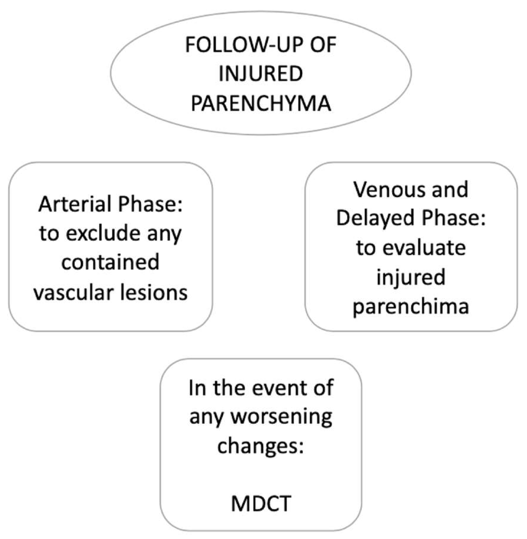
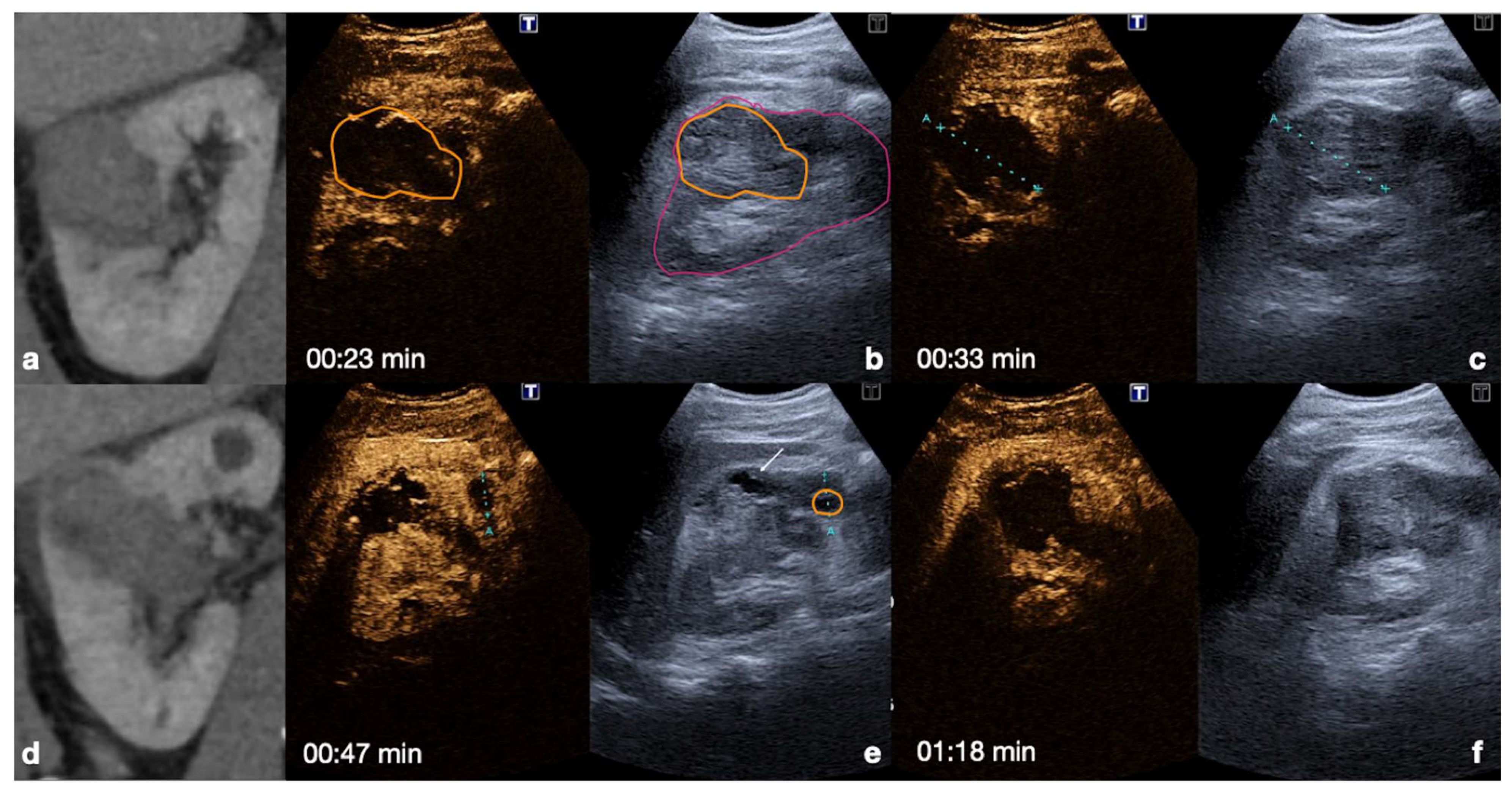
2.2.2. Follow-Up of Conservatively Managed Abdominal Trauma
2.3. Findings
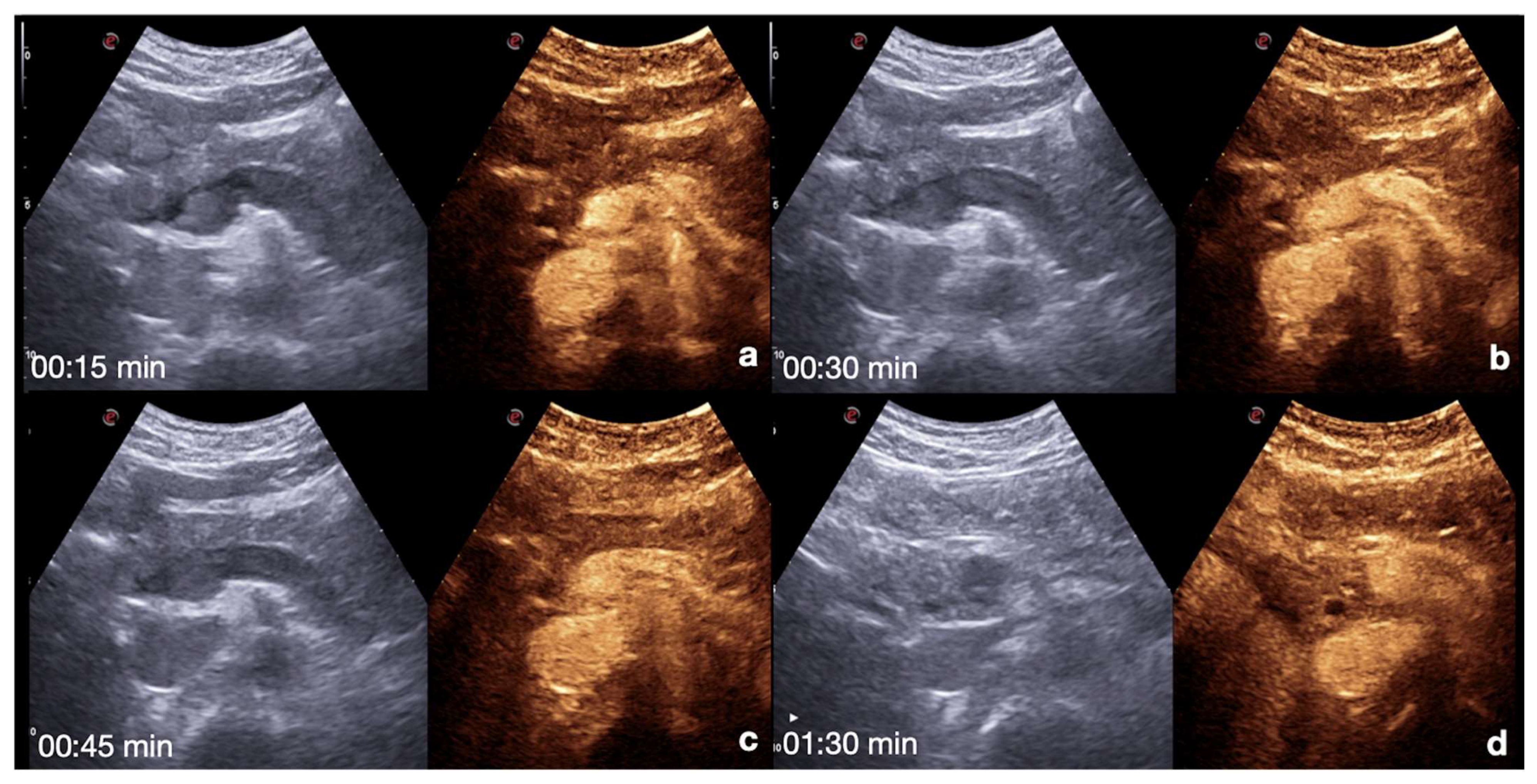
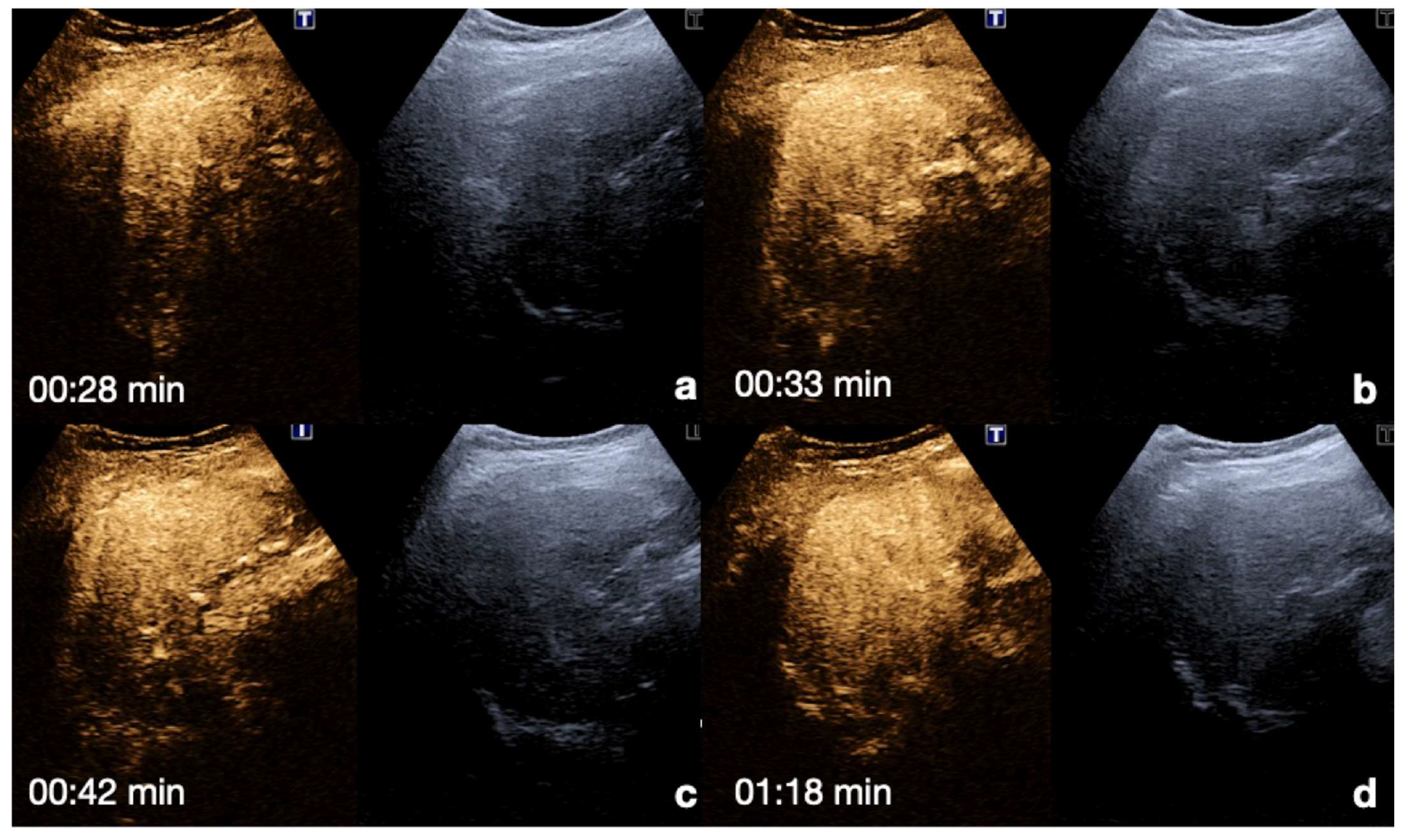
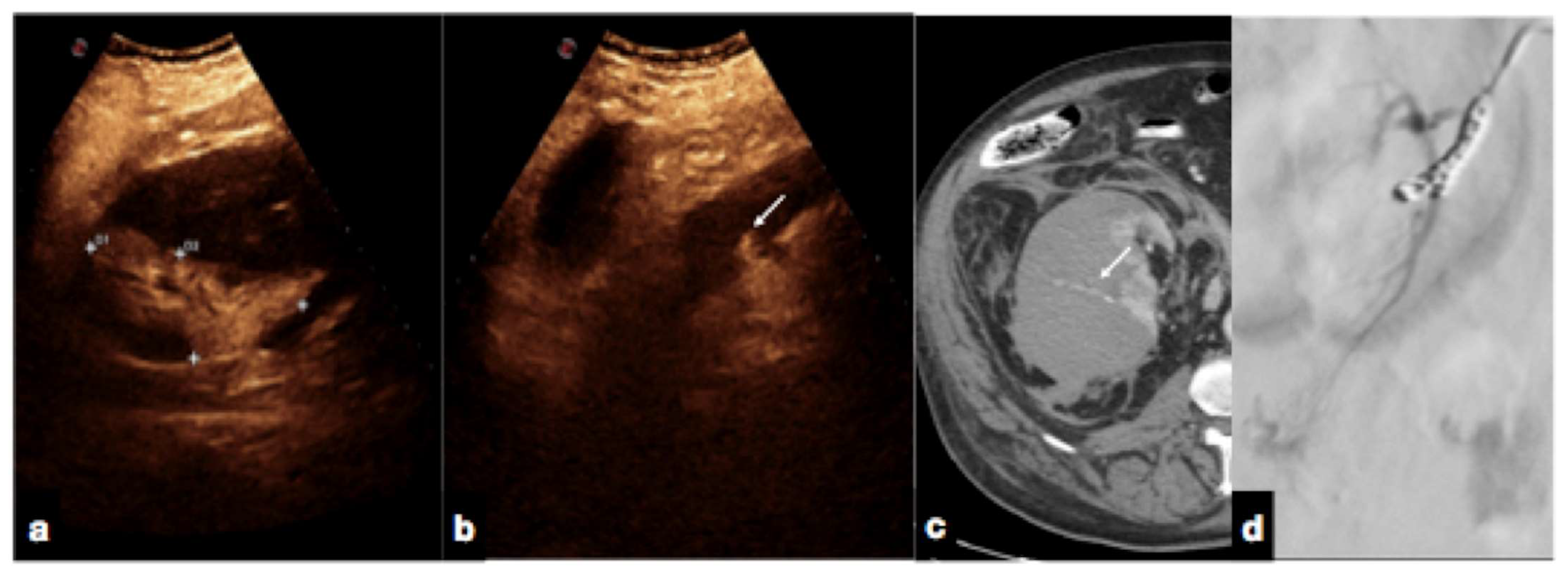
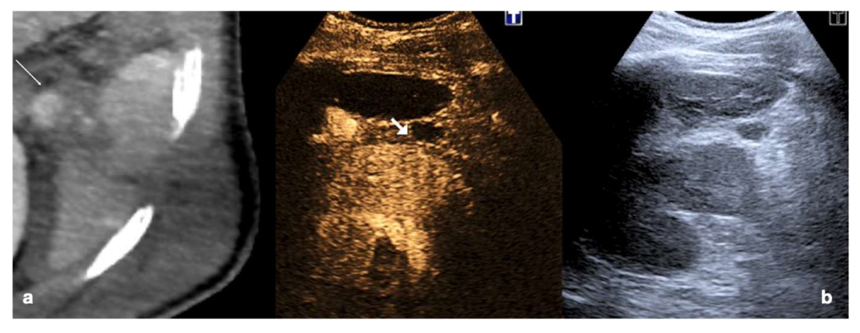
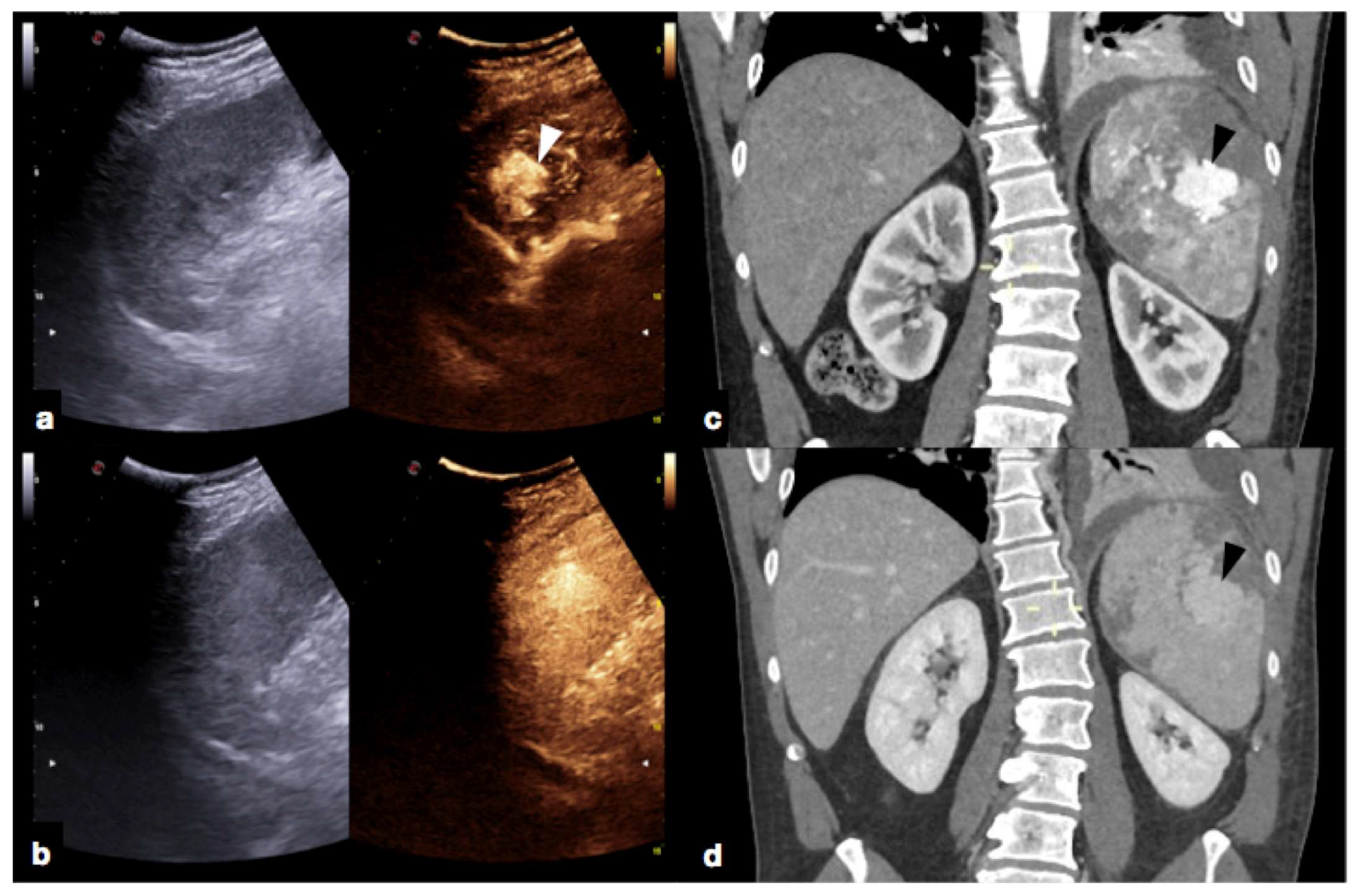
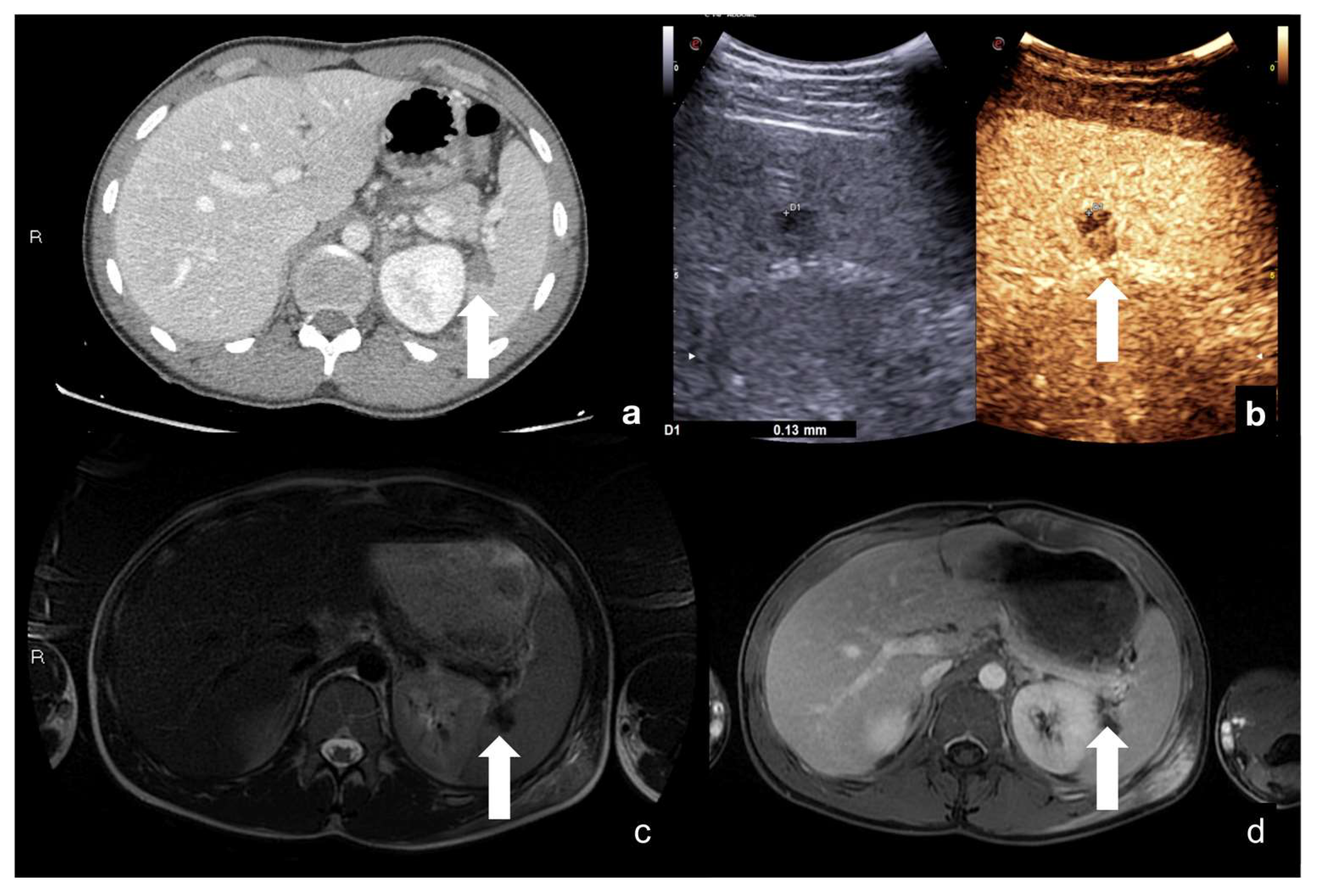
2.3.1. Solid Organ Injuries May Involve the Parenchyma and the Vessel
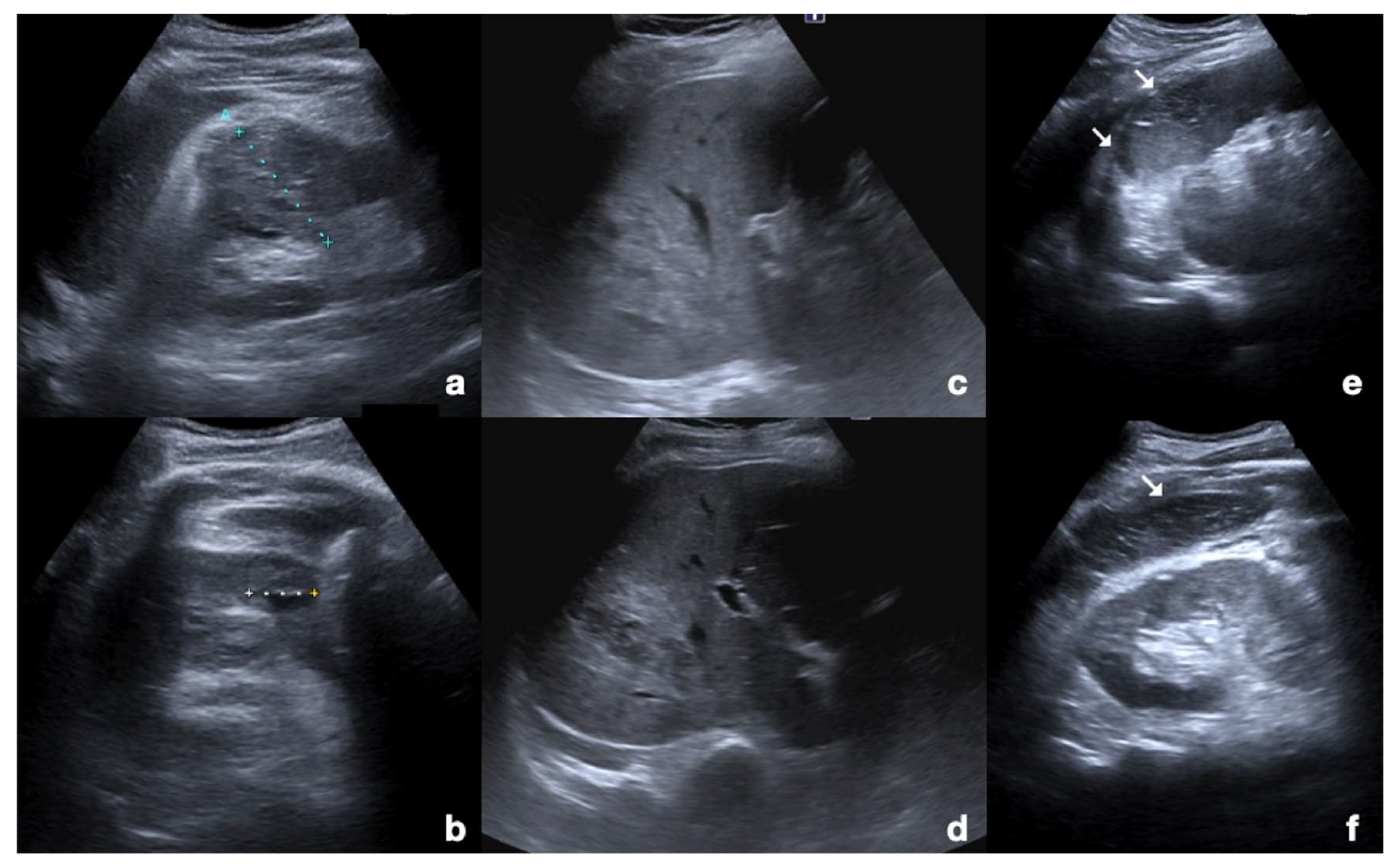
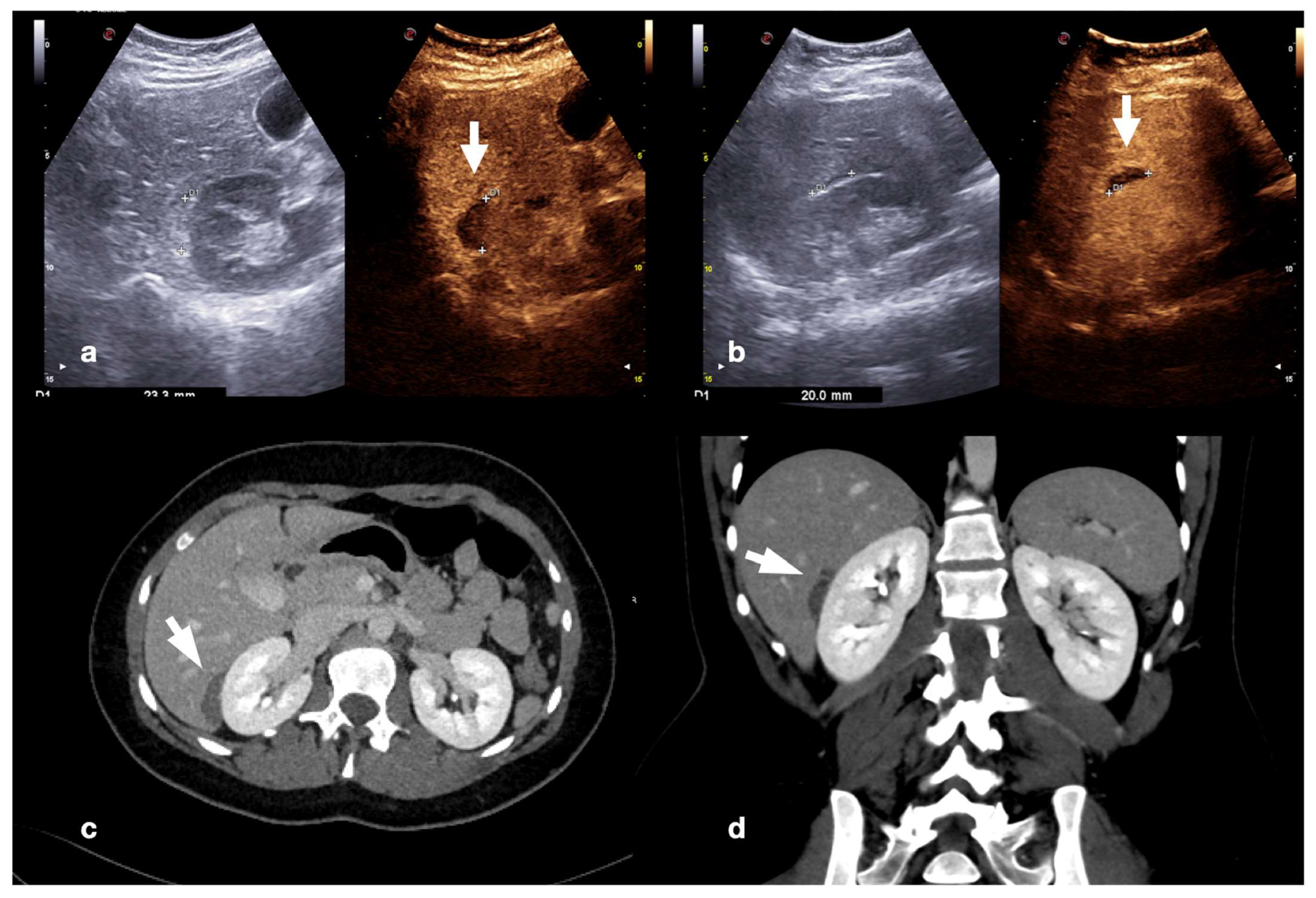
- Parenchymal injuries:
2.3.2. Vascular Injuries
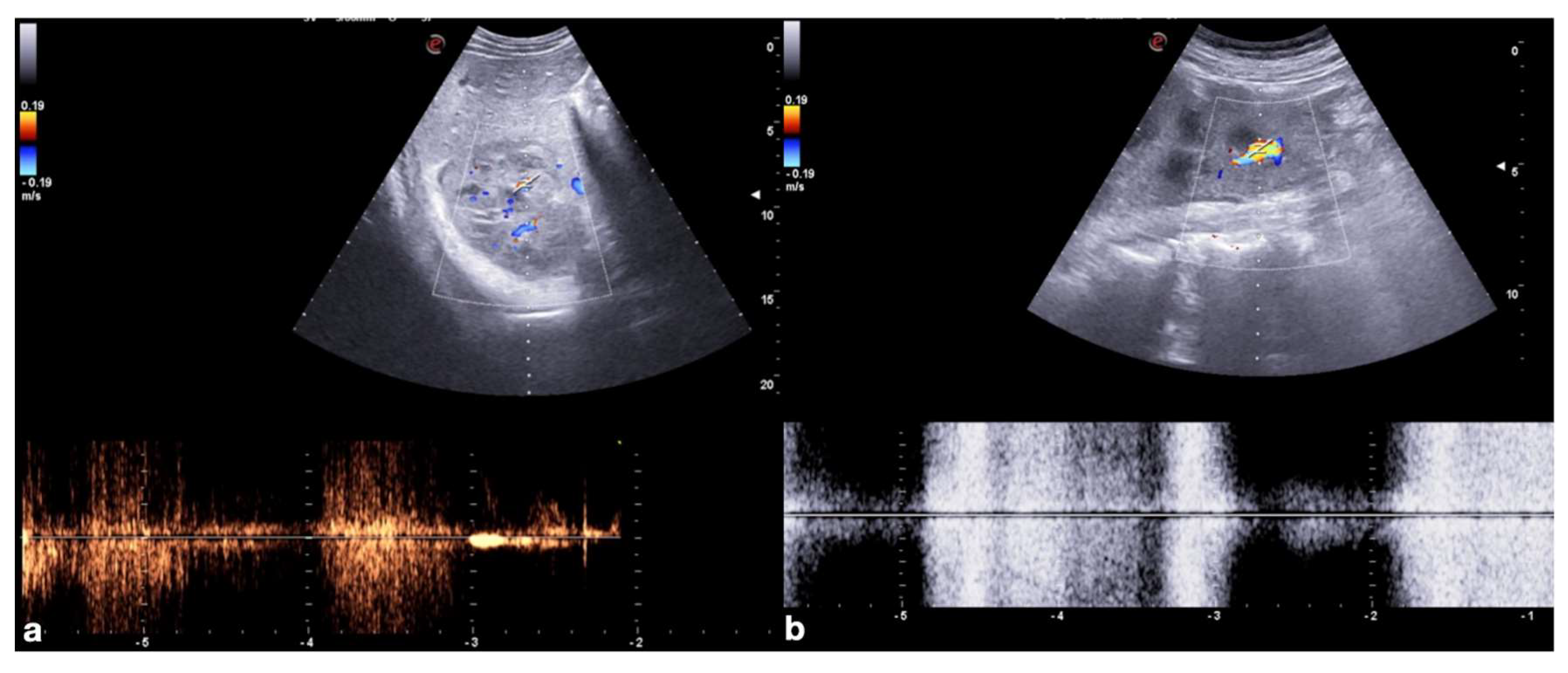
- Active bleeding:
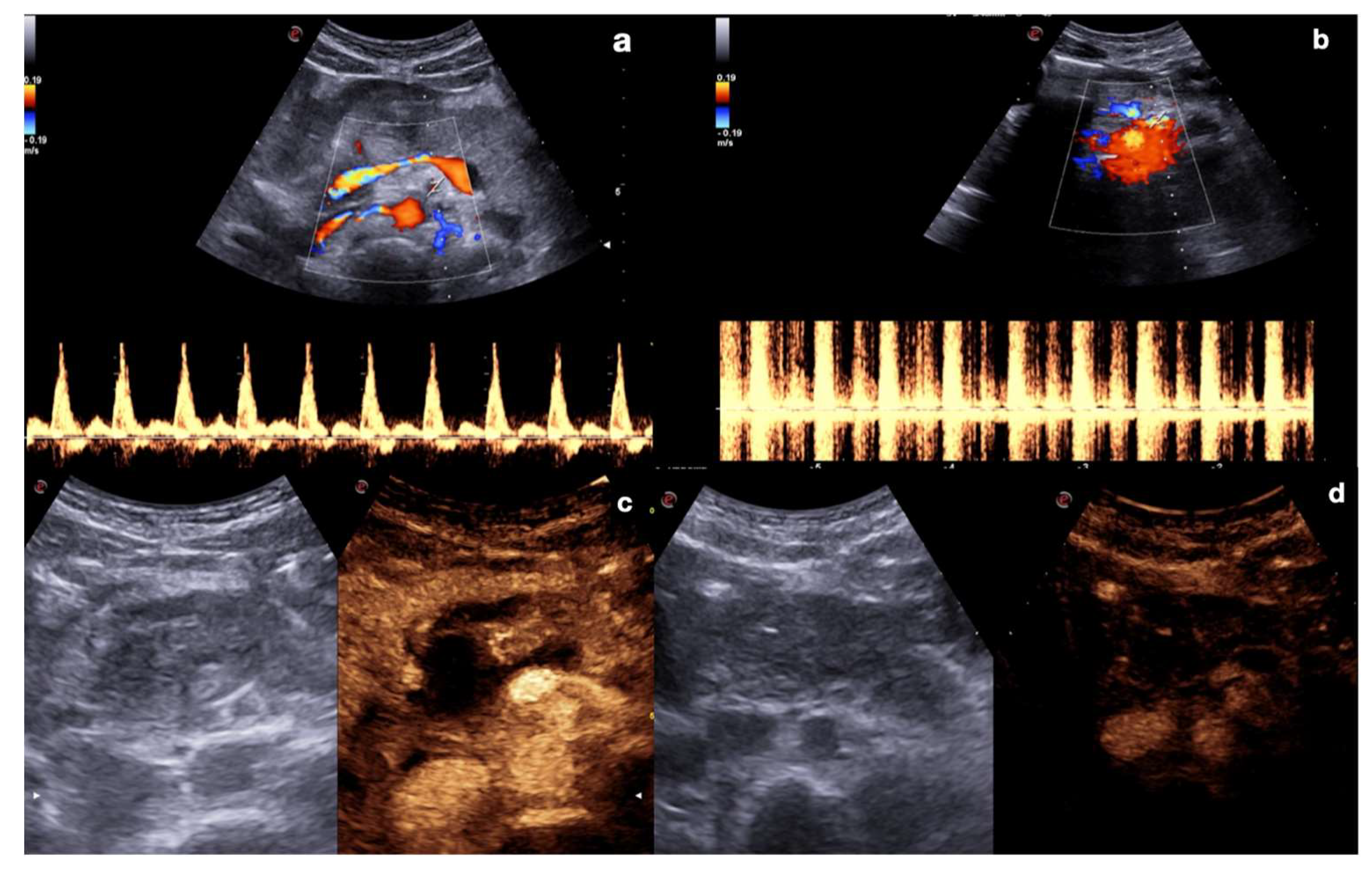
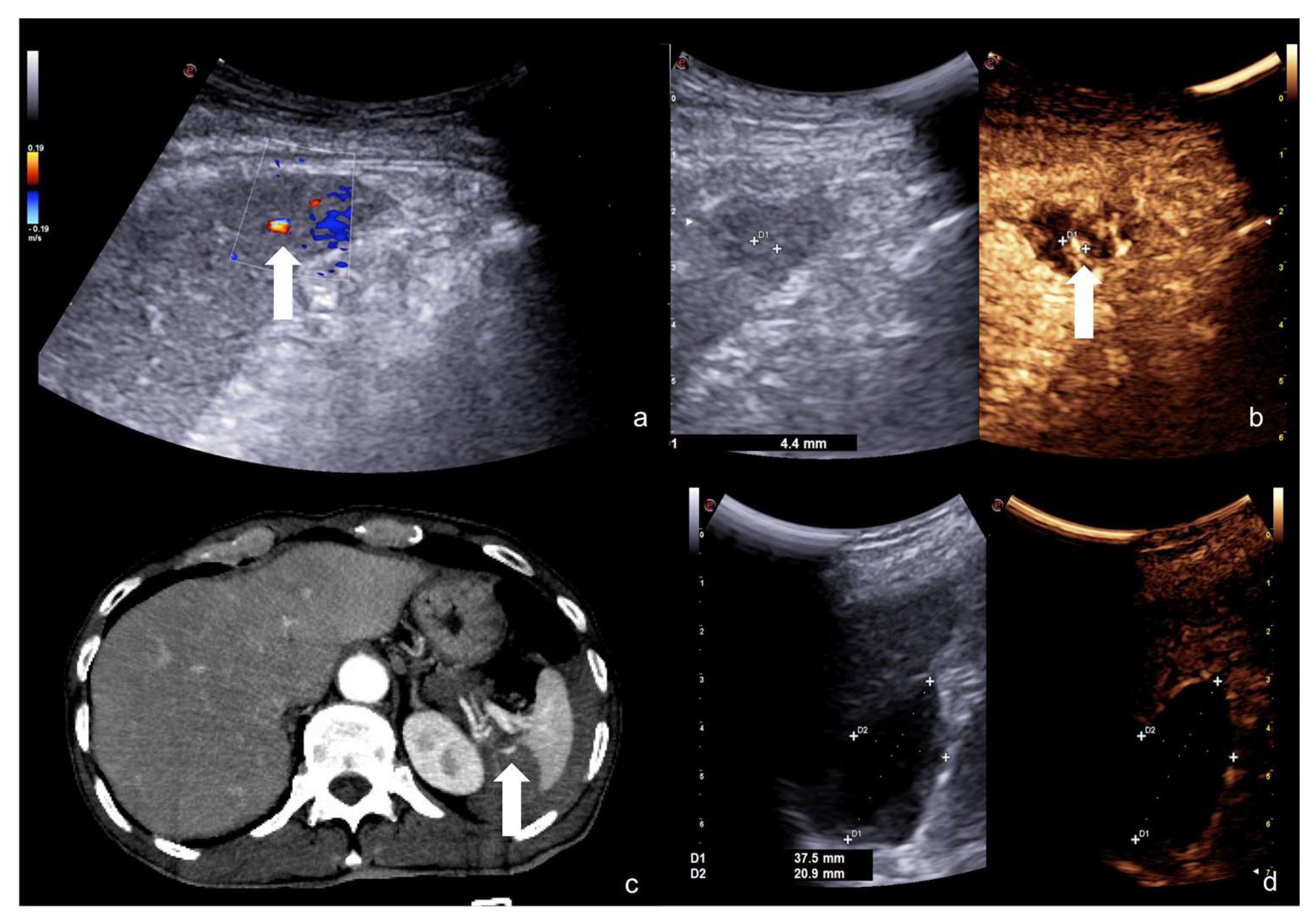
- Contained vascular injuries:
3. Advantages and Disadvantages
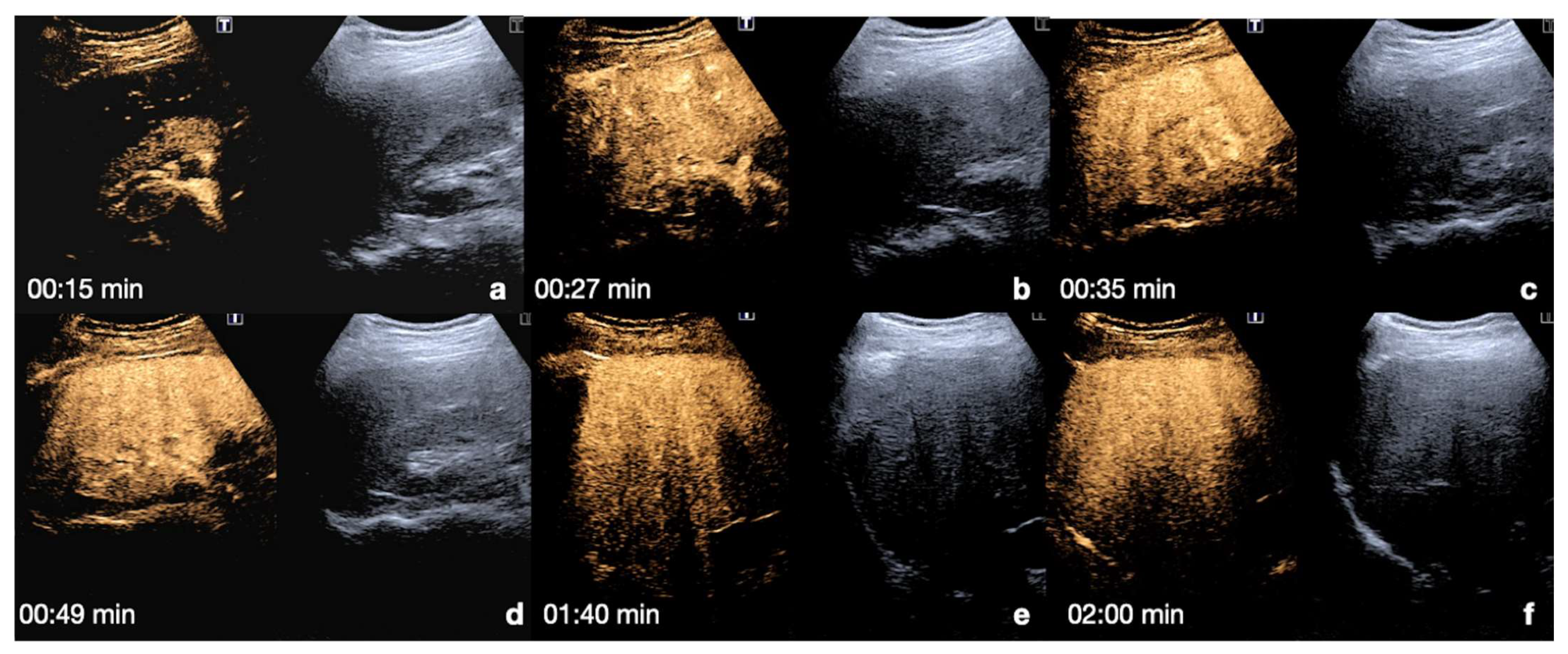
4. Case Series: Step-by-Step Practical Applications, Tips and Tricks during CEUS Follow-Up of Conservatively Managed Abdominal Trauma
4.1. Step 1
4.2. Step 2
4.3. Step 3
4.4. Step 4
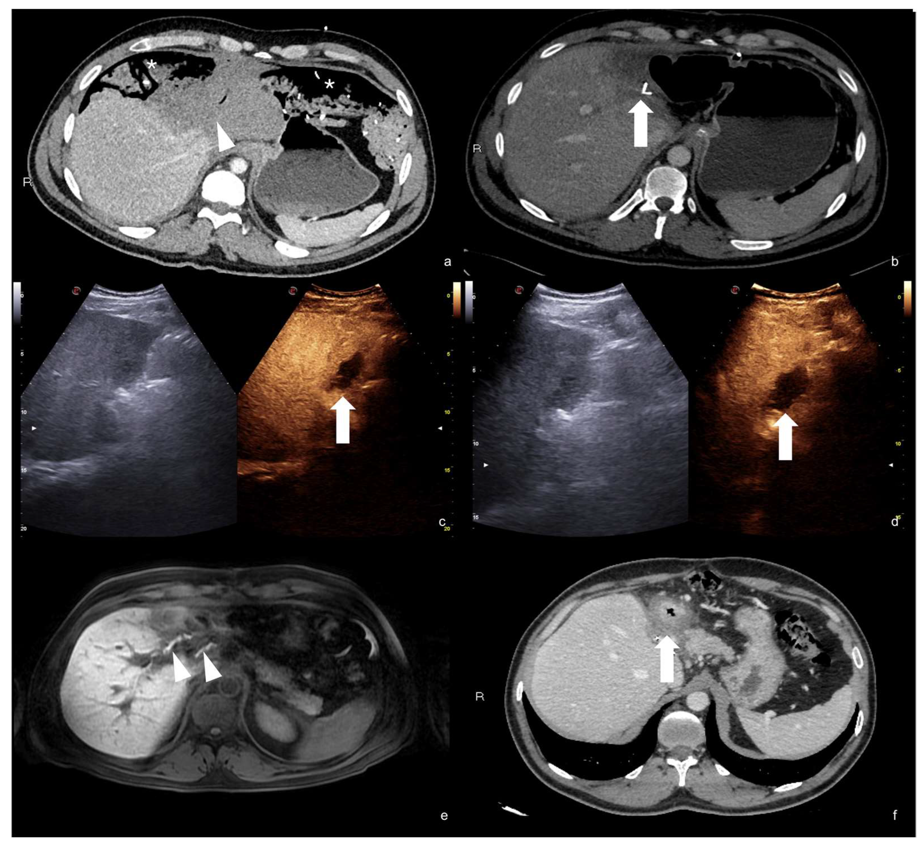
4.5. Step 5
5. Conclusions
Author Contributions
Funding
Institutional Review Board Statement
Informed Consent Statement
Data Availability Statement
Acknowledgments
Conflicts of Interest
References
- Iacobellis, F.; Ierardi, A.M.; Mazzei, M.A.; Biasina, A.M.; Carrafiello, G.; Nicola, R.; Scaglione, M. Dual-phase CT for the assessment of acute vascular injuries in high-energy blunt trauma: The imaging findings and management implications. Br. J. Radiol. 2016, 89, 20150952. [Google Scholar] [CrossRef]
- Iacobellis, F.; Abu-Omar, A.; Crivelli, P.; Galluzzo, M.; Danzi, R.; Trinci, M.; Dell’Aversano Orabona, G.; Conti, M.; Romano, L.; Scaglione, M. Current Standards for and Clinical Impact of Emergency Radiology in Major Trauma. Int. J. Environ. Res. Public Health 2022, 19, 539. [Google Scholar] [CrossRef] [PubMed]
- Iacobellis, F.; Romano, L.; Rengo, A.; Danzi, R.; Scuderi, M.G.; Brillantino, A.; Scaglione, M. CT Protocol Optimization in Trauma Imaging: A Review of Current Evidence. Curr. Radiol. Rep. 2020, 8, 8. [Google Scholar] [CrossRef]
- Di Serafino, M.; Severino, R.; Scavone, C.; Gioioso, M.; Coppola, V.; Brigida, R.; Lisanti, F.; Rocca, R.; Scarano, E. Prevention Strategies of Contrast Medium Induced Nephropathy (CIN): A Review of the Current Literature. Open J. Nephrol. 2016, 6, 98–110. [Google Scholar] [CrossRef]
- Pinto, A.; Russo, A.; Reginelli, A.; Iacobellis, F.; Di Serafino, M.; Giovine, S.; Romano, L. Gunshot Wounds: Ballistics and Imaging Findings. Semin. Ultrasound CT MRI 2019, 40, 25–35. [Google Scholar] [CrossRef]
- Brillantino, A.; Iacobellis, F.; Robustelli, U.; Villamaina, E.; Maglione, F.; Colletti, O.; De Palma, M.; Paladino, F.; Noschese, G. Non operative management of blunt splenic trauma: A prospective evaluation of a standardized treatment protocol. Eur. J. Trauma Emerg. Surg. 2015, 42, 593–598. [Google Scholar] [CrossRef]
- Brillantino, A.; Iacobellis, F.; Festa, P.; Mottola, A.; Acampora, C.; Corvino, F.; Del Giudice, S.; Lanza, M.; Armellino, M.; Niola, R.; et al. Non-Operative Management of Blunt Liver Trauma: Safety, Efficacy and Complications of a Standardized Treatment Protocol. Bull. Emerg. Trauma 2019, 7, 49–54. [Google Scholar] [CrossRef]
- Miele, V.; Piccolo, C.L.; Sessa, B.; Trinci, M.; Galluzzo, M. Comparison between MRI and CEUS in the follow-up of patients with blunt abdominal trauma managed conservatively. Radiol. Med. 2015, 121, 27–37. [Google Scholar] [CrossRef]
- Iacobellis, F.; Di Serafino, M.; Brillantino, A.; Mottola, A.; Del Giudice, S.; Stavolo, C.; Festa, P.; Patlas, M.N.; Scaglione, M.; Romano, L. Role of MRI in early follow-up of patients with solid organ injuries: How and why we do it? Radiol. Med. 2021, 126, 1328–1334. [Google Scholar] [CrossRef]
- Sessa, B.; Trinci, M.; Ianniello, S.; Menichini, G.; Galluzzo, M.; Miele, V. Blunt abdominal trauma: Role of contrast-enhanced ultrasound (CEUS) in the detection and staging of abdominal traumatic lesions compared to US and CE-MDCT. Radiol. Med. 2014, 120, 180–189. [Google Scholar] [CrossRef]
- Zhang, Z.; Hong, Y.; Liu, N.; Chen, Y. Diagnostic accuracy of contrast enhanced ultrasound in patients with blunt abdominal trauma presenting to the emergency department: A systematic review and meta-analysis. Sci. Rep. 2017, 7, 4446. [Google Scholar] [CrossRef] [PubMed]
- Dormagen, J.; Meyerdierks, O.; Gaarder, C.; Naess, P.; Sandvik, L.; Klow, N.-E. Contrast-enhanced ultrasound of the injured spleen after embolization comparison with computed tomography. Ultraschall Med. 2011, 32, 485–491. [Google Scholar] [CrossRef] [PubMed]
- Sidhu, P.; Cantisani, V.; Dietrich, C.F.; Gilja, O.H.; Saftoiu, A.; Bartels, E.; Bertolotto, M.; Calliada, F.; Clevert, D.A.; Cosgrove, D.; et al. The EFSUMB Guidelines and Recommendations for the Clinical Practice of Contrast-Enhanced Ultrasound (CEUS) in Non-Hepatic Applications: Update 2017 (Long Version). Ultraschall Med. 2018, 39, e2–e44. [Google Scholar] [CrossRef] [PubMed]
- Piccolo, C.L.; Trinci, M.; Pinto, A.; Brunese, L.; Miele, V. Role of contrast-enhanced ultrasound (CEUS) in the diagnosis and management of traumatic splenic injuries. J. Ultrasound 2018, 21, 315–327. [Google Scholar] [CrossRef] [PubMed]
- Gulati, M.; King, K.G.; Gill, I.S.; Pham, V.; Grant, E.; Duddalwar, V.A. Contrast-enhanced ultrasound (CEUS) of cystic and solid renal lesions: A review. Abdom Imaging 2015, 40, 1982–1996. [Google Scholar] [CrossRef]
- Kloth, C.; Kratzer, W.; Schmidberger, J.; Beer, M.; Clevert, D.A.; Graeter, T. Ultrasound 2020–Diagnostics & Therapy: On the Way to Multimodal Ultrasound: Contrast-Enhanced Ultrasound (CEUS), Microvascular Doppler Techniques, Fusion Imaging, Sonoelastography, Interventional Sonography. Rofo 2020, 193, 23–32. [Google Scholar] [CrossRef]
- Jang, H.-J.; Kim, T.K.; Burns, P.N.; Wilson, S.R. CEUS: An essential component in a multimodality approach to small nodules in patients at high-risk for hepatocellular carcinoma. Eur. J. Radiol. 2015, 84, 1623–1635. [Google Scholar] [CrossRef]
- Granata, A.; Zanoli, L.; Insalaco, M.; Valentino, M.; Pavlica, P.; Di Nicolò, P.P.; Scuderi, M.; Fiorini, F.; Fatuzzo, P.M.; Bertolotto, M. Contrast-enhanced ultrasound (CEUS) in nephrology: Has the time come for its widespread use? Clin. Exp. Nephrol. 2014, 19, 606–615. [Google Scholar] [CrossRef]
- Cantisani, V.; Wilson, S.R. CEUS: Where are we in 2015? Eur. J. Radiol. 2015, 84, 1621–1622. [Google Scholar] [CrossRef] [PubMed]
- Cantisani, V.; Di Leo, N.; David, E.; Clevert, D.-A. Role of CEUS in Vascular Pathology. Ultraschall Med. 2021, 42, 348–366. [Google Scholar] [CrossRef] [PubMed]
- Sidhu, P.; Cantisani, V.; Deganello, A.; Dietrich, C.; Duran, C.; Franke, D.; Harkanyi, Z.; Kosiak, W.; Miele, V.; Ntoulia, A.; et al. Role of Contrast-Enhanced Ultrasound (CEUS) in Paediatric Practice: An EFSUMB Position Statement. Ultraschall Med. 2016, 38, 33–43. [Google Scholar] [CrossRef] [PubMed]
- Cozzi, D.; Agostini, S.; Bertelli, E.; Galluzzo, M.; Papa, E.; Scevola, G.; Trinci, M.; Miele, V. Contrast-Enhanced Ultrasound (CEUS) in Non-Traumatic Abdominal Emergencies. Ultrasound Int. Open 2020, 6, E76–E86. [Google Scholar] [CrossRef]
- Trinci, M.; Danti, G.; Di Maurizio, M.; Tursini, S.; Briganti, V.; Galluzzo, M.; Miele, V. Can contrast enhanced ultrasound (CEUS) be useful in the diagnosis of ovarian torsion in pediatric females? A preliminary monocentric experience. J. Ultrasound 2021, 24, 505–514. [Google Scholar] [CrossRef] [PubMed]
- Yusuf, G.T.; Fang, C.; Huang, D.; Sellars, M.E.; Deganello, A.; Sidhu, P. Endocavitary contrast enhanced ultrasound (CEUS): A novel problem solving technique. Insights Imaging 2018, 9, 303–311. [Google Scholar] [CrossRef]
- Huang, D.Y.; Yusuf, G.T.; Daneshi, M.; Ramnarine, R.; Deganello, A.; Sellars, M.E.; Sidhu, P. Contrast-enhanced ultrasound (CEUS) in abdominal intervention. Abdom. Radiol. 2018, 43, 960–976. [Google Scholar] [CrossRef] [PubMed]
- Miele, V.; Piccolo, C.L.; Galluzzo, M.; Ianniello, S.; Sessa, B.; Trinci, M. Contrast-enhanced ultrasound (CEUS) in blunt abdominal trauma. Br. J. Radiol. 2016, 89, 20150823. [Google Scholar] [CrossRef]
- Cokkinos, D.; Antypa, E.; Stefanidis, K.; Tserotas, P.; Kostaras, V.; Parlamenti, A.; Tavernaraki, K.; Piperopoulos, P.N. Contrast-Enhanced Ultrasound for Imaging Blunt Abdominal Trauma–Indications, Description of the Technique and Imaging Review. Ultraschall Med. 2012, 33, 60–67. [Google Scholar] [CrossRef]
- Trinci, M.; Piccolo, C.L.; Ferrari, R.; Galluzzo, M.; Ianniello, S.; Miele, V. Contrast-enhanced ultrasound (CEUS) in pediatric blunt abdominal trauma. J. Ultrasound 2018, 22, 27–40. [Google Scholar] [CrossRef]
- Esposito, F.; DI Serafino, M.; Sgambati, P.; Mercogliano, F.; Tarantino, L.; Vallone, G.; Oresta, P. Ultrasound contrast media in paediatric patients: Is it an off-label use? Regulatory requirements and radiologist’s liability. Radiol. Med. 2011, 117, 148–159. [Google Scholar] [CrossRef][Green Version]
- Iacobellis, F.; Acampora, C.; Ponticiello, G.; Scuderi, M.; Cinque, T.; Pinto, A.; Gagliardi, N.; Romano, L. Technique and advantages of contrast-enhanced ultrasound versus MDCT in the follow-up of solid organ injuries. Med. Pharmacol. 2021, 2021120479. [Google Scholar] [CrossRef]
- Iacobellis, F.; Laccetti, E.; Tamburrini, S.; Altiero, M.; Iaselli, F.; DI Serafino, M.; Gagliardi, N.; Danzi, R.; Rengo, A.; Romano, L.; et al. Role of multidetector computed tomography in the assessment of pancreatic injuries after blunt trauma: A multicenter experience. Gland Surg. 2019, 8, 184–196. [Google Scholar] [CrossRef] [PubMed]
- Menichini, G.; Sessa, B.; Trinci, M.; Galluzzo, M.; Miele, V. Accuracy of contrast-enhanced ultrasound (CEUS) in the identification and characterization of traumatic solid organ lesions in children: A retrospective comparison with baseline US and CE-MDCT. Radiol. Med. 2015, 120, 989–1001. [Google Scholar] [CrossRef] [PubMed]
- Iacobellis, F.; Iacobellis, F.; Scaglione, M.; Scaglione, M.; Brillantino, A.; Brillantino, A.; Scuderi, M.G.; Scuderi, M.G.; Giurazza, F.; Giurazza, F.; et al. The additional value of the arterial phase in the CT assessment of liver vascular injuries after high-energy blunt trauma. Emerg. Radiol. 2019, 26, 647–654. [Google Scholar] [CrossRef] [PubMed]
- Cokkinos, D.D.; Antypa, E.; Kalogeropoulos, I.; Tomais, D.; Ismailos, E.; Matsiras, I.; Benakis, S.; Piperopoulos, P.N. Contrast-enhanced ultrasound performed under urgent conditions. Indications, review of the technique, clinical examples and limitations. Insights Imaging 2012, 4, 185–198. [Google Scholar] [CrossRef]
- Pinto, F.; Valentino, M.; Romanini, L.; Basilico, R.; Miele, V. The role of CEUS in the assessment of haemodynamically stable patients with blunt abdominal trauma. Radiol. Med. 2014, 120, 3–11. [Google Scholar] [CrossRef]
- Pinto, F.; Miele, V.; Scaglione, M.; Pinto, A. The use of contrast-enhanced ultrasound in blunt abdominal trauma: Advantages and limitations. Acta Radiol. 2014, 55, 776–784. [Google Scholar] [CrossRef]








| Time and Flash Technique | Advantages |
|---|---|
| <0 s | Choose the best scan view in B-Mode and US Doppler studies (color, power, and pulse Doppler). |
| 0 s | Injection of 2 mL of UCA followed by 10mL of saline solution. |
| 10–20 s (early) 20–40 s (late) | Arterial phase: best depiction of contained vascular injuries, such as pseudoaneurysms and arteriovenous fistulas in the early phase. |
| 2–6 min | Venous-late phases: distribution of the contrast in the whole organ. Best time to depict parenchymal injuries. |
| Flash mode | Destruction of bubbles and possibility to re-evaluate an area of interest. |
| Main Organs to Explore | Enhancement Characteristic |
|---|---|
| Kidney | Quick enhancement of the cortex after injection. Pyramids enhancement after 30 s. No excretory phase. Good evaluation up to 2.5 min. |
| Liver | Arterial phase: 10–40 s Hepatic and portal phases: 40–120 s Sinusoidal phase: 120–300 s Dual vascular supply permits homogeneous enhancement. |
| Pancreas | Arterial phase: 15–30 s Venous phase: 30–120 s The best moment to detect organ injury: venous phase. |
| Adrenal glands | Arterial phase: 20–40 s Homogeneous enhancement up to 5 min. |
| Spleen | Arterial phase: 12–20 s. Venous phase: 40–60 s up to 5–7 min. The best moment to detect organ injury: venous phase. |
Publisher’s Note: MDPI stays neutral with regard to jurisdictional claims in published maps and institutional affiliations. |
© 2022 by the authors. Licensee MDPI, Basel, Switzerland. This article is an open access article distributed under the terms and conditions of the Creative Commons Attribution (CC BY) license (https://creativecommons.org/licenses/by/4.0/).
Share and Cite
Di Serafino, M.; Iacobellis, F.; Schillirò, M.L.; Ronza, R.; Verde, F.; Grimaldi, D.; Dell’Aversano Orabona, G.; Caruso, M.; Sabatino, V.; Rinaldo, C.; et al. The Technique and Advantages of Contrast-Enhanced Ultrasound in the Diagnosis and Follow-Up of Traumatic Abdomen Solid Organ Injuries. Diagnostics 2022, 12, 435. https://doi.org/10.3390/diagnostics12020435
Di Serafino M, Iacobellis F, Schillirò ML, Ronza R, Verde F, Grimaldi D, Dell’Aversano Orabona G, Caruso M, Sabatino V, Rinaldo C, et al. The Technique and Advantages of Contrast-Enhanced Ultrasound in the Diagnosis and Follow-Up of Traumatic Abdomen Solid Organ Injuries. Diagnostics. 2022; 12(2):435. https://doi.org/10.3390/diagnostics12020435
Chicago/Turabian StyleDi Serafino, Marco, Francesca Iacobellis, Maria Laura Schillirò, Roberto Ronza, Francesco Verde, Dario Grimaldi, Giuseppina Dell’Aversano Orabona, Martina Caruso, Vittorio Sabatino, Chiara Rinaldo, and et al. 2022. "The Technique and Advantages of Contrast-Enhanced Ultrasound in the Diagnosis and Follow-Up of Traumatic Abdomen Solid Organ Injuries" Diagnostics 12, no. 2: 435. https://doi.org/10.3390/diagnostics12020435
APA StyleDi Serafino, M., Iacobellis, F., Schillirò, M. L., Ronza, R., Verde, F., Grimaldi, D., Dell’Aversano Orabona, G., Caruso, M., Sabatino, V., Rinaldo, C., & Romano, L. (2022). The Technique and Advantages of Contrast-Enhanced Ultrasound in the Diagnosis and Follow-Up of Traumatic Abdomen Solid Organ Injuries. Diagnostics, 12(2), 435. https://doi.org/10.3390/diagnostics12020435


