
Male Breast Cancer Review. A Rare Case of Pure DCIS: Imaging Protocol, Radiomics and Management


Abstract
1. Introduction
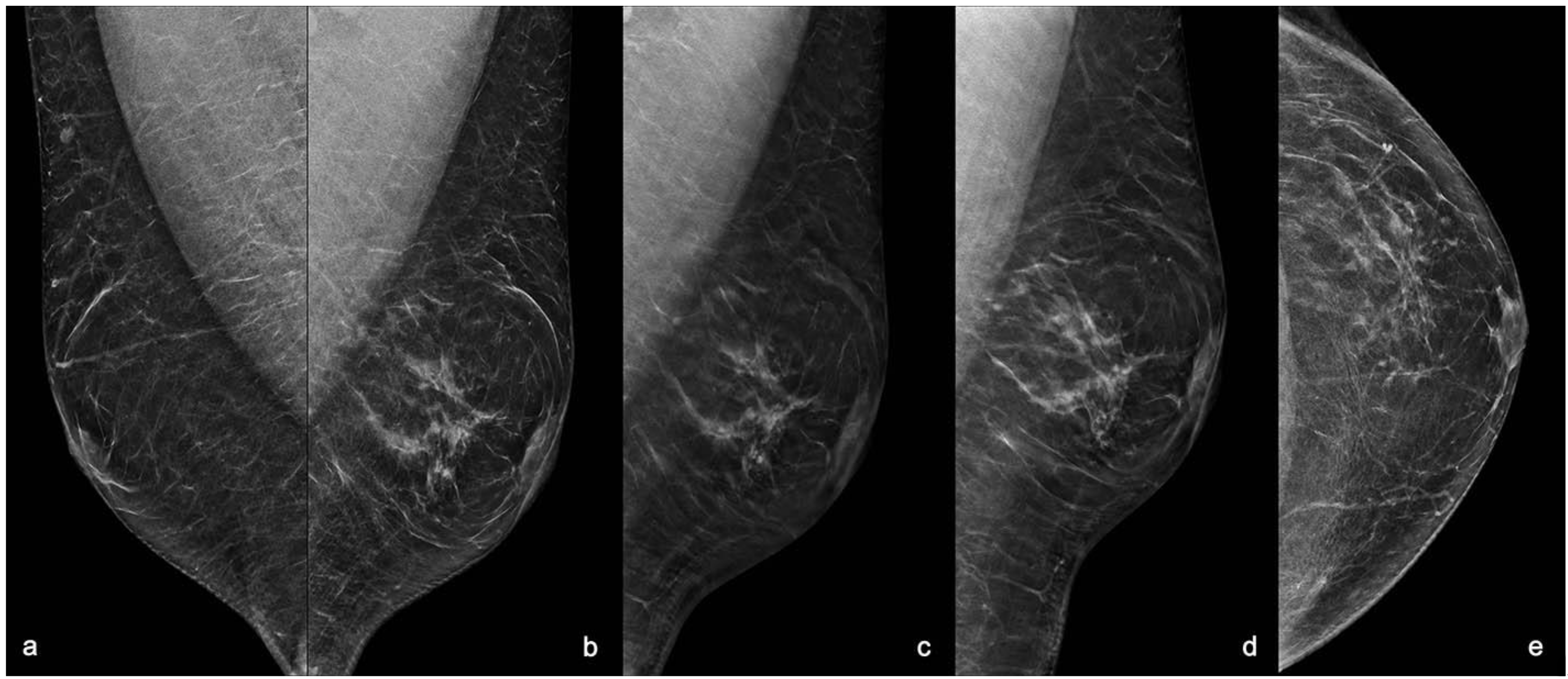
2. Case Report
3. Discussion
4. Conclusions
Author Contributions
Funding
Institutional Review Board Statement
Informed Consent Statement
Data Availability Statement
Conflicts of Interest
References
- Fentiman, I.S.; Fourquet, A.; Hortobagyi, G.N. Male breast cancer. Lancet 2006, 367, 595–604. [Google Scholar] [CrossRef]
- Gucalp, A.; Traina, T.A.; Eisner, J.R.; Parker, J.S.; Selitsky, S.R.; Park, B.H.; Elias, A.D.; Baskin-Bey, E.S.; Cardoso, F. Male breast cancer: A disease distinct from female breast cancer. Breast Cancer Res. Treat. 2019, 173, 37–48. [Google Scholar] [CrossRef]
- Brents, M.; Hancock, J. Ductal Carcinoma In situ of the Male Breast. Breast Care 2016, 11, 288–290. [Google Scholar] [CrossRef] [PubMed]
- Ruddy, K.J.; Winer, E.P. Male breast cancer: Risk factors, biology, diagnosis, treatment, and survivorship. Ann. Oncol. 2013, 24, 1434–1443. [Google Scholar] [CrossRef]
- D’Orsi, C.J.; Sickles, E.A.; Mendelson, E.B.; Morris, E.A. ACR BI-RADS® Atlas, Breast Imaging Reporting and Data System, 5th ed.; American College of Radiology: Reston, VA, USA, 2013. [Google Scholar]
- Field, A.S.; Raymond, W.A.; Rickard, M.; Arnold, L.; Brachtel, E.F.; Chaiwun, B.; Chen, L.; Di Bonito, L.; Kurtycz, D.F.I.; Lee, A.H.S.; et al. The International Academy of Cytology Yokohama System for Reporting Breast Fine-Needle Aspiration Biopsy Cytopathology. Acta Cytol. 2019, 63, 257–273. [Google Scholar] [CrossRef] [PubMed]
- Field, A.S.; Raymond, W.A.; Rickard, M.; Schmitt, F. Breast fine needle aspiration biopsy cytology: The potential impact of the International Academy of Cytology Yokohama System for Reporting Breast Fine Needle Aspiration Biopsy Cytopathology and the use of rapid on-site evaluation. J. Am. Soc. Cytopathol. 2020, 9, 103–111. [Google Scholar] [CrossRef] [PubMed]
- Lee, A.H.S.; Carder, P.; Deb, R.; Ellis, I.O.; Howe, M.; Jenkins, J.A.; Pinder, S.E. Guidelines for Non-Operative Diagnostic Procedures and Reporting in Breast Cancer Screening; The Royal College of Pathologists Publications G-150: London, UK, 2021. [Google Scholar]
- Nofal, M.N.; Yousef, A.J. The diagnosis of male breast cancer. Neth. J. Med. 2019, 77, 356–359. [Google Scholar]
- Khan, N.A.J.; Tirona, M. An updated review of epidemiology, risk factors, and management of male breast cancer. Med. Oncol. 2021, 38, 39. [Google Scholar] [CrossRef]
- Coopey, S.B.; Kartal, K.; Li, C.; Yala, A.; Barzilay, R.; Faulkner, H.R.; King, T.A.; Acevedo, F.; Garber, J.E.; Guidi, A.J.; et al. Atypical ductal hyperplasia in men with gynecomastia: What is their breast cancer risk? Breast Cancer Res. Treat. 2019, 175, 1–4. [Google Scholar] [CrossRef]
- Vagios, I.; Nonni, A.; Liakea, A.; Constantinidou, A.; Kontos, M. Intraductal papilloma of the male breast: A case report and review of the literature. J. Surg. Case. Rep. 2019, 2019, rjz023. [Google Scholar] [CrossRef]
- Bharti, S.; Bharti, J.N.; Vishnoi, J.R.; Soudamini, A.B. A rare case of intraductal papilloma with atypical ductal hyperplasia in a male breast: A pathological diagnosis. J. Fam. Community Med. 2020, 27, 216–218. [Google Scholar] [CrossRef] [PubMed]
- Giordano, S.H.; Cohen, D.S.; Buzdar, A.U.; Perkins, G.; Hortobagyi, G.N. Breast carcinoma in men: A population-based study. Cancer 2004, 101, 51–57. [Google Scholar] [CrossRef] [PubMed]
- Speirs, V.; Shaaban, A.M. The rising incidence of male breast cancer. Breast Cancer Res. Treat 2009, 115, 429–430. [Google Scholar] [CrossRef]
- White, J.; Kearins, O.; Dodwell, D.; Horgan, K.; Hanby, A.M.; Speirs, V. Male breast carcinoma: Increased awareness needed. Breast Cancer Res. 2011, 13, 219. [Google Scholar] [CrossRef] [PubMed]
- Cardoso, F.; Bartlett, J.M.S.; Slaets, L.; van Deurzen, C.H.M.; van Leeuwen-Stok, E.; Porter, P.; Linderholm, B.; Hedenfalk, I.; Schröder, C.; Martens, J.; et al. Characterization of male breast cancer: Results of the EORTC 10085/TBCRC/BIG/NABCG International Male Breast Cancer Program. Ann. Oncol. 2018, 29, 405–417. [Google Scholar] [CrossRef] [PubMed]
- Bohli, M.; Tebra Mrad, S.; Zrafi, I.; Bouaouina, N. Cancer du sein chez l’homme: Quelles particularités? Cancer/Radiothérapie 2017, 21, 701. [Google Scholar] [CrossRef]
- Methamem, M.; Ghadhab, I.; Hidar, S.; Briki, R. Breast cancer in men: A serie of 45 cases and literature review. Pan Afr. Med. J. 2020, 36, 183. [Google Scholar] [CrossRef]
- Gao, Y.; Heller, S.L.; Moy, L. Male Breast Cancer in the Age of Genetic Testing: An Opportunity for Early Detection, Tailored Therapy, and Surveillance. Radiographics 2018, 38, 1289–1311. [Google Scholar] [CrossRef]
- Woods, R.W.; Salkowski, L.R.; Elezaby, M.; Burnside, E.S.; Strigel, R.M.; Fowler, A.M. Image-based screening for men at high risk for breast cancer: Benefits and drawbacks. Clin. Imaging 2020, 60, 84–89. [Google Scholar] [CrossRef]
- Biganzoli, L.; Calabrese, M.; Conte, B.; Cortesi, L.; Criscitiello, C.; Del Mastro, L.; Fiorentino, A.; Levaggi, A.; Montemurro, F.; Marchiò, C.; et al. Linee Guida AIOM-Neoplasie Della Mammella; Edizione: Treviso, Italy, 2020. [Google Scholar]
- Nguyen, C.; Kettler, M.D.; Swirskt, M.E.; Miller, V.I.; Scott, C.; Krause, R.; Hadro, J.A. Male Breast Disease: Pictorial Review with Radiologic-Pathologic Correlation. RadioGraphics 2013, 33, 763–779. [Google Scholar] [CrossRef]
- Mathew, J.; Perkins, G.H.; Stephens, T.; Middleton, L.P.; Yang, W.T. Primary breast cancer in men: Clinical, imaging, and pathologic findings in 57 patiens. AJR Am. J. Roentgenol. 2008, 191, 1631–1639. [Google Scholar] [CrossRef]
- Hittmair, A.P.; Lininger, R.A.; Tavassoli, F. A: Ductal carcinoma in situ (DCIS) in the male breast. A morphologic study of 84 cases of pure DCIS and 30 cases of DCIS associated with invasive carcinoma—A preliminary report. Cancer 1998, 83, 2139–2149. [Google Scholar] [CrossRef]
- Erhan, Y.; Zekioglu, O.; Erhan, Y. Invasive lobular carcinoma of the male breast. Can. J. Surg. 2006, 49, 365–366. [Google Scholar]
- Coroneos, C.J.; Hamm, C. Ductal carcinoma in situ in a 25-year-old man presenting with apparent unilateral gynecomastia. Curr. Oncol. 2010, 17, 133–137. [Google Scholar] [CrossRef] [PubMed]
- Foerster, R.; Schroeder, L.; Foerster, F.; Wulff, V.; Schubotz, B.; Baaske, D.; Rudlowski, C. Metastatic male breast cancer: A retrospective cohort analysis. Breast Care 2014, 9, 267–271. [Google Scholar] [CrossRef] [PubMed]
- Irwig, L.; Macaskill, P.; Houssami, N. Evidence relevant to the investigation of breast symptoms: The triple test. Breast 2002, 11, 2015–2220. [Google Scholar] [CrossRef]
- Muñoz Carrasco, R.; Alvarez Benito, M.; Muñoz Gomariz, E.; Raya Povedano, J.L.; Martínez Paredes, M. Mammography and ultrasound in the evaluation of male breast disease. Eur. Radiol. 2010, 20, 2797–2805. [Google Scholar] [CrossRef] [PubMed]
- Evans, G.F.; Anthony, T.; Turnage, R.H.; Schumpert, T.D.; Levy, K.R.; Amirkhan, R.H.; Campbell, T.J.; Lopez, J.; Appelbaum, A.H. The diagnostic accuracy of mammography in the evaluation of male breast disease. Am. J. Surg. 2001, 181, 96–100. [Google Scholar] [CrossRef]
- Adibelli, Z.H.; Oztekin, O.; Postaci, H.; Uslu, A. The Diagnostic Accuracy of Mammography and Ultrasound in the Evaluation of Male Breast Disease: A New Algorithm. Breast Care 2009, 4, 255–259. [Google Scholar] [CrossRef] [PubMed]
- Volpe, C.M.; Raffetto, J.D.; Collure, D.W.; Hoover, E.L.; Doerr, R.J. Unilateral male breast masses: Cancer risk and their evaluation and management. Am. Surg. 1999, 65, 250–253. [Google Scholar]
- Hines, S.L.; Tan, W.; Larson, J.M.; Thompson, K.M.; Jorn, H.K.; Files, J.A. A practical approach to guide clinicians in the evaluation of male patients with breast masses. Geriatrics 2008, 63, 19–24. [Google Scholar] [PubMed]
- Munn, S. When should men undergo mammography? AJR Am. J. Roentgenol. 2002, 178, 1419–1420. [Google Scholar] [CrossRef] [PubMed]
- Chau, A.; Jafarian, N.; Rosa, M. Male Breast: Clinical and Imaging Evaluations of Benign and Malignant Entities with Histologic Correlation. Am. J. Med. 2016, 129, 776–791. [Google Scholar] [CrossRef] [PubMed]
- Taylor, K.; Ames, V.; Wallis, M. The diagnostic value of clinical examination and imaging used as part of an age-related protocol when diagnosing male breast disease: An audit of 1141 cases from a single centre. Breast 2013, 22, 268–272. [Google Scholar] [CrossRef]
- Expert Panel on Breast Imaging; Niell, B.L.; Lourenco, A.P.; Moy, L.; Baron, P.; Didwania, A.D.; di Florio-Alexander, R.M.; Heller, S.L.; Holbrook, A.I.; Le-Petross, H.T.; et al. ACR Appropriateness Criteria® Evaluation of the Symptomatic Male Breast. J. Am. Coll. Radiol. JACR 2018, 15, S313–S320. [Google Scholar] [CrossRef]
- Marino, M.A.; Gucalp, A.; Leithner, D.; Keating, D.; Avendano, D.; Bernard-Davila, B.; Morris, E.A.; Pinker, K.; Jochelson, M.S. Mammographic screening in male patients at high risk for breast cancer: Is it worth it? Breast Cancer Res. Treat. 2019, 177, 705–711. [Google Scholar] [CrossRef] [PubMed]
- Ciatto, S.; Houssami, N.; Bernardi, D.; Caumo, F.; Pellegrini, M.; Brunelli, S.; Tuttobene, P.; Bricolo, P.; Fantò, C.; Valentini, M.; et al. Integration of 3D digital mammography with tomosynthesis for population breast-cancer screening (STORM): A prospective comparison study. Lancet Oncol. 2013, 14, 583–589. [Google Scholar] [CrossRef]
- Skaane, P.; Bandos, A.I.; Gullien, R.; Eben, E.B.; Ekseth, U.; Haakenaasen, U.; Izadi, M.; Jebsen, I.N.; Jahr, G.; Krager, M.; et al. Comparison of digital mammography alone and digital mammography plus tomosynthesis in a population-based screening program. Radiology 2013, 267, 47–56. [Google Scholar] [CrossRef]
- Ray, K.M.; Turner, E.; Sickles, E.A.; Joe, B.N. Suspicious findings at digital breast Tomosynthesis occult to conventional digital mammography: Imaging features and pathology findings. Breast J. 2015, 21, 538–542. [Google Scholar] [CrossRef]
- Partyka, L.; Lourenco, A.P.; Mainiero, M.B. Detection of mammographically occult architectural distortion on digital breast tomosynthesis screening: Initial clinical experience. AJR Am. J. Roentgenol. 2014, 203, 216–222. [Google Scholar] [CrossRef]
- Durand, M.A.; Wang, S.; Hooley, R.J.; Raghu, M.; Philpotts, L.E. Tomosynthesis-detected architectural distortion: Management algorithm with radiologic-pathologic correlation. Radiographics 2016, 36, 311–321. [Google Scholar] [CrossRef]
- Freer, P.E.; Niell, B.; Rafferty, E.A. Preoperative Tomosynthesis guided needle localization of mammographically and sonographically occult breast lesions. Radiology 2015, 275, 377–383. [Google Scholar] [CrossRef]
- Bernardi, D.; Macaskill, P.; Pellegrini, M.; Valentini, M.; Fantò, C.; Ostillio, L.; Tuttobene, P.; Luparia, A.; Houssami, N. Breast cancer screening with tomosynthesis (3D mammography) with acquired or synthetic 2D mammography compared with 2D mammography alone (STORM-2): A population-based prospective study. Lancet Oncol. 2016, 17, 1105–1113. [Google Scholar] [CrossRef]
- Sonnenblick, E.B.; Margolies, L.R.; Szabo, J.R.; Jacobs, L.M.; Patel, N.; Lee, K.A. Digital breast tomosynthesis of gynecomastia and associated findings-a pictorial review. Clin. Imaging. 2014, 38, 565–570. [Google Scholar] [CrossRef]
- Rudlowski, C. Male Breast Cancer. Breast Care 2008, 3, 183–189. [Google Scholar] [CrossRef]
- Héquet, D.; Mzoughi, S.; Rouzier, R.; Guccione, E. Androgen Receptors in Breast Cancer: Expression, Value and Therapeutic Perspectives. Bull Cancer 2017, 104, 363–369. [Google Scholar] [CrossRef]
- Appelbaum, A.H.; Evans, G.F.; Levy, K.R.; Amirkhan, R.H.; Schumpert, T.D. Mammographic appearances of male breast disease. Radiographics 1999, 19, 559–568. [Google Scholar] [CrossRef] [PubMed]
- Günhan-Bilgen, I.; Bozkaya, H.; Ustün, E.; Memiş, A. Male breast disease: Clinical, mammographic, and ultrasonographic features. Eur. J. Radiol. 2002, 43, 246–255. [Google Scholar] [CrossRef]
- Yitta, S.; Singer, C.I.; Toth, H.B.; Mercado, C.L. Image presentation. Sonographic appearances of benign and malignant male breast disease with mammographic and pathologic correlation. J. Ultrasound Med. 2010, 29, 931–947. [Google Scholar] [CrossRef] [PubMed]
- Şafak, K.Y. Mammography Findings of Male Breast Diseases. J. Breast Health 2015, 11, 106–110. [Google Scholar] [CrossRef][Green Version]
- Hines, S.L.; Tan, W.W.; Yasrebi, M.; DePeri, E.R.; Perez, E.A. The role of mammography in male patients with breast symptoms. Mayo. Clin. Proc. 2007, 82, 297–300. [Google Scholar] [CrossRef]
- Kim, S.H.; Kim, Y.S. Ultrasonographic and Mammographic Findings of Male Breast Disease. J. Ultrasound Med. 2019, 38, 243–252. [Google Scholar] [CrossRef] [PubMed]
- Gaur, S.; Dialani, V.; Slanetz, P.J.; Eisenberg, R.L. Architectural distortion of the breast. AJR Am. J. Roentgenol. 2013, 201, W662–W670. [Google Scholar] [CrossRef] [PubMed]
- Chopier, J.; Roedlich, M.N.; Mathelin, C. Imagerie mammaire du syndrome de masse, distorsion architecturale et asymétrie: Recommandations pour la pratique clinique [Breast imaging of mass, architectural distortion and asymmetry: Clinical practice guidelines]. J. Gynecol. Obs. Biol. Reprod. (Paris) 2015, 44, 947–959. [Google Scholar] [CrossRef]
- Bahl, M.; Baker, J.A.; Kinsey, E.N.; Ghate, S.V. Architectural Distortion on Mammography: Correlation with Pathologic Outcomes and Predictors of Malignancy. AJR Am. J. Roentgenol. 2015, 205, 1339–1345. [Google Scholar] [CrossRef]
- Pujara, A.C.; Hui, J.; Wang, L.C. Architectural distortion in the era of digital breast tomosynthesis: Outcomes and implications for management. Clin. Imaging 2019, 54, 133–137. [Google Scholar] [CrossRef]
- AlSharif, S.; Alshamrani, K.M.; Scaranelo, A.; Khoumais, N.; Subahi, A.; Mesurolle, B. Unusual Male Breast Lesions. J. Clin. Imaging. Sci. 2021, 11, 21. [Google Scholar] [CrossRef]
- Isley, L.M.; Leddy, R.J.; Rumboldt, T.; Bernard, J.M. Asymptomatic Incidental Ductal Carcinoma in situ in a Male Breast Presenting with Contralateral Gynecomastia. J. Clin. Imaging. Sci. 2012, 2, 9. [Google Scholar] [CrossRef]
- Centers for Disease Control and Prevention. Male Breast Cancer Incidence and Mortality, United States—2013–2017; USCS Data Brief, No 19; Centers for Disease Control and Prevention, US Department of Health and Human Services: Atlanta, GA, USA, 2020. [Google Scholar]
- AIRTUM Working Group; AIOM Working Group; PASSI Working Group; SIAPEC-IAP Working Group. I Numeri del Cancro in Italia, 9th ed.; Intermedia Editore: Brescia, Italy, 2019. [Google Scholar]
- Li, X.; Yang, J.; Krishnamurti, U.; Huo, L.; Ward, K.C.; O’Regan, R.; Peng, L. Hormone Receptor-Positive Breast Cancer Has a Worse Prognosis in Male Than in Female Patients. Clin. Breast Cancer 2017, 17, 356–366. [Google Scholar] [CrossRef]
- Johansson, I.; Killander, F.; Linderholm, B.; Hedenfalk, I. Molecular profiling of male breast cancer-Lost in translation? Int. J. Biochem. Cell Biol. 2014, 53, 526–535. [Google Scholar] [CrossRef]
- Meijer-van Gelder, M.E.; Look, M.P.; Bolt-de Vries, J.; Peters, H.A.; Klijn, J.G.; Foekens, J.A. Clinical relevance of biologic factors in male breast cancer. Breast Cancer Res. Treat. 2001, 68, 249–260. [Google Scholar] [CrossRef] [PubMed]
- Di Lauro, L.; Barba, M.; Pizzuti, L.; Vici, P.; Sergi, D.; Di Benedetto, A.; Mottolese, M.; Speirs, V.; Santini, D.; De Maria, R.; et al. Androgen receptor and antiandrogen therapy in male breast cancer. Cancer Lett. 2015, 368, 20–25. [Google Scholar] [CrossRef] [PubMed]
- Goss, P.E.; Reid, C.; Pintilie, M.; Lim, R.; Miller, N. Male breast carcinoma: A review of 229 patients who presented to the Princess Margaret Hospital during 40 years: 1955–1996. Cancer 1999, 85, 629–639. [Google Scholar] [CrossRef]
- Ge, Y.; Sneige, N.; Eltorky, M.A.; Wang, Z.; Lin, E.; Gong, Y.; Guo, M. Immunohistochemical characterization of subtypes of male breast carcinoma. Breast Cancer Res. 2009, 11, R28. [Google Scholar] [CrossRef]
- Fentiman, I.S. Surgical options for male breast cancer. Breast Cancer Res. Treat. 2018, 172, 539–544. [Google Scholar] [CrossRef]
- Macdonald, G.; Paltiel, C.; Olivotto, I.A.; Tyldesley, S. A comparative analysis of radiotherapy use and patient outcome in males and females with breast cancer. Ann. Oncol. 2005, 16, 1442–1448. [Google Scholar] [CrossRef]
- Giordano, S.H.; Perkins, G.H.; Broglio, C.; Garcia, S.G.; Middleton, L.P.; Buzdar, A.U.; Hortobagyi, G.N. Adjuvant systemic therapy for male breast carcinoma. Cancer 2005, 104, 2359–2364. [Google Scholar] [CrossRef]
- Li, Z.; Yu, L.; Wang, X.; Yu, H.; Gao, Y.; Ren, Y.; Wang, G.; Zhou, X. Diagnostic Performance of Mammographic Texture Analysis in the Differential Diagnosis of Benign and Malignant Breast Tumors. Clin. Breast Cancer 2018, 18, e621-7. [Google Scholar] [CrossRef]
- Yip, S.S.; Aerts, H.J. Applications and limitations of radiomics. Phys Med Biol. 2016, 61, R150–R166. [Google Scholar] [CrossRef]
- Liu, Z.; Li, Z.; Qu, J.; Zhang, R.; Zhou, X.; Li, L.; Sun, K.; Tang, Z.; Jiang, H.; Li, H.; et al. Radiomics of Multiparametric MRI for Pretreatment Prediction of Pathologic Complete Response to Neoadjuvant Chemotherapy in Breast Cancer: A Multicenter Study. Clin. Cancer Res. 2019, 25, 3538–3547. [Google Scholar] [CrossRef]
- Li, H.; Zhu, Y.; Burnside, E.S.; Drukker, K.; Hoadley, K.A.; Fan, C.; Conzen, S.D.; Whitman, G.J.; Sutton, E.J.; Net, J.M.; et al. MR Imaging Radiomics Signatures for Predicting the Risk of Breast Cancer Recurrence as Given by Research Versions of MammaPrint, Oncotype DX, and PAM50 Gene Assays. Radiology 2016, 281, 382–391. [Google Scholar] [CrossRef]
- Mayerhoefer, M.E.; Materka, A.; Langs, G.; Häggström, I.; Szczypiński, P.; Gibbs, P.; Cook, G. Introduction to Radiomics. J. Nucl. Med. 2020, 61, 488–495. [Google Scholar] [CrossRef]
- Huang, Y.; Xiao, Q.; Sun, Y.; Wang, Z.; Li, Q.; Wang, H.; Gu, Y. An Approach Based on Mammographic Imaging and Radiomics for Distinguishing Male Benign and Malignant Lesions: A Preliminary Study. Front Oncol. 2021, 10, 607235. [Google Scholar] [CrossRef] [PubMed]
- Muñoz Carrasco, R.; Álvarez Benito, M.; del Rivin Campo, E. Value of mammography and breast ultrasound in male patients with nipple discharge. Eur. J. Radiol. 2013, 82, 478–484. [Google Scholar] [CrossRef] [PubMed]
- Drummond, M.F.; Sculpher, M.J.; Claxton, K.; Stoddart, G.L.; Torrance, G.W. Methods for the Economic Evaluation of Health Care Programmes; Oxford University Press: Oxford, UK, 2015. [Google Scholar]
- NICE. Guide to the Methods of Technology Appraisal; NICE: London, UK, 2013. [Google Scholar]
- Whitehead, S.J.; Ali, S. Health outcomes in economic evaluation: The QALY and utilities. Br. Med. Bull. 2010, 96, 5–21. [Google Scholar] [CrossRef]
- Bromley, H.L.; Petrie, D.; Mann, G.B.; Nickson, C.; Rea, D.; Roberts, T.E. Valuing the health states associated with breast cancer screening programmes: A systematic review of economic measures. Soc. Sci. Med. 2019, 228, 142–154. [Google Scholar] [CrossRef]
- Xu, X.; Grossetta Nardini, H.K.; Ruger, J.P. Micro-costing studies in the health and medical literature: Protocol for a systematic review. Syst. Rev. 2014, 3, 47. [Google Scholar] [CrossRef] [PubMed]


| MATERIALS | Genomic DNA from Venipuncture |
|---|---|
| PANEL OF GENES | ATM, APC, BARD1, BRCA1, BRCA2, BRIP1, CDH1, CHEK2, EPCAM, FAM175A, MLH1, MRE11A, MSH2, MSH6, MUTYH, NBN, PALB2, PIK3CA, PMS2, PMS2CL, PTEN, RAD50, RAD51C, RAD51D, STK11, TP53, XRCC2. |
| RESULTS | Molecular analysis performed did not identify pathogenic variants of sequence and rearrangements in the investigated genes. |
| COMMENTS | The analysis did not reveal the presence of clinical-relevant genetic alterations of the genes investigated. |
| METHODS | Method 1: Next Generation Sequencing (NGS) and CNV analysis (MiSeq/NextSeq, Illumina), made with the Hereditary Cancer Solution kit (HCS, Sophia Genetics), of the coding exons and of the adjacent intronic sequences (+/−25 bp) of the investigated genes. Minimal coverage per locus: 100×. Data analysis made with software DDM (current version, Sophia Genetics). Method 2 (confirmation): Sanger sequencing of interested region; analysis of electropherogram with Sequencer 5.4 (Gene Codes Corporation, Ann Arbor, Michigan). Sensitivity and specificity > 99%. Reference Genome: GRCh37/hg19. Numeration starting from ATG (A = +1) |
| NOTE | The nomenclature used for the description of the variant is in accordance with the current Human Genome Variation Society (HGVS) guidelines (v20.05), May 2020. The interpretation of the clinical significance is in accordance with the disease database of reference (ClinVar) and “Standards and guidelines for the interpretation of sequence variants: a joint consensus recommendation of the American College of Medical Genetics and Genomics and the Association for Molecular Pathology”. Richards et al., 2015. Benign variants (c1) are not listed in the report. The method does not identify tissue mosaicism. |
Publisher’s Note: MDPI stays neutral with regard to jurisdictional claims in published maps and institutional affiliations. |
© 2021 by the authors. Licensee MDPI, Basel, Switzerland. This article is an open access article distributed under the terms and conditions of the Creative Commons Attribution (CC BY) license (https://creativecommons.org/licenses/by/4.0/).
Share and Cite
Tari, D.U.; Morelli, L.; Guida, A.; Pinto, F. Male Breast Cancer Review. A Rare Case of Pure DCIS: Imaging Protocol, Radiomics and Management. Diagnostics 2021, 11, 2199. https://doi.org/10.3390/diagnostics11122199
Tari DU, Morelli L, Guida A, Pinto F. Male Breast Cancer Review. A Rare Case of Pure DCIS: Imaging Protocol, Radiomics and Management. Diagnostics. 2021; 11(12):2199. https://doi.org/10.3390/diagnostics11122199
Chicago/Turabian StyleTari, Daniele Ugo, Luigi Morelli, Antonella Guida, and Fabio Pinto. 2021. "Male Breast Cancer Review. A Rare Case of Pure DCIS: Imaging Protocol, Radiomics and Management" Diagnostics 11, no. 12: 2199. https://doi.org/10.3390/diagnostics11122199
APA StyleTari, D. U., Morelli, L., Guida, A., & Pinto, F. (2021). Male Breast Cancer Review. A Rare Case of Pure DCIS: Imaging Protocol, Radiomics and Management. Diagnostics, 11(12), 2199. https://doi.org/10.3390/diagnostics11122199