Abstract
Background: Tribulus terrestris is a medicinal plant used in traditional medicine to treat certain illnesses. Though past efforts mostly focused on the fruits and roots, current research examined the phytochemical composition and bioactivity of leaf extract (LE) and seed extract (SE). Methods: GC-MS compared phytochemical profiles, and total phenolic and flavonoid content were determined. The extracts were tested for antibacterial activity (disc diffusion, MIC/MBC), antioxidant potential (DPPH, ABTS+), cytotoxicity (MTT assay in MCF-7 and HepG2 cells), and anti-diabetic activity (α-amylase and α-glucosidase inhibition). Expression of apoptotic genes was also investigated. Results: The LE had a superior phytochemical composition, with greater phenolic and flavonoid levels. Compared to SE, it exhibited considerably higher antibacterial activity (MIC = 6.25–25 μg/mL), antioxidant potential (IC50 = 90.71–113.41 μg/mL), cytotoxicity (IC50 = 105.12–126.14 μg/mL), and enzyme inhibition (IC50 = 84–96.62 μg/mL). The LE also drastically reduced the expression of anti-apoptotic genes Bcl-2 and Bcl-xL in cancer cells. T. terrestris LE has significantly higher bioactive potential than SE in a range of pharmacological arenas due to its superior phytochemically complete profile. Conclusions: The findings indicate the LE as a promising candidate for the development of standardized phytotherapeutically active compounds.
1. Introduction
Tribulus terrestris, commonly known as Gokshur or Gokharu or puncture vine, is a herbaceous plant of the family Zygophyllaceae that is widely distributed in mild temperate and tropical regions, including parts of Europe, the United States, China, India, and the Mediterranean [,,]. T. terrestris is utilized in traditional medicine as a tonic, aphrodisiac, palliative, astringent, stomachic, antihypertensive, diuretic, lithotriptic, and urinary disinfectant. The herb’s dried fruit is an effective treatment for most genitourinary tract disorders. It is a crucial part of Gokshuradi Guggul, a potent Ayurvedic treatment that promotes normal genitourinary tract function and gets rid of kidney stones. In Ayurveda, T. terrestris has been used for ages to cure sexual debility, venereal infections, and impotence. The herb is used as a traditional remedy for impotence in Bulgaria. The Indian Ayurvedic Pharmacopoeia assigns cardiotonic qualities to the fruit and root in addition to all these uses. The fruits were used in ancient Chinese medicine to treat morbid leukorrhea, sexual dysfunction, eye problems, edema, and abdominal distension. T. terrestris is recognized as a highly esteemed medicinal substance in the Shennong Pharmacopoeia, which is the earliest known pharmacological text in China, for its ability to rejuvenate the liver and treat conditions such as chest fullness, mastitis, flatulence, acute conjunctivitis, headaches, and vitiligo. In Unani medicine, T. terrestris is utilized as a diuretic, a mild laxative, and a general tonic [,,].
Geographical regions play a crucial role in determining the composition of herbal pharmaceuticals. According to Dinchev et al. [] (2008), proto-tribestin was found solely in samples sourced from Bulgaria, Turkey, Greece, Macedonia, Iran, and Serbia, with no traces of protodioscin in samples from Vietnam and India. This indicates that the compound could serve as a marker for the European strain of T. terrestris []. Lazarova et al. (2011) [] revealed that there were significant variations across samples taken from the same country; for example, dioscin was not found in some samples taken from Bulgaria, and the chemicals’ amounts varied greatly. Because furostanol bidesmosides were converted into their spirostanol monodesmosides analogs during extraction, the outcome could be connected to the extraction techniques Lazarova and associates. Sarvin et al. (2018) demonstrated that a more extended extraction period (60 min) produced a greater yield, although Lazarova et al. (2011) [], carried out the extraction by sonication for 15 min using 50% aqueous acetonitrile as a solvent []. Fruits, leaves, stems, and roots were found to contain the β-Carboline indole alkaloids harman, harmine, and harmalol; however, only the roots, stems, and leaves contained harmaline [].
Due to its complex phytochemical profile, T. terrestris holds significant potential for medicinal applications. Steroid saponins and flavonoids are among the best-known and mostresearched of the plant’s many bioactive compounds []. The main active components of saponins are furostanol and spirostanol types, such as protodioscin and protogracillin. Their concentrations can vary significantly based on the plant’s geographic origin, the portion consumed, and the time of harvest []. The plant’s pharmacological qualities are also influenced by alkaloids such as harmane and norharmane, flavonoids derived from quercetin, kaempferol, and isorhamnetin, and other compounds like phytosterols and phenolic acids [,].
Recent scientific research has uncovered a wide range of pharmacological properties, supporting many of its traditional uses. Numerous studies have demonstrated the diuretic, anti-urolithic, anti-inflammatory, antioxidant, hepatoprotective, immunomodulatory, and antidiabetic qualities of T. terrestris extracts [,,]. Although clinical evidence of its effectiveness in significantly raising human testosterone levels is still up for debate and needs more validation, its reputation for enhancing sexual function has been a significant focus of research, with studies examining its effects on erectile dysfunction and sperm parameters []. Furthermore, current research suggests its potential in other areas, such as cytotoxic activity against cancer cell lines, including liver cancer HepG2 cells, and wound-healing ability in diabetic models [].
Even though traditional knowledge and growing phytopharmacological databases are available, there is a definite foundation for additional scientific research. There are relatively few thorough comparison studies examining distinctly different anatomical sections, such as the leaves and seeds, while the majority of published research has concentrated on the bioactivity of the roots and fruits. Furthermore, although a variety of biological activities have been identified, more research is needed to understand the mechanisms underlying these actions. Therefore, the goal of the current study was to compare the phytochemical and pharmacological profiles of the ethanolic extracts of T. terrestris leaf and seed, characterize their composition, and evaluate their cytotoxic, antioxidant, antibacterial, and antidiabetic properties.
2. Materials and Methods
2.1. Preparation of Ethanol SE and LE of T. terrestris
The leaves and seeds of T. terrestris were collected from El-Kharj region, Saudi Arabia. It was identified at the Botany and Microbiology Department’s Herbarium, King Saud University’s College of Science (KSU NO-147345). The leaves and seeds of Saudi T. terrestris were meticulously washed with distilled water, then air-dried at room temperature before being processed into a powder with an electric mixer. After that, the 20 g of plant powder was extracted using 100 mL of absolute ethanol. To remove contaminants and solid residues from the extract, Whatman No. 1 filter sheets were utilized. The extracts were then dried and concentrated using rotary vacuum evaporation (Yamato BO410, Yamato Scientific Co., Ltd., Tokyo, Japan). Finally, the dried extract was refrigerated at 4 °C for further use. The extract yield % was calculated using the following formula: Yield (%) = Weights of solvent-free extract (g) × 100/dried extract weight [].
2.2. Determination of Bioactive Components
Bioactive components of T. terrestris LE and SE were identified by GC-MS (Santa Clara, CA, USA) using an Agilent Technologies system. To minimize the possibility of artifacts originating from plastic materials, all vials used were made of glass, and procedural blanks were run under identical conditions to ensure that no contamination peaks overlapped with those of the plant extracts. Briefly, 2 mg of each dried extract was dissolved in 2 mL of high-performance liquid chromatography (HPLC)-grade methanol, filtered through a 0.22 µm PTFE membrane filter, and 1.5 µL of this filtrate was injected via an autosampler injection system of GC-MS 7890B GC system from Agilent Technologies (Santa Clara, CA, USA). The products were identified using the database-integrated software (NIST MS). The identification of the sample components was achieved using Gas Chromatography coupled with a mass selective detector (GC-MS). For the separation of target compounds, a DB-5 MS fused silica capillary column (30 m × 0.25 mm, 0.25 μm) was used, with helium as the carrier gas at 1 mL/min (for 1 min). The oven temperature program began with 3 min at 40 °C, increasing by 7.5 °C per minute to 280 °C (held for 5 min), then to 290 °C (held for 1 min). The injector and detector temperatures were set to 200 °C and 300 °C. Data were collected in electron impact (EI) mode at 70 eV, scanning m/z 91–283. The split injection ratio was 1:10 (1 μL volume), and the total run time was 60 min. The MS detector was set as follows: Acquisition scan type, mass ranging from 40 to 500 g/mol, scan speed 1.56, 8 min solvent delay, and 230 °C MS source temperature. Compounds were identified by comparing spectra with Wiley and NIST mass libraries, considering matches above 90% determined.
2.3. Determination of Total Phenolic Content (TPC) and Total Flavonoid Content (TFC)
The TPC values of T. terrestris LE and SE were determined using the Folin–Ciocalteu standard technique [] with slight modifications. In brief, a solution of 0.1 mL of plant extract (1 mg/mL) and 3 mL of distilled water was mixed. Five minutes later, 2 mL of 20% (w/v) sodium carbonate (Na2CO3) was added. At 25 °C, the reaction mixture was incubated in the dark for 30 min. Utilizing a spectrophotometer (U2001 U2001 UV-VIS-Spectrophotometer, Hitachi, Tokyo, Japan), the absorbance at 725 nm was measured. Gallic acid was used as a reference, and the results are presented as milligrams of gallic acid equivalents per gram of dry weight (mg GAE/g DW).
The aluminum chloride (AlCl3) colorimetric assay, as described in reference [], was used for TFC measurement with slight modifications. Briefly, 1 mL of 2% AlCl3 was mixed with 500 μL of the leaf and seed extracts (2 mg/mL). The mixture was then mixed with 3 mL of sodium acetate solution (50 g/L). Following that, two acetic acid drops were added. Following a one-hour dark incubation period at 25 °C, the mixture’s absorbance was measured at 420 nm using a spectrophotometer (U2001 UV-VIS-Spectrophotometer, Hitachi, Japan). Quercetin was chosen as the standard, and the TFC was expressed as quercetin equivalents in milligrams per gram of dry sample (mg QE/g DW).
2.4. Antibacterial Activity
2.4.1. Disc Diffusion Method
The antibacterial activity of the LE and SE of T. terrestris was assessed by disc diffusion assay as previously described [], against 3 Gram-positive bacteria: Staphylococcus aureus (MTCC-29213), Staphylococcus epidermidis (MTCC-12228), Bacillus subtilis, (MTCC-10400) and 3 Gram-negative bacteria; Escherichia coli (ATCC-25922), P. aeruginosa, (MTCC-27853), Klebsiella pneumonia (MTCC-13883). Muller-Hinton broth (MHB) was used to cultivate the examined bacteria, which were then incubated for 24 h at 37 °C. Mueller-Hinton agar (MHA) was then stirred with a 0.1 mL bacterial solution (at a McFarland turbidity of 0.5). A sterile corkborer was used to punch 5 mm-diameter holes at equal intervals to form wells. This was followed by adding various concentrations of T. terrestris LE and SE (100, 200, 400, and 800 µg/mL) to the wells. Chloramphenicol (25 µg/mL) served as the positive control, while Muller-Hinton Broth (HMB) served as the negative control. All of the plates were incubated at 4 °C for two hours, then incubated at 37 °C for twenty-four hours to promote microbial growth and estimate the zone of inhibition surrounding each well. The millimetres (mm) of the inhibitory zone that formed around the discs were measured. Three separate tests were conducted.
2.4.2. Determination of Minimum Inhibitory Concentration (MIC) and Minimum Bactericidal Concentration (MBC)Values
The MIC of the LE and SE were determined against the previously mentioned bacterial strains using the microdilution broth technique in a 96-well microplate. The assay used 2,3,5-triphenyltetrazolium chloride (TTC) as described by Basri and Sandra (2016), with minor modifications []. Two hundred microliters of MHB medium were added to each well of the microplate. The LE and SE were tested using a two-fold serial dilution (1.95–1000 µg/mL). Chloramphenicol (25 µg/mL) served as the positive control for the MIC assay. After adjusting the bacterial cell solution to 106 CFU/mL, 10 µL was added to each well. Microplates were incubated at 37 °C for 24 h. Then, 20 µL of TTC (2 mg/mL) was added to each well. The appearance of a red color indicated bacterial proliferation. The lowest concentration with no observable color change was recorded as the MIC. To determine the MBC, 100 µL from wells without color change were cultured on MHA and incubated at 37 °C for another 24 h [].
2.5. Antioxidant Activity
2.5.1. Diphenyl-1-Picrylhydrazyl (DPPH) Radical Scavenging Activity
The ABTS+ cation radical decolorization technique was used to assess the antioxidant efficacy of T. terrestris LE and SE. This approach was carried out in accordance with the protocol outlined in the earlier study []. Various concentrations of T. terrestris LE and SE were prepared (100, 200, 400, and 800 µg/mL). To each concentration of the extract, 2 mL of the 0.08 mM DPPH solution was added, and the mixture was vigorously agitated. Ascorbic acid (200 μL, 100–800 μg/mL) was used as the standard control. The reaction mixtures were allowed to rest for 30 min at 25 °C. After the incubation period, the optical density (OD) of the samples was measured at 517 nm using a U2001 UV-VIS (U2001) Hitachi, Japan). The IC50 values for ascorbic acid and Moringa oleifera extract, indicating the concentration required to reduce the initial DPPH concentration by 50%, were determined using Graph Pad Prism software (version 5.0, La Jolla, CA, USA). The antioxidant activity of the T. terrestris LE and SE was evaluated using the following formula: DPPH radical scavenging activity (%) = (OD of control − absorbance of the extract)/OD of control × 100.
2.5.2. 2,2′-Azino-bis(3-ethylbenzothiazoline-6-sulfonic acid) (ABTS) Assay
To evaluate the antioxidant activity of T. terrestris LE and SE, the ABTS+ cation radical decolorization assay was used. This method was performed according to the procedure described in a previous study []. The concentrations of ascorbic acid and T. terrestris LE and SE were established as follows: 100, 200, 400, and 800 µg/mL. The ABTS solution (192 mg in 50 mL of distilled water) was mixed with 140 mM. The ABTS solution (192 mg in 50 mL of distilled water) was mixed with the 140 mM K2S2O8 solution (K2S2O8) in the dark for 12–16 h at 25 °C to generate the ABTS cation radical (ABTS+). This radical solution was subsequently diluted in ethanol (1:89, V/V) to obtain an OD of approximately 0.70 ± 0.02 at 734 nm. The assay involved the mixing of 1 mL of the diluted ABTS+ solution with 1 mL of each concentration of T. terrestris LE and SE, or ascorbic acid. A spectrophotometer was employed to measure the OD at 734 nm after the reaction mixtures were allowed to equilibrate at 30 °C. The readouts of ABTS+ % and IC50 were presented as described above.
2.6. Cell Culture and Cytotoxicity Assays
The cytotoxic properties of extracts derived from the LE and SE of T. terrestris were evaluated in vitro utilizing human hepatoma HepG2 (ATCC HB-8065) and breast cancer MCF-7 (ATCC HTB-22) cellular models [,]. The cells are maintained at 37 °C in 5% CO2 in Dulbecco’s modified Eagle medium (DMEM) supplemented with 1% penicillin-streptomycin and fetal calf serum (FCS). The 3-(4,5-dimethylthiazol-2-yl)-2,5-diphenyl-2H-tetrazolium bromide (MTT) assay was used to assess the cell viability []. Various concentrations of T. terrestris LE and SE (125, 250, 500, and 1000 µg/mL) were added to the culture medium, and the cells were incubated for 24 h at 37 °C with 5% CO2. The positive control was cisplatin (30 µg/mL). The cells were not exposed to T. terrestris LE and SE, which acted as negative control cells. After incubation, 10 µL of the MTT solution (5 mg/mL) was applied to each well. Each well received 10 µL of the MTT solution (5 mg/mL) following incubation. The cell culture plates were subjected to an operational shaker (MPS-1, Biosan, London, UK) at 150 rpm for 5 min to mix the components effectively, followed by an incubation period of 2–4 h. The MTT solution was then removed, and 100 µL of Dimethyl sulfoxide (DMSO) was added to each well. The optical density (OD) for each treatment was measured at 570 nm using an ELX-808 microplate reader (BioTek Laboratories, LLC., Shoreline, WA, USA), with a reference wavelength of 620 nm. The percentage of cell viability and cell death was calculated using the following formulas:
The cell viability (%) = [(OD of treated cells − absorbance of the extract)/OD of Untreated cells (control)] × 100. GraphPad Prism software (version 5.0, La Jolla, CA, USA) was used to calculate the IC50 value, with the mean value ± SD for data processing [].
2.7. Antidiabetic Activity
2.7.1. Determination of α-Amylase Inhibitory Activity
The inhibitory activity of α-amylase by extracts from LE and SE of T. terrestris was assessed utilizing the 3,5-dinitrosalicylic acid (DNSA) method []. In summary, the plant extracts were diluted to achieve concentrations between 50 and 1000 μg/mL with a buffer solution (0.02 M Na2HPO4/NaH2PO4; 0.006 M NaCl; pH 6.9). The mixture was incubated for 10 min at 37 °C after combining 200 µL of each extract with 200 µL of the Molychem α-amylase solution (2 units/mL). Subsequently, each tube was filled with 200 µL of a 1% starch solution (w/v) and incubated at 37 °C for 3 min. To halt the reaction, 200 µL of DNSA reagent (composed of 12 g of sodium potassium tartrate tetrahydrate in 8.0 mL of 2 M NaOH and 20 mL of 96 mM 3,5-DNSA solution) was added, followed by heating for 10 min at 85 °C in a water bath. The positive control consisted of 100 μL of 400 µg/mL acarbose (Bayer). After allowing the sample to cool to room temperature and diluting it with 5 mL of distilled water, the optical density at 540 nm was recorded using aU2001 UV-VIS-Spectrophotometer (U2001 UV-VIS-Spectrophotometer, Hitachi, Japan). The following formula computed the inhibition of α-amylase as a percentage.
The inhibitory activity of extract (%) = [(X − Y)/X] × 100, where X denotes the reaction occurring without the extract, while Y indicates the enhancement in absorbance when the extract is present.
The IC50 values were determined using GraphPad Prism software (version 5.0, La Jolla, CA, USA).
2.7.2. Determination of α-Glucosidase Inhibitory Activity
The α-glucosidase inhibitory activity of T. terrestris LE and SE was measured using yeast α-glucosidase and p-nitrophenyl-α-D-glucopyranoside (pNPG) as previously reported []. To get 0.5 to 5.0 mg/mL final concentration, 50 μL of α-glucosidase (1 U/mL) produced in 0.1 M phosphate buffer (pH 6.9) and 250 μL of 0.1 M phosphate buffer were added to the ethanol flower extract of M. recutita L. or Acarbose (a positive control) (100 μL of 2 to 20 mg/mL). The mixture was pre-incubated for twenty minutes at 37 °C. Ten μL of 10 mM pNPG made in 0.1 M phosphate buffer (pH 6.9) were added after pre-incubation and left for 30 min at 37 °C. The reactions were terminated by adding 650 μL of 1 M sodium carbonate, and the absorbance was measured at 405 nm using A UV-vis spectrophotometer (U2001 UV-vis Spectrophotometer, Hitachi, Japan). The percentage of inhibition of enzyme activity and the IC50 values were calculated as described above.
2.8. Statistical Analysis
GraphPad Prism (version 5.0, La Jolla, CA, USA) was used to analyze the results. The mean value ± standard deviation (SD) of three independent experiments in triplicate was presented. Student’s unpaired t-test was used to compare the means of two independent groups. For non-Gaussian variables, the Mann–Whitney U test was used to compare the groups. Significant differences between the means of the measurements were determined using an LSD test, with p ≤ 0.05 considered statistically significant. All measurements were performed in triplicate.
3. Results
3.1. Extraction Yields
The yield of the T. terrestris LE and SE was 12.13% and 12.47%, respectively, based on the applied operating mode and the dry matter weight computation (w/w).
3.2. Chemical Composition of the T. terrestris LE and SE
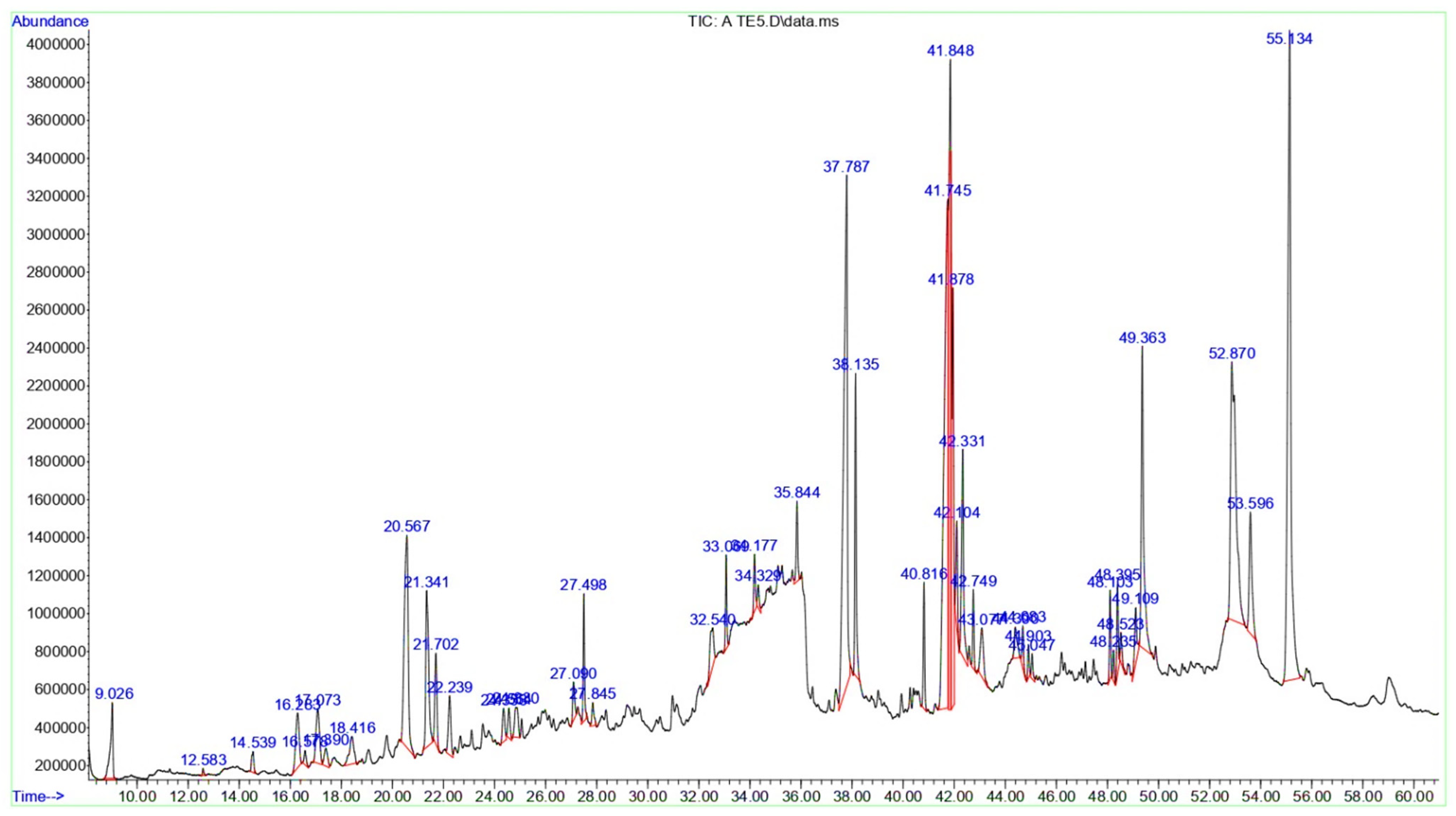
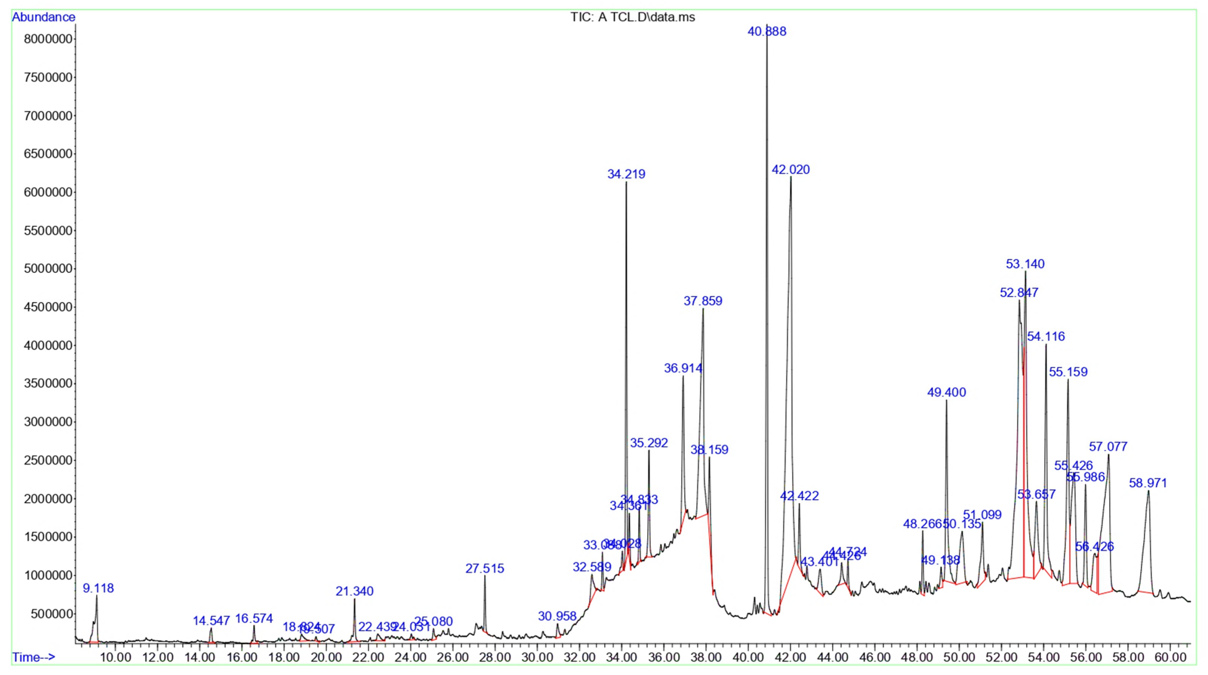
GC-MS analyses were performed in triplicate for both extracts, yielding extremely consistent chromatographic profiles with relative peak-area standard deviations < 5%. There were no contamination peaks in solvent blanks, indicating that the detected components came from plant sources. The bioactive compounds in T. terrestris LE were identified using GC–MS. The GC–MS chromatogram depicting these bioactive compounds is displayed in Figure 1, with their peak retention time (RT), peak area (%), molecular formula (MF), and molecular weight (MW) listed in Table 1. 42 bioactive compounds were identified in T. terrestris LE, expressed as percentages of the peak area relative to the overall peak area. The predominant constituent in the T. terrestris leaves was Linolenic acid, which accounted for 16.32% of the total. The following constituents were Vitamin E (13.49%), Campesterol (7.46%), Linolenic acid, methyl ester (6.99%), Phytol (6.47%), and n-Hexadecanoic acid (6.09%). Collectively, the main categories in the GC-MS analysis of T. terrestris LE were Lipids and Fatty Acid Derivatives (37.08%), followed by Terpenoids and Steroids (26.48%) and Glycosyl Compounds and Carbohydrates (8.31%).
Figure 1.
The GC-MS chromatograms of T. terrestris LE. All spectral peaks correlate with the identified chemicals, with a major peak indicating the primary constituent of the extract.
Table 1.
GC-MS compounds in T. terrestris LE.
However, a total of 46 bioactive components have been discovered in the T. terrestris SE, with Oleamide being the predominant compound, representing 13.01% of the total. The following compounds were cis,cis-Linoleic acid (12.05%), n-Hexadecanoic acid (11.38%), and 1-Monolinolein (8.68%), Linolenic acid (7.69%), and Linoleic acid ethyl ester (5.33%) (Table 2 and Figure 2). Collectively, the main categories in the GC-MS analysis of T. terrestris SE were the Lipids and Fatty Acid Derivatives composed the highest percentages (55.71%), followed by Glycerolipids (16.54%), and Glycosyl Compounds and Carbohydrates (3.08%), where Terpenoids and Steroids were represented by only 1.89%.
Table 2.
GC-MS compounds in T. terrestris SE.
Figure 2.
The GC-MS chromatograms of T. terrestris SE. A prominent peak in the spectrum indicates the major component of the extract, and every peak reflects a recognized chemical.
3.3. TPC and TFC of T. terrestris LE and SE
The TPC and TFC assay results revealed a significant difference in phytochemical content between the T. terrestris LE and SE. The TPC for the T. terrestris LE was higher than that of the T. terrestris SE (68.12 mg GAE/g DW). Similarly, the TFC for the T. terrestris LE was found to be (87 mg QE/g DW) higher than that of the T. terrestris SE (61 mg QE/g DW).
3.4. Antibacterial Effects of T. terrestris LE and SE
The disc diffusion technique was used to assess the antibacterial properties of the ethanol extracts of T. terrestris LE and SE against various bacterial strains. Table 3 and Table 4 demonstrate this extract’s ability to prevent the growth of the tested bacteria. The findings demonstrated that the extracts inhibited the growth of bacterial strains in a dose-dependent manner at various concentrations. The antibacterial activity of T. terrestris LE and SE increased gradually with higher concentration; at 400 and 800 μg/mL, the inhibitory zones began to increase significantly (p < 0.05), although not as much as the positive control (25 µg/mL of chloramphenicol). The T. terrestris LE showed more antibacterial activity with MIC 6.25 ± 0.00–25 ± 0.00 μg/mL than T. terrestris SE with MIC = 12.50 ± 0.00–50 ± 0.00 μg/mL. Gram-negative bacteria, particularly K. pneumoniae were more vulnerable to T. terrestris LE or SE.
Table 3.
The inhibitory zone (mm), MIC (μg/mL), and MBC (μg/mL) of T. terrestris LE.
Table 4.
The inhibitory zone (mm), MIC (μg/mL), and MBC (μg/mL) of T. terrestris SE.
3.5. DPPH and ABTS+ Radical Scavenging Activity
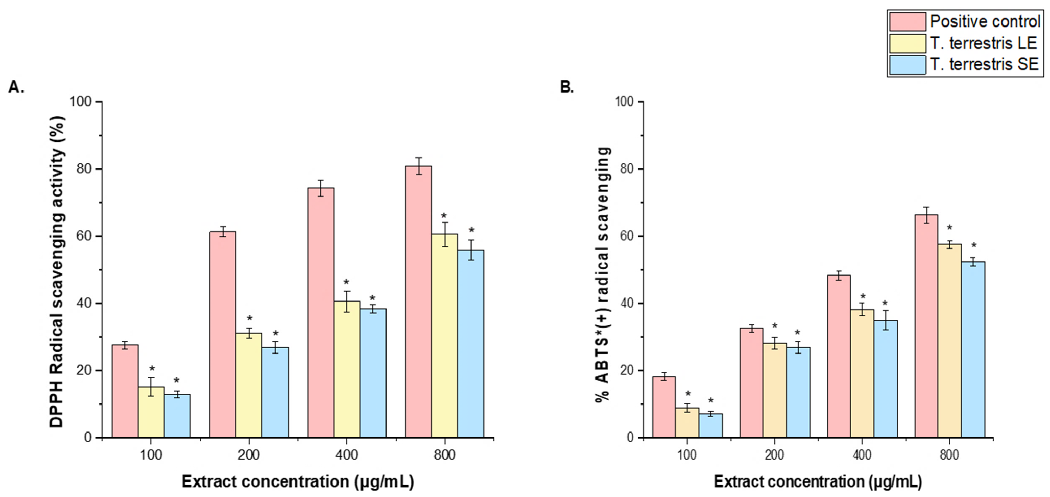
DPPH and ABTS+ assays were performed to evaluate the radical-scavenging properties of the T. terrestris LE and SE, expressed as IC50 values, and compared with ascorbic acid, the representative positive control for these assays. Based on the IC50 values in the DPPH and ABTS+ assays. The T. terrestris LE with IC50 value (113.41 μg/mL) demonstrated higher antioxidant ability than the T. terrestris SE IC50 = 162.42 μg/mL). However, the IC50 value of both the T. terrestris LE and SE is lower than the antioxidant level of the positive control (IC50 = 28.11 μg/mL) (Figure 3A,B).
Figure 3.
Antioxidant activity of T. terrestris LE and SE. (A) DPPH reducing power and (B) ABTS+ scavenging activity at various concentrations (100–800 μg/mL). An ascorbic acid (100–800 µg/mL) was used as a positive control. The mean value of 3 independent experiments is presented. The scavenging activity of T. terrestris LE and SE was significantly lower (*) than the positive control at a significance level of p < 0.05. + = radical cation.
Likewise, the IC50 value for the T. terrestris LE (90.71 μg/mL) exhibited superior antioxidant activity compared to the IC50 value of the T. terrestris SE (176.11 μg/mL) in the ABTS + radical assay. However, both the T. terrestris LE and SE have IC50 values that are lower than the antioxidant level of the positive control (IC50 = 24.18 μg/mL) (Figure 3A,B). The observed variation in DPPH and ABTS+ radical-scavenging activity between the LE and SE of T. terrestris may be due to differences in the quantity of phytochemicals in each extract.
3.6. Cell Cytotoxicity
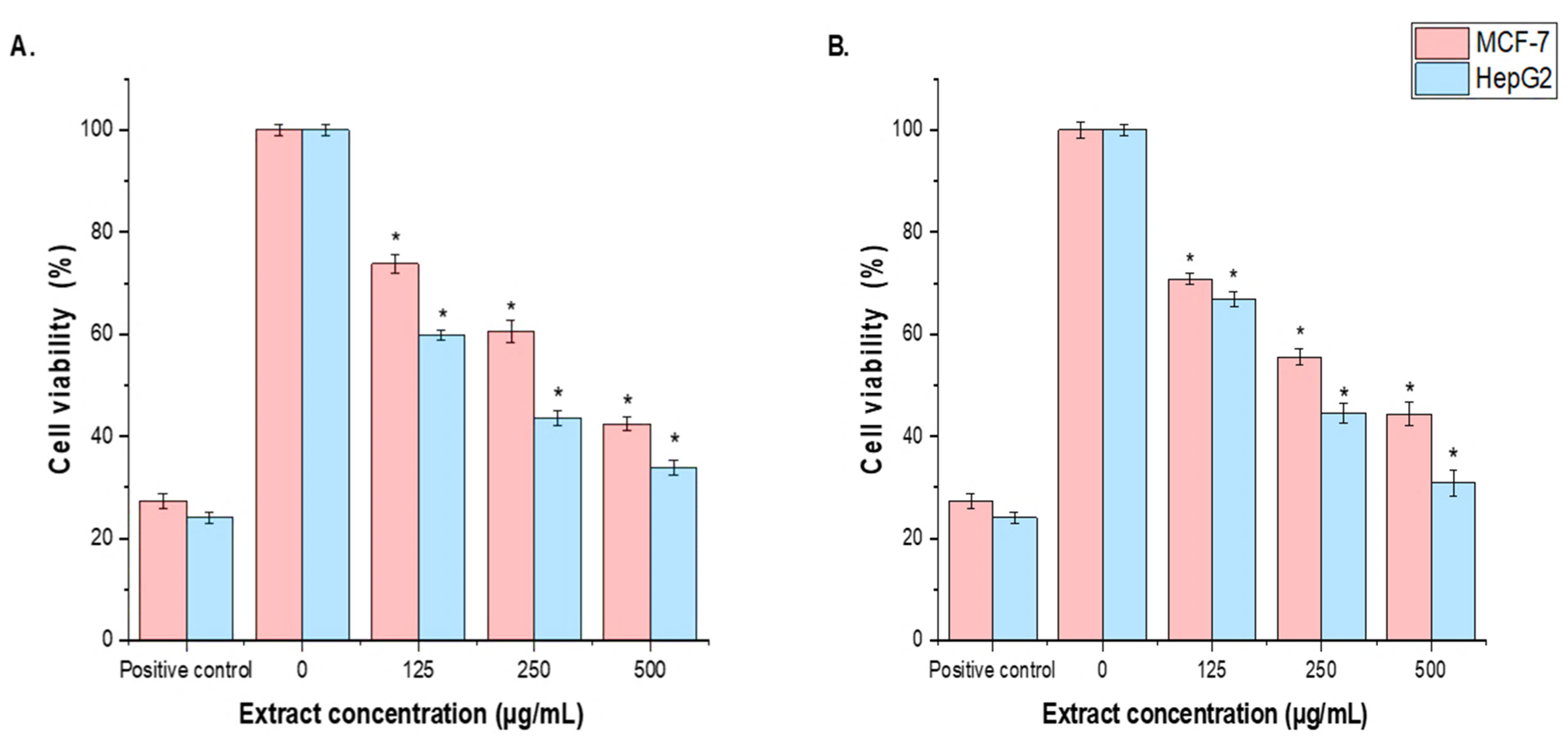
The cytotoxicity activity of T. terrestris LE and SE against MCF-7 and HepG2 cells was assessed using the MTT assay. T. terrestris LE and SE were tested for their anti-carcinogenic properties at concentrations of 125, 250, 500, and 1000 µg/mL. The cytotoxic activity of T. terrestris leaf and seed extracts on MCF-7 and HepG2 cells was concentration-dependent. At 125 μg/mL of T. terrestris LE and SE, cellular proliferation of MCF-7 and HepG2 lines was significantly (p < 0.05) inhibited compared to the untreated control cells, although less than the positive control (30 µg/mL of cisplatin). The cytotoxic effects of the T. terrestris LE were more potent than those of the T. terrestris SE, with IC50 values of 126.14 μg/mL and 105.12 μg/mL against MCF-7 and HepG2 cells, respectively, in both tests. Interestingly, the current investigation showed that HepG2 cells were more responsive to LE and SE than MCF-7 cells (Figure 4A,B).
Figure 4.
The effect of T. terrestris (A), LE and (B), SE on MCF-7 and HepG2 cell viabilities using the MTT assay. Cells were treated with the T. terrestris LE and SE (125, 250, 500, and1000 µg/mL) for 24 h. Mean values ± SD of three independent experiments are shown. (* p < 0.05 compared to non-treated cells (negative control)).
Ethanol extracts from T. terrestris seeds and leaves were evaluated for their effects on HepG2-induced apoptotic signaling and MCF-7. The MCF-7 and HepG2 cell lines treated with seed extract showed higher levels of mRNA expression, according to the rRT-PCR study. Anti-apoptotic genes (Bcl-xL and Bcl-2) were expressed less in the MCF-7 and HepG2 cell lines treated with plant extract than in the control group (p < 0.05) (Figure 5A,B).
Figure 5.
Effect of T. terrestris (A), LE and (B), SE on MCF-7 and HepG2 cells and determination of pro- and anti-apoptosis marker genes (caspase-3, 8, and 9, Bax, Bcl-2, and Bcl-Xl genes). The values represent the mean ± SD from ± trials (* p < 0.01).
3.7. In Vitro Antidiabetic Activities of T. terrestris LE and SE
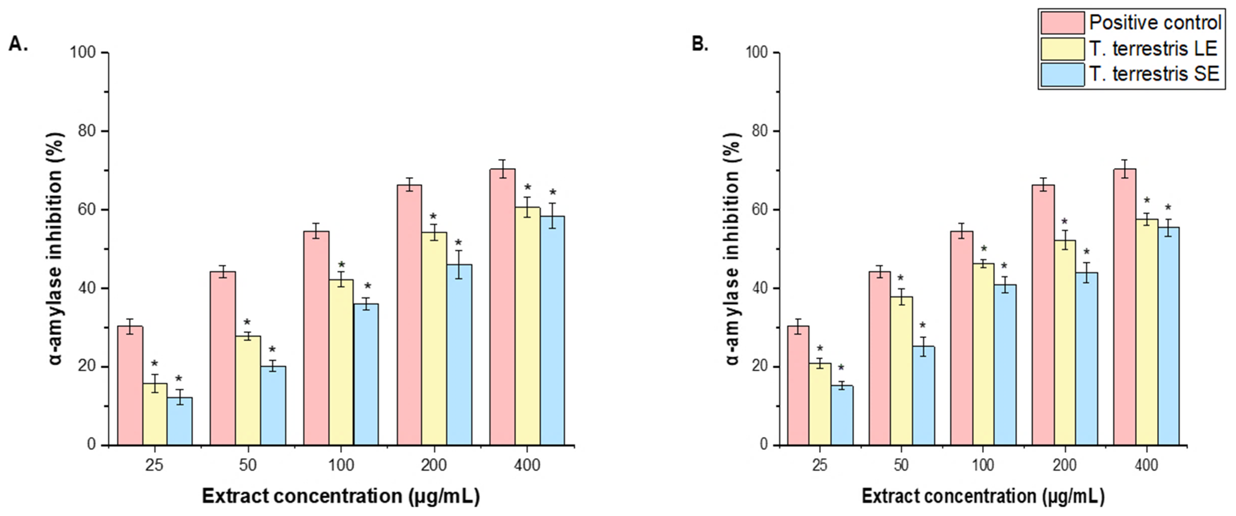
The antidiabetic activities of T. terrestris LE and SE were determined using the inhibitory effects on α-amylase and α-glucosidase enzymatic activities. The results are expressed as IC50 values. The T. terrestris LE showed a considerable inhibitory effect against α-amylase and α-glucosidase (IC50 = 84 ± 2.84 μg/mL and 96.62 ± 1.13 μg/mL, respectively) (Figure 6A), whereas T. terrestris SE inhibited α-amylase (IC50 = 113 ± 0.14 μg/mL), α-glucosidase (IC50 = 126.62 ± 1.12 μg/mL) (Figure 6B). The LE showed a significant inhibitory action against both enzymes (IC50 = 84 ± 2.84 μg/mL and 96.62 ± 1.13 μg/mL, respectively) in comparison to the T. terrestris SE, which had IC50 values of 113 ± 0.14 μg/mL for α-amylase and 126.62 ± 1.12 μg/mL for α-glucosidase. Conversely, both the LE and SE of T. terrestris presented IC50 values for the two enzymes analyzed, α-amylase (IC50 = 34.41 ± 0.18 μg/mL) and α-glucosidase (IC50 = 42.52 ± 1.28 μg/mL), that were less than the positive control, which was noted at 28.11 μg/mL (as shown in Figure 6A,B).
Figure 6.
Effect of T. terrestris (A), LE and (B), SEon α-amylase and α-glucosidase inhibitory activities at various concentrations (25–400 μg/mL). The results are the mean values of three replicates. The results are the mean ± SD of three experiments (* p < 0.05 compared to the acarbose, positive control).
4. Discussion
The GC-MS analysis of T. terrestris LE and SE revealed a complex phytochemical profile dominated by specific chemical classes, many of which are known to have biologically relevant properties. In the current study, Linolenic acid was represented in the LE and SE by 16.32% and 12.05%, respectively. In the LE, Vitamin E, Campesterol, and Phytol were the most represented compounds by 13.49%, 7.46%, and 6.47%, respectively. The highest expression levels in the SE were 13.01%, 11.38%, and 8.68% for oleamide, n-hexadecanoic acid (palmitic acid), and 1-monolinolein, respectively. The potential for antibacterial properties of the LE and SE is greatly enhanced by the high concentration of fatty acids in both [,]. Fatty acid components in a recent study on the fruit extract of T. terrestris demonstrated possible antibacterial activity against methicillin-resistant S. epidermidis (MRSE) according to HPLC fractionation []. The presence of Vitamin E and terpenoids such as Phytol in the LE is consistent with the potent antioxidant activity reported for several T. terrestris extracts. Studies have indicated that ethanol and methanol extracts of the plant showed strong radical scavenging capacities in DPPH, FRAP, and ABTS assays, which are crucial for alleviating oxidative stress [,,]. The presence of Oleamide in the SE and other fatty acids in both extracts is consistent with previous findings about their effect on the inflammatory pathways []. The high concentration of sterols in LE, such as Campesterol and Stigmasterol, is notable. These substances are structurally similar to the steroidal saponins (e.g., dioscin), which have been identified as the principal anticancer agents in T. terrestris, triggering apoptosis in cancer cells []. n-Hexadecanoic acid and monoacylglycerols, such as 1-Monolinolein, have been shown to inhibit enzymes, including α-glucosidase []. T. terrestris extracts, particularly ethanol extracts, have been shown to have a substantial inhibitory effect against α-glycosidase and cholinesterases (AChE and BChE), which are essential for managing diabetes and Alzheimer’s disease, respectively []. An in vivo investigation verified the antihyperglycemic efficacy of an aqueous extract of T. terrestris, which dramatically reduced blood glucose and glycated hemoglobin (HbA1c) levels in diabetic rats [].
The phytochemical profile is consistent with previous investigations on T. terrestris, while exhibiting noticeable variations. According to a detailed examination, the chemical makeup of the plant varies greatly depending on the plant component, geographic origin, and extraction solvent. For example, although the LE has Vitamin E and β-Lactose, fruit extracts from other locations typically contain steroidal saponins such as protodioscin []. These findings, indicating a significant concentration of fatty acids and lipids in the seeds, are consistent with the plant’s biology, as seeds commonly store energy as lipids []. Ethyl iso-allocholate and digitoxin were found to be more prevalent in the GC-MS analysis of alcoholic extracts of T. terrestris leaves and fruits, according to another study []. T. terrestris’ medicinal potential reflects the Zygophyllaceae family’s overall pharmacological significance. The presence of potent secondary metabolites, such as saponins, flavonoids, and alkaloids, is a common feature across many genera in this family, supporting their use in traditional medicine systems worldwide []. The GC-MS profiles provide a preliminary screening of T. terrestris’ volatile and semi-volatile metabolites. While some fatty acids, sterols, and terpenoids were clearly detected, which is compatible with other previous studies [,], the presence of certain chemicals (e.g., oleamide or arachidonic acid derivatives) should be regarded with caution, as they could be the result of minor analytical errors or derivatization reactions during the GC-MS procedure. However, careful use of glassware and procedural blanks reduced the risk of contamination as mentioned in the Section 2 .
In the current study, both extracts showed higher TPC and TFC values, but these were higher in the LE compared to the SE. That suggests a phytochemical basis for T. terrestris’ powerful biological activities. The flavonoids in T. terrestris are mainly derivatives of quercetin, kaempferol, and isorhamnetin []. A previous study showed that a difference in the polarity of the extraction solvents might affect the amounts of flavonoids and phenols measured, where the ethanolic extract resulted in higher values (TPC: 51 ± 0.7 mg. GAE/g and TFC: 66.5 ± 0.4 mg QE/g) compared to aceton (TPC: 47 ± 1.5 mg. GA. E/g and TFC: 52.5 ± 0.5 mg QE/g) and chloroform (TPC: 37 ± 1.2 mg. GA. E/g and TFC: 43 ± 1.5 mg QE/g) extracts []. According to another study, the TPC of various T. terrestris LE varied between 17.93 and 32.00 mg GAE/g, with the methanolic extract having the highest TFC at 27.62 mgRE/g []. So, TFC levels in the LE not only confirm a higher concentration of polyphenolic antioxidants, but also clearly suggest that the leaves are a more attractive source for leveraging the plant’s established anti-inflammatory and anticancer effects than the SE.
The preliminary phytochemical research on T. terrestris revealed the presence of saponins, flavonoids, glycosides, alkaloids, and tannins []. Data from the literature indicate that T. terrestris from various geographical locations has varying saponin contents and compositions []. The chemistry and bioactivity of saponins in T. terrestris were investigated by Kostova et al. According to their findings, this plant commonly contains furostanol and spirostanol saponins of the tigogenin, neotigogenin, gitogenin, neogitogenin, hecogenin, neohecogenin, diosgenin, chlorogenin, ruscogenin, and sarsasapogenin kinds. Four sulfated saponins of the diosgenin and tigogenin types were also identified. Furostanol glycosides, such as protodioscin and protogracillin, are mostly found; protodioscin is the most prevalent saponin. Spirostanol glycosides are found in trace amounts [,]. According to Wu et al., there are almost 1.5 times as many major flavonoids as main saponins. This suggested that the flavonoid content of T. terrestris should be investigated, developed, and further utilized []. In leaf extracts from four Tribulus species, Louveaux et al. used HPLC to identify eighteen flavonoids (caffeoyl derivatives, quercetin glycosides, including rutin and kaempferol glycosides) [].
The antioxidant activity of T. terrestris LE and SE differs significantly. The LE (IC50 = 113.41 μg/mL in DPPH; 90.71 μg/mL in ABTS+) outperformed the SE (IC50 = 162.42 μg/mL in DPPH; 176.11 μg/mL in ABTS+). The DPPH assay assesses hydrogen-atom transfer activity, while the ABTS+ assay detects electron-transfer capacity [], resulting in different sensitivity profiles for the same complex mixture. This found bioactivity is consistent with the plant’s known phytochemistry and the Zygophyllaceae family’s overall pharmacological profile. Previous studies showed that T. terrestris’ primary antioxidants are steroidal saponins (e.g., protodioscin) and flavonoids (e.g., rutin, quercetin, isorhamnetin) [,]. That a methanolic extract of T. terrestris had significant phenolic (341.3 mg GAE/g) and flavonoid (209 mg QE/g) content, which correlates with strong antioxidant activity. The LE is more potent in the current study, showing that it contains a larger concentration of these phenolic and flavonoid chemicals than the SE. From another point of view, saponin-rich T. terrestris fractions showed higher antioxidant and anti-glycation activity than crude extracts [,]. They showed that increasing the saponin content from 40% to 72.8% resulted in an 89.89% reduction in advanced glycation end-product and improved performance in several antioxidant tests. Furthermore, while there have been few systematic comparative studies about the antioxidant activities of Zygophyllaceae members, this family is known for producing a wide range of bioactive chemicals of known antioxidant potential. Different extracts from Zygophyllum Geslini Coss [], Zygophyllum simplex L. [], and Zygophyllum coccineum L. [], showed significant antioxidant activities due to their robust chemical profile. The antioxidant potential of these extracts might be explained through various routes, including direct free radical scavenging and enhancing the body’s endogenous defence by increasing glutathione levels. In the study conducted by Shetty et al. (2024) [], they found that T. terrestris’ nephroprotective and hepatoprotective benefits in rats were achieved by regulating the inflammatory marker IL-6 and lowering oxidative stress. The LE’s enhanced antioxidant activity is most likely owing to a greater concentration of steroidal saponins and flavonoids [].
The current study evaluated the antibacterial properties of T. terrestris LE and SE. The results showed that the LE was more potent than that of the SE with MICs of 6.25–25 μg/mL and 12.50–50 μg/mL, respectively. The most susceptible strains were E. coli for the seed extract and K. pneumoniae for both. On the other side, S. aureus and S. epidermidis were the most resistant to the LE, while B. subtilis was the most resistant to the SE. The findings are consistent with the previously documented antibacterial profile of T. terrestris. Al-Bayati and Al-Mola (2008) [], conducted a study in Iraq. They found that, to the root extract, the methanolic leaf had the lowest MIC values against B. subtilis (0.31 mg/mL) and K. pneumoniae (0.31 mg/mL). According to a different study, the antibacterial activity of T. terrestris fruit extract against Streptococcus mutans, Streptococcus sanguis, Actinomyces viscosus, Enterococcus faecalis, S. aureus, and Escherichia coli was enhanced when combined with Glycyrrhiza glabra and Capsella bursa-pastoris []. A recent study found that the methanolic extract of T. terrestris fruit significantly inhibited the acidification activity of S. mutans and Lactobacillus acidophilus, two oral microbes that cause cavities in teeth (Azarm et al., 2024) []. It is believed that T. terrestris’s potent antibacterial properties stem from its diverse phytochemical makeup. Numerous bioactive compounds, such as alkaloids, flavonoids, and saponins, are said to be present in the plant. The compounds from a fruit fraction were effective against MRSE in the study by Zhu et al. (2017) []. By targeting the penicillin-binding protein 2a (PBP2a) transpeptidase, a critical protein mediating methicillin resistance, these bioactive compounds may inhibit bacterial growth, according to a recent study that used molecular docking tools []. So, these findings provide strong experimental evidence to support the traditional use of T. terrestris and highlight its leaves as a ting source of antibacterial agents with various modes of action.
The results demonstrated that T. terrestris LE and SE exhibit concentration-dependent cytotoxicity against MCF-7 and HepG2 cell lines. A similar study found substantial benefits across multiple cancer types. A methanolic extract of T. terrestris exhibited cytotoxicity against lung cancer A549 cells (IC50 = 179.62 μg/mL) and MCF-7 cells []. Moreover, exceptional efficacy has been found against additional lines, such as human ovarian adenocarcinoma A2780 cells (IC50 = 3.69 μg/mL) [], colorectal cancer HT-29 cells (IC50 = 7.1 μg/mL), and prostate cancer LNCaP cells (IC50 = 0.3 μg/mL) []. The gene expression data revealed downregulation of the anti-apoptotic genes Bcl-2 and Bcl-xL, which is central to the mechanism of action. Previous studies found that T. terrestris extracts greatly increased the activity of important executioner enzymes, caspase-3 and caspase-8 [,], and upregulate key pro-apoptotic proteins such as Bax and p53 [], further shifting the cellular balance toward programmed cell death. The mitochondrial (intrinsic) apoptotic pathway in cancer cells is successfully started by this dual action of blocking survival signals (Bcl-2/Bcl-xL) and activating cell death executors (caspases, Bax). Multiple studies have shown that T. terrestris extracts have much lower cytotoxicity against non-malignant cells. This includes PBMCs and normal fibroblast cells (L929 and HSkMC) [,], which were significantly more viable than cancer cells after treatment. However, a word of caution is required; one study on human cells found that high amounts of aqueous fruit extract (40–80 mg/L) may have genotoxic effects, such as producing micronuclei and chromosomal abnormalities [,] demonstrated that Tribulus macropterus, a close relative of T. terrestris, exhibited similar cytotoxic activity against HepG2 liver cancer cells, attributed to the presence of cytotoxic cholestane and pregnane glycosides. Nyctanthes arbor-tristis (Oleaceae) also exhibits alkaloid extracts that induce apoptosis by changing the Bax/Bcl-2 ratio []. This shows that many therapeutic plants use the same successful approach to target the apoptotic machinery.
T. terrestris LE and SE have been shown to inhibit α-amylase and α-glucosidase, providing quantitative evidence for their traditional usage in diabetes management. The observed differences in efficacy between plant sections, in contrast to a typical medicine, provide valuable information for medicinal development. A previous study discovered that a methanolic root extract of T. terrestris significantly inhibited α-glucosidase []. Another study showed that ethanol extracts of T. terrestris block the α-glycosidase enzyme, a critical target for regulating postprandial blood glucose levels, and that this effect was frequently linked to flavonoid concentration in the plant []. Research suggests that the plant’s strong saponin content stimulates insulin production from pancreatic β-cells and enhances glucose absorption in peripheral tissues, in addition to blocking carbohydrate-digesting enzymes []. Furthermore, a 2024 study found that a mixed aqueous extract of T. terrestris and Curcuma amada was highly efficient in diabetic rats, drastically decreasing mean blood glucose and glycated hemoglobin (HbA1c) while improving body weight and plasma insulin levels []. An 8-week in vivo study found that the aqueous fruit extract of T. terrestris was more effective than metformin in restoring normal blood glucose levels and rehabilitating the histological architecture of hepatocytes in diabetic rats, owing to the herb’s potent antioxidant activities, which included direct reactive oxygen species (ROS) scavenging and regeneration of key antioxidant enzymes such as catalase (CAT) and glutathione-S-transferase (GST) []. Another study discovered that extracts with high protodioscin content (TT-HPC) were more successful at lowering raised blood glucose levels in diabetic rats than those with low protodioscin concentration (TT-LPC) []. This indicates that the concentration of specific saponins plays a crucial role in the plant’s hypoglycemic effect and suggests that T. terrestris may synergistically combine with other medicinal plants.
This work presents a fresh and comparative phytochemical and pharmacological profile of T. terrestris, focusing on the LE and SE. The significant contribution is the direct evidence that the LE, frequently disregarded in favor of the fruit, exhibits higher bioactivity in numerous domains, including antibacterial, antioxidant, cytotoxic, and antidiabetic assays. The downregulation of Bcl-2 and Bcl-xL genes in cancer cells, combined with the plant’s inhibitory activity against α-glucosidase and α-amylase, provides new insights into its traditional uses and positions it as a promising source for future therapeutic development.
We acknowledge the shortcomings and limitations in terms of the methodology we used in our present study. To ensure consistency in retention times and spectral matches, each GC-MS run for both leaf and seed extracts should be performed in triplicate. To further validate the newly identified compounds, retention indices of the components should be calculated in relation to the retention periods of a series of n-alkanes (two standard mixes, C8–C20 and C21–C40) using linear interpolation. It is also recognized that steroidal saponins and other glycosidic chemicals, which are the primary bioactive principles of T. terrestris, are non-volatile and thus cannot be detected successfully by GC-MS. Future research will use complementary techniques such as LC-MS/MS and HPLC-ELSD to accomplish a thorough characterization and quantification of steroidal glycosides. These additional tests will offer a more thorough chemical fingerprint of T. terrestris and validate the early findings presented here.
5. Conclusions
The study demonstrates that T. terrestris contains high concentrations of bioactive compounds with significant multifunctional pharmacological potential. The LE consistently proved more potent than the SE, exhibiting vigorous antibacterial activity against Gram-negative pathogens, notable antioxidant capacity, potent cytotoxicity against MCF-7 and HepG2 cancer cells through apoptosis induction, and effective inhibition of key carbohydrate-digesting enzymes. The enhanced efficacy of the LE is closely related to its higher phenolic, flavonoid, and terpenoid content. These results support the ethnopharmacological uses of T. terrestris and highlight the LE as a valuable yet underutilized component for future nutraceutical and pharmaceutical research and applications.
Author Contributions
Conceptualization, S.A.A. and I.M.A.; methodology, I.M.A.; software, I.M.A.; validation, S.A.A., I.M.A., M.A.F. and A.K.A.; formal analysis, R.M.A. (Rawan M. Alshalan); investigation, M.A.F. and A.K.A.; resources, R.M.A. (Reem M. Aljowaie); data curation, R.M.A. (Reem M. Aljowaie); writing—original draft preparation, S.A.A. and I.M.A.; writing—review and editing, M.A.F., A.M.A. and R.M.A. (Rawan M. Alshalan); visualization, S.A.A. and A.K.A.; supervision, S.A.A.; project administration, I.M.A.; funding acquisition, A.M.A. All authors have read and agreed to the published version of the manuscript.
Funding
This research received no external funding.
Institutional Review Board Statement
Not applicable.
Informed Consent Statement
Not applicable.
Data Availability Statement
Data is contained within the article.
Acknowledgments
The authors thank the Ongoing Research Funding Program, (ORF-2025-933), King Saud University, Riyadh, Saudi Arabia.
Conflicts of Interest
The authors declare no conflicts of interest.
References
- Chhatre, S.; Nesari, T.; Somani, G.; Kanchan, D.; Sathaye, S. Phytopharmacological overview of Tribulus terrestris. Pharmacogn. Rev. 2014, 8, 45. [Google Scholar] [CrossRef]
- Gupta, R. Ethnobotanical studies on medicinal plant: Gokhru (Tribulus terrestris). Int. J. Herb. Med. 2017, 5, 73–74. [Google Scholar]
- Nebieridze, V.; Skhirtladze, A.; Kemertelidze, E.; Ganzera, M. Nucleosides from Tribulus terrestris. Chem. Nat. Compd. 2017, 53, 1010–1011. [Google Scholar] [CrossRef]
- Zhu, W.; Du, Y.; Meng, H.; Dong, Y.; Li, L. A review of traditional pharmacological uses, phytochemistry, and pharmacological activities of Tribulus terrestris. Chem. Cent. J. 2017, 11, 60. [Google Scholar] [CrossRef]
- Ștefănescu, R.; Tero-Vescan, A.; Negroiu, A.; Aurică, E.; Vari, C.-E. A comprehensive review of the phytochemical, pharmacological, and toxicological properties of Tribulus terrestris L. Biomolecules 2020, 10, 752. [Google Scholar] [CrossRef]
- Dovchinsuren, B.; Damdinsuren, A.; Bayarkhuu, B.; Purevkhuu, M.; Batjargal, O.; Badarch, B.; Dorjbal, E. Tribulus terrestris L. in traditional Mongolian medicine: Medicinal Applications, Phytochemistry, Pharmacology. Pharmacogn. J. 2025, 17, 171–178. [Google Scholar] [CrossRef]
- Dinchev, D.; Janda, B.; Evstatieva, L.; Oleszek, W.; Aslani, M.R.; Kostova, I. Distribution of steroidal saponins in Tribulus terrestris from different geographical regions. Phytochemistry 2008, 69, 176–186. [Google Scholar] [CrossRef] [PubMed]
- Batovska, D.; Panova, N.; Gerasimova, A.; Tumbarski, Y.; Ivanov, I.; Dincheva, I.; Yotkovska, I.; Gentscheva, G.; Nikolova, K. Chamomile Matters: Species-and Producer-Dependent Variation in Bulgarian Matricaria recutita L. and Chamaemelum nobile L. Essential Oils and Their Cosmetic Potential. Cosmetics 2025, 12, 123. [Google Scholar] [CrossRef]
- Lazarova, I.; Ivanova, A.; Mechkarova, P.; Peev, D.; Valyovska, N. Intraspecific variability of biologically active compounds of different populations of Tribulus terrestris L. (Zygophyllaceae) in South Bulgaria. Biotechnol. Biotechnol. Equip. 2011, 25, 2352–2356. [Google Scholar] [CrossRef]
- Sarvin, B.; Stekolshchikova, E.; Rodin, I.; Stavrianidi, A.; Shpigun, O. Optimization and comparison of different techniques for complete extraction of saponins from T. terrestris. J. Appl. Res. Med. Aromat. Plants 2018, 8, 75–82. [Google Scholar] [CrossRef]
- Saeed, M.; Munawar, M.; Bi, J.B.; Ahmed, S.; Ahmad, M.Z.; Kamboh, A.A.; Arain, M.A.; Naveed, M.; Chen, H. Promising phytopharmacology, nutritional potential, health benefits, and traditional usage of Tribulus terrestris L. herb. Heliyon 2024, 10, e25549. [Google Scholar] [CrossRef]
- Wang, Z.-F.; Wang, B.-B.; Zhao, Y.; Wang, F.-X.; Sun, Y.; Guo, R.-J.; Song, X.-B.; Xin, H.-L.; Sun, X.-G. Furostanol and spirostanol saponins from Tribulus terrestris. Molecules 2016, 21, 429. [Google Scholar] [CrossRef]
- Shahid, M.; Riaz, M.; Talpur, M.; Pirzada, T. Phytopharmacology of Tribulus terrestris. J. Biol. Regul. Homeost. Agents 2016, 30, 785–788. [Google Scholar]
- Zhang, H.; Kuang, Y.; Shahrajabian, M.H.; Wang, N.; Sun, W. Study of puncture vine (Tribulus terrestris L.), and the important roles of its chemical ingredients. Not. Sci. Biol. 2025, 17, 12194. [Google Scholar] [CrossRef]
- Sirotkin, A.V.; Kolesarova, A. Puncture vine (Tribulus terrestris L.) in control of health and reproduction. Physiol. Res. 2021, 70, S657. [Google Scholar]
- Vilar Neto, J.d.O.; de Moraes, W.M.A.M.; Pinto, D.V.; da Silva, C.A.; Caminha, J.d.S.R.; Nunes Filho, J.C.C.; Reis, C.E.G.; Prestes, J.; Santos, H.O.; De Francesco Daher, E. Effects of Tribulus (Tribulus terrestris L.) Supplementation on Erectile Dysfunction and Testosterone Levels in Men—A Systematic Review of Clinical Trials. Nutrients 2025, 17, 1275. [Google Scholar] [CrossRef]
- Khalid, A.; Algarni, A.S.; Homeida, H.E.; Sultana, S.; Javed, S.A.; Rehman, Z.U.; Abdalla, H.; Alhazmi, H.A.; Albratty, M.; Abdalla, A.N. Phytochemical, cytotoxic, and antimicrobial evaluation of Tribulus terrestris L., Typha domingensis pers., and Ricinus communis L.: Scientific evidences for folkloric uses. Evid. Based Complement. Altern. Med. 2022, 2022, 6519712. [Google Scholar] [CrossRef]
- Abbas, A.; Naqvi, S.A.R.; Rasool, M.H.; Noureen, A.; Mubarik, M.S.; Tareen, R.B. Phytochemical analysis, antioxidant and antimicrobial screening of Seriphidium oliverianum plant extracts. Dose Response 2021, 19, 15593258211004739. [Google Scholar] [CrossRef]
- Wolfe, K.L.; Liu, R.H. Apple peels as a value-added food ingredient. J. Agric. Food Chem. 2003, 51, 1676–1683. [Google Scholar] [CrossRef]
- Ordonez, A.; Gomez, J.; Vattuone, M. Antioxidant activities of Sechium edule (Jacq.) Swartz extracts. Food Chem. 2006, 97, 452–458. [Google Scholar] [CrossRef]
- Salem, N.; Kefi, S.; Tabben, O.; Ayed, A.; Jallouli, S.; Feres, N.; Hammami, M.; Khammassi, S.; Hrigua, I.; Nefisi, S. Variation in chemical composition of Eucalyptus globulus essential oil under phenological stages and evidence synergism with antimicrobial standards. Ind. Crops Prod. 2018, 124, 115–125. [Google Scholar] [CrossRef]
- Basri, D.F.; Sandra, V. Synergistic interaction of methanol extract from Canarium odontophyllum Miq. Leaf in combination with oxacillin against methicillin-resistant Staphylococcus aureus (MRSA) ATCC 33591. Int. J. Microbiol. 2016, 2016, 5249534. [Google Scholar] [CrossRef]
- Aljeldah, M.M.; Yassin, M.T.; Mostafa, A.A.-F.; Aboul-Soud, M.A. Synergistic Antibacterial Potential of Greenly Synthesized Silver Nanoparticles with Fosfomycin Against Some Nosocomial Bacterial Pathogens. Infect. Drug Resist. 2022, 15, 125–142. [Google Scholar] [CrossRef]
- Tian, M.; Wu, X.; Lu, T.; Zhao, X.; Wei, F.; Deng, G.; Zhou, Y. Phytochemical analysis, antioxidant, antibacterial, cytotoxic, and enzyme inhibitory activities of Hedychium flavum rhizome. Front. Pharmacol. 2020, 11, 572659. [Google Scholar] [CrossRef]
- Yu, X.; Zhao, M.; Liu, F.; Zeng, S.; Hu, J. Antioxidants in volatile Maillard reaction products: Identification and interaction. LWT-Food Sci. Technol. 2013, 53, 22–28. [Google Scholar] [CrossRef]
- Mosmann, T. Rapid colorimetric assay for cellular growth and survival: Application to proliferation and cytotoxicity assays. J. Immunol. Methods 1983, 65, 55–63. [Google Scholar] [CrossRef]
- Alkhudhayri, A.A.; Wahab, R.; Siddiqui, M.A.; Ahmad, J. Selenium nanoparticles induce cytotoxicity and apoptosis in human breast cancer (MCF-7) and liver (HEPG2) cell lines. Nanosci. Nanotechnol. Lett. 2020, 12, 324–330. [Google Scholar] [CrossRef]
- Ghasemi, M.; Turnbull, T.; Sebastian, S.; Kempson, I. The MTT assay: Utility, limitations, pitfalls, and interpretation in bulk and single-cell analysis. Int. J. Mol. Sci. 2021, 22, 12827. [Google Scholar] [CrossRef]
- Al-Dhabi, N.A.; Valan Arasu, M. Quantification of phytochemicals from commercial Spirulina products and their antioxidant activities. Evid. Based Complement. Altern. Med. 2016, 2016, 7631864. [Google Scholar] [CrossRef]
- Wickramaratne, M.N.; Punchihewa, J.; Wickramaratne, D. In-vitro alpha amylase inhibitory activity of the leaf extracts of Adenanthera pavonina. BMC Complement. Altern. Med. 2016, 16, 466. [Google Scholar] [CrossRef]
- Kim, Y.-M.; Wang, M.-H.; Rhee, H.-I. A novel α-glucosidase inhibitor from pine bark. Carbohydr. Res. 2004, 339, 715–717. [Google Scholar] [CrossRef]
- Al-Bayati, F.A.; Al-Mola, H.F. Antibacterial and antifungal activities of different parts of Tribulus terrestris L. growing in Iraq. J. Zhejiang Univ. Sci. B 2008, 9, 154–159. [Google Scholar] [CrossRef]
- Soleimanpour, S.; Sedighinia, F.S.; Afshar, A.S.; Zarif, R.; Ghazvini, K. Antibacterial activity of Tribulus terrestris and its synergistic effect with Capsella bursa-pastoris and Glycyrrhiza glabra against oral pathogens: An in-vitro study. Avicenna J. Phytomed. 2015, 5, 210. [Google Scholar]
- Alzahrani, K.J. Tribulus terrestris Fruit Extract: Bioactive Compounds, ADMET Analysis, and Molecular Docking with Penicillin-Binding Protein 2a Transpeptidase of Methicillin-Resistant Staphylococcus epidermidis. Curr. Issues Mol. Biol. 2025, 47, 52. [Google Scholar] [CrossRef]
- Ammar, N.M.; El-Hawary, S.; Mohamed, D.A.; Afifi, M.S.; Ghanem, D.M.; Awad, G. Phytochemical and biological studies of Tribulus terrestris L. growing in Egypt. Int. J. Pharmacol. 2018, 14, 248–259. [Google Scholar] [CrossRef]
- Abbas, M.W.; Hussain, M.; Akhtar, S.; Ismail, T.; Qamar, M.; Shafiq, Z.; Esatbeyoglu, T. Bioactive compounds, antioxidant, anti-inflammatory, anti-cancer, and toxicity assessment of Tribulus terrestris—In vitro and in vivo studies. Antioxidants 2022, 11, 1160. [Google Scholar] [CrossRef]
- Kaya, E.; Aydın, T.; Sağlamtaş, R. Evaluation of antioxidant activities and inhibition effects of Tribulus terrestris L. extracts on some metabolic enzymes. S. Afr. J. Bot. 2024, 170, 156–162. [Google Scholar] [CrossRef]
- Kaliaperumal, K.; Bhat, B.A.; Subramanian, K.; Ramakrishnan, T.; Chakravarthy, E.; Al-Keridis, L.A.; Ahmad, I.; Alabdallah, N.M.; Saeed, M.; Karunakaran, R. In-vivo anti-hyperglycemic effect of herbal extracts Tribulus terrestris (L.) and Curcuma amada (R) on streptozotocin-induced diabetic rats and its associated histopathological studies. Heliyon 2024, 10, e24009. [Google Scholar] [CrossRef]
- Saxena, S.; Chakraborty, D. Antimicrobial Activity and GC-MS Analysis of Bioactive Compounds of Tribulus terrestris L. Asian J. Res. Bot. 2023, 6, 352–361. [Google Scholar]
- Hal, D.M.; Eltamany, E.; Abdelhameed, R.F.; Ibrahim, A.K.; Badr, J. Chemical review on Zygophyllum genus. Rec. Pharm. Biomed. Sci. 2022, 6, 105–129. [Google Scholar] [CrossRef]
- Williams, C.; Mbuyane, L.L.; Bauer, F.F.; Mokwena, L.; Divol, B.; Buica, A. A gas chromatography-mass spectrometry method for the determination of fatty acids and sterols in yeast and grape juice. Appl. Sci. 2021, 11, 5152. [Google Scholar] [CrossRef]
- Schlag, S.; Huang, Y.; Vetter, W. GC/EI-MS method for the determination of phytosterols in vegetable oils. Anal. Bioanal. Chem. 2022, 414, 1061–1071. [Google Scholar] [CrossRef]
- Siddique, M.H.; Andleeb, R.; Ashraf, A.; Zubair, M.; Fakhar-e-Alam, M.; Hayat, S.; Muzammil, S.; Atif, M.; Shafeeq, S.; Afzal, M. Integration of in silico and in vitro approaches to evaluate antioxidant and anticancer properties of Tribulus terrestris extracts. Arab. J. Chem. 2022, 15, 103984. [Google Scholar] [CrossRef]
- Uysal, S.; Senkardes, I.; Jekő, J.; Cziáky, Z.; Zengin, G. Chemical characterization and pharmacological profile of Tribulus terrestris extracts: A novel source of cosmeceuticals and pharmaceuticals. Biochem. Syst. Ecol. 2023, 107, 104600. [Google Scholar] [CrossRef]
- Affuf, A.; Karpenko, J.N.; Gulyaev, D.K.; Molokhova, E.I.; Blinova, O.L.; Gileva, A.A.; Belonogova, V.D. Phytochemical study of Tribulus terrestris L. Pharm. Pharmacol. 2019, 7, 346–355. [Google Scholar] [CrossRef]
- Kostova, I.; Dinchev, D. Saponins in Tribulus terrestris–chemistry and bioactivity. Phytochem. Rev. 2005, 4, 111–137. [Google Scholar] [CrossRef]
- Xu, Y.-J.; Xu, T.-H.; Zhou, H.-O.; Li, B.; Xie, S.-X.; Si, Y.-S.; Liu, Y.; Liu, T.-H.; Xu, D.-M. Two new furostanol saponins from Tribulus terrestris. J. Asian Nat. Prod. Res. 2010, 12, 349–354. [Google Scholar] [CrossRef]
- Wu, T.-S.; Shi, L.-S.; Kuo, S.-C. Alkaloids and other constituents from Tribulus terrestris. Phytochemistry 1999, 50, 1411–1415. [Google Scholar] [CrossRef]
- Louveaux, A.; Jay, M.; El Hadi, O.T.M.; Roux, G. Variability in flavonoid compounds of four Tribulus species: Does it play a role in their identification by desert locust Schistocerca gregaria? J. Chem. Ecol. 1998, 24, 1465–1481. [Google Scholar] [CrossRef]
- Platzer, M.; Kiese, S.; Herfellner, T.; Schweiggert-Weisz, U.; Miesbauer, O.; Eisner, P. Common trends and differences in antioxidant activity analysis of phenolic substances using single electron transfer based assays. Molecules 2021, 26, 1244. [Google Scholar] [CrossRef]
- Figueiredo, C.C.M.; Gomes, A.C.; Granero, F.O.; Junior, J.L.B.; Silva, L.P.; Ruiz, A.L.T.G.; da Silva, R.M.G. Antiglycation and antitumoral activity of Tribulus terrestris dry extract. Avicenna J. Phytomed. 2021, 11, 224. [Google Scholar] [PubMed]
- Figueiredo, C.C.M.; da Costa Gomes, A.; Granero, F.O.; Junior, J.L.B.; Silva, L.P.; Ximenes, V.F.; da Silva, R.M.G. Antiglycation and antioxidant activities of the crude extract and saponin fraction of Tribulus terrestris before and after microcapsule release. J. Integr. Med. 2022, 20, 153–162. [Google Scholar] [CrossRef] [PubMed]
- Touaibia, M.; Dhoha, A. Biochemical characterization, antioxidant and antidiabetic properties of Zygophyllum geslini Coss extracts. Int. J. Biol. Chem. Sci. 2024, 18, 1105–1114. [Google Scholar] [CrossRef]
- Abdallah, H.M.; Esmat, A. Antioxidant and anti-inflammatory activities of the major phenolics from Zygophyllum simplex L. J. Ethnopharmacol. 2017, 205, 51–56. [Google Scholar] [CrossRef] [PubMed]
- El-Afify, S.M.; El-Metwaly, M.A.; Abbas, M.A.; El-Amier, Y.A. In vitro assessment of antioxidant and cytotoxic activities of Zygophyllum coccineum L. methanolic extract. Egypt. J. Chem. 2024, 67, 393–401. [Google Scholar]
- Shetty, N.; Holla, S.; Nayak, V.; Shenoy, V.B.; Mohandas, R.K. Antioxidant and anti-inflammatory activity by modulating IL-6 as a potential mechanism in the nephroprotective and hepatoprotective properties of Tribulus terrestris. Res. Pharm. Sci. 2024, 19, 376–386. [Google Scholar] [CrossRef]
- Azarm, A.; Ayoobi, F.; Zare-Bidaki, M.; Taheri, M.; Zarandi, E.R. Antibacterial and antibiofilm activities of tribulus terrestris methanolic extract against Streptococcus mutans, Streptococcus sobrinus, and Lactobacillus acidophilus: An in vitro study. Dent. Res. J. 2024, 21, 57. [Google Scholar] [CrossRef]
- Alshabi, A.M.; Alkahtani, S.A.; Shaikh, I.A.; Orabi, M.A.; Abdel-Wahab, B.A.; Walbi, I.A.; Habeeb, M.S.; Khateeb, M.M.; Shettar, A.K.; Hoskeri, J.H. Tribulus terrestris cytotoxicity against breast cancer MCF-7 and lung cancer A549 cell lines is mediated via activation of apoptosis, caspase-3, DNA degradation, and suppressing Bcl-2 activity. Separations 2022, 9, 383. [Google Scholar] [CrossRef]
- Pourali, M.; Yaghoobi, M.M.; Sormaghi, M.H.S. Cytotoxic, anti-proliferative and apoptotic effects of Tribulus terrestris L. fruit extract on human prostate cancer Lncap and colon cancer HT-29 Cell Lines. Jundishapur J. Nat. Pharm. Prod. 2017, 12, e33561. [Google Scholar]
- Patel, A.; Soni, A.; Siddiqi, N.J.; Sharma, P. An insight into the anticancer mechanism of Tribulus terrestris extracts on human breast cancer cells. 3 Biotech 2019, 9, 58. [Google Scholar] [CrossRef]
- Kim, H.J.; Kim, J.C.; Min, J.S.; Kim, M.-j.; Kim, J.A.; Kor, M.H.; Yoo, H.S.; Ahn, J.K. Aqueous extract of Tribulus terrestris Linn induces cell growth arrest and apoptosis by down-regulating NF-κB signaling in liver cancer cells. J. Ethnopharmacol. 2011, 136, 197–203. [Google Scholar] [CrossRef]
- Qari, S.H.; El-Assouli, S. Evaluation of cytological and genetic effects of Tribulus terrestris fruit aqueous extract on cultured human lymphocytes. Saudi J. Biol. Sci. 2019, 26, 91–95. [Google Scholar] [CrossRef] [PubMed]
- Parekh, S.; Arkatkar, A.; Soni, A.; Patel, P.; Mishra, K. Nyctanthes arbor-tristis alkaloids activates p53 independent cell death receptor and necroptosis pathways in HepG2 cells. 3 Biotech 2023, 13, 167. [Google Scholar] [CrossRef] [PubMed]
- Al-Eisa, R.A.; Tag, H.M.; ElNaggar, M.S.; Abdelrazek, H.M.; El-Shenawy, N.S. Evaluation of Tribulus terrestris extracts relative to metformin on oxidative stress and Histopathology of the liver for diabetic male rats. Diabetology 2022, 3, 46–55. [Google Scholar] [CrossRef]
Disclaimer/Publisher’s Note: The statements, opinions and data contained in all publications are solely those of the individual author(s) and contributor(s) and not of MDPI and/or the editor(s). MDPI and/or the editor(s) disclaim responsibility for any injury to people or property resulting from any ideas, methods, instructions or products referred to in the content. |
© 2025 by the authors. Licensee MDPI, Basel, Switzerland. This article is an open access article distributed under the terms and conditions of the Creative Commons Attribution (CC BY) license (https://creativecommons.org/licenses/by/4.0/).