Abstract
Background: The anatomy laboratory can provide the ideal setting for the preclinical phase of neurosurgical research. Our purpose is to comprehensively and critically review the preclinical anatomical quantification methods used in cranial neurosurgery. Methods: A systematic review was conducted following the PRISMA guidelines. The PubMed, Ovid MEDLINE, and Ovid EMBASE databases were searched, yielding 1667 papers. A statistical analysis was performed using R. Results: The included studies were published from 1996 to 2023. The risk of bias assessment indicated high-quality studies. Target exposure was the most studied feature (81.7%), mainly with area quantification (64.9%). The surgical corridor was quantified in 60.9% of studies, more commonly with the quantification of the angle of view (60%). Neuronavigation-based methods benefit from quantifying the surgical pyramid features that define a cranial neurosurgical approach and allowing post-dissection data analyses. Direct measurements might diminish the error that is inherent to navigation methods and are useful to collect a small amount of data. Conclusion: Quantifying neurosurgical approaches in the anatomy laboratory provides an objective assessment of the surgical corridor and target exposure. There is currently limited comparability among quantitative neurosurgical anatomy studies; sharing common research methods will provide comparable data that might also be investigated with artificial intelligence methods.
1. Introduction
In recent years, evidence-based medicine has gained significant importance in surgery. In this scenario, the IDEAL (Development, Exploration, Assessment, and Long-term study) paradigm was the first promoter of evidence-based surgery [1,2,3,4]. IDEAL describes the different phases and challenges of research in surgery and includes a specific phase of preclinical research that can be performed in the anatomy laboratory. Quantitative anatomical research in neurosurgery still poses the following considerable challenges: despite the evolving innovation in surgical technologies (e.g., microscope, endoscopic-assisted techniques, robotics-assisted procedure), objective and shared methods to compare different surgical approaches are often lacking. These seem particularly important in neurosurgery, as even minor differences in surgical technique can significantly affect patient outcomes [5].
Over the last three decades, different quantitative methods have been reported in anatomical neurosurgical research. However, the heterogeneity and multitude of these methods and the different measured parameters complicate the panorama of neurosurgical anatomical quantification. This paper aims to provide a systematic and critical review of the current literature on preclinical anatomical quantification and the comparison of cranial neurosurgical approaches, analyze the proposed research methods and the studied features, and discuss their advantages and disadvantages.
2. Materials and Methods
2.1. Literature Search
The systematic review was performed per the Preferred Reporting Items for Systematic Reviews and Meta-Analysis (PRISMA) guidelines [6]. Two authors performed a systematically comprehensive literature search of the databases PubMed, Ovid MEDLINE, and Ovid EMBASE. The first literature search was performed on 10 April 2023, and the search was updated on 10 May 2023. A combination of keyword searches was performed to generate a search strategy. The search keywords, including “anatomy”, “quantification”, “neurosurgery”, “approach”, “comparison”, and “surgery”, were used in both AND and OR combinations. Studies were retrieved using the following Medical Subject Heading (MeSH) terms and Boolean operators: (“neurosurgical” OR “neurologic surgery”) AND (“approaches” OR “open” OR “microsurgery” OR “endoscopy” OR “endonasal” NOT “transorbital”) AND (“anatomy” OR “anatomical studies” OR “preclinical” OR “quantitative” NOT “qualitative”) AND (“comparison” OR “quantification” OR “methods” OR “conservative”). Other pertinent articles were identified through reference analysis of selected papers. A search filter was set to show only publications over the designated period, 1990–2023.
All studies were selected based on the following inclusion criteria: (1) English language; (2) articles that quantify and compare anatomical features of different neurosurgical approaches in the anatomy laboratory; (3) articles that quantify and compare anatomical features of different neurosurgical approaches in a virtual environment. The following exclusion criteria were employed: (1) studies that qualitatively compare surgical approaches; (2) studies reporting on neurosurgical approaches other than cranial.
The list of identified studies was imported into Endnote X9, and duplicates were removed. Two independent researchers (E.A. and L.D.M.) checked the results according to the inclusion and exclusion criteria. A third reviewer (A.F.) resolved all disagreements. Then, eligible articles were subject to full-text screening.
2.2. Data Extraction
For each study, we abstracted the following information: year of publication, quantified feature, quantified parameter, method, tool, and pros and cons of each technique.
2.3. Outcomes
Our primary outcomes were measurements related to the surgical corridor and target exposure. As for the surgical corridor, the following parameters were extrapolated from the analyzed studies: volume, surgical freedom or maneuverability, surgical window, and angle of view. Considering the target exposure, the following measurement techniques have been collected: anatomical structures visualization, linear measurements, areas, and volumes.
2.4. Risk of Bias Assessment
The Newcastle–Ottawa Scale (NOS) was used to assess the quality of the included studies. Quality assessment was performed by assessing the selection criteria, comparability of the study, and outcome assessment. The ideal score was 9. Higher scores indicated better quality of studies. Studies receiving 7 or more points were considered high-quality studies. Two authors performed the quality assessment independently. When discrepancies arose, papers were re-examined by the third author.
2.5. Statistical Analysis
Descriptive statistics were reported, including ranges and percentages. All statistical analyses were performed using the R statistical package v3.4.1 (http://www.r-project.org (accessed on 1 July 2023)).
3. Results
3.1. Literature Review
A total of 1667 papers were identified after duplicate removal. After title and abstract analysis, 200 articles were identified for full-text analysis. Eligibility was ascertained for 114 articles. The remaining 86 articles were excluded for the following reasons: (1) studies were not comparative (37 articles), (2) studies reporting only on qualitative comparison (39 articles), (3) overview studies (5 articles), (4) studies lacking methods details (4 articles), and (5) studies reporting on neurosurgical approaches other than cranial (1 article). All studies included in the analysis had at least one or more outcome measures available. Figure 1 shows the flow chart according to the PRISMA statement.
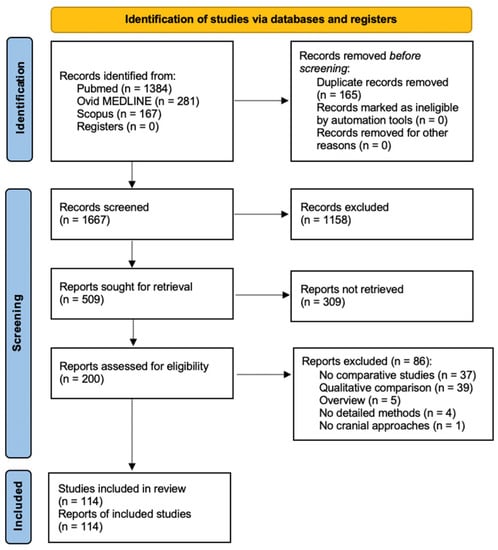
Figure 1.
PRISMA flow diagram depicting the literature search process [6].
3.2. Review Data and Outcomes
A total of 114 articles were included in our systematic review. The year of publication ranged from 1996 to 2023 as follows: four articles were published before 2000 (3.5%), 17 articles were published from 2000 to 2004 (14.9%), 32 articles from 2005 to 2009 (28.1%), 29 from 2010 to 2014 (25.5%), 20 from 2015 to 2019 (17.5%), and 12 from 2020 to May 2023 (10.5%). Table 1 lists all the articles included in our review ordered per year of publication.

Table 1.
List of all the articles included in our systematic review ordered per year of publication.
The surgical corridor and target were quantified on 69 articles (60.5%) and 94 articles (82.5%), respectively.
The quantified parameters of the surgical corridor were the angle of view in 42 articles (60.9%), surgical freedom or maneuverability in 20 (29%), and its volume in 16 (23.2%); the surgical window was quantified in 4 (5.8%) articles.
Target exposure was quantified by measuring the exposed area (61 articles; 64.9%) or linear distances (32 articles; 34%); semi-quantitative methods, based on visualization, were used in 14 articles (14.9%).
Table 2 and Table 3 summarize the quantified parameters, with respective methods and tools, and the advantages and disadvantages of each reported technique.
Table 2.
Summary of different methods and tools, with corresponding pros and cons, described in the literature for quantifying surgical corridor parameters.
Table 3.
Summary of different methods and tools, with corresponding pros and cons, described in the literature for quantifying target exposure parameters.
4. Discussion
This systematic literature review has collected and analyzed all the studies published since 1996 reporting the anatomical quantification and comparison of neurosurgical approaches.
With its constant advancements in surgical innovation and technology, neurosurgery requires objective methods to compare various surgical approaches [5,74,126,127]. Even minor differences in surgical technique can significantly impact patient outcomes in this field, and personal experience alone is no longer enough for ideal surgical decision-making. As shown by the various studies analyzed, quantifying neurosurgical approaches also aids in interpreting research results and promotes evidence-based medicine.
The systematic review revealed that, even if the first quantitative anatomical studies in neurosurgery were published in 1996, most were published after 2004 and mainly concentrated on target exposure analysis, thanks to the implementation of new technologies and dedicated software applied to preclinical research. It emerged that the quantitative measurements were initially limited to providing partial measurements of the surgical volumes, and the analysis of the surgical corridor has moved toward target exposure with the analysis of the exposure area (Figure 2).
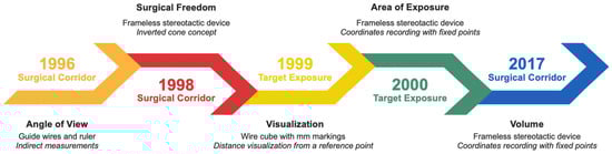
Figure 2.
Timeline depicting the evolution of neurosurgical approaches quantification and comparison.
It also emerged that the angle of view was the most frequently quantified parameter related to the surgical corridor, with 60% of the articles reporting its measurement. On the contrary, the surgical window was quantified in fewer articles, suggesting the difficulty of replicating this measurement. Regarding target exposure measurements, the exposure area was the most frequently quantified parameter, followed by linear measurements and visualization methods. These findings underscore the significance of assessing the extent of target exposure and the accuracy of surgical maneuvers during different neurosurgical approaches.
The systematic review also included a risk of bias assessment using the Newcastle–Ottawa Scale (NOS). The NOS allowed for evaluating the quality of the included studies based on selection criteria, comparability of the study, and outcome assessment. This assessment ensured that the included studies were reliable and provided robust evidence for quantifying and comparing neurosurgical approaches.
Choosing the proper research method is also paramount in quantitative anatomical studies. Direct measurements might be the best option to collect a relatively small amount of data with limited error, e.g., the length of a nerve exposed from different approaches. Neuronavigation-based methods, developed with dedicated software, allow the straightforward quantification of all the features that define the surgical pyramid, which is specific to each cranial neurosurgical approach. They can provide real-time data acquisition but also have the advantage of post-dissection data analyses, including the definition of the area of interest exposed by a specific approach.
Using standardized measurement techniques, researchers can accurately analyze and compare outcomes across different studies, enhancing the reliability and validity of their findings. This might contribute to accumulating robust evidence to guide clinical decision-making and improve patient outcomes. Furthermore, anatomical quantification facilitates the development of strategic surgical roadmaps, especially for deep-sited regions and complex targets. Additionally, quantifying neurosurgical approaches is essential for promoting new surgical strategies. For example, quantitative anatomical research has been critical in documenting the potential advantages of transnasal endoscopic transclival approaches [99,109].
While anatomical quantitative neurosurgical studies share similar research objectives, they have different research methods and are not comparable. Furthermore, despite incorporating modern technology into the research methodology, there often needs to be more adherence to scientific principles, resulting in a limited broad applicability of the findings. To address these issues, advancements in information technology and use big data analysis techniques through artificial intelligence methods are being increasingly implemented in quantitative neurosurgical anatomy research [128,129]. The final goal is to establish an evidence-based approach and achieve greater standardization and reliability in the research process.
Over the years, our research group has published several anatomical quantitative studies [96,98,99,100,106,107,109,110,113,114,116,127,130], focusing on the quantification of both the surgical volume and the exposure area. In accordance with our experience and with the aim of promoting standardization of the methods of quantification, we detail the minimum instrumentation necessary for an anatomical laboratory that wants to carry out quantitative studies. In detail:
- (1)
- Specimens:
- A minimum number of specimens equal to or greater than 5 so that the sample size of the data obtained allows the obtaining of statistically strong results;
- Better alcohol-fixed specimens, as they have a greater preservation of the anatomical tissues and the respect of the relationships between the neurovascular structures, they convert more over time.
- (2)
- Computed tomography scan:
- 1 × 1. frame with contiguous slices, both at 1 and 3 mm;
- Parameters: gantry of 0°, scan window diameter of at least 225 mm and pixel size of more than 0.44 × 0.44;
- Images recorded in DICOM format.
- (3)
- Surgical instruments and tools:
- Microscopes;
- Endoscope with 0° and angled optics (at least 30° and 45°);
- Straight and curved microscopic and endoscopic instruments.
- (4)
- Neuronavigation:
- Radiological software (e.g., RadiAnt, Philips, OsiriX, Horos);
- Navigation system composed by a navigation hardware and a dedicated navigation software (e.g., ApproachViewer, part of GTx-UHN—GuidedTherapeutics software developed at University Health Network—Toronto, Canada).
- (5)
- Quantification:
- 3D rendering software (e.g., ITK-Snap, 3D Slicer);
- Digital surface calculator (e.g., Autodesk Meshmixer);
- Software able to intersect surgical volume and target surface to derive the exposure area (e.g., ApproachViewer, part of GTx-UHN—GuidedTherapeutics software developed at University Health Network—Toronto, Canada).
- (6)
- Statistical analysis:
- Software for statistical analysis (e.g., R-Studio);
- Ideal is the collaboration and support of a biostatistician.
5. Conclusions
The quantification of neurosurgical approaches can assess target exposure and different surgical corridor parameters, including volume, angle of view, surgical freedom, and surgical window. These measurements can provide valuable insights into the feasibility and effectiveness of a specific approach, helping surgeons decide the best surgical approach for a specific patient. Neuronavigation-based research methods have the advantage of being relatively straightforward in data collection while also providing the possibility of post-dissection analyses. More standardization is needed to collect data that are comparable across different studies.
Author Contributions
Conceptualization, F.D. and M.M.F.; methodology, F.D., E.A. and L.D.M.; software, L.D.M.; validation, A.O., L.L. and A.F.; formal analysis, L.D.M.; investigation, E.A. and L.D.M.; resources, E.A.; data curation, L.D.M.; writing—original draft preparation, E.A.; writing—review and editing, L.D.M., P.P.M., G.M.D.P. and G.F.D.; visualization, A.F. and L.L.; supervision, M.M.F. and F.D.; project administration, E.A. and L.D.M. All authors have read and agreed to the published version of the manuscript.
Funding
This research received no external funding.
Institutional Review Board Statement
Not applicable.
Informed Consent Statement
Not applicable.
Data Availability Statement
The authors confirm that the data supporting the findings of this study are available within the article.
Acknowledgments
We thank Marco Fontanella and Francesco Doglietto for their support and guidance.
Conflicts of Interest
The authors declare no conflict of interest. All co-authors have seen and agree with the contents of the manuscript and there is no financial interest to report.
References
- McCulloch, P.; Cook, J.A.; Altman, D.G.; Heneghan, C.; Diener, M.K. IDEAL Framework for Surgical Innovation 1: The Idea and Development Stages. BMJ 2013, 346, f3012. [Google Scholar] [CrossRef]
- McCulloch, P.; Altman, D.G.; Campbell, W.B.; Flum, D.R.; Glasziou, P.; Marshall, J.C.; Nicholl, J.; Balliol Collaboration; Aronson, J.K.; Barkun, J.S.; et al. No Surgical Innovation without Evaluation: The IDEAL Recommendations. Lancet 2009, 374, 1105–1112. [Google Scholar] [CrossRef] [PubMed]
- Ergina, P.L.; Barkun, J.S.; McCulloch, P.; Cook, J.A.; Altman, D.G.; IDEAL Group. IDEAL Framework for Surgical Innovation 2: Observational Studies in the Exploration and Assessment Stages. BMJ 2013, 346, f3011. [Google Scholar] [CrossRef]
- Cook, J.A.; McCulloch, P.; Blazeby, J.M.; Beard, D.J.; Marinac-Dabic, D.; Sedrakyan, A.; IDEAL Group. IDEAL Framework for Surgical Innovation 3: Randomised Controlled Trials in the Assessment Stage and Evaluations in the Long Term Study Stage. BMJ 2013, 346, f2820. [Google Scholar] [CrossRef] [PubMed]
- Doglietto, F.; Radovanovic, I.; Ravichandiran, M.; Agur, A.; Zadeh, G.; Qiu, J.; Kucharczyk, W.; Fernandez, E.; Fontanella, M.M.; Gentili, F. Quantification and Comparison of Neurosurgical Approaches in the Preclinical Setting: Literature Review. Neurosurg. Rev. 2016, 39, 357–368. [Google Scholar] [CrossRef] [PubMed]
- Page, M.J.; McKenzie, J.E.; Bossuyt, P.M.; Boutron, I.; Hoffmann, T.C.; Mulrow, C.D.; Shamseer, L.; Tetzlaff, J.M.; Akl, E.A.; Brennan, S.E.; et al. The PRISMA 2020 Statement: An Updated Guideline for Reporting Systematic Reviews. BMJ 2021, 372, n71. [Google Scholar] [CrossRef]
- Honeybul, S.; Neil-Dwyer, G.; Lees, P.D.; Evans, B.T.; Lang, D.A. The Orbitozygomatic Infratemporal Fossa Approach: A Quantitative Anatomical Study. Acta Neurochir. 1996, 138, 255–264. [Google Scholar] [CrossRef]
- Honeybul, S.; Neil-Dwyer, G.; Lang, D.A.; Evans, B.T.; Weller, R.O.; Gill, J. The Extended Transbasal Approach: A Quantitative Anatomical and Histological Study. Acta Neurochir. 1999, 141, 251–259. [Google Scholar] [CrossRef]
- Ammirati, M.; Bernardo, A. Analytical Evaluation of Complex Anterior Approaches to the Cranial Base: An Anatomic Study. Neurosurgery 1998, 43, 1398–1407; discussion 1407–1408. [Google Scholar] [CrossRef]
- Spencer, W.R.; Das, K.; Nwagu, C.; Wenk, E.; Schaefer, S.D.; Moscatello, A.; Couldwell, W.T. Approaches to the Sellar and Parasellar Region: Anatomic Comparison of the Microscope versus Endoscope. Laryngoscope 1999, 109, 791–794. [Google Scholar] [CrossRef]
- Spektor, S.; Anderson, G.J.; McMenomey, S.O.; Horgan, M.A.; Kellogg, J.X.; Delashaw, J.B. Quantitative Description of the Far-Lateral Transcondylar Transtubercular Approach to the Foramen Magnum and Clivus. J. Neurosurg. 2000, 92, 824–831. [Google Scholar] [CrossRef] [PubMed]
- Horgan, M.A.; Anderson, G.J.; Kellogg, J.X.; Schwartz, M.S.; Spektor, S.; McMenomey, S.O.; Delashaw, J.B. Classification and Quantification of the Petrosal Approach to the Petroclival Region. J. Neurosurg. 2000, 93, 108–112. [Google Scholar] [CrossRef] [PubMed]
- Evans, J.J.; Hwang, Y.S.; Lee, J.H. Pre- versus Post-Anterior Clinoidectomy Measurements of the Optic Nerve, Internal Carotid Artery, and Opticocarotid Triangle: A Cadaveric Morphometric Study. Neurosurgery 2000, 46, 1018–1021; discussion 1021–1023. [Google Scholar]
- Das, K.; Spencer, W.; Nwagwu, C.I.; Schaeffer, S.; Wenk, E.; Weiss, M.H.; Couldwell, W.T. Approaches to the Sellar and Parasellar Region: Anatomic Comparison of Endonasal-Transsphenoidal, Sublabial-Transsphenoidal, and Transethmoidal Approaches. Neurol. Res. 2001, 23, 51–54. [Google Scholar] [CrossRef]
- Wanebo, J.E.; Chicoine, M.R. Quantitative Analysis of the Transcondylar Approach to the Foramen Magnum. Neurosurgery 2001, 49, 934–941; discussion 941–943. [Google Scholar] [CrossRef] [PubMed]
- Chanda, A.; Nanda, A. Partial Labyrinthectomy Petrous Apicectomy Approach to the Petroclival Region: An Anatomic and Technical Study. Neurosurgery 2002, 51, 147–159; discussion 159–160. [Google Scholar] [CrossRef]
- Nanda, A.; Vincent, D.A.; Vannemreddy, P.S.S.V.; Baskaya, M.K.; Chanda, A. Far-Lateral Approach to Intradural Lesions of the Foramen Magnum without Resection of the Occipital Condyle. J. Neurosurg. 2002, 96, 302–309. [Google Scholar] [CrossRef]
- Batay, F.; Vural, E.; Karasu, A.; Al-Mefty, O. Comparison of the Exposure Obtained by Endoscope and Microscope in the Extended Trans-Sphenoidal Approach. Skull Base 2002, 12, 119–124. [Google Scholar] [CrossRef]
- Gonzalez, L.F.; Crawford, N.R.; Horgan, M.A.; Deshmukh, P.; Zabramski, J.M.; Spetzler, R.F. Working Area and Angle of Attack in Three Cranial Base Approaches: Pterional, Orbitozygomatic, and Maxillary Extension of the Orbitozygomatic Approach. Neurosurgery 2002, 50, 550–555; discussion 555–557. [Google Scholar]
- Devlin, M.A.; Hoffmann, K.D.; Johnson, W.D. Comparison of Mandibular Surgical Techniques for Accessing Cranial Base Vascular Lesions. Skull Base 2003, 13, 65–72. [Google Scholar] [CrossRef]
- Suhardja, A.; Agur, A.M.R.; Cusimano, M.D. Anatomical Basis of Approaches to Foramen Magnum and Lower Clival Meningiomas: Comparison of Retrosigmoid and Transcondylar Approaches. Neurosurg. Focus 2003, 14, e9. [Google Scholar] [CrossRef] [PubMed]
- Mortini, P.; Roberti, F.; Kalavakonda, C.; Nadel, A.; Sekhar, L.N. Endoscopic and Microscopic Extended Subfrontal Approach to the Clivus: A Comparative Anatomical Study. Skull Base 2003, 13, 139–147. [Google Scholar] [CrossRef] [PubMed][Green Version]
- Andaluz, N.; Van Loveren, H.R.; Keller, J.T.; Zuccarello, M. Anatomic and Clinical Study of the Orbitopterional Approach to Anterior Communicating Artery Aneurysms. Neurosurgery 2003, 52, 1140–1148; discussion 1148–1149. [Google Scholar] [PubMed]
- Hsu, F.P.K.; Anderson, G.J.; Dogan, A.; Finizio, J.; Noguchi, A.; Liu, K.C.; McMenomey, S.O.; Delashaw, J.B. Extended Middle Fossa Approach: Quantitative Analysis of Petroclival Exposure and Surgical Freedom as a Function of Successive Temporal Bone Removal by Using Frameless Stereotaxy. J. Neurosurg. 2004, 100, 695–699. [Google Scholar] [CrossRef] [PubMed]
- Youssef, A.S.; Abdel Aziz, K.M.; Kim, E.-Y.; Keller, J.T.; Zuccarello, M.; van Loveren, H.R. The Carotid-Oculomotor Window in Exposure of Upper Basilar Artery Aneurysms: A Cadaveric Morphometric Study. Neurosurgery 2004, 54, 1181–1187; discussion 1187–1189. [Google Scholar] [CrossRef] [PubMed]
- Acharya, R.; Shaya, M.; Kumar, R.; Caldito, G.C.; Nanda, A. Quantification of the Advantages of the Extended Frontal Approach to Skull Base. Skull Base 2004, 14, 133–142; discussion 141–142. [Google Scholar] [CrossRef] [PubMed][Green Version]
- Tanriover, N.; Ulm, A.J.; Rhoton, A.L.; Yasuda, A. Comparison of the Transvermian and Telovelar Approaches to the Fourth Ventricle. J. Neurosurg. 2004, 101, 484–498. [Google Scholar] [CrossRef]
- Figueiredo, E.G.; Deshmukh, P.; Zabramski, J.M.; Preul, M.C.; Crawford, N.R.; Siwanuwatn, R.; Spetzler, R.F. Quantitative Anatomic Study of Three Surgical Approaches to the Anterior Communicating Artery Complex. Neurosurgery 2005, 56, 397–405; discussion 397–405. [Google Scholar] [CrossRef]
- Post, N.; Russell, S.M.; Jafar, J.J. Role of Uncal Resection in Optimizing Transsylvian Access to the Basilar Apex: Cadaveric Investigation and Preliminary Clinical Experience in Eight Patients. Neurosurgery 2005, 56, 274–280; discussion 274–280. [Google Scholar] [CrossRef]
- Balasingam, V.; Anderson, G.J.; Gross, N.D.; Cheng, C.-M.; Noguchi, A.; Dogan, A.; McMenomey, S.O.; Delashaw, J.B.; Andersen, P.E. Anatomical Analysis of Transoral Surgical Approaches to the Clivus. J. Neurosurg. 2006, 105, 301–308. [Google Scholar] [CrossRef]
- Siwanuwatn, R.; Deshmukh, P.; Figueiredo, E.G.; Crawford, N.R.; Spetzler, R.F.; Preul, M.C. Quantitative Analysis of the Working Area and Angle of Attack for the Retrosigmoid, Combined Petrosal, and Transcochlear Approaches to the Petroclival Region. J. Neurosurg. 2006, 104, 137–142. [Google Scholar] [CrossRef] [PubMed]
- Figueiredo, E.G.; Deshmukh, V.; Nakaji, P.; Deshmukh, P.; Crusius, M.U.; Crawford, N.; Spetzler, R.F.; Preul, M.C. An Anatomical Evaluation of the Mini-Supraorbital Approach and Comparison with Standard Craniotomies. Neurosurgery 2006, 59, 212–220; discussion 220. [Google Scholar] [CrossRef] [PubMed]
- Deshmukh, V.R.; Figueiredo, E.G.; Deshmukh, P.; Crawford, N.R.; Preul, M.C.; Spetzler, R.F. Quantification and Comparison of Telovelar and Transvermian Approaches to the Fourth Ventricle. Neurosurgery 2006, 58, 202–206; discussion 206–207. [Google Scholar] [CrossRef] [PubMed]
- Figueiredo, E.G.; Zabramski, J.M.; Deshmukh, P.; Crawford, N.R.; Preul, M.C.; Spetzler, R.F. Anatomical and Quantitative Description of the Transcavernous Approach to Interpeduncular and Prepontine Cisterns. Technical Note. J. Neurosurg. 2006, 104, 957–964. [Google Scholar] [CrossRef] [PubMed]
- Figueiredo, E.G.; Zabramski, J.M.; Deshmukh, P.; Crawford, N.R.; Spetzler, R.F.; Preul, M.C. Comparative Analysis of Anterior Petrosectomy and Transcavernous Approaches to Retrosellar and Upper Clival Basilar Artery Aneurysms. Neurosurgery 2006, 58, 13–21; discussion 13–21. [Google Scholar] [CrossRef] [PubMed]
- Liu, J.K.; Fukushima, T.; Sameshima, T.; Al-Mefty, O.; Couldwell, W.T. Increasing Exposure of the Petrous Internal Carotid Artery for Revascularization Using the Transzygomatic Extended Middle Fossa Approach: A Cadaveric Morphometric Study. Neurosurgery 2006, 59, 309–318; discussion 318–319. [Google Scholar] [CrossRef] [PubMed]
- Tanriover, N.; Ulm, A.J.; Rhoton, A.L.; Kawashima, M.; Yoshioka, N.; Lewis, S.B. One-Piece versus Two-Piece Orbitozygomatic Craniotomy: Quantitative and Qualitative Considerations. Neurosurgery 2006, 58, 229–237; discussion 237. [Google Scholar] [CrossRef]
- Andaluz, N.; Beretta, F.; Bernucci, C.; Keller, J.T.; Zuccarello, M. Evidence for the Improved Exposure of the Ophthalmic Segment of the Internal Carotid Artery after Anterior Clinoidectomy: Morphometric Analysis. Acta Neurochir. 2006, 148, 971–975; discussion 975–976. [Google Scholar] [CrossRef]
- Beretta, F.; Hemida, S.A.; Andaluz, N.; Zuccarello, M.; Keller, J.T. Exposure of the Cervical Internal Carotid Artery: Surgical Steps to the Cranial Base and Morphometric Study. Neurosurgery 2006, 59, 25–34; discussion 25–34. [Google Scholar] [CrossRef]
- Catapano, D.; Sloffer, C.A.; Frank, G.; Pasquini, E.; D’Angelo, V.A.; Lanzino, G. Comparison between the Microscope and Endoscope in the Direct Endonasal Extended Transsphenoidal Approach: Anatomical Study. J. Neurosurg. 2006, 104, 419–425. [Google Scholar] [CrossRef]
- Sincoff, E.H.; Delashaw, J.B. Petroclival Surgery. J. Neurosurg. 2006, 104, 4–5; discussion 5–6. [Google Scholar] [CrossRef] [PubMed]
- Safavi-Abbasi, S.; Zabramski, J.M.; Deshmukh, P.; Reis, C.V.; Bambakidis, N.C.; Theodore, N.; Crawford, N.R.; Spetzler, R.F.; Preul, M.C. Moving toward the Petroclival Region: A Model for Quantitative and Anatomical Analysis of Tumor Shift. J. Neurosurg. 2007, 107, 797–804. [Google Scholar] [CrossRef] [PubMed]
- Figueiredo, E.G.; Deshmukh, P.; Nakaji, P.; Crusius, M.U.; Crawford, N.; Spetzler, R.F.; Preul, M.C. The Minipterional Craniotomy: Technical Description and Anatomic Assessment. Neurosurgery 2007, 61, 256–264; discussion 264–265. [Google Scholar] [CrossRef] [PubMed]
- Wu, C.; Lan, Q. Quantification of the Presigmoid Transpetrosal Keyhole Approach to Petroclival Region. Chin. Med. J. 2008, 121, 740–744. [Google Scholar] [CrossRef] [PubMed]
- Jittapiromsak, P.; Little, A.S.; Deshmukh, P.; Nakaji, P.; Spetzler, R.F.; Preul, M.C. Comparative Analysis of the Retrosigmoid and Lateral Supracerebellar Infratentorial Approaches along the Lateral Surface of the Pontomesencephalic Junction: A Different Perspective. Neurosurgery 2008, 62, 279–287; discussion 287–288. [Google Scholar] [CrossRef] [PubMed]
- Fatemi, N.; Dusick, J.R.; Malkasian, D.; McArthur, D.L.; Emerson, J.; Schad, W.; Kelly, D.F. A Short Trapezoidal Speculum for Suprasellar and Infrasellar Exposure in Endonasal Transsphenoidal Surgery. Neurosurgery 2008, 62, 325–329; discussion 329–330. [Google Scholar] [CrossRef] [PubMed]
- Mandelli, C.; Porras, L.; López-Sánchez, C.; Sicuri, G.M.; Lomonaco, I.; García-Martínez, V. The Partial Labyrinthectomy Petrous Apicectomy Approach to Petroclival Meningiomas. A Quantitative Anatomic Comparison with Other Approaches to the Same Region. Neurocirugia 2008, 19, 133–142. [Google Scholar] [CrossRef]
- Kuriakose, M.A.; Sorin, A.; Sharan, R.; Fishman, A.J.; Babu, R.; Delacure, M.D. Quantitative Evaluation of Transtemporal and Facial Translocation Approaches to Infratemporal Fossa. Skull Base 2008, 18, 17–27. [Google Scholar] [CrossRef][Green Version]
- D’Ambrosio, A.L.; Mocco, J.; Hankinson, T.C.; Bruce, J.N.; van Loveren, H.R. Quantification of the Frontotemporal Orbitozygomatic Approach Using a Three-Dimensional Visualization and Modeling Application. Neurosurgery 2008, 62, 251–260; discussion 260–261. [Google Scholar] [CrossRef]
- Dzierzanowski, J.; Słoniewski, P.; Rut, M. Morphometry of the Pterional and Pterional-Orbitozygomatic Approaches to the Basilar Artery Bifurcation by the Use of Neuronavigation Systems: A New Technical Concept. Folia Morphol. 2008, 67, 267–272. [Google Scholar]
- Pillai, P.; Baig, M.N.; Karas, C.S.; Ammirati, M. Endoscopic Image-Guided Transoral Approach to the Craniovertebral Junction: An Anatomic Study Comparing Surgical Exposure and Surgical Freedom Obtained with the Endoscope and the Operating Microscope. Neurosurgery 2009, 64, 437–442; discussion 442–444. [Google Scholar] [CrossRef] [PubMed]
- Li, Z.-Q.; Lan, Q. Microsurgical anatomy and quantitative assessment of suboccipital median transcerebellomedullary fissure keyhole approach. Zhonghua Yi Xue Za Zhi 2009, 89, 2754–2758. [Google Scholar] [PubMed]
- Jittapiromsak, P.; Deshmukh, P.; Nakaji, P.; Spetzler, R.F.; Preul, M.C. Comparative Analysis of Posterior Approaches to the Medial Temporal Region: Supracerebellar Transtentorial versus Occipital Transtentorial. Neurosurgery 2009, 64, 35–42; discussion 42–43. [Google Scholar] [CrossRef]
- Chang, S.W.; Wu, A.; Gore, P.; Beres, E.; Porter, R.W.; Preul, M.C.; Spetzler, R.F.; Bambakidis, N.C. Quantitative Comparison of Kawase’s Approach versus the Retrosigmoid Approach: Implications for Tumors Involving Both Middle and Posterior Fossae. Neurosurgery 2009, 64, 44–51; discussion 51–52. [Google Scholar] [CrossRef]
- Filipce, V.; Pillai, P.; Makiese, O.; Zarzour, H.; Pigott, M.; Ammirati, M. Quantitative and Qualitative Analysis of the Working Area Obtained by Endoscope and Microscope in Various Approaches to the Anterior Communicating Artery Complex Using Computed Tomography-Based Frameless Stereotaxy: A Cadaver Study. Neurosurgery 2009, 65, 1147–1152; discussion 1152–1153. [Google Scholar] [CrossRef] [PubMed]
- Doglietto, F.; Lauretti, L.; Frank, G.; Pasquini, E.; Fernandez, E.; Tschabitscher, M.; Maira, G. Microscopic and Endoscopic Extracranial Approaches to the Cavernous Sinus: Anatomic Study. Neurosurgery 2009, 64, 413–421; discussion 421–422. [Google Scholar] [CrossRef] [PubMed]
- Baird, C.J.; Conway, J.E.; Sciubba, D.M.; Prevedello, D.M.; Quiñones-Hinojosa, A.; Kassam, A.B. Radiographic and Anatomic Basis of Endoscopic Anterior Craniocervical Decompression: A Comparison of Endonasal, Transoral, and Transcervical Approaches. Neurosurgery 2009, 65, 158–163; discussion 163–164. [Google Scholar] [CrossRef]
- Alvernia, J.E.; Lanzino, G.; Melgar, M.; Sindou, M.P.; Mertens, P. Is Exposure of the Superior Sagittal Sinus Necessary in the Interhemispheric Approach? Neurosurgery 2009, 65, 962–964; discussion 964–965. [Google Scholar] [CrossRef]
- Roth, J.; Singh, A.; Nyquist, G.; Fraser, J.F.; Bernardo, A.; Anand, V.K.; Schwartz, T.H. Three-Dimensional and 2-Dimensional Endoscopic Exposure of Midline Cranial Base Targets Using Expanded Endonasal and Transcranial Approaches. Neurosurgery 2009, 65, 1116–1128; discussion 1128–1130. [Google Scholar] [CrossRef]
- Wu, A.; Zabramski, J.M.; Jittapiromsak, P.; Wallace, R.C.; Spetzler, R.F.; Preul, M.C. Quantitative Analysis of Variants of the Far-Lateral Approach: Condylar Fossa and Transcondylar Exposures. Neurosurgery 2010, 66, 191–198; discussion 198. [Google Scholar] [CrossRef]
- Agrawal, A.; Cavalcanti, D.D.; Garcia-Gonzalez, U.; Chang, S.W.; Crawford, N.R.; Sonntag, V.K.H.; Spetzler, R.F.; Preul, M.C. Comparison of Extraoral and Transoral Approaches to the Craniocervical Junction: Morphometric and Quantitative Analysis. World Neurosurg. 2010, 74, 178–188. [Google Scholar] [CrossRef] [PubMed]
- Wu, A.; Chang, S.W.; Deshmukh, P.; Spetzler, R.F.; Preul, M.C. Through the Choroidal Fissure: A Quantitative Anatomic Comparison of 2 Incisions and Trajectories (Transsylvian Transchoroidal and Lateral Transtemporal). Neurosurgery 2010, 66, 221–228; discussion 228–229. [Google Scholar] [CrossRef] [PubMed]
- Safavi-Abbasi, S.; de Oliveira, J.G.; Deshmukh, P.; Reis, C.V.; Brasiliense, L.B.C.; Crawford, N.R.; Feiz-Erfan, I.; Spetzler, R.F.; Preul, M.C. The Craniocaudal Extension of Posterolateral Approaches and Their Combination: A Quantitative Anatomic and Clinical Analysis. Oper. Neurosurg. 2010, 66, 54–64. [Google Scholar] [CrossRef] [PubMed]
- Beretta, F.; Andaluz, N.; Chalaala, C.; Bernucci, C.; Salud, L.; Zuccarello, M. Image-Guided Anatomical and Morphometric Study of Supraorbital and Transorbital Minicraniotomies to the Sellar and Perisellar Regions: Comparison with Standard Techniques. J. Neurosurg. 2010, 113, 975–981. [Google Scholar] [CrossRef] [PubMed]
- Jittapiromsak, P.; Sabuncuoglu, H.; Deshmukh, P.; Spetzler, R.F.; Preul, M.C. Accessing the Recesses of the Fourth Ventricle: Comparison of Tonsillar Retraction and Resection in the Telovelar Approach. Neurosurgery 2010, 66, 30–39; discussion 39–40. [Google Scholar] [CrossRef] [PubMed][Green Version]
- Boari, N.; Roberti, F.; Biglioli, F.; Caputy, A.J.; Mortini, P. Quantification of Clival and Paraclival Exposure in the Le Fort I Transmaxillary Transpterygoid Approach: A Microanatomical Study. J. Neurosurg. 2010, 113, 1011–1018. [Google Scholar] [CrossRef] [PubMed]
- Zador, Z.; Lu, D.C.; Arnold, C.M.; Lawton, M.T. Deep Bypasses to the Distal Posterior Circulation: Anatomical and Clinical Comparison of Pretemporal and Subtemporal Approaches. Neurosurgery 2010, 66, 92–100; discussion 100–101. [Google Scholar] [CrossRef]
- Wang, H.; Zhang, R.; Yu, W.; Zhong, P.; Tan, D. The Posterior Subtemporal Keyhole Approach Combined with the Transchoroidal Approach to the Ambient Cistern: Microsurgical Anatomy and Image-Guided Quantitative Analysis. Acta Neurochir. 2010, 152, 1933–1942. [Google Scholar] [CrossRef]
- Vince, G.H.; Herbold, C.; Coburger, J.; Westermaier, T.; Drenckhahn, D.; Schuetz, A.; Kunze, E.; Solymosi, L.; Roosen, K.; Matthies, C. An Anatomical Assessment of the Supracerebellar Midline and Paramedian Approaches to the Inferior Colliculus for Auditory Midbrain Implants Using a Neuronavigation Model on Cadaveric Specimens. J. Clin. Neurosci. 2010, 17, 107–112. [Google Scholar] [CrossRef]
- Seker, A.; Inoue, K.; Osawa, S.; Akakin, A.; Kilic, T.; Rhoton, A.L. Comparison of Endoscopic Transnasal and Transoral Approaches to the Craniovertebral Junction. World Neurosurg. 2010, 74, 583–602. [Google Scholar] [CrossRef]
- Cavalcanti, D.D.; García-González, U.; Agrawal, A.; Crawford, N.R.; Tavares, P.L.M.S.; Spetzler, R.F.; Preul, M.C. Quantitative Anatomic Study of the Transciliary Supraorbital Approach: Benefits of Additional Orbital Osteotomy? Neurosurgery 2010, 66, 205–210. [Google Scholar] [CrossRef] [PubMed]
- Wang, S.; Salma, A.; Ammirati, M. Posterior Interhemispheric Transfalx Transprecuneus Approach to the Atrium of the Lateral Ventricle: A Cadaveric Study. J. Neurosurg. 2010, 113, 949–954. [Google Scholar] [CrossRef]
- Salma, A.; Alkandari, A.; Sammet, S.; Ammirati, M. Lateral Supraorbital Approach vs Pterional Approach: An Anatomic Qualitative and Quantitative Evaluation. Neurosurgery 2011, 68, 364–372; discussion 371–372. [Google Scholar] [CrossRef] [PubMed]
- Lin, H.; Zhao, G. A Comparative Anatomic Study of a Modified Temporal-Occipital Transtentorial Transpetrosal-Ridge Approach and a Transpetrosal Presigmoid Approach. World Neurosurg. 2011, 75, 495–502. [Google Scholar] [CrossRef]
- Sabuncuoğlu, H.; Jittapiromsak, P.; Cavalcanti, D.D.; Spetzler, R.F.; Preul, M.C. Accessing the Basilar Artery Apex: Is the Temporopolar Transcavernous Route an Anatomically Advantageous Alternative? Skull Base 2011, 21, 23–30. [Google Scholar] [CrossRef] [PubMed][Green Version]
- Kinoshita, M.; Nakada, M.; Tanaka, S.; Ozaki, N.; Hamada, J.; Hayashi, Y. Transcrusal Approach to the Retrochiasmatic Region with Special Reference to Temporal Lobe Retraction: An Anatomical Study. Acta Neurochir. 2011, 153, 659–665. [Google Scholar] [CrossRef] [PubMed]
- Russo, V.M.; Graziano, F.; Russo, A.; Albanese, E.; Ulm, A.J. High Anterior Cervical Approach to the Clivus and Foramen Magnum: A Microsurgical Anatomy Study. Neurosurgery 2011, 69, 103–114; discussion 115–116. [Google Scholar] [CrossRef] [PubMed]
- Kinoshita, M.; Tanaka, S.; Nakada, M.; Ozaki, N.; Hamada, J.-I.; Hayashi, Y. What Bone Part Is Important to Remove in Accessing the Suprachiasmatic Region with Less Frontal Lobe Retraction in Frontotemporal Craniotomies. World Neurosurg. 2012, 77, 342–348. [Google Scholar] [CrossRef]
- Yeremeyeva, E.; Salma, A.; Chow, A.; Ammirati, M. Microscopic and Endoscopic Anterior Communicating Artery Complex Anatomy as Seen through Keyhole Approaches. J. Clin. Neurosci. 2012, 19, 1422–1425. [Google Scholar] [CrossRef]
- Russo, V.M.; Graziano, F.; Quiroga, M.; Russo, A.; Albanese, E.; Ulm, A.J. Minimally Invasive Supracondylar Transtubercular (MIST) Approach to the Lower Clivus. World Neurosurg. 2012, 77, 704–712. [Google Scholar] [CrossRef]
- Tang, C.-T.; Kurozumi, K.; Pillai, P.; Filipce, V.; Chiocca, E.A.; Ammirati, M. Quantitative Analysis of Surgical Exposure and Maneuverability Associated with the Endoscope and the Microscope in the Retrosigmoid and Various Posterior Petrosectomy Approaches to the Petroclival Region Using Computer Tomograpy-Based Frameless Stereotaxy. A Cadaveric Study. Clin. Neurol. Neurosurg. 2013, 115, 1058–1062. [Google Scholar] [CrossRef]
- McLaughlin, N.; Ma, Q.; Emerson, J.; Malkasian, D.R.; Martin, N.A. The Extended Subtemporal Transtentorial Approach: The Impact of Trochlear Nerve Dissection and Tentorial Incision. J. Clin. Neurosci. 2013, 20, 1139–1143. [Google Scholar] [CrossRef] [PubMed]
- Guan, M.; Wang, J.; Feng, D.; Fu, P.; Chen, L.; Li, M.; Zhang, Q.; Samii, A.; Samii, M.; Kong, F.; et al. Anatomical Study of Endoscope-Assisted Far Lateral Keyhole Approach to the Ventral Craniocervical Region with Neuronavigational Guidance. Chin. Med. J. 2013, 126, 1707–1713. [Google Scholar] [PubMed]
- Ambekar, S.; Amene, C.; Sonig, A.; Guthikonda, B.; Nanda, A. Quantitative Comparison of Retrosigmoid Intradural Suprameatal Approach and Retrosigmoid Transtentorial Approach: Implications for Tumors in the Petroclival Region. J. Neurol. Surg. B Skull Base 2013, 74, 300–304. [Google Scholar] [CrossRef] [PubMed]
- Tang, C.-T.; Baidya, N.B.; Ammirati, M. Endoscope-Assisted Neurovascular Decompression of the Trigeminal Nerve: A Cadaveric Study. Neurosurg. Rev. 2013, 36, 403–410. [Google Scholar] [CrossRef] [PubMed]
- Cheng, C.-M.; Noguchi, A.; Dogan, A.; Anderson, G.J.; Hsu, F.P.K.; McMenomey, S.O.; Delashaw, J.B. Quantitative Verification of the Keyhole Concept: A Comparison of Area of Exposure in the Parasellar Region via Supraorbital Keyhole, Frontotemporal Pterional, and Supraorbital Approaches. J. Neurosurg. 2013, 118, 264–269. [Google Scholar] [CrossRef] [PubMed]
- Wilson, D.A.; Williamson, R.W.; Preul, M.C.; Little, A.S. Comparative Analysis of Surgical Freedom and Angle of Attack of Two Minimal-Access Endoscopic Transmaxillary Approaches to the Anterolateral Skull Base. World Neurosurg. 2014, 82, e487–e493. Available online: https://pubmed.ncbi.nlm.nih.gov/23395852/ (accessed on 8 June 2023). [CrossRef]
- De Notaris, M.; Prats-Galino, A.; Enseñat, J.; Topczewski, T.; Ferrer, E.; Cavallo, L.M.; Cappabianca, P.; Solari, D. Quantitative Analysis of Progressive Removal of Nasal Structures during Endoscopic Suprasellar Approach. Laryngoscope 2014, 124, 2231–2237. [Google Scholar] [CrossRef]
- Jacquesson, T.; Simon, E.; Berhouma, M.; Jouanneau, E. Anatomic Comparison of Anterior Petrosectomy versus the Expanded Endoscopic Endonasal Approach: Interest in Petroclival Tumors Surgery. Surg. Radiol. Anat. 2015, 37, 1199–1207. [Google Scholar] [CrossRef]
- Jacquesson, T.; Berhouma, M.; Tringali, S.; Simon, E.; Jouanneau, E. Which Routes for Petroclival Tumors? A Comparison Between the Anterior Expanded Endoscopic Endonasal Approach and Lateral or Posterior Routes. World Neurosurg. 2015, 83, 929–936. [Google Scholar] [CrossRef]
- Tripathi, M.; Deo, R.C.; Suri, A.; Srivastav, V.; Baby, B.; Kumar, S.; Kalra, P.; Banerjee, S.; Prasad, S.; Paul, K.; et al. Quantitative Analysis of the Kawase versus the Modified Dolenc-Kawase Approach for Middle Cranial Fossa Lesions with Variable Anteroposterior Extension. J. Neurosurg. 2015, 123, 14–22. [Google Scholar] [CrossRef] [PubMed]
- Kim, Y.-D.; Elhadi, A.M.; Mendes, G.A.C.; Maramreddy, N.; Agrawal, A.; Kalb, S.; Nakaji, P.; Spetzler, R.F.; Preul, M.C. Quantitative Study of the Opticocarotid and Carotid-Oculomotor Windows for the Interpeduncular Fossa, before and after Internal Carotid Artery Mobilization and Posterior Communicating Division. Neurosurgery 2015, 11 (Suppl. 2), 162–179; discussion 179–180. [Google Scholar] [CrossRef] [PubMed]
- Yang, J.; Zhang, F.; Xu, A.; Li, H.; Ding, Z. Comparison of Surgical Exposure and Maneuverability Associated with Microscopy and Endoscopy in the Retrolabyrinthine and Transcrusal Approaches to the Retrochiasmatic Region: A Cadaveric Study. Acta Neurochir. 2016, 158, 703–710. [Google Scholar] [CrossRef] [PubMed]
- Lee, J.-S.; Scerrati, A.; Zhang, J.; Ammirati, M. Quantitative Analysis of Surgical Exposure and Surgical Freedom to the Anterosuperior Pons: Comparison of Pterional Transtentorial, Orbitozygomatic, and Anterior Petrosal Approaches. Neurosurg. Rev. 2016, 39, 599–605. [Google Scholar] [CrossRef]
- Jägersberg, M.; Brodard, J.; Qiu, J.; Mansouri, A.; Doglietto, F.; Gentili, F.; Kucharczyk, W.; Fasel, J.; Schaller, K.; Radovanovic, I. Quantification of Working Volumes, Exposure, and Target-Specific Maneuverability of the Pterional Craniotomy and Its Minimally Invasive Variants. World Neurosurg. 2017, 101, 710–717.e2. [Google Scholar] [CrossRef] [PubMed]
- Schreiber, A.; Ferrari, M.; Rampinelli, V.; Doglietto, F.; Belotti, F.; Lancini, D.; Ravanelli, M.; Rodella, L.F.; Fontanella, M.M.; Nicolai, P. Modular Endoscopic Medial Maxillectomies: Quantitative Analysis of Surgical Exposure in a Preclinical Setting. World Neurosurg. 2017, 100, 44–55. [Google Scholar] [CrossRef] [PubMed]
- Vitorino Araujo, J.L.; Veiga, J.C.E.; Wen, H.T.; de Andrade, A.F.; Teixeira, M.J.; Otoch, J.P.; Rhoton, A.L.; Preul, M.C.; Spetzler, R.F.; Figueiredo, E.G. Comparative Anatomical Analysis of the Transcallosal-Transchoroidal and Transcallosal-Transforniceal-Transchoroidal Approaches to the Third Ventricle. J. Neurosurg. 2017, 127, 209–218. [Google Scholar] [CrossRef] [PubMed]
- Belotti, F.; Doglietto, F.; Schreiber, A.; Ravanelli, M.; Ferrari, M.; Lancini, D.; Rampinelli, V.; Hirtler, L.; Buffoli, B.; Bolzoni Villaret, A.; et al. Modular Classification of Endoscopic Endonasal Transsphenoidal Approaches to Sellar Region: Anatomic Quantitative Study. World Neurosurg. 2018, 109, e281–e291. [Google Scholar] [CrossRef]
- Doglietto, F.; Ferrari, M.; Mattavelli, D.; Belotti, F.; Rampinelli, V.; Kheshaifati, H.; Lancini, D.; Schreiber, A.; Sorrentino, T.; Ravanelli, M.; et al. Transnasal Endoscopic and Lateral Approaches to the Clivus: A Quantitative Anatomic Study. World Neurosurg. 2018, 113, e659–e671. [Google Scholar] [CrossRef]
- Muhanna, N.; Chan, H.; Qiu, J.; Daly, M.; Khan, T.; Doglietto, F.; Kucharczyk, W.; Goldstein, D.P.; Irish, J.C.; de Almeida, J.R. Volumetric Analysis of Endoscopic and Maxillary Swing Surgical Approaches for Nasopharyngectomy. J. Neurol. Surg. B Skull Base 2018, 79, 466–474. [Google Scholar] [CrossRef]
- Peraio, S.; Chumas, P.; Nix, P.; Phillips, N.; Tyagi, A. From above or from below? That Is the Question. Comparison of the Supraorbital Approach with the Endonasal Approach. A Cadaveric Study. Br. J. Neurosurg. 2018, 32, 548–552. [Google Scholar] [CrossRef] [PubMed]
- Wu, P.; Colasanti, R.; Lee, J.; Scerrati, A.; Ercan, S.; Zhang, J.; Ammirati, M. Quantitative Evaluation of Different Far Lateral Approaches to the Cranio-Vertebral Junction Using the Microscope and the Endoscope: A Cadaveric Study Using a Tumor Model. Acta Neurochir. 2018, 160, 695–705. [Google Scholar] [CrossRef] [PubMed]
- Di Somma, A.; Torales, J.; Cavallo, L.M.; Pineda, J.; Solari, D.; Gerardi, R.M.; Frio, F.; Enseñat, J.; Prats-Galino, A.; Cappabianca, P. Defining the Lateral Limits of the Endoscopic Endonasal Transtuberculum Transplanum Approach: Anatomical Study with Pertinent Quantitative Analysis. J. Neurosurg. 2018, 130, 848–860. [Google Scholar] [CrossRef] [PubMed]
- Bozkurt, B.; Yağmurlu, K.; Belykh, E.; Tayebi Meybodi, A.; Staren, M.S.; Aklinski, J.L.; Preul, M.C.; Grande, A.W.; Nakaji, P.; Lawton, M.T. Quantitative Anatomic Analysis of the Transcallosal-Transchoroidal Approach and the Transcallosal-Subchoroidal Approach to the Floor of the Third Ventricle: An Anatomic Study. World Neurosurg. 2018, 118, 219–229. [Google Scholar] [CrossRef] [PubMed]
- Belykh, E.; Yağmurlu, K.; Lei, T.; Safavi-Abbasi, S.; Oppenlander, M.E.; Martirosyan, N.L.; Byvaltsev, V.A.; Spetzler, R.F.; Nakaji, P.; Preul, M.C. Quantitative Anatomical Comparison of the Ipsilateral and Contralateral Interhemispheric Transcallosal Approaches to the Lateral Ventricle. J. Neurosurg. 2018, 128, 1492–1502. [Google Scholar] [CrossRef] [PubMed]
- Doglietto, F.; Belotti, F.; Qiu, J.; Roca, E.; Radovanovic, I.; Agur, A.; Kucharczyk, W.; Schreiber, A.; Villaret, A.B.; Nicolai, P.; et al. Endonasal and Transoral Approaches to the Craniovertebral Junction: A Quantitative Anatomical Study. Acta Neurochir. Suppl. 2019, 125, 37–44. [Google Scholar] [CrossRef] [PubMed]
- Ferrari, M.; Schreiber, A.; Mattavelli, D.; Lombardi, D.; Rampinelli, V.; Doglietto, F.; Rodella, L.F.; Nicolai, P. Surgical Anatomy of the Parapharyngeal Space: Multiperspective, Quantification-Based Study. Head Neck 2019, 41, 642–656. [Google Scholar] [CrossRef]
- Da Silva, S.A.; Yamaki, V.N.; Solla, D.J.F.; de Andrade, A.F.; Teixeira, M.J.; Spetzler, R.F.; Preul, M.C.; Figueiredo, E.G. Pterional, Pretemporal, and Orbitozygomatic Approaches: Anatomic and Comparative Study. World Neurosurg. 2019, 121, e398–e403. [Google Scholar] [CrossRef]
- Agosti, E.; Saraceno, G.; Qiu, J.; Buffoli, B.; Ferrari, M.; Raffetti, E.; Belotti, F.; Ravanelli, M.; Mattavelli, D.; Schreiber, A.; et al. Quantitative Anatomical Comparison of Transnasal and Transcranial Approaches to the Clivus. Acta Neurochir. 2020, 162, 649–660. [Google Scholar] [CrossRef]
- Saraceno, G.; Agosti, E.; Qiu, J.; Buffoli, B.; Ferrari, M.; Raffetti, E.; Belotti, F.; Ravanelli, M.; Mattavelli, D.; Schreiber, A.; et al. Quantitative Anatomical Comparison of Anterior, Anterolateral and Lateral, Microsurgical and Endoscopic Approaches to the Middle Cranial Fossa. World Neurosurg. 2020, 134, e682–e730. [Google Scholar] [CrossRef]
- Topczewski, T.E.; Di Somma, A.; Pineda, J.; Ferres, A.; Torales, J.; Reyes, L.; Morillas, R.; Solari, D.; Cavallo, L.M.; Cappabianca, P.; et al. Endoscopic Endonasal and Transorbital Routes to the Petrous Apex: Anatomic Comparative Study of Two Pathways. Acta Neurochir. 2020, 162, 2097–2109. [Google Scholar] [CrossRef]
- Martínez-Pérez, R.; Albonette-Felicio, T.; Hardesty, D.A.; Prevedello, D.M. Comparative Anatomical Analysis between the Minipterional and Supraorbital Approaches. J. Neurosurg. 2020, 134, 1276–1284. [Google Scholar] [CrossRef] [PubMed]
- Agosti, E.; Turri-Zanoni, M.; Saraceno, G.; Belotti, F.; Karligkiotis, A.; Rocca, G.; Buffoli, B.; Raffetti, E.; Hirtler, L.; Rezzani, R.; et al. Quantitative Anatomic Comparison of Microsurgical Transcranial, Endoscopic Endonasal, and Transorbital Approaches to the Spheno-Orbital Region. Oper. Neurosurg. 2021, 21, E494–E505. [Google Scholar] [CrossRef] [PubMed]
- Agosti, E.; Saraceno, G.; Rampinelli, V.; Raffetti, E.; Veiceschi, P.; Buffoli, B.; Rezzani, R.; Giorgianni, A.; Hirtler, L.; Alexander, A.Y.; et al. Quantitative Anatomic Comparison of Endoscopic Transnasal and Microsurgical Transcranial Approaches to the Anterior Cranial Fossa. Oper. Neurosurg. 2022, 23, e256–e266. [Google Scholar] [CrossRef] [PubMed]
- Houlihan, L.M.; Abramov, I.; Loymak, T.; Jubran, J.H.; Staudinger Knoll, A.J.; Farhadi, D.S.; Naughton, D.; Howshar, J.T.; O’Sullivan, M.G.J.; Lawton, M.T.; et al. Volumetric 3-Dimensional Analysis of the Supraorbital vs Pterional Approach to Paramedian Vascular Structures: Comprehensive Assessment of Surgical Maneuverability. Oper. Neurosurg. 2022, 22, 66–74. [Google Scholar] [CrossRef] [PubMed]
- Serioli, S.; Agosti, E.; Buffoli, B.; Raffetti, E.; Alexander, A.Y.; Salgado-López, L.; Hirtler, L.; Rezzani, R.; Maroldi, R.; Draghi, R.; et al. Microsurgical Transcranial Approaches to the Posterior Surface of Petrosal Portion of the Temporal Bone: Quantitative Analysis of Surgical Volumes and Exposed Areas. Neurosurg. Rev. 2023, 46, 48. [Google Scholar] [CrossRef] [PubMed]
- Martins Coelho, V.d.P.; Saquy Rassi, M.; Colli, B.O. Retrosigmoid versus Retrolabyrinthine Posterior Petrosal Route to the Petroclival Area: Quantitative Assessment of Endoscope-Assisted Approaches and Correlations with Morphometric Features. World Neurosurg. 2023, 173, e462–e471. [Google Scholar] [CrossRef]
- Alexander, A.Y.; Agosti, E.; Leonel, L.C.P.C.; Lanzino, G.; Peris-Celda, M. Comparison Between the Supracerebellar Infratentorial and Precuneal Interhemispheric, Transtentorial Approaches to the Cerebellomesencephalic Fissure: An Anatomoradiological Study and Illustrative Cases. Oper. Neurosurg. 2023, 25, e6–e14. [Google Scholar] [CrossRef]
- Lin, B.-J.; Ju, D.-T.; Tseng, K.-Y.; Liu, W.-H.; Tang, C.-T.; Hueng, D.-Y.; Chen, Y.-H.; Hsia, C.-C.; Chen, G.-J.; Ma, H.-I.; et al. Endoscopically Assisted Presigmoid Retrolabyrinthine Approach to the Lateral Mesencephalic Sulcus: A Cadaveric Study with Comparison to the Variant Supracerebellar Infratentorial Approaches. Neurosurg. Rev. 2023, 46, 73. [Google Scholar] [CrossRef]
- Revuelta Barbero, J.M.; Porto, E.; Prevedello, D.M.; Noiphithak, R.; Yanez-Siller, J.C.; Martinez-Perez, R.; Pradilla, G. Quantitative Comparative Analysis of the Endoscope-Assisted Expanded Retrosigmoid Approach and the Far-Lateral Approach to the Inframeatal Area: An Anatomic Study With Surgical Implications. Oper. Neurosurg. 2023, 24, e187–e200. [Google Scholar] [CrossRef]
- Schwartz, M.S.; Anderson, G.J.; Horgan, M.A.; Kellogg, J.X.; McMenomey, S.O.; Delashaw, J.B. Quantification of Increased Exposure Resulting from Orbital Rim and Orbitozygomatic Osteotomy via the Frontotemporal Transsylvian Approach. J. Neurosurg. 1999, 91, 1020–1026. [Google Scholar] [CrossRef] [PubMed]
- İçke, S.; Erbayraktar, S.; Ösün, A.; Kirişoğlu, Ü.; Güner, M. Anatomo-Radiological Comparison of the Cloward’s Technique and Medial Facetectomy. Turk. Neurosurg. 1998, 8, 13–21. [Google Scholar]
- Chanda, A.; Nanda, A. Anatomical Study of the Orbitozygomatic Transsellar-Transcavernous-Transclinoidal Approach to the Basilar Artery Bifurcation. J. Neurosurg. 2002, 97, 151–160. [Google Scholar] [CrossRef] [PubMed]
- Ulm, A.J.; Tanriover, N.; Kawashima, M.; Campero, A.; Bova, F.J.; Rhoton, A. Microsurgical Approaches to the Perimesencephalic Cisterns and Related Segments of the Posterior Cerebral Artery: Comparison Using a Novel Application of Image Guidance. Neurosurgery 2004, 54, 1313–1327; discussion 1327–1328. [Google Scholar] [CrossRef] [PubMed]
- Kawashima, M.; Rhoton, A.L.; Matsushima, T. Comparison of Posterior Approaches to the Posterior Incisural Space: Microsurgical Anatomy and Proposal of a New Method, the Occipital Bi-Transtentorial/Falcine Approach. Neurosurgery 2008, 62, 1136–1149. [Google Scholar] [CrossRef] [PubMed]
- Couldwell, W.T. Microsurgical anatomy of membranous layers of the pituitary gland and the expression of extracellular matrix collagenous proteins. Acta Neurochir. 2011, 153, 2443. [Google Scholar] [CrossRef]
- Doglietto, F.; Qiu, J.; Ravichandiran, M.; Radovanovic, I.; Belotti, F.; Agur, A.; Zadeh, G.; Fontanella, M.; Kucharczyk, W.; Gentili, F. Quantitative Comparison of Cranial Approaches in the Anatomy Laboratory: A Neuronavigation Based Research Method. World J. Methodol. 2017, 7, 139–147. [Google Scholar] [CrossRef] [PubMed]
- Raju, B.; Jumah, F.; Ashraf, O.; Narayan, V.; Gupta, G.; Sun, H.; Hilden, P.; Nanda, A. Big Data, Machine Learning, and Artificial Intelligence: A Field Guide for Neurosurgeons. J. Neurosurg. 2020, 1–11. [Google Scholar] [CrossRef]
- Houlihan, L.M.; Naughton, D.; O’Sullivan, M.G.J.; Lawton, M.T.; Preul, M.C. Toward “Bigger” Data for Neurosurgical Anatomical Research: A Single Centralized Quantitative Neurosurgical Anatomy Platform. Neurosurg. Rev. 2022, 46, 22. [Google Scholar] [CrossRef]
- Rampinelli, V.; Agosti, E.; Saraceno, G.; Ferrari, M.; Taboni, S.; Mattavelli, D.; Schreiber, A.; Tomasoni, M.; Gualtieri, T.; Ravanelli, M.; et al. Endoscopic Subtemporal Epidural Key-Hole Approach: Quantitative Anatomic Analysis of Three Surgical Corridors. World Neurosurg. 2021, 152, e128–e137. [Google Scholar] [CrossRef]


Disclaimer/Publisher’s Note: The statements, opinions and data contained in all publications are solely those of the individual author(s) and contributor(s) and not of MDPI and/or the editor(s). MDPI and/or the editor(s) disclaim responsibility for any injury to people or property resulting from any ideas, methods, instructions or products referred to in the content. |
© 2023 by the authors. Licensee MDPI, Basel, Switzerland. This article is an open access article distributed under the terms and conditions of the Creative Commons Attribution (CC BY) license (https://creativecommons.org/licenses/by/4.0/).