Article

Uraninite from the Guangshigou Pegmatite-Type Uranium Deposit in the North Qinling Orogen, Central China: Its Occurrence, Alteration and Implications for Post-Caledonian Uranium Circulation

1
State Key Laboratory of Nuclear Resources and Environment, East China University of Technology, Nanchang 330013, China
2
Information Engineering School, Jiangxi College of Applied Technology, Ganzhou 341000, China
*
Authors to whom correspondence should be addressed.
Minerals 2021, 11(7), 729; https://doi.org/10.3390/min11070729
Submission received: 4 June 2021 / Revised: 25 June 2021 / Accepted: 2 July 2021 / Published: 5 July 2021
(This article belongs to the Special Issue Geochemistry, Mineral Chemistry and Geochronology of Uranium Deposits)

Abstract

:
The Guangshigou deposit is the largest pegmatite-type uranium deposit in the Shangdan domain of the North Qinling Orogenic Belt, which is characterized by the enrichment of uraninite hosted in biotite granitic pegmatites. At Guangshigou, uraninite commonly occurs as mineral inclusions in quartz, K-feldspar and biotite or in interstices of these rock-forming minerals with magmatic characteristics (e.g., U/Th < 100, high ThO2, Y2O3 and REE2O3 contents and low concentrations of CaO, FeO and SiO2). It crystallized at 407.6 ± 2.9 Ma from fractionated calc-alkaline high-K pegmatitic melts under conditions of 470–700 °C and 2.4–3.4 kbar as deduced by the compositions of coexisting peritectic biotite. The primary uranium mineralization took place during the Late Caledonian post-collisional extension in the North Qinling Orogen. After this magmatic event, uraninite has experienced multiple episodes of fluid-assisted metasomatism, which generated an alteration halo of mineral assemblages. The alteration halo (or radiohalo) was the result of the combined effects of metamictization and metasomatism characterized by an assemblage of goethite, coffinite and an unidentified aluminosilicate (probably clay minerals) around altered uraninite. This fluid-assisted alteration was concomitant with the albitization of K-feldspar subsequently followed by the coffinitization of uraninite during the major period of 84.9–143.6 Ma, as determined by U-Th-Pb chemical ages. Further investigations revealed that the metasomatic overprinting on uraninite initially and preferentially took place along microcracks or cavities induced by metamictization and promoted their amorphization, followed by the release of U and Pb from structure and the incorporation of K, Ca and Si from the fluids, finally resulting in various degrees of uraninite coffinitization. The released U and Pb were transported by alkali-rich, relatively oxidizing fluids and then re-precipitated locally as coffinite and an amorphous U-Pb-rich silicate under low to moderate temperature conditions (85–174 °C). The compositional changes in primary uraninite, its structure amorphization together with the paragenetic sequence of secondary phases, therefore, corroborate a combined result of intense metamictization of uraninite and an influx of alkali–metasomatic fluids during the Late Mesozoic Yanshanian magmatic event in the region. Hence, the remobilization and circulation of uranium in the North Qinling Orogen was most likely driven by post-Caledonian magmatism and hydrothermal activities related to large-scale tectonic events. In this regards, Paleozoic pegmatite-type uranium mineralization may represent a significant uranium source for Mesozoic hydrothermal mineralization identified in the Qinling Orogenic Belt.

Graphical Abstract

1. Introduction

Uraninite (ideally UO2) is one of the most common uranium oxides widespread in various types of uranium deposits worldwide. In practice, uraninite always contains “impurities” such as Pb, Ca, Si, Th, Zr, Fe, REEs and vacancies in its crystal structure, thus its chemical formula should be realistically revised as (U4+1−x−y−z−vU6+xREE3+yM2+z4−v)O2+x−(0.5y)−z−2v [1]. Owing to its high U and Th contents, uraninite is an ideal candidate for age determination by a variety of microanalytical geochronological methods, such as secondary ion mass spectrometry (SIMS), laser ablation inductively coupled plasma mass spectrometry (LA-ICPMS) and electron probe microanalysis (EPMA) [2,3,4]. Uraninite is also a typical radioactive mineral, which is characterized by self-irradiation damage resulting from spontaneous decay of actinides (e.g., Th and U) inside [5]. Accumulation of damage over a long-term geological period can result in disordering of uraninite structure and further amorphization or metamictization [6,7,8]. In addition to self-damage, the metamictization of magmatic uraninite can cause structural distortions or external damages (radial fractures) to the neighboring minerals, which mainly include biotite, quartz, chlorite and feldspar in which they are embedded [9,10,11]. The metamictization and the induced fracturing make both uraninite and its host minerals more susceptible to post-crystallization interactions with fluids [12]. During this process, hydrothermal modifications would affect the chemical (e.g., Pb, Ca, Si and other elements) and isotopic (e.g., U, Pb and O isotopes) compositions of uraninite and generate concentric rings filling fractures in host minerals with secondary phases (e.g., chlorite and coffinite, [13,14,15,16]). These concentric rings are termed as “pleochroic halo” or “radiohalos”, and they represent potential sites for fluids pathway and secondary phase precipitation. Previous studies have demonstrated that the post-irradiation fluid-assisted alteration of natural uraninite and their related secondary phases in “radiohalos” provide a good opportunity to study the remobilization of dissolved elements (including REEs and U) in uraninite, the nature of hydrothermal fluids and the resistance of host minerals for containment of radioactive waste [17,18,19,20]. For example, differential microscale hydrothermal mobility of Pb and U within one single uraninite crystal was measured from the Cigar Lake uranium deposit, Canada [16], and radiohalos composed of chlorite and K-feldspar around uraninite from the Mangalwar complex, India, were interpreted to form under low-temperature, K-rich fluid conditions [21]. However, few nano-scale works ([16] and references therein) have been done to evaluate the redistribution of elements liberated from uraninite and characterize the nature of fluids that were involved during the post-crystallization stage.
The Guangshigou uranium deposit from the Shangdan uraniferous province in the North Qinling Orogenic Belt (NQB) is the largest intrusive type, pegmatite-related uranium deposit in China (>3000 t U metal; [22]). Uraninite is the primary host mineral for its U resources. Previous studies mainly concentrated on the magmatic processes related to this uranium deposit, including its petrogenesis, magma origin, geochronology and mechanisms for the uranium mineralization [23,24,25,26,27]. A general conclusion was made that the U-rich granitic pegmatites in the Guangshigou deposit originated from fractionated high-K calc-alkaline pegmatitic magma derived from low-degree partial melting of ancient basaltic lower crust, and that subsequent assimilation–fractional crystallization processes between the U-rich pegmatitic magma and wall rocks of the Qinling Group were responsible for the intense U mineralization that occurred at 412 ± 3 Ma [28], during the Late Caledonian post-collisional extension in the NQB. After the formation of this deposit, the Qinling Orogenic Belt experienced several post-Caledonian large-scale tectonic events [28,29,30]. However, the relations between these post-crystallization events and the uranium mineralization in the Guangshigou deposit, as well as potential uraninite alteration and associated uranium circulation have been overlooked. In the recent petrographic observations presented in Guo et al. [28], extensive radiohalos around uraninite grains in the Guangshigou deposit were discovered, which is suggesting that post-crystallization events may have also affected the redistribution of U and other elements during the post-magmatic stage. In this study, we present a detailed mineralogical investigation of Guangshigou uraninite and its associated radiohalos. The main goals of our study are to explore nano-scale elements redistribution, characterize the nature and role of post-crystallization hydrothermal events during uraninite alteration and to constrain the evolution of the uranium mineralization in the Guangshigou deposit and the North Qinling Orogenic Belt.

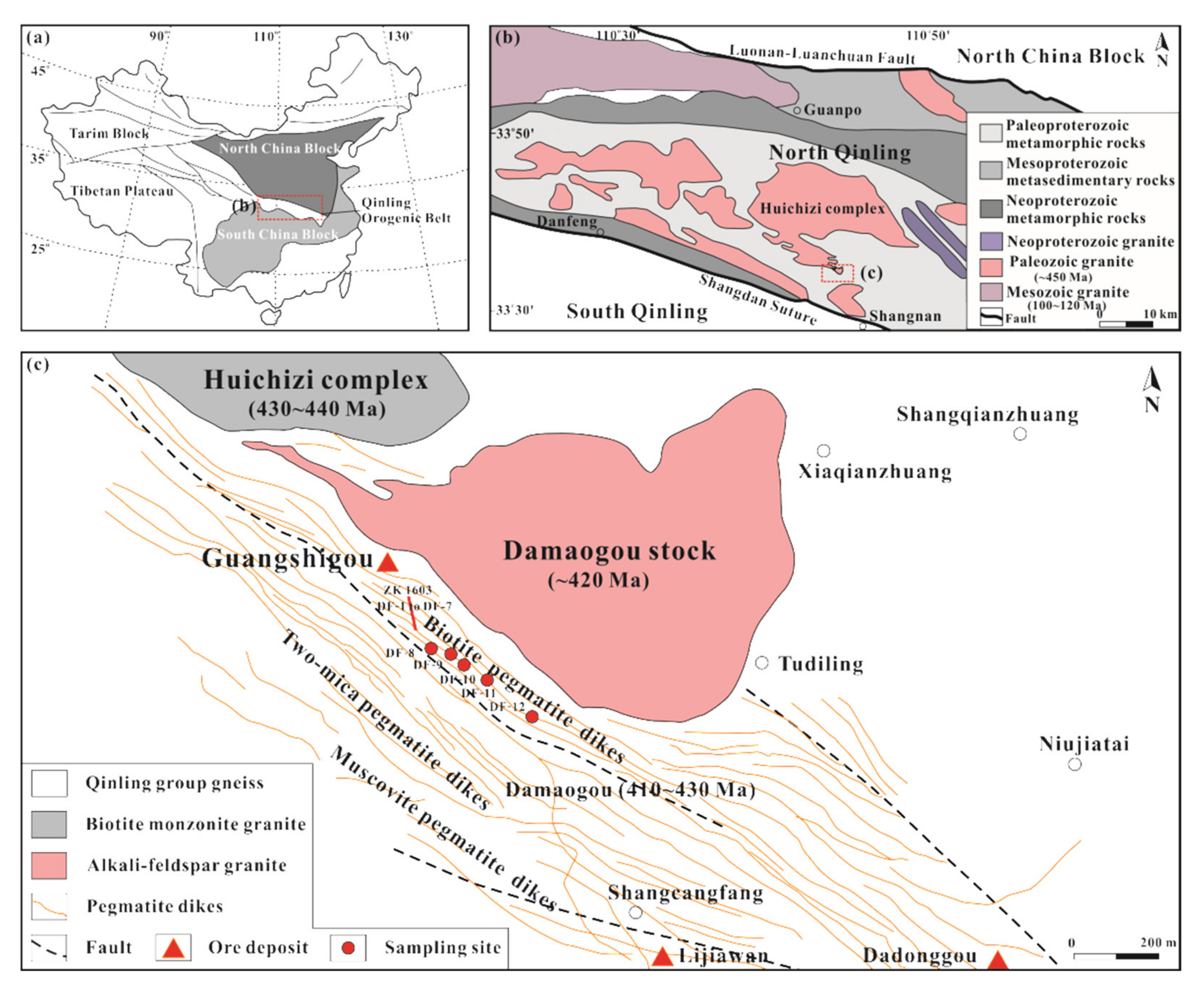
2. Geological Setting

The Qinling Orogenic Belt extends east–west for more than 1500 km across central China, and it separates the North China Block from the South China Block (Figure 1a, [31,32]). The overall orogenic belt has undergone multiple stages of tectonic–magmatic activities (Neoproterozoic, Paleozoic and Mesozoic) and ultimately forms as the product of the collision between the North and South China Blocks. It represents one of the most significant units of the China Central Orogenic Systems [27,29,30]. The Qinling Orogenic Belt consists of the Shangdan and Mianlue sutures and four thrust-fault or suture-bound belts from north to south, namely (1) the southern margin of the North China Block, (2) North Qinling Orogenic Belt (NQB), (3) South Qinling Orogenic Belt (SQB) and (4) the northern margin of the South China Block (Figure 1b, [33,34]). The NQB is bounded by the Luonan–Luanchuan fault zone in the north and the Shangdan Suture in the south (Figure 1b). It is primarily composed of Proterozoic to Paleozoic medium- to high-grade metasedimentary and metavolcanic rocks, including the Qinling Group, Kuanping Group, Erlangping Group, Songshugou Complex and Danfeng Group, which are separated from each other by thrust faults or ductile shear zones [35]. The SQB is bounded by the Shangdan Suture in the north and by the Mianlue Suture in the south and comprises a thick pile of Late Proterozoic to Triassic sedimentary package lying over an Early Proterozoic crystalline basement [32].
The Qinling Group is the oldest and most representative unit of the Precambrian basement of the NQB. It is composed of biotite–plagioclase and garnet–sillimanite gneisses, mica–quartz schists, graphite-bearing marbles and amphibolites or garnet amphibolites with some eclogites [30]. The Qinling Group was previously thought to have formed during the Paleoproterozoic period [27], but recent zircon U-Pb geochronology of 1400–1600 Ma, 962–1062 Ma and 850–950 Ma indicated a Mesoproterozoic–Neoproterozoic origin [36,37]. Voluminous Early Paleozoic (mostly Caledonian) granites and pegmatites intruded in the Qinling Group, leading to a WNW-trending magmatic belt (Figure 1c). They were emplaced in two stages during the Early Silurian and Late Silurian–Early Devonian [38]: (i) Early Silurian granitoids, represented by the Huichizi (434 ± 7 Ma; [39], Figure 1c), Kuanping (452.8 ± 2 Ma; [40]) and Shangnan (428 ± 7 Ma; [39]), which formed in a collisional and a following uplifting setting, and (ii) Late Silurian–Early Devonian granitoids occur as high-K calc-alkaline granites accompanied by ductile deformation and undeformed pegmatites, which are mainly represented by the Chenjiazhuang granite and biotite pegmatite (415 ± 27 Ma and 407 Ma, respectively; [41,42]), Huichizi and Damaogou granitic stock (422–441 Ma and 418–420 Ma, respectively; [23,38]; Figure 1c). These granitoids formed after the main collisional deformation in the NQB. Then, the NQB experienced several tectono-magmatic events during the Mesozoic, characterized by two major emplacement peak ages at 220–190 Ma and 160–110 Ma [43,44,45].
The Guangshigou U-bearing granitic pegmatites are spatially distributed along the contact zone with plagioclase-biotite gneiss of the Qinling Group in the southern margin of the Huichizi granite batholith and Damaogou granitic stock (Figure 1c; [38]). These granitic pegmatites show three petrographic facies that are concentrically arranged around the Damaogou granite stock and Huichizi granite batholith, varying from (i) an inner zone of U-rich biotite granitic pegmatite near the intrusions, through (ii) an intermediate zone of muscovite–biotite granitic pegmatite and to (iii) an outer zone of U-poor muscovite granitic pegmatite [22]. The Guangshigou uranium deposit has an estimated reserve of 3700 tons of UO2 with an ore grade of 0.050–0.199 wt.% [22]. The ore bodies consist of U-rich biotite granitic pegmatite dikes or layers of 80–770 m in length and 2–3 m in thickness with local enrichment of uraninite. These dikes intruded the Qinling Group at 415 ± 2 Ma [25,38,46], and coeval disseminated magmatic uraninite dated by Guo et al. [28] at 412 ± 3 Ma represents the U mineralization in the Guanshigou deposit. Moreover, it was recently characterized that the pegmatitic melts originated from high-K calc-alkaline magma derived from the partial melting of Proterozoic basaltic crust [28,38], and that the late assimilation between biotite pegmatitic melts and wall rock, as well as fractional crystallization process, was the primary mechanism for the uranium mineralization [38].

3. Sampling and Analytical Methods

Twelve hand specimens of uranium ores were collected from the Guangshigou biotite pegmatites from a drill hole (ZK1603) with depths from 9.7 m to 169.2 m (DF-1 to DF-7) and horizontal tunnels with depths of 985 m and 1080 m (DF-8 to DF-12) in the Shangdan uraniferous province (Figure 1c). Then, uraninite grains and radiohalos from polished thin sections of these ore samples were carefully examined using an optical microscope, followed by backscattered electron (BSE) imaging for detailed mineral assemblage determinations and microtexture observation using a field emission electron probe microanalyzer (FE-EPMA, JEOL JXA-8530F Plus) equipped with five wavelength-dispersive spectrometers (WDS) and an energy dispersive spectrometer (EDS, OXFORD INSTRUMENTS X-MAX 60) at the State Key Laboratory of Nuclear Resources and Environment, East China University of Technology, China.

3.1. Mineral Analysis and Characteristic X-ray Element Mapping by EPMA

The chemical compositions of different uraninite grains and other associated minerals were also analyzed by the same FE-EPMA (JEOL JXA-8530F Plus). The accelerating voltage, beam current and beam size were operated at 15 kV, 20 nA and 2 μm, respectively. Peaks and backgrounds for Ti, U, Th, Al, REEs, Y, Sc Ti, Mn, Sr and Pb were measured with counting times of 20 and 10 s, and those for P, Si, Mg, Ca, Fe, Na, K and F were measured with counting times of 10 and 5 s. All data were corrected by standard ZAF correction procedures. The following mineral standards were used for quantitative elemental calibrations: uraninite (U, Th and Pb), monazite (LREEs and P), rutile (Ti), REE-phosphate (Sm-Yb and Y), jadeite (Na, Si), strontium–barium niobate (Sr, Ba)Nb2O6 (Sr), topaz (Al, F), olivine (Mg), plagioclase (Ca), magnetite (Fe), pyrophanite (Mn), sanidine (K) and pure Sc metal. The analytical uncertainties were <1% for major elements and <10% for trace elements. To better show the elements redistribution during the hydrothermal alteration, characteristic X-ray maps of S, Si, U, Th, Al, Y, Ca, Fe, Pb, Na and K were scanned by the same FE-EPMA at an accelerating voltage of 15 kV, a beam current of 20 nA and size of 0.5 μm for each scanning step.

3.2. U-Th-Pb Chemical Age Determination on Uraninite, Coffinite and Uranothorite by EPMA

In order to constrain the timing of uraninite crystallization and its post-crystallization alteration characterized by coffinite [U(SiO4)1−x(OH)4x], U-Th-Pb chemical age determination was performed on non-altered uraninite (SiO2 < 0.50 wt.% and without microfractures, preferentially core of uraninite to avoid Pb loss), coffinite and uranothorite [(Th,U)SiO4] grains (the late two mostly < 20 μm) from all twelve samples, using the FE-EPMA at an accelerating voltage of 15 kV, a beam current of 100 nA and a beam size of 1 μm. Counting time of peaks and backgrounds for U, Th and Pb were 100 s and 50 s, and those were 20 s and 10 s for all the other elements. The chosen characteristic X-ray lines were U Mβ, Th Mα and Pb Mα. The standard used for data calibration and quality control was a homogeneous uraninite of 90.20 wt.% UO2, 6.20 wt.% ThO2 and 1.75 wt.% PbO provided by Zhang et al. [47]. Under these conditions, the calculated detection limits (1σ) were about 125 ppm for U, 95 ppm for Th and 140 ppm for Pb. U-Th-Pb chemical age of each uraninite, coffinite and uranothorite grain was calculated from the U, Th and Pb contents obtained by EPMA using age determination software provided by Guo et al. [48], which was modified from multiple iterations in Bowles [49] and Pommier et al. [50]. A minimum relative error of 2% (2σ) was applied to U, Th and Pb determinations to account for other error sources, such as standardization, instrument drift and matrix corrections, which has been described in similar age-calculation methods [51,52]. Then, the weighted average age of these uraninite grains was calculated using the modified Isoplot 4.15 program after Ludwig [53] on the basis of U-Th-Pb chemical age of each uraninite grain.

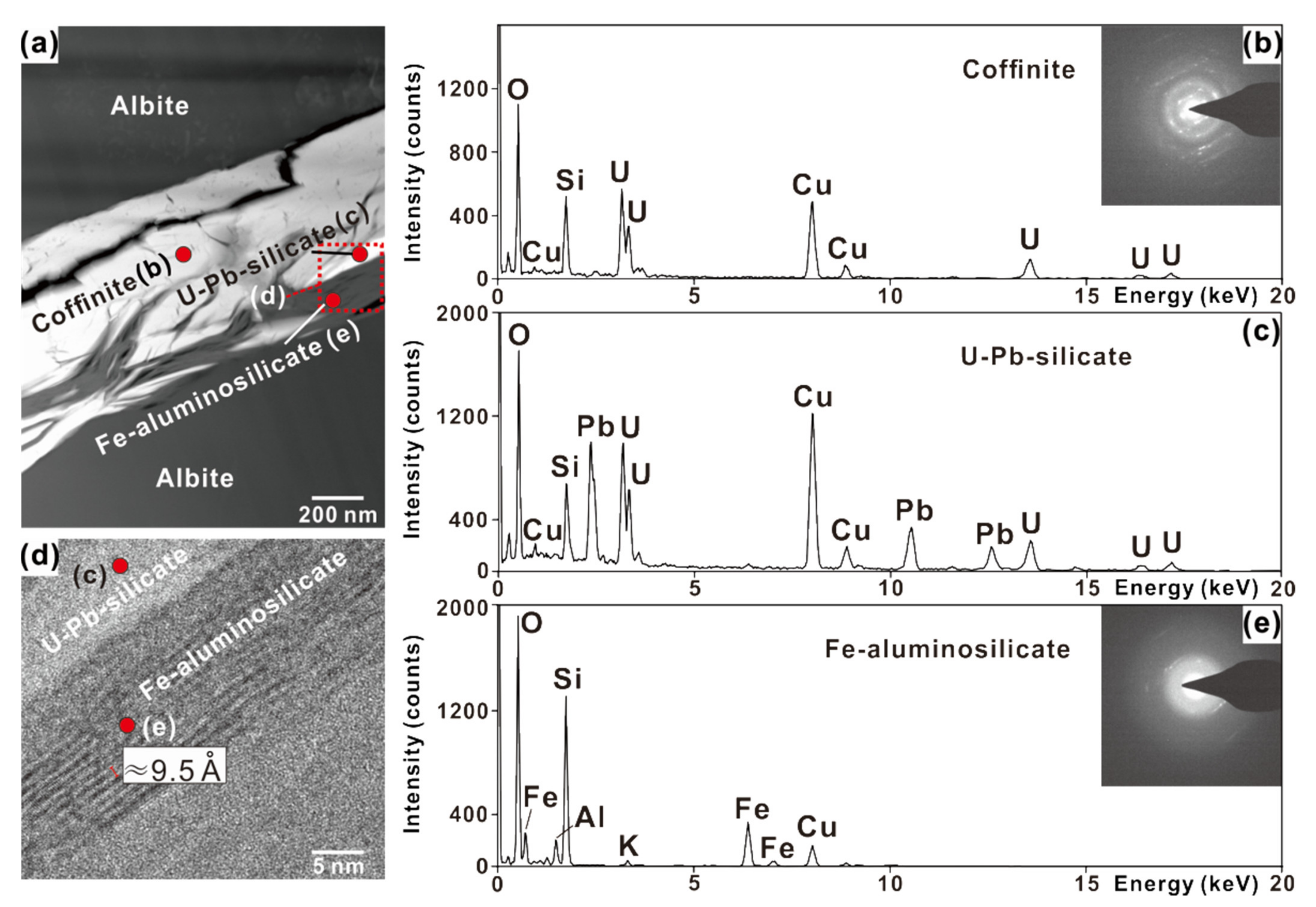
3.3. Nanoscale Observations of Uraninite Alteration by TEM

Two ultra-thin foils of altered uraninite were excavated with a focused ion beam (FIB) system from the thin section, which contains altered uraninite. The FIB preparation was performed on the FEI Scios Dual Beam at the Institute of Geochemistry, Chinese Academy of Sciences, Guiyang, China. First, a platinum layer of 2 μm was deposited on the sample to protect the surface. Then, initial sectioning was conducted under the working voltage of 30 kV and current of 5 nA. After the foil was lifted out from the polished section and attached to a copper grid using Pt deposition, it was milled with a beam current in the order of 1 nA, 500 pA, 300 pA and 100 pA, respectively. The final foil was approximately 60 nm in thickness. The texture and mineralogy of mineral assemblages were studied using a FEI Tecnai F20 transmission electron microscope (TEM) at the State Key Laboratory for Mineral Deposits Research, Nanjing University, China. The TEM measurement was conducted under the working voltage of 200 kV. A broad variety of observations were performed including energy-dispersive X-ray analyses, X-ray elemental mapping, selected area electron diffraction (SEAD), fast Fourier transform (FFT) of the high-resolution TEM (HRTEM) and high-angle annular dark field-scanning transmission electron microscope (HAADF-STEM).

4. Results

4.1. Petrography of the Guangshigou Biotite Pegmatites

The Guangshigou pegmatite dikes of 0.2–3 m thick intruded alkali feldspar granites of the Damaogou stock and biotite–plagioclase gneiss of the Qinling Group (Figure 2a). In the Guangshigou deposit, the uranium mineralization is dominantly hosted in biotite granitic pegmatite. The pegmatite mainly consists of centimeter-sized K-feldspar (further identified as microcline and perthite), biotite, quartz and minor plagioclase (Figure 2b–d). Late metasomatism is frequently observed in biotite pegmatite, which is characterized by chlorite and sericite as partial replacements of primary biotite and K-feldspar (Figure 2e). Accessory mineral assemblages consist of zircon, uraninite, monazite, thorite, apatite, xenotime, rutile, ilmenite, pyrite, molybdenite and galena. In most cases, these accessory minerals occur in interstices among rock-forming minerals, while mineral inclusions of rutile, monazite and zircon (<200 μm across) are sometimes enclosed in large biotite crystals with dark aureoles around (Figure 2f).
Uraninite is the predominant U-bearing mineral in the Guangshigou biotite pegmatite. Disseminated uraninite grains commonly show euhedral to subhedral crystal habit with a grain size of several hundred microns (Figure 2g). They occur as inclusions in major rock-forming minerals such as feldspar, quartz and biotite (Figure 3a), along fractures within pegmatite, or in interstices at the boundaries between rock-forming minerals (Figure 2g–i). Moreover, uraninite also coexists with other accessories including monazite, zircon, apatite, xenotime and molybdenite (Figure 3b), which show intergrowth relationships, thus, indicative of their coeval crystallization. Radial cracks and fractures are present both within and at the boundaries between the host minerals and uraninite (Figure 2h). In addition, radiohalos of several tens of microns in width around uraninite grains are frequently observed (Figure 2i). These fractures and radiation-induced destruction rims are associated with secondary minerals such as coffinite, geothite and unidentified alteration phases, which need to be further examined.

4.2. Mineralogy of Non-Altered Uraninite

As described above, magmatic uraninite commonly occurs as inclusions enclosed in or in interstices between major rock-forming minerals coexisting with calcite, pyrite and molybdenite (Figure 3a–c). Euhedral uraninite grains are relatively non-altered and characterized by homogeneous compositions and little leaching of their U, Th and Pb contents, as well as the absence of incorporation of K, Ca and Si (Figure 4). In few cases of poorly developed alteration, only a little U is liberated from uraninite, which is re-precipitated as a relatively rare Ca-Fe-U-bearing aluminosilicate closely associated with goethite (Figure 3d). Prior to in situ mineral analysis, non-metamict uraninite grains with low fracturing degree were selected for EPMA measurements. Uraninite with SiO2 < 0.50 wt.% and without microfractures was considered as non-altered uraninite. The non-altered uraninite grains have relatively homogeneous chemical composition (Supplementary Materials, Table S1; Figure 5). In detail, UO2 contents vary from 84.45 to 91.67 wt.% with an average of 86.64 wt.% (Figure 5a), and the concentrations of ThO2 and PbO display narrow compositional ranges from 1.94 to 4.29 wt.% with U/Th = 20.12–47.20 (weight ratio) and from 4.73 to 5.34 wt.%, respectively (Table S1). Although compositional variations exist among different crystals of uraninite, the composition within one single grain remains homogeneous and no obvious compositional zonings or core-rim textures are observed (Figure 4). In addition to major elements, the Guangshigou uraninite also has low to relatively high concentrations in REE (0.50–2.36 wt.% REE2O3, including Y, Figure 5f). Relatively homogeneous CaO contents give the range from below the detection limits to 0.50 wt.% (LOD ≈ 200 ppm), suggesting that minor calcium was most likely incorporated during primary crystallization. Similarly, very low Si concentrations (mean value = 0.06 wt.%; Figure 5a) show evidence for a low degree of post-crystallization alteration.

4.3. Mineralogy of Magmatic Biotite

Biotite is very common in the Guangshigou uraninite-bearing biotite pegmatites, and its composition can be used to evaluate crystallization conditions. Crystallization temperatures for biotite grains show homogeneous values in the range of 640–700 °C (Table S2) based on the Ti-in-biotite geothermometer in Henry et al. [54]. In addition, the total Al content of biotite also has a relation with the crystallization pressure of the granitic rocks by the empirical Equation (1):
P (kbar) = 3.03 × TAl − 6.53(±0.33)
where TAl denotes the total aluminum content in biotite on the basis of 22 oxygen atoms [55]. Following this equation, the initial crystallization pressure calculated from the total aluminum in biotite ranges from 2.403 to 3.421 kbar with 2.975 kbar in average (Table S2). The crystallization temperature and pressure deduced from the biotite composition in the Guangshigou biotite pegmatites are very consistent with those of the Chenjiazhuang pegmatite-type uranium deposit (656–717 °C and 2.7 kbar in average, [56]), which is located approximately 30 km northwest of the Guangshigou pegmatites.

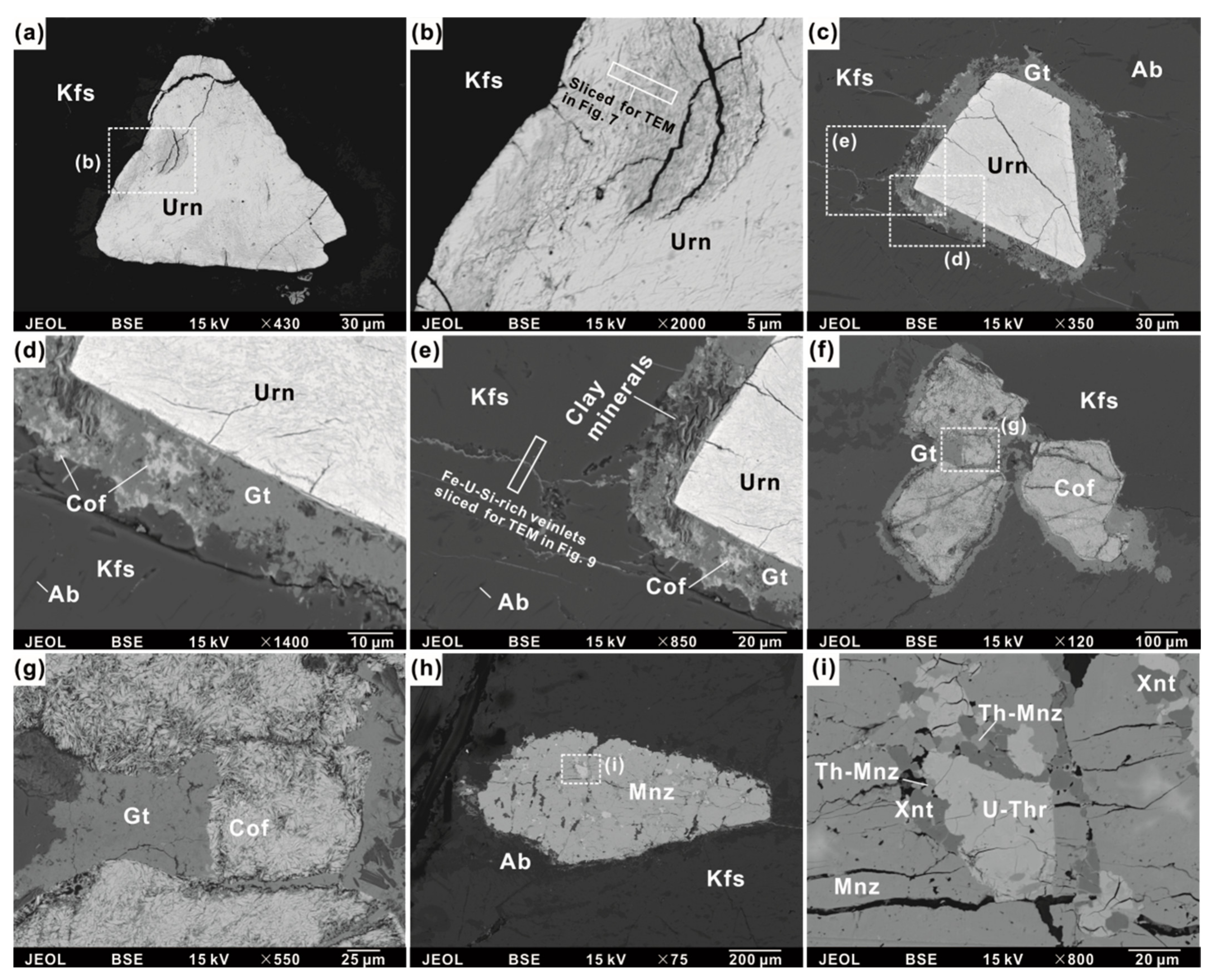
4.4. Alteration of Uraninite and Mineralogy of Radiohalos

Further observations show that most uraninite crystals in the Guangshigou biotite pegmatite have undergone various degrees of metamictization and post-crystallization hydrothermal alteration. These processes are characterized by the element redistributions and exchanges within uraninite, the replacement of uraninite by a series of secondary minerals and the generation of radiohalos around uraninite.

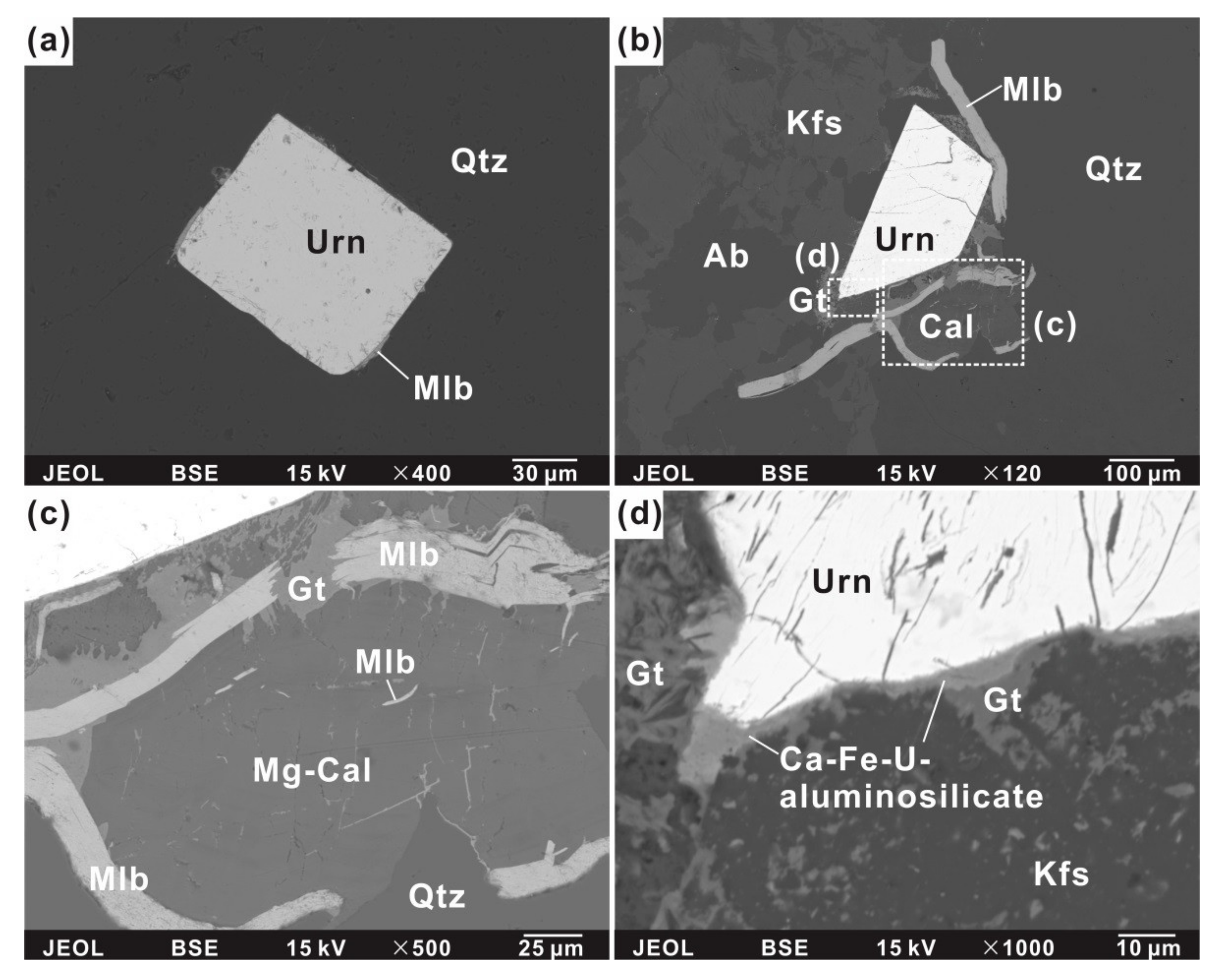
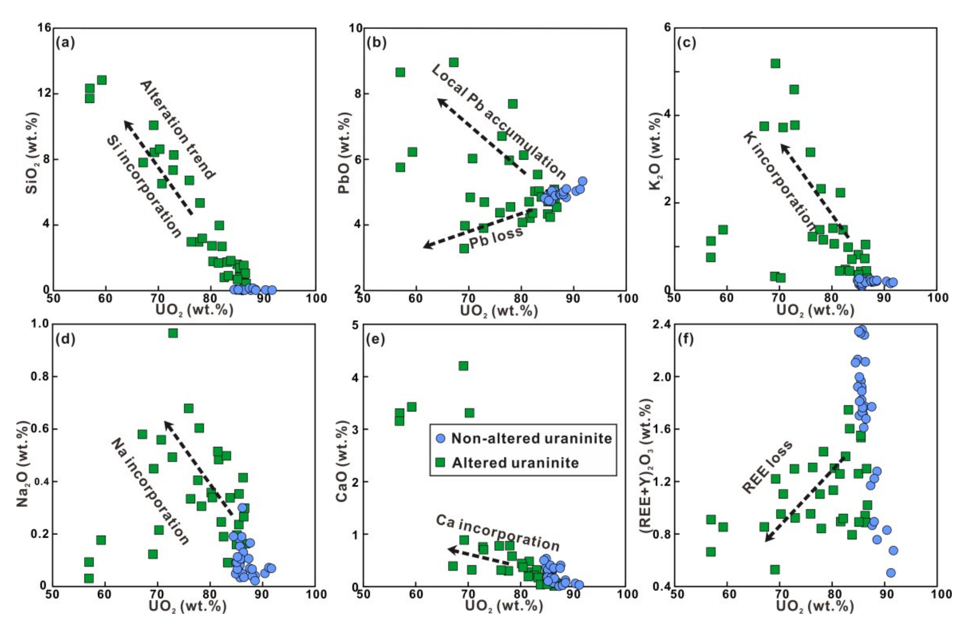
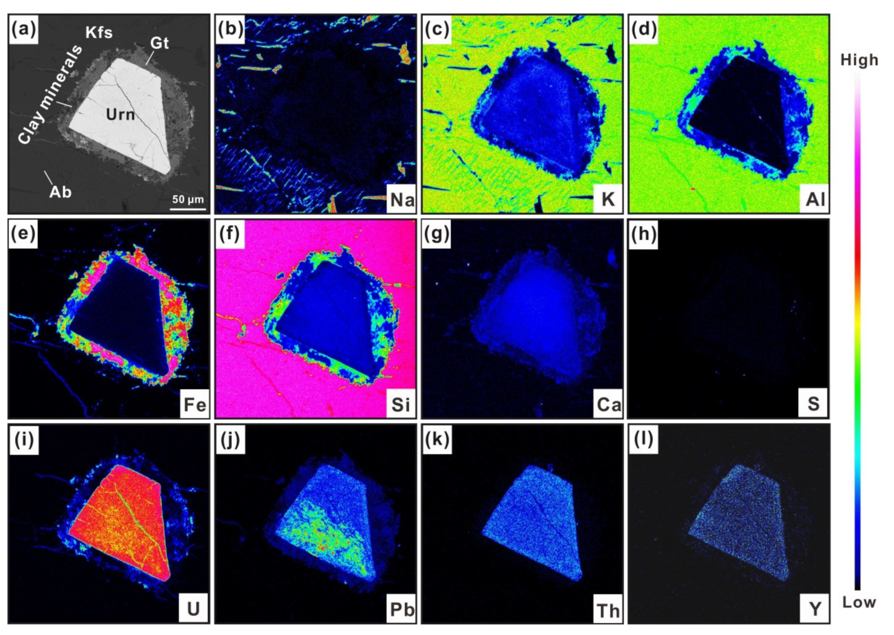
4.4.1. Uraninite Alteration and Element Redistributions

Compared with non-altered uraninite (Figure 3a), altered uraninite grain is crosscut by fractures or cracks of several hundred microns throughout the entire crystal, especially well developed in its rims (Figure 6a). Another important feature is the compositional heterogeneity of the altered uraninite grain that is characterized by relatively dark areas along and in the vicinity of fractures compared with bright non-altered parts of uraninite under the BSE mode of the EPMA (Figure 6a–e). These dark, altered areas show highly variable of U, Pb, Si, K, Na, Ca and REE (including Y) relative to non-altered parts. In detail, altered areas contain lower UO2 (56.91–86.73 wt. % with an average of 77.41 wt.%) and REE2O3 (including Y2O3, 0.53–1.75 wt.% with an average of 1.11 wt.%) and higher SiO2 (0.43–12.84 wt.% with an average of 4.42 wt.%), K2O (0.28–5.19 wt.% with an average of 1.49 wt.%), Na2O (0.03–0.97 wt.% with an average of 0.35 wt.%) and CaO (0.01–4.21 wt.% with an average of 0.83 wt.%, Table S1; Figure 5). Altered uraninite also has variable PbO contents (3.28–8.96 wt.% PbO) displaying much wider ranges that can be either higher or lower compared with those in non-altered parts (4.73–5.34 wt.% PbO, Figure 5b). In addition, the alteration degree of the Guangshigou uraninite is positively correlated to its SiO2 contents (Figure 5a), showing an alteration trend from the uraninite end member (ideally UO2) to the coffinite end member (U(SiO4)1-x(OH)4x, i.e., coffinization; [16]).
As the alteration develops, the proportion of dark zonation as well as the degree of coffinization gradually increases (Figure 6c–e), and in some grains, coffinite totally replaces the uraninite crystal with only minor residual uraninite left (Figure 6f,g). Besides, more nanoscale fractures and inclusions are observed in the dark zonation, which probably resulted from integrated effects of uraninite metamictization and hydrothermal alteration. Further, HAADF-STEM observations and SAED patterns of boundary between dark (altered) and bright (non-altered) parts of uraninite show that the majority of both parts are still well crystalline, as demonstrated by the presence of the regular lattice fringes with identical interplanar distance of 2.732–2.734 Å and 3.156–3.159 Å (Figure 7a). The SAED pattern and energy-dispersive X-ray (EDX) analyses of mineral inclusions enclosed in uraninite suggest a polycrystalline, potentially Fe-U-rich aluminosilicate phase (Figure 7b). Uraninite is gradually becoming amorphous towards fractures or cavities, as illustrated by the fast Fourier transform (FFT) of the high-resolution TEM (HRTEM) image (Figure 7c,d), accompanying the growing enrichments in Si and F, but depletion in U with increasing semi-quantitative Si/U ratios (Figure 7e–g). It suggests that the initial alteration and amorphization of uraninite and the element exchanges preferentially occur along microfractures of uraninite.
As previously stated, the altered uraninite is ultimately replaced by coffinite to form pseudomorph (Figure 6f,g). Coffinite is the predominant secondary mineral in pseudomorph in the form of randomly oriented, fibrous aggregates. These aggregates may represent the further development of nanoscale fractures on the surface of uraninite. Grains of coffinite are mainly composed of 59.95–70.44 wt.% UO2, 1.14–6.85 wt.% ThO2, 0.74–4.23 wt.% PbO, 10.65–15.68 wt.% SiO2 and 1.89–6.26 wt.% CaO (Table S3). In addition to uraninite that is altered into coffinite, monazite associated with uraninite has also undergone hydrothermal alteration and been locally replaced by hydrothermal uranothorite, Th-rich monazite and xenotime (Figure 6h,i). Compared with magmatic, Th-free monazite, ThO2 contents in hydrothermal monazite show compositional ranges of 5.85–10.62 wt.% ThO2. UO2 and ThO2 of ~2.00 wt.% are also detected in coexisting xenotime (Table S4). Uranothorite grains here contain much higher ThO2 contents. In detail, they mainly contain 39.81–54.59 wt.% ThO2, 8.98–23.32 wt.% UO2 and 16.10–18.43 wt.% SiO2 (Table S3). Experimental and empirical studies have shown that the equilibrium partitioning of the rare-earth elements and yttrium between coexisting monazite and xenotime is pressure and temperature dependent [57,58,59]. The partitioning of yttrium in monazite is experimentally calibrated as a geothermometer by Gratz and Heinrich [59], expressed as Equation (2):
T = ln 100 X Ymz 1.459 + 0.0852 P 2.2745 × 10 3
where T is the equilibration temperature in °C, XYmz is the mole fraction of yttrium in monazite, and P is the pressure in kbar. Assuming that the pressure of 2.975 kbar obtained by biotite is constant, temperatures calculated for coexisting monazite and xenotime range from 85 to 174 °C (Table S4). Therefore, it suggests that the hydrothermal alteration of uraninite and uranothorite probably happened under relatively low temperatures.

4.4.2. Mineralogy of Radiohalos Around Uraninite

Radiohalos around uraninite are commonly 10–50 μm in width, and they are mainly characterized by a mineral assemblage including goethite + coffinite + a Ca-Fe-U-bearing aluminosilicate around uraninite showing various degrees of metamictization and hydrothermal alteration.
In radiohalos associated with higher degree of post-crystallization alteration (coffinization relative to uraninite partially crosscut by microfractures), alkali-metasomatism is common in neighboring K-feldspar, which is mainly characterized by their replacement in albite (Figure 6c and Figure 8b). Overall, the characteristic X-ray element maps of altered uraninite show the incorporation of K, Ca, Si and minor Fe along the rim, the leaching of U and Pb and the relatively low mobility of Th and Y during the alteration processes, which is in agreement with compositional variations in altered uraninite (Table S1; Figure 5 and Figure 8). Moreover, Pb is heterogeneously redistributed within one single grain of altered uraninite and within radiohalos (Figure 8j). Goethite is one of the major secondary minerals occurring in radiohalos as anhedral aggregates of around 10 μm across (Figure 6d–g). It occasionally contains some small coffinite inclusions (<2 μm in size). If all Fe is regarded as ferric iron, total oxides of goethite show narrow compositional ranges of 89.94–92.24 wt.% with homogeneous contents of Fe2O3 (78.31–84.71 wt.%), SiO2 (4.64–7.87 wt.%), minor Al2O3 (0.98–1.69 wt.%) and PbO (0.85–1.38 wt.%) and undetectable ions (e.g., OH-) or H2O (Table S5). In addition, nearly all goethite grains contain trace amounts of K (0.13–1.09 wt.% K2O), Na (0.04–0.39 wt.% K2O), U (0.08–1.49 wt.% UO2, mostly <0.30 wt.%) and Th (0.03–0.28 wt.% ThO2). Of note, compared with U and Th, more Pb contents are detected in goethite.
The unidentified aluminosilicate is observed in almost all radiohalos as layered aggregates associated with goethite (Figure 6e). Small coffinite inclusions are also occasionally enclosed in it. This aluminosilicate contains depleted total oxides (83.00–89.10 wt.%) with variable Fe (18.54–34.60 wt.% FeO), Al (14.35–20.48 wt.% Al2O3) and Si (30.28–42.55 wt.% SiO2) contents (Table S6). It also has minor concentrations of K (1.20–3.76 wt.% K2O), Na (0.15–0.55 wt.% Na2O), Ca (0.29–1.26 wt.% CaO), Mg (1.66–3.51 wt.% MgO) and F (0.29–0.82 wt.% F). Compared with a stoechiometric chlorite composition, higher SiO2 and lower FeO in aluminosilicate indicate a different mineral phase. Similar to goethite, Pb (0.71–1.78 wt.% PbO) in aluminosilicate is higher than U (0.10–0.92 wt.% UO2) and Th (0–0.17 wt.% ThO2), and that is in accordance with Pb migration from the altered uraninite as illustrated in Figure 8.
In addition to radiohalos, we also observed that some veinlets of 0.5–1 μm in width extend outwards from the altered uraninite and radiohalos along borders between neighboring albite or K-feldspar (Figure 6e and Figure 9a). Further HAADF-STEM and EDX analyses of these veinlets indicate that they are rich in U, Si, Al, Fe and trace Pb and composed of three phases including major coffinite and minor U-Pb-rich silicate and Fe-rich aluminosilicate. Coffinite is poorly crystallized as characterized by the presence of week lattice fringes and diffuse selected area electron diffraction (SAED) patterns (Figure 9b). Needle-like U-Pb-rich silicate is the brightest under HAADF-STEM imaging (Figure 9c). Although the size of U-Pb-rich silicate is too small to obtain its SAED patterns, further fast Fourier transform (FFT) of the high-resolution TEM (HRTEM) image and EDX analyses reveal that the U-Pb-rich silicate is completely amorphous as characterized by irregular structural array (Figure 9d). By contrast, the Fe-rich aluminosilicate is relatively well crystallized with approximately 9.5 Å d-spacing in HRTEM image (Figure 9d). The contradictory presence of diffuse diffraction patterns with sparse lattice fringes in Figure 9e is attributed to the fragility of its crystal structure exposed under the high working voltage. It is probably compositionally identical to the above unidentified aluminosilicate, since both of them have similar Si, Al, Fe and K compositions. Furthermore, the wide interplanar distance of the Fe-rich aluminosilicate probably suggests that it belongs to one kind of clay mineral that usually has large d-spacing.

4.5. U-Th-Pb Chemical Age Determination of Uraninite, Coffinite and Uranothorite

Textural and compositional features of uraninite characterized its magmatic origin (e.g., euhedral to subhedral crystals with U/Th < 100, high ThO2, Y2O3 and REE2O3 contents and low concentrations of CaO, FeO and SiO2), while the presence of coffinite after uraninite and uranothorite after monazite recorded the post-crystallization alteration related to the overlapping hydrothermal activity. Based on Pb, U and Th contents in non-altered grains, chemical age estimates were calculated to constrain the timing of uraninite crystallization relatively to the precipitation of coffinite and uranothorite. In this study, two major age groups are obtained from all twelve samples by statistics, which are characterized by a first group of relatively narrow ages ranging from 394.8 to 420.6 Ma (mean value at 407.6 ± 2.9 Ma, n = 31) determined on uraninite, that the magmatic uranium mineralization in the Guangshigou deposit formed during this period, and a second group with more scattered ages varying from 84.9 to 143.6 Ma (mean value at 111.7 Ma, n = 38, Table S3; Figure 10). Moreover, two additional minor peak ages of coffinite and uranothorite may also be identified at ca. 310 Ma (from 285.6 to 326.0 Ma, n = 6) and ca. 210 Ma (from 186.0 to 217.6 Ma, n = 6). Chemical age determinations display multiple peak ages at 310 Ma, 210 Ma and 112 Ma, hence suggesting multiple events of post-crystallization hydrothermal alteration. Few ages of coffinite older than the crystallization age of uraninite may be due to the presence of micro/nano inclusions of galena, a high content of common Pb and/or radiogenic Pb diffusion.

5. Discussion

5.1. Timing of Uranium Mineralization and Post-Crystallization Alteration in the Guangshigou Biotite Pegmatite

For the past decades, dozens of in situ geochronological analyses on various minerals from the Guangshigou biotite pegmatites have been carried out to determine the ages of pegmatite emplacement and uranium mineralization, and these ages can be primarily divided into two groups: (i) a first group of 400–420 Ma is represented by U-Pb ages of 415.6 ± 1.5 Ma and 413.6 ± 2.4 Ma on zircon, 405 ± 3 Ma, 403 ± 3 Ma and 412 ± 3 Ma on uraninite by LA-ICP-MS and SIMS [28,38,46], U-Th-Pb chemical ages of 398.0 ± 4.2 Ma, 407.6 ± 8.0 Ma and 407–415 Ma on Th-rich uraninite performed by EPMA (normally ThO2 > 2.00 wt.%, [26,60,61]); (ii) a second group in the range 346–386 Ma with a peak at ca. 370–390 Ma is summarized by U-Th-Pb chemical ages on Th-poor uraninite (normally ThO2 < 0.40 wt.%, Table S1, [26,38]). In our study, Th-rich uraninite grains gave a weighted average age of 407.6 ± 2.9 Ma, which is consistent within errors with previous age determinations of magmatic uraninite and characterizing an Early Devonian stage for the formation of the pegmatite-type U mineralization in the Guangshigou deposit, hence belonging to the Late Caledonian post-collisional extension in the North Qinling area [28]. In Yuan et al. [26,38], the Th-poor uraninite was interpreted as an alteration product after Th-rich uraninite, and the younger age of 346–386 Ma was attributed to radiogenic Pb loss during post-crystallization hydrothermal process. However, compared with the relatively immobile behavior of Th in minerals such as uraninite, monazite and bastnäsite that are used for age dating, Pb is more prone to being leached out during the fluid-assisted modification process, hence generally leading to radiogenic Pb loss [16,60]. For instance, in the Guangshigou uraninite, element maps presented in Figure 8 and uraninite data in Table S1 have illustrated the uneven redistribution or depletion of Pb, whereas most of its Th content was preserved during the alteration process. Therefore, the significant Th depletion and the relatively preserved Pb content in the Th-poor uraninite described in Yuan et al. [26,38] is contradictory to the common characteristics of uraninite alteration, if it is assumed as an alteration product after Th-rich uraninite in the Guangshigou deposit. In addition, the incorporation of Ca, Si and Fe (especially Si, can be up to several wt.%) and the Pb loss, which are common in altered uraninite [16,62], are absent in the Guangshigou Th-poor uraninite with Σ(CaO + FeO + SiO2) contents < 0.50 wt.% in Yuan et al. [26,38] (Table S1). Moreover, the close association of secondary thorite and Th-poor uraninite were not observed in our study, if they are recrystallized from alteration process of primary Th-rich uraninite. Therefore, we tend to believe that the younger ages of 346–386 Ma obtained on Th-poor uraninite in Yuan et al. [26,38] probably represent another episode of uranium mineralization events in the Guangshigou pegmatites, and that the Th-poor uraninite is not the direct, fluid-modified product after primary Th-rich uraninite. Furthermore, the relatively high U/Th ratios (i.e., >100; [38]) and the minimum age gap of about 25 Ma between the Th-poor uraninite described in Yuan et al. [38] and the magmatic Th-rich uraninite dated in situ by SIMS at 412 ± 3 Ma (in Guo et al. [28]) suggest that this second group of uraninite has a hydrothermal origin.
Due to the radiogenic Pb loss in uraninite related to various degrees of post-crystallization alteration, younger and dispersive ages are commonly obtained on altered uraninite [16], hence preventing constraints on hydrothermal overprinting episodes. Moreover, U-Th-Pb chemical dating does not allow measuring common Pb contents, which could induce older age estimates on uraninite (e.g., [63]). One way to overcome this limitation is to apply statistical age clustering obtained by U-Th-Pb isotopic or chemical dating to their alteration or hydrothermal products (e.g., hydrothermal coffinite, uraninite and uranothorite). This method has been successfully applied to trace post-ore hydrothermal activities worldwide, as exampled by studies conducted on the Bayan Obo REE deposit and Miaoya REE-Nb deposit of China [64,65], Cigar Lake, McArthur River and Virgin River U deposits of Canada [62,66]. Coffinite and uranothorite that represent the predominant secondary uranium mineral after uraninite and monazite alteration in the Guangshigou pegmatite yielded two minor peaks of chemical age estimates at ca. 310 Ma and 210 Ma and a major peak age at 112 Ma. The first peak at ca. 310 Ma may represent a first stage of hydrothermal alteration of the Guangshigou deposit that could be related to the early Permian episode of granite intrusion in the North Qinling Area (e.g., the 296 Ma Tieyupu and 283 Ma Gaoshansi granites; [38]) that occurred during the northward subduction of the Mianlue oceanic arc crust beneath the Shandan arc crust and the North Qinling unit [67]. The second peak at ca. 210 Ma is consistent with the Late Triassic–Early Jurassic tectono-magmatic event (i.e., the final subduction and collision of the North China Block and South China Block, [45,67]) that was recorded in the NQB (220–190 Ma), which may have been responsible for a second stage of hydrothermal alteration in the Guangshigou pegmatite. Finally, ages around 112 Ma likely represent a more dominant episode of hydrothermal activity that was responsible for major uraninite alteration and coffinite crystallization. It is concomitant with the intense tectono-magmatic activities and U-Mo-polymetallic mineralization that occurred during the Late Yanshanian period (100–140 Ma) in the North Qinling Block [68,69], which was dominated by continental extension driven by the subduction of the paleo pacific plate. In comparison, the Huayangchuan U-polymetallic deposit in the North Qinling Block also recorded a hydrothermal uranium mineralization around 130–140 Ma that postdated the main carbonatite-related uranium mineralization, which yielded Triassic ages of 210–230 Ma [70,71,72]. Therefore, coffinite and uranothorite after uraninite and monazite in the Guanshigou deposit recorded multiple stages of post-Caledonian hydrothermal alteration that are related to major tectono-magmatic episodes and uranium circulation events in the NQB.

5.2. Processes Responsible for Uraninite Alteration and Radiohalo Formation

Textural and compositional features (high Th, Y, REE but low Ca contents) of uraninite from the Guangshigou biotite pegmatites indicate a magmatic origin, as also characterized in detail by Guo et al. [28]. Then, the uraninite crystals underwent various degrees of post-crystallization alteration, which is demonstrated by the variable, low to high Si contents (0.43–12.84 wt.% SiO2) in altered uraninite [16]. Furthermore, alteration of natural uraninite is an integrated product of self metamictization and hydrothermal overprinting events, which are characterized by the incorporation of impurities (e.g., Ca, Fe and Si), the depletion of Pb and U and the generation of a series of secondary minerals (e.g., coffinite, hematite, chlorite and clay minerals [21,73]). Here, the progressive development of radial microfractures through time, from the uraninite grains spreading out into the surrounding rock-forming minerals, combined with amorphization of the uraninite structure during metamictization facilitated the fluid-rock chemical exchange of elements (e.g., Zhang et al. [73]; Bonnetti et al. [74]) during the successive episodes of hydrothermal alteration proposed in Section 5.1. In Guangshigou pegmatites, at the initiation stage of the post-ore alteration, uraninite is preferentially altered along microfractures and cavities, accompanied by the increasing incorporation of Si and F (Figure 5a and Figure 7f,g), while the main body of the crystal remains well crystalline. In this step, only a minor amount of U is leached out of primary uraninite (Table S1; Figure 8i), which is locally re-precipitated as Fe-U-rich aluminosilicate inclusions.
In the Guangshigou deposit, the hydrothermal alteration of uraninite and the associated U leaching showed evidence for alkali-metasomatism, as demonstrated by the albitization of K-feldspar and the incorporation of impurities such as K, Na and Ca in altered uraninite composition (Figure 5 and Figure 8). Actually, alkali–metasomatic fluids are one of the most significant carries for uranium transport to form uranium deposits worldwide, as exampled by uranium deposits in the Aricheng South, Guyana [75] and the central Ukrainian Shields [76]. High oxygen fugacity is a prerequisite for the uranium remobilization by fluids [77]. In the Guangshigou deposit, the abundance of goethite in radiohalos suggests a relatively high oxygen fugacity of the fluids that were responsible for the post-Caledonian hydrothermal alteration of the U-rich pegmatites. Moreover, TEM results show that U and Pb leached out of uraninite are transported along fractures over several tens to several hundreds of microns and redeposited locally as a U-Pb-rich silicate or U-Pb-rich oxide phase (Figure 9a,c). Specifically, in Fe-Pb-O-S system, when T= 100 °C, the stability field changing from Fe2+-Pb-sulfide to Fe3+-Pb-oxide implies the drop of log(ƒS2) to below −40 log units and the rise log(ƒO2) to above -55 log units, respectively [78,79], but it is still unable to trigger the large-scale remobilization of uranium as soluble ions. Temperature constraints from monazite–xenotime thermometry suggest low temperature conditions (85–174 °C) for local coffinitization after uraninite. Aqueous inclusions associated with uraninite have distinctly higher homogenization temperatures (470 to 580 °C in Feng et al. [22]), implying that post-ore alteration was related to separated fluid events. Although the chemical dating on coffinite and uranothorite suggested several stages of post-Caledonian alteration of uraninite from the Guangshigou pegmatites, as the large majority of the age estimates (84.9–143.6 Ma; Figure 10a) belong to the Cretaceous, Late Yanshanian stage identified in Section 5.1, we can reasonably assume that the characteristics of the fluids related to the hydrothermal alteration discussed previously are predominantly representative of this event. In addition, this latest hydrothermal event was most likely responsible for the re-opening and reset of the U-Pb system in the host pegmatites, hence explaining why the two first peak ages recorded by coffinite and uranothorite only have a minor representation in our dataset. Therefore, the main alteration (coffinitization) of uraninite as well as the remobilization of uranium were most probably driven by relatively oxidizing, low-temperature and alkali-rich fluids during the hydrothermal activity of the Late Yanshanian crustal extension in the NQB.

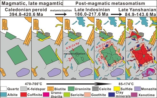
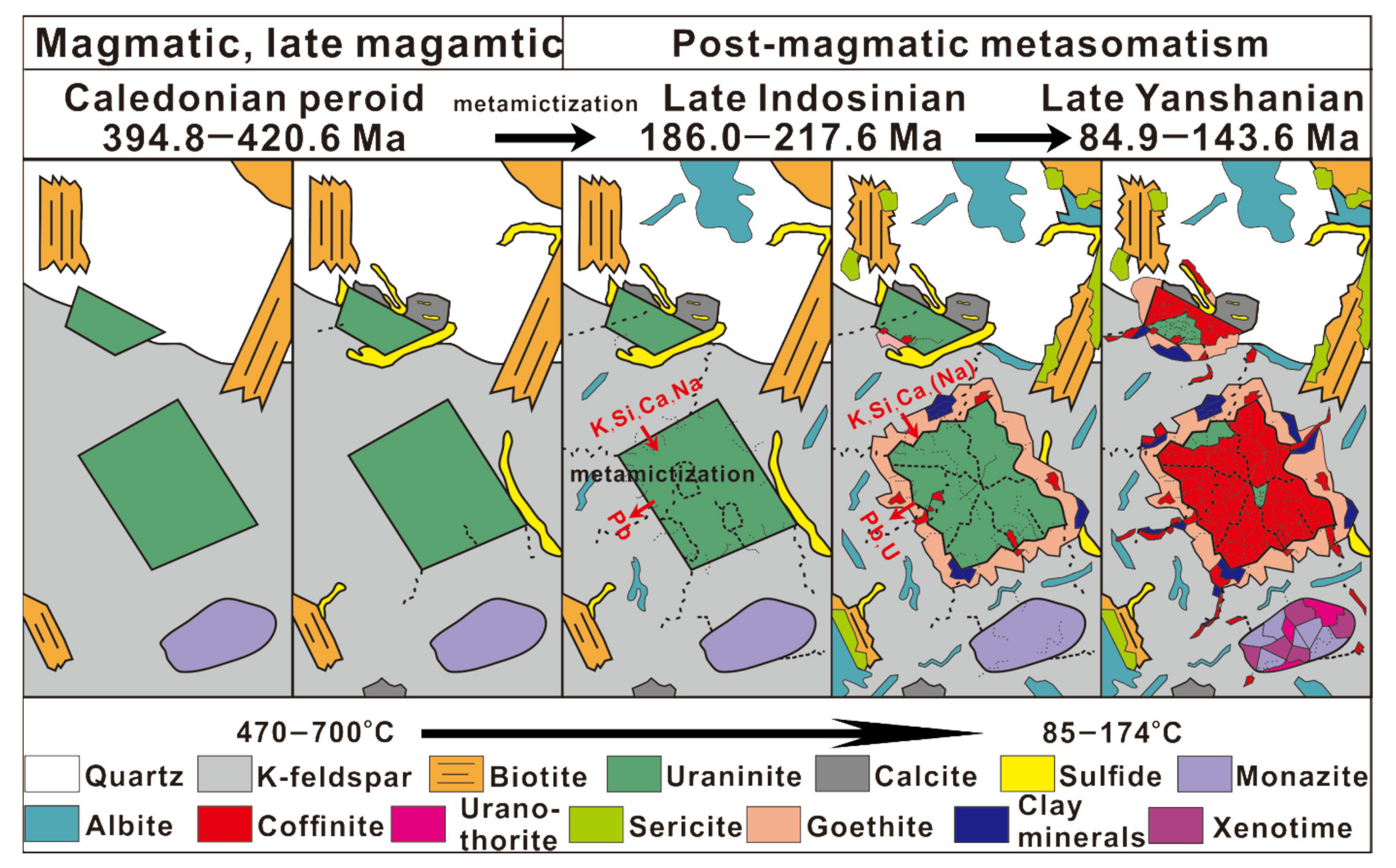
5.3. An Integrated Model for the Guangshigou Uraninite Alteration and Its Implications for Uranium Circulation in the Qinling Orogenic Belt

The Guangshigou uraninite crystallized from pegmatitic melts during the Early Devonian, Late Caledonian period (407.6 ± 2.9 Ma in this study; 412 ± 3 Ma in Guo et al. [28]) under relatively high temperatures (470 to 580 °C, [22]). After the primary crystallization of uraninite, three episodes of post-ore hydrothermal alteration were recorded by coffinite chemical compositions, including two first stages during the Permian (ca. 310 Ma) and the Late Triassic period (ca. 210 Ma), and a last predominant event that occurred during the Late Yanshanian (at 112 Ma; Figure 10a). An integrated model for uraninite alteration and uranium circulation is summarized in Figure 11. With the increasing degree of uraninite metamictization, the hydrothermal event that occurred during the Late Yanshanian crustal extension was characterized by the circulation of relatively oxidizing, low-temperature (85–174 °C) and alkali-rich fluids, which promoted the local coffinitization of the Guangshigou uraninite, as well as remobilization of U and Pb. In the meantime, impurities such as Na, K, Ca and Si from the fluids were incorporated into the uraninite structure during its alteration. A part of alkalis could also come from neighboring rock-forming minerals through albitization and sericitization of K-feldspar as well as chloritization of biotite. Although we observed relatively limited U remobilization along fractures in the vicinity of uraninite in the Guangshigou pegmatites (Figure 6 and Figure 8), uraninite is an easily leachable mineral by percolation of oxidizing fluids [80,81,82] as indicated by coffinite pseudomorph after uraninite (Figure 6f,g). Therefore, this primary stock of uraninite may have represented a significant source of U for post-Caledonian hydrothermal mineralization that formed in the Qinling Orogen (e.g., Zheng et al. [83]), as it was also demonstrated in other major U districts in South China [73,74] or in the European Hercynian Belt [84].
The Qinling Orogenic Belt hosts the most important uranium mineralization belt in central China, which includes various types of uranium deposit (mainly granite-type, pegmatite-type, carbonatite-type and metasedimentary rock-type uranium mineralization) that formed at relatively narrow ages of 400–430 Ma, 200–230 Ma and 100–140 Ma, corresponding to three periods of large-scale tectono-magmatic events (the Caledonian extension-related magmatism, the Indosinian compression process and Yanshanian magmatism) in the Qinling Orogenic Belt [30,85]. For instance, recent studies have revealed that multi-stage uranium mineralization took place within individual deposits in the Qinling Orogen: (i) the Huayangchuan U-polymetallic deposit characterized by Triassic carbonatite-related primary U mineralization at 220–230 Ma followed by a late stage of hydrothermal mineralization at 120–140 Ma [70,71,83], (ii) the Miaoya REE-Nb-(U) deposit characterized by Caledonian magmatic mineralization at 420–440 Ma followed by hydrothermal remobilization at 220–240 Ma [65,86] and (iii) the Guangshigou U deposit in this study with a Late Caledonian magmatic U mineralization followed by multiple stages of hydrothermal alteration at ca. 310 Ma, ca. 210 Ma and 112 Ma. In these deposits, the late uranium mineralization show obvious fluid overprinting characteristics such as recrystallization of uraninite after the dissolution of early uraninite and pyrochlore group minerals, as well as the coffinitization after uraninite. It suggests that the remobilization and circulation of uranium in the Qinling Orogenic Belt could be driven by the regional post-Caledonian magmatism and hydrothermal activities related to large-scale tectonic events. Such multi-stage, magmatic–hydrothermal uranium mineralization, through the fluid-aided release of uranium from primary uranium-bearing minerals alteration, was also reported in the granite-related uranium deposits in South China [73,87]. Finally, in the Huayangchuan U-polymetallic deposit, Zheng et al. [83] proposed that the uranium-rich carbonatite emplaced during the Triassic was ultimately enriched in uranium by the local contamination with Proterozoic uraninite-bearing pegmatite wall rocks, hence demonstrating the potential role of uraninite-bearing pegmatite as a major source of U in the province.

6. Conclusions

The uranium mineralization of the Guangshigou biotite pegmatite, characterized by magmatic uraninite, took place at 407.6 ± 2.9 Ma during the Late Caledonian post-collisional extension in the North Qinling Belt. After uraninite crystallization, it has experienced multiple stages of hydrothermal alteration at ca. 310 Ma, ca. 210 Ma and 112 Ma, which corresponds to regional tectonic events including: (i) the Middle Hercynian northward subduction of the Mianlue oceanic arc crust beneath the Shandan arc crust and the North Qinling unit, (ii) the Indosinian subduction and collision of the North China Block and South China Block and (iii) the Late Yanshanian continental extension driven by the subduction of the paleo pacific plate.
The third stage represent a more dominant episode of hydrothermal activity that was responsible for major uraninite alteration characterized by the coffinitization after uraninite and the generation of radiohalo composed of goethite + coffinite + an unidentified aluminosilicate phase (probably clay minerals). The alteration preferentially took place along microcracks or cavities induced by metamictization and promoted their amorphization, followed by the release of U and Pb from structure, the incorporation of K, Ca and Si from the fluids, finally resulting in various degrees of uraninite coffinitization. The released U and Pb were transported by alkali-rich, relatively oxidizing fluids and then re-precipitated locally as coffinite and an amorphous U-Pb-rich silicate under low to moderate temperature conditions (85–174 °C). This study suggests that Paleozoic pegmatite-type uranium mineralization may represent a significant uranium source for Mesozoic hydrothermal mineralization identified in the Qinling Orogenic Belt.

Supplementary Materials

The following are available online at https://www.mdpi.com/article/10.3390/min11070729/s1, Table S1: Electron-microprobe results for non-altered and altered uraninite in the Guangshigou deposit (wt.%), Table S2: Electron-microprobe results for biotite in the Guangshigou deposit (wt.%), Table S3: Electron-microprobe results for coffinite and uranothorite in the Guangshigou deposit (wt.%), Table S4: Electron-microprobe results for Th-rich monazite and coexisting xenotime in the Guangshigou biotite pegmatites (wt.%), Table S5: Electron-microprobe results for goethite around altered uraninite in the Guangshigou biotite pegmatites (wt.%), Table S6: Electron-microprobe results for the unidentified aluminosilicate in the Guangshigou biotite pegmatites (wt.%).

Author Contributions

B.W. and Z.-S.Z. wrote the manuscript; C.B. and G.-L.G. helped manuscript editing and discussion; data processing and figure drawing were carried out by Y.L.; G.-L.L., Y.-Q.H., and Z.-Y.Y. helped field works and experiments. All authors have read and agreed to the published version of the manuscript.

Funding

This study was financially supported by the National Natural Science Foundation of China (Grant No. 42063006, 41702033, 41862008 and 41972071) and Foundation of State Key Laboratory of Nuclear Resources and Environment (East China University of Technology, Grant No. Z1907).

Institutional Review Board Statement

Not applicable.

Informed Consent Statement

Not applicable.

Data Availability Statement

Not applicable.

Acknowledgments

We appreciate Yuan-Yun Wen and Jia-Ni Chen for their help in FIB sample preparation and TEM measurement.

Conflicts of Interest

The authors declare no conflict of interest.

References

  1. Janeczek, J.; Ewing, R.C. Structural formula of uraninite. J. Nucl. Mater. 1992, 190, 128–132. [Google Scholar] [CrossRef]
  2. Votyakov, S.L.; Ivanov, K.S.; Khiller, V.V.; Bochkarev, V.S.; Erokhin, Y.V. Chemical microprobe Th-U-Pb age dating of monazite and uraninite grains from granites of the Yamal crystalline basement. Dokl. Earth Sci. 2011, 439, 994–997. [Google Scholar] [CrossRef]
  3. Luo, J.C.; Hu, R.Z.; Fayek, M.; Li, C.S.; Bi, X.W.; Abdu, Y.; Chen, Y.W. In-situ SIMS uraninite U-Pb dating and genesis of the Xianshi granite-hosted uranium deposit, South China. Ore Geol. Rev. 2015, 65, 968–978. [Google Scholar] [CrossRef]
  4. Zhong, F.J.; Pan, J.Y.; Qi, J.M.; Yan, J.; Liu, W.Q.; Li, H.D. New in-situ LA-ICP-MS U-Pb ages of uraninite from the Mianhuakeng uranium deposit, northern Guangdong Province, China: Constraint on the metallogenic mechanism. Acta Geol. Sin. Engl. 2018, 92, 852–854. [Google Scholar] [CrossRef]
  5. Ewing, R.C.; Weber, W.J.; Clinard, F.W., Jr. Radiation effects in nuclear waste forms for high-level radioactive waste. Prog. Nucl. Energ. 1995, 29, 63–127. [Google Scholar] [CrossRef]
  6. Ewing, R.C.; Chakoumakos, B.C.; Lumpkin, G.R.; Murakami, T.; Greegor, R.B.; Lytle, F.W. Metamict minerals–natural analogues for radiation damage effects in ceramic nuclear waste forms. Nucl. Instrum. Meth. 1988, B32, 487–497. [Google Scholar] [CrossRef]
  7. Ewing, R.C.; Meldrum, A.; Wang, L.; Wang, S. Radiation-induced amorphization. In Reviews in Minaralogy and Geochemistry, 39; Ribbe, P.H., Ed.; Mineralogical Society of America and the Geochemical Society: Washington, DC, USA, 2000; pp. 319–361. [Google Scholar]
  8. Pal, D.C.; Chaudhuri, T. Radiation damage-controlled localization of alteration haloes in albite: Implications for alteration types and patterns vis-à-vis mineralization and element mobilization. Miner. Petrol. 2016, 110, 823–843. [Google Scholar] [CrossRef]
  9. Nasdala, L.; Wenzel, M.; Andrut, M.; Wirth, R.; Blaum, P. The nature of radiohaloes in biotite: Experimental studies and modeling. Am. Mineral. 2001, 86, 498–512. [Google Scholar] [CrossRef]
  10. Nasdala, L.; Wildner, M.; Wirth, R.; Groschopf, N.; Pal, D.C.; Möller, A. Alpha particle haloes in chlorite and cordierite. Miner. Petrol. 2006, 86, 1–27. [Google Scholar] [CrossRef]
  11. Pal, D.C. Concentric rings of radioactive halo in chlorite, Turamdih uranium deposit, Singhbhum Shear Zone, Eastern India: A possible result of 238U chain decay. Curr. Sci. India 2004, 87, 662–667. [Google Scholar]
  12. Seydoux-Guillaume, A.M.; Montel, J.M.; Wirth, R.; Moine, B. Radiation damages in diopside and calcite crystals from uranothorianite inclusions. Chem. Geol. 2009, 261, 318–332. [Google Scholar] [CrossRef] [Green Version]
  13. Fayek, M.; Burns, P.; Guo, Y.X.; Ewing, R.C. Microstructures associated with uraninite alteration. J. Nucl. Mater. 2000, 277, 204–210. [Google Scholar] [CrossRef]
  14. Montel, J.M.; Giot, R. Fracturing around radioactive minerals: Elastic model and applications. Phys. Chem. Miner. 2013, 40, 635–645. [Google Scholar] [CrossRef]
  15. Ozha, M.K.; Pal, D.C.; Mishra, B.; Desapati, T.; Shaji, T.S. Geochemistry and chemical dating of uraninite in the Samarkiya area, central Rajasthan, northwestern India-Implication for geochemical and temporal evolution of uranium mineralization. Ore Geol. Rev. 2017, 88, 23–42. [Google Scholar] [CrossRef]
  16. Martz, P.; Mercadier, J.; Perret, J.; Villeneuve, J.; Deloule, E.; Cathelineau, M.; Quirt, D.; Doney, A.; Ledru, P. Post-crystallization alteration of natural uraninites: Implications for dating, tracing, and nuclear forensics. Geochim. Cosmochim. Acta 2019, 249, 139–159. [Google Scholar] [CrossRef]
  17. Ewing, R.C. The metamict state: 1993-the centennial. N. Nucl. Instrum. Meth. 1994, B91, 22–29. [Google Scholar] [CrossRef]
  18. Ewing, R.C. Nuclear waste forms for actinides. Proc. Natl. Acad. Sci. USA 1999, 96, 3432–3439. [Google Scholar] [CrossRef] [PubMed] [Green Version]
  19. Procházka, V.; Seydoux-Guillaume, A.M.; Trojek, T.; Goliáš, V.; Korbelová, Z.; Matějka, D.; Novotná, P. Alteration halos around radioactive minerals in plutonic and metamorphic rocks of the northern Moldanubian area, Bohemian massif. Eur. J. Mineral. 2011, 66, 689–708. [Google Scholar] [CrossRef]
  20. Seydoux-Guillaume, A.M.; Montel, J.M.; Bingen, B.; Bosse, V.; De Parseval, P.; Paquette, J.L.; Wirth, R. Low-temperature alteration of monazite: Fluid mediated coupled dissolution-precipitation, irradiation damage, and disturbance of the U-Pb and Th-Pb chronometers. Chem. Geol. 2012, 330, 140–158. [Google Scholar] [CrossRef]
  21. Ozha, M.K.; Mishra, B.; Jeyagopal, A.V. Reaction aureoles around uraninites within biotite and plagioclase: Evidence of low-temperature sequential fluid alteration and LREE-mobilization from monazite. Mineral. Mag. 2016, 80, 567–584. [Google Scholar] [CrossRef]
  22. Feng, M.Y.; Rong, J.S.; Sun, Z.F.; Xu, Z.Y.; Xie, H.J. Pegmatitic Uranium Deposit in the North Qinling; Atomic Energy Press: Beijing, China, 1996. (In Chinese) [Google Scholar]
  23. Zuo, W.Q.; Sa, Y.Z.; Chen, B.; Luo, Z.S.; Zhang, Z.S. U-Pb istopic dating of zircon from Damaogou granite stock of Guangshigou uranium deposit in Danfeng area and its significance. Uran. Geol. 2010, 26, 222–227. (In Chinese) [Google Scholar]
  24. Chen, Y.W.; Bi, X.W.; Hu, R.Z.; Dong, S.H.; Cheng, D.J.; Feng, Z.S. Mineral chemistry of biotite and its implications for uranium mineralization in Guangshigou pegmatite-type uranium deposit, south Shaanxi Province. J. Mineral. Petrol. 2013, 33, 17–28. (In Chinese) [Google Scholar]
  25. Chen, Y.W.; Hu, R.Z.; Bi, X.W.; Luo, J.C. Genesis of the Guangshigou pegmatite-type uranium deposit in the North Qinling Orogenic Belt, China. Ore Geol. Rev. 2019, 115, 103165. [Google Scholar] [CrossRef]
  26. Yuan, F.; Jiang, S.Y.; Liu, J.J.; Zhang, S.; Xiao, Z.B.; Liu, G.; Hu, X.J. Geochronology and geochemistry of uraninite and coffinite: Insights into ore-forming process in the pegmatite-hosted uraniferous province, North Qinling, central China. Minerals 2019, 9, 552. [Google Scholar] [CrossRef] [Green Version]
  27. Zhang, G.W.; Zhang, B.R.; Yuan, X.C. Qinling Orogenic Belt and Continental Dynamics; Science Press: Beijing, China, 2001; pp. 1–855. (In Chinese) [Google Scholar]
  28. Guo, G.L.; Bonnetti, C.; Zhang, Z.S.; Li, G.L.; Yan, Z.B.; Wu, J.H.; Wu, Y.; Liu, X.; Wu, B. SIMS U-Pb dating of uraninite from the Guangshigou uranium deposit: Constraints on the Paleozoic pegmatite-type uranium mineralization in the North Qinling Orogen, China. Minerals 2021, 11, 402. [Google Scholar] [CrossRef]
  29. Zhang, C.L.; Liu, L.; Zhang, G.W.; Wang, T.; Chen, D.L.; Yuan, H.L.; Liu, X.M.; Yan, Y.X. Determination of Neoproterozoic post-collisional granites in the north Qinling Mountains and its tectonic significance. Earth Sci. Front. 2004, 11, 33–42. (In Chinese) [Google Scholar]
  30. Ratschbacher, L.; Hacker, B.R.; Calvert, A.; Webb, L.E.; Grimmer, J.C.; McWilliams, M.O.; Ireland, T.; Dong, S.W.; Hu, J.M. Tectonic of the Qinling (Central China): Tectonostratigraphy, geochronology, and deformation history. Tectonophysics 2003, 366, 1–53. [Google Scholar] [CrossRef]
  31. Meng, Q.R.; Zhang, G.W. Timing of collision of the North and South China blocks: Controversy and reconciliation. Geology 1999, 27, 123–126. [Google Scholar] [CrossRef]
  32. Wang, X.; Wang, T.; Zhang, C. Neoproterozoic, Paleozoic, and Mesozoic granitoid magmatism in the Qinling Orogen, China: Constraints on orogenic process. J. Asian Earth Sci. 2013, 72, 129–151. [Google Scholar] [CrossRef]
  33. Li, Y.; Yang, J.S.; Dilek, Y.; Zhang, J.; Pei, X.Z.; Chen, S.Y.; Xu, X.Z.; Li, J.Y. Crustal architecture of the Shangdan suture zone in the early Paleozoic Qinling orogenic belt, China: Record of subduction initiation and backarc basin development. Gondwana Res. 2015, 27, 733–744. [Google Scholar] [CrossRef]
  34. Yu, H.; Zhang, H.F.; Li, X.H.; Zhang, J.; Santosh, M.; Yang, Y.H.; Zhou, D.W. Tectonic evolution of the North Qinling Orogen from subduction to collision and exhumation: Evidence from zircons in metamorphic rocks of the Qinling Group. Gondwana Res. 2016, 30, 65–78. [Google Scholar] [CrossRef]
  35. Dong, Y.P.; Zhang, G.W.; Neubauer, F.; Liu, X.M.; Genser, J.; Hauzenberger, C. Tectonic evolution of the Qinling orogeny, China: Review and synthesis. J. Asian Earth Sci. 2011, 41, 213–237. [Google Scholar] [CrossRef]
  36. Yang, L.; Chen, F.K.; Yang, Y.Z.; Li, S.Q.; Zhu, X.Y. Zircon U-Pb ages of the Qinling Group in Danfeng area: Recording Mesoproterozoic and Neoproterozoic magmatism and early Paleozoic metamorphism in the North Qinling terrain. Acta Petrol. Sin. 2010, 26, 1589–1603. (In Chinese) [Google Scholar]
  37. Diwu, C.R.; Sun, Y.; Zhao, Y.; Liu, B.X.; Lai, S.C. Geochronological, geochemical, and Nd-Hf isotopic studies of the Qinling complex, Central China: Implications for the evolutionary history of the north Qinling Orogenic Belt. Geosci. Front. 2014, 5, 499–513. [Google Scholar] [CrossRef] [Green Version]
  38. Yuan, F.; Liu, J.J.; Carranza, E.J.M.; Zhai, D.G.; Wang, Y.H.; Zhang, S.; Sha, Y.Z.; Liu, G.; Wu, J. The Guangshigou uranium deposit, northern Qinling Orogen, China: A product of assimilation-fractional crystallization of pegmatitic magma. Ore Geol. Rev. 2018, 99, 17–41. [Google Scholar] [CrossRef]
  39. Wang, T.; Wang, X.; Tian, W.; Zhang, C.; Li, W.; Li, S. North Qinling Paleozoic granite associations and their variation in space and time: Implications for orogenic processes in the orogens of central China. Sci. China Ser. Earth Sci. 2009, 52, 1359–1384. (In Chinese) [Google Scholar] [CrossRef]
  40. Wang, T.; Wang, X.; Zhang, G.; Pei, X.; Zhang, C. Remnants of a Neoproterozoic collisional orogenic belt in the core of the Phanerozoic Qinling orogenic belt (China). Gondwana Res. 2003, 6, 699–710. [Google Scholar]
  41. Zhao, R.; Li, W.; Jiang, C.; Wang, J.; Wang, B.; Xi, Z. The LA-ICP-MS zircon U-Pb Dating, Petro-geochemical characteristics of Huanglongmiao Monzogranite in Danfeng area in eastern Qingling Mts. And their geological significance. Geol. Rev. 2014, 60, 1123–1132. [Google Scholar]
  42. Xu, Z. Characteristics and geneses of Rössing type uranium mineralization in Chenjiazhuang granite, Danfeng, Shanxi. Uranium Geol. 1988, 4, 257–265. (In Chinese) [Google Scholar]
  43. Wang, X.; Wang, T.; Jahn, B.M.; Nenggao, H.U.; Chen, W. Tectonic significance of late Triassic post-collisional lamprophyre dikes from the Qinling Mountains, China. Geol. Mag. 2007, 144, 837–848. [Google Scholar] [CrossRef]
  44. Cao, J.; Ye, H.S.; Li, Z.; Zhang, X.K.; Wang, P.; He, W. Geochronology, geochemistry and petrogenesis of the Mogou alkalic pluton in the East Qinling orogenci belt. Acta Petrol. Min. 2015, 34, 665–684. (In Chinese) [Google Scholar]
  45. Zhang, W.; Chen, W.T.; Gao, J.F.; Chen, H.K.; Li, J.H. Two episodes of REE mineralization in the Qinling Orogenic Belt, Central China: In-situ U-Th-Pb dating of bastäsite and monazite. Miner. Deposita 2019, 54, 1265–1280. [Google Scholar] [CrossRef]
  46. Wu, Y.; Qin, M.K.; Guo, D.F.; Fan, G.; Liu, Z.Y.; Guo, G.L. The Latest In-Situ uraninite U-Pb age of the Guangshigou uranium deposit, Northern Qinling Orogen, China: Constraint on the Metallogenic Mechanism. Acta Geol. Sin-Engl. 2018, 92, 389–391. [Google Scholar]
  47. Zhang, W.L.; Wang, R.C.; Hua, R.M.; Chen, X.M. Chemical Th-U-total Pb isochro dating of accessory minerals: Principle and application to zircon from the Piaotang muscovite granite in the Xihuashan complex, South China. Geol. Rev. 2003, 49, 253–260. (In Chinese) [Google Scholar]
  48. Guo, G.L.; Pan, J.Y.; Liu, C.D.; Guo, F.S. Chemical dating technique on the electron-probe microanalysis and its application on earth science. J. East China Inst. Technol. 2005, 28, 39–42. (In Chinese) [Google Scholar]
  49. Bowles, J.F.W. Age dating of individual grains of uraninite in rocks from electron microprobe analysis. Chem. Geol. 1990, 83, 47–53. [Google Scholar] [CrossRef]
  50. Pommier, A.; Cocherie, A.; Legendre, O. EPMA dating: A program for age calculation from electron microprobe measurements of U-Th-Pb. In Proceedings of the EGS-AGU-EUG Joint Assembly, Abstracts from the Meeting Held in Nice, France, 6–11 April 2003. abstract id. 9054. [Google Scholar]
  51. Cocherie, A.; Legendre, O. Potential minerals for determining U–Th–Pb chemical age using electron microprobe. Lithos 2007, 93, 288–309. [Google Scholar] [CrossRef]
  52. Cross, A.; Jaireth, S.; Rapp, R.; Armstrong, R. Reconnaissance-style EPMA chemical U-Th-Pb dating of uraninite. Aust. J. Earth Sci. 2011, 58, 675–683. [Google Scholar] [CrossRef]
  53. Ludwig, K.R. Isoplot; a Plotting and Regression Program for Radiogenic-Isotope Data; Version 2.53, U.S. Geological Survey Open File Report 91-0445; U.S. Geological Survey: Reston, VA, USA, 1991; p. 39. [Google Scholar]
  54. Henry, D.J.; Guidotti, C.V.; Thomson, J.A. The Ti-saturation surface for low-medium pressure metapelitic biotites: Implications for geothermometry and Ti-substitution mechanism. Am. Mineral. 2005, 90, 316–328. [Google Scholar] [CrossRef]
  55. Uchida, E.; Endo, S.; Makino, M. Relationship between solidification depth of granitic rocks and formation of hydrothermal ore deposits. Resour. Geol. 2006, 57, 47–56. [Google Scholar] [CrossRef]
  56. Ling, J.L.; Tian, H.H.; Wang, J.G.; Hu, X.J. Characteristics and genesis of biotite in Chenjiazhuang pegmatite type uranium deposit. J. Mineral. Petrol. 2018, 38, 13–19. [Google Scholar]
  57. Gratz, R.; Heinrich, W. Monazite-xenotime thermobarometry: Experimental calibration of the miscibility gap in the binary system CePO4-YPO4. Am. Mineral. 1997, 82, 772–780. [Google Scholar] [CrossRef]
  58. Gratz, R.; Heinrich, W. Monazite-xenotime thermometry. III. Experimental calibration of the partitioning of gadolinium between monazite and xenotime. Eur. J. Mineral. 1998, 10, 579–588. [Google Scholar] [CrossRef] [Green Version]
  59. Heinrich, W.; Andrehs, G.; Franz, G. Monazite-xenotime miscibility gap thermometry. I. An empirical calibration. J. Metamorphic. Geol. 1997, 15, 3–16. [Google Scholar] [CrossRef]
  60. Guo, G.L.; Zhang, Z.S.; Liu, X.D.; Feng, Z.S.; Lai, D.R.; Zhou, W.T. EPMA chemical U-Th-Pb dating of uraninite in Guangshigou uranium deposit. J. East China Inst. Technol. 2012, 35, 309–314. [Google Scholar]
  61. Ge, X.K. Electron Probe Chemical Dating Development and Its Application in Uranium and U-Bearing Mineral Research. Ph.D. Thesis, Bejing Research Institute of Uranium Geology, Bejing, China, 2013; pp. 1–202. (In Chinese). [Google Scholar]
  62. Alexandre, P.; Kyser, T.K. Effects of cationic substitutions and alteration in uraninite, and implications for the dating of uranium deposits. Can. Mineral. 2005, 43, 1005–1017. [Google Scholar] [CrossRef]
  63. Bonnetti, C.; Liu, X.D.; Mercadier, J.; Cuney, M.; Wu, B.; Li, G.L. Genesis of the volcanic-related Be-U-Mo Baiyanghe deposit, West Junggar (NW China), constrained by mineralogical, trace element and U-Pb isotope signature of the primary U mineralization. Ore Geol. Rev. 2021, 128, 103921. [Google Scholar] [CrossRef]
  64. Li, X.C.; Yang, K.F.; Spandler, C.; Fan, H.R.; Zhou, M.F.; Hao, J.L.; Yang, Y.H. The effect of fluid-aided modification on the Sm-Nd and Th-Pb geochronology of monazite and bastnäsite: Implication for resolving complex isotopic age data in REE ore systems. Geochim. Cosmochim. Acta 2021, 300, 1–24. [Google Scholar] [CrossRef]
  65. Wu, B.; Hu, Y.Q.; Christophe, B.; Xu, C.; Wang, R.C.; Zhang, Z.S.; Li, Z.Y.; Yin, R. Hydrothermal alteration of pyrochlore group minerals from the Miaoya carbonatite complex, central China and its implications for Nb mineralization. Ore Geol. Rev. 2021, 132, 104059. [Google Scholar] [CrossRef]
  66. Fayek, M.; Kyser, T.K.; Riciputi, L.R. U and Pb isotope analyses of uranium minerals by ion microprobe and the geochronology of the McArthur River and Sue Zone uranium deposits, Saskatchewan, Canada. Can. Mineral. 2002, 40, 1553–1569. [Google Scholar] [CrossRef] [Green Version]
  67. Wu, Y.B.; Zheng, Y.F. Tectonic evolution of a composite collision orogeny: An overview on the Qinling-Tongbai-Hong’an-Dabie-Sulu orogenic belt in central China. Gondwana Res. 2013, 23, 1402–1428. [Google Scholar] [CrossRef]
  68. Chen, Y.J.; Chen, H.Y.; Zaw, K.; Pirajno, F.; Zhang, Z.J. Geodynamic settings and tectonic model of skarn gold deposits in China: A overview. Ore Geol. Rev. 2007, 31, 139–169. [Google Scholar] [CrossRef]
  69. Chen, Y.J. Indosinian tectonic setting, magmatism and metallogenesis in Qinling Orogen, central China. Geol. China 2010, 37, 854–865. (In Chinese) [Google Scholar]
  70. He, S.; Li, Z.Y.; Hui, X.C.; Guo, J. 40Ar/39Ar Geochronology of biotite in Huayangchuan uranium-polymetallic deposit in Shanxi Province and its geological significance. Uran. Geol. 2016, 32, 159–164. (In Chinese) [Google Scholar]
  71. Huang, H.; Pan, J.Y.; Hong, B.Y.; Kang, Q.Q.; Zhong, F.J. EPMA chemical U-Th-Pb dating of uraninite in Huayangchuan U-polymetallic deposit of Shaanxi Province and its geological significance. Miner. Deposit 2020, 39, 351–368. (In Chinese) [Google Scholar]
  72. Xue, S.; Ling, M.X.; Liu, Y.L.; Kang, Q.Q.; Huang, R.F.; Zhang, Z.K.; Sun, W.D. The formation of the giant Huayangchuan U-Nb deposit associated with carbonatite in the Qingling Orogen Belt. Ore Geol. Rev. 2020, 122, 103498. [Google Scholar] [CrossRef]
  73. Zhang, L.; Chen, Z.Y.; Wang, F.Y.; White, N.C.; Zhou, T.F. Release of uranium from uraninite in granites through alteration: Implications for the source of granite-related uranium ores. Econ. Geol. 2021. [Google Scholar] [CrossRef]
  74. Bonnetti, C.; Liu, X.D.; Mercadier, J.; Cuney, M.; Deloule, E.; Villeneuve, J.; Liu, W.Q. The genesis of granite-related hydrothermal unranium deposit in the Xiazhuang and Zhuguang ore fields, North Guangdong Province, SE China: Insight from mineralogical, trace elements and U-Pb isotopes signatures of the U mineralization. Ore Geol. Rev. 2018, 92, 588–612. [Google Scholar] [CrossRef]
  75. Alexandre, P. Mineralogy and geochemistry of the sodium metasomatism-related uranium occurrence of Aricheng South, Guyana. Miner. Deposita 2010, 45, 351–367. [Google Scholar] [CrossRef]
  76. Cuney, M.; Emetz, A.; Mercadier, J.; Mykchaylov, V.; Shunko, V.; Yuslenko, A. Uranium deposits associated with Na-metasomatism from central Ukraine: A review of some of the major deposits and genetic constraints. Ore Geol. Rev. 2012, 44, 82–106. [Google Scholar] [CrossRef]
  77. Keppler, H.; Wyllie, P.J. Role of fluids in transport and fractionation of uranium and thorium in magmatic processes. Nature 1990, 348, 531–533. [Google Scholar] [CrossRef]
  78. Savary, V.; Pagel, M. The effects of water radiolysis on local redox conditions in the Oklo, Gabon, natural fission reactors 10 and 16. Geochim. Cosmochim. Acta 1997, 61, 4479–4494. [Google Scholar] [CrossRef]
  79. Deditius, A.P.; Smith, F.N.; Utsunomiya, S.; Ewing, R.C. Role of vein-phases in nanoscale sequestration of U, Nb, Ti, and Pb during the alteration of pyrochlore. Geochim. Cosmochim. Acta 2015, 150, 226–252. [Google Scholar] [CrossRef] [Green Version]
  80. Förster, H.J. The chemical composition of uraninite in Variscan granite of the Erzgebirge, Germany. Mineral Soc. 1999, 63, 239–252. [Google Scholar] [CrossRef]
  81. Friedrich, M.H.; Cuney, M.; Poty, B. Uranium geochemistry in peraluminous leucogranites, in: Concentration mechanisms of uranium in geological environments-a conference report. Uranium 1987, 3, 353–385. [Google Scholar]
  82. Tartèse, R.; Boulvais, P.; Poujol, M.; Glouagen, E.; Cunry, M. Uranium mobilization from the Variscan Questember syntectonic granite during hydrothermal alteration fluid-rock interations at depth. Econ. Geol. 2021, 108, 379–386. [Google Scholar] [CrossRef]
  83. Zheng, H.; Chen, H.Y.; Wu, C.; Jiang, H.J.; Gao, C.; Kang, Q.Q.; Yang, C.S.; Wang, D.Q.; Lai, C.K. Genesis of the supergiant Huayangchuan carbonatite-hosted uranium-polymetallic deposit in the Qinling Orogen, Central China. Gondwana Res. 2020, 86, 250–265. [Google Scholar] [CrossRef]
  84. Cuney, M.; Friedrich, M.; Blumenfeld, P.; Bourgignon, A.; Boiron, M.C.; Vigneresse, J.L.; Poty, B. Metallogenesis in the French part of the Variscan orogeny. Part I: U-preconcentrations in the pre-Variscan and Variscan formations-A comparison with Sn, W and Au. Tectonophysics 1990, 177, 39–57. [Google Scholar] [CrossRef]
  85. Zhang, G.W.; Zhang, Z.Q.; Dong, Y.P. Nature of main tectono-lithostratigraphic units of the Qinling Orogen: Implications for the tectonic evolution. Acta Petrol. Sin. 1995, 11, 101–114. (In Chinese) [Google Scholar]
  86. Ying, Y.C.; Chen, W.; Lu, J.; Jiang, S.Y.; Yang, Y.H. In situ U-Th-Pb ages of the Miaoya carbonatite complex in the South Qinling orogenic belt, central China. Lithos 2017, 290, 159–171. [Google Scholar] [CrossRef]
  87. Zhang, L.; Chen, Z.Y.; Tian, Z.J.; Huang, G.L. The application of electron microprobe dating method on uranium minerals in Changjiang granite, northern Guangdong. Roc. Miner. Anal. 2016, 35, 98–107. (In Chinese) [Google Scholar]
Figure 1. (a,b) Simplified geotectonic map showing the location and major units of the North Qinling Orogenic Belt (modified after Wang et al. [32]) and (c) schematic geological map of the Guangshigou uranium deposit (modified after Zuo et al. [23]).
Minerals 11 00729 g001
Figure 2. Photographs and optical microphotographs showing petrographic characteristics of rock-forming minerals and uraninite from the Guangshigou biotite pegmatite. (a) The Guangshigou pegmatite penetrates the alkali feldspar granite of the Damaogou complex. (b) The Guangshigou biotite pegmatite mainly consists of centimeter-sized K-feldspar, biotite, and quartz. (c) Hand specimen of the biotite pegmatite from the drill hole ZK1603. (d) K-feldspar in the biotite pegmatite is further identified as microcline and perthite. (e) Biotite is partially replaced by chlorite and sericite. (f) Radiohalos around zircon which is enclosed in biotite. (g,h) Euhedral uraninite grains enclosed in K-feldspar with fractures. (i) Radiohalos around uraninite. Abbreviations: Bt—biotite, Chl—chlorite, Kfs—K-feldspar, Mc—microcline, Per—perthite, Ser—sericite, Qtz—quartz, Urn—uraninite, Zrn—zircon.
Minerals 11 00729 g002
Figure 3. Backscattered electron images of uraninite and associated minerals from the Guangshigou deposit. (a) Uraninite included in quartz. (b) Uraninite associated with molybdenite and calcite along interstices of quartz and K-feldspar. (c,d) Details of (b) showing molybdenite inclusion in Mg-bearing calcite and a Ca-Fe-U-aluminosilicate together with goethite around uraninite Abbreviations: Ab—albite, Cal—calcite, Cof—coffinite, Gt—goethite, Kfs—K-feldspar, Mlb—molybdenite, Qtz—quartz, Urn—uraninite.
Minerals 11 00729 g003
Figure 4. (a) Backscattered electron image showing the association of molybdenite, calcite and goethite around non-altered uraninite. (bl) X-ray compositional maps of Na, K, Al, Fe, Si, Ca, S, U, Pb, Th and Y in the same area as (a). Of note, U, Pb, Th and Y in uraninite are homogeneously distributed and do not show evidence for post-crystallization alteration. Abbreviations: Ab—albite, Cal—calcite, Gt—goethite, Kfs—K-feldspar, Mlb—molybdenite, Qtz—quartz, Urn—uraninite.
Minerals 11 00729 g004
Figure 5. Compositional variations for U, Pb, Si, K, Na, Ca and REE (including Y) between non-altered and altered uraninite from the Guangshigou deposit. (a) UO2 vs. SiO2. (b) UO2 vs. PbO. (c) UO2 vs. K2O. (d) UO2 vs. Na2O. (e) UO2 vs. CaO. (f) UO2 vs. REE2O3 (including Y2O3).
Minerals 11 00729 g005
Figure 6. Backscattered electron images of altered uraninite grains and associated minerals from the Guangshigou deposit. (a,b) Dark, altered zone along fractures in uraninite. (c) Radiohalo around altered uraninite included in K-feldspar, and K-feldspar is partially replaced by albite. (d,e) Details of (c) showing the radiohalo around uraninite mainly composed of goethite, a Fe-aluminosilicate (probably clay minerals) and coffinite and Fe-U-Si-rich veinlets extending outwards from the altered uraninite. (f,g) Radiohalos mainly composed of goethite around altered uraninite showing a high degree of coffinitization. (h,i) Monazite is replaced by hydrothermal Th-rich monazite, xenotime and uranothorite. Abbreviations: Ab—albite, Cal—calcite, Cof—coffinite, Gt—goethite, Kfs—K-feldspar, Mlb—molybdenite, Mnz—monazite, Qtz—quartz, Thr—thorite, Urn—uraninite, Xnt—xenotime.
Minerals 11 00729 g006
Figure 7. TEM images of transitional zones between the non-altered and altered part of uraninite in Figure 6b. (a,b) HAADF-STEM image, SAED patterns and EDX analysis show the well-crystalline main body of uraninite and a polycrystalline, potential Fe-U-rich aluminosilicate inclusion at the beginning of alteration. (cg) HAADF-STEM and FFT of HRTEM images further show that uraninite is becoming more amorphous towards edges or cavities with increasing Si/U ratios by EDX analyses. Of note, d-spacing of 3.2 Å in (f) is close to that of well-crystalline uraninite in (a). Signal of Cu in EDX is from copper grid, rather than from the sample.
Minerals 11 00729 g007
Figure 8. (a) Backscattered electron image showing the radiohalo and veinlets extending outwards from an altered uraninite grain. (bl) X-ray compositional maps of Na, K, Al, Fe, Si, Ca, S, U, Pb, Th and Y in the same area as (a). Of note, Pb and U show heterogeneous redistribution within and outside the uraninite. Abbreviations: Ab—albite, Gt—goethite.
Minerals 11 00729 g008
Figure 9. TEM images of Fe-U-Si-rich veinlet extending outwards from the altered uraninite in Figure 6e. (a) HAADF-STEM image shows that the vein is mainly composed of three phases. (b) SAED patterns and EDX analysis show the presence of poorly crystallized coffinite as main body of the vein. (c) EDX spectrum identifies needle-like U-Pb-rich silicate of part (c) in (a). (d,e) FFT of HRTEM image and EDX spectrum further show the border between needle-like U-Pb-rich silicate (part c) and Fe-aluminosilicate (part e) in (a), and that Fe-rich aluminosilicate is relatively well crystallized with approximately 9.5 Å d-spacing, which is probably indicative of clay minerals. Signal of Cu in EDX is from copper grid, rather than from the sample.
Minerals 11 00729 g009
Figure 10. (a) Histogram showing U-Th-Pb age variations of uraninite, coffinite and uranothorite from the Guangshigou deposit, data from Guo et al. [28] and Yuan et al. [38] are selected for comparison. (b,c) U-Th-Pb age variations of uraninite and their weighted average value plotted on Isoplot chart (b) and histogram (c) based on 31 measurements on uraninite from the Guangshigou deposit. Abbreviations: Ap—apatite, Urn—uraninite.
Minerals 11 00729 g010
Figure 11. A model showing the uraninite evolution from magmatic crystallization through multiple hydrothermal alterations combined with metamictization processes in the Guangshigou deposit.
Minerals 11 00729 g011
Publisher’s Note: MDPI stays neutral with regard to jurisdictional claims in published maps and institutional affiliations.

