You are currently viewing a new version of our website. To view the old version click .
Water
  • Article
  • Open Access

5 November 2025

Who Ate Whom—Competition and Predation in a Freshwater Microcosm

,
,
,
,
,
,
and
1
Department of Biology, Faculty of Science, University of Zagreb, Horvatovac 102a, HR-10000 Zagreb, Croatia
2
IV. Gimnazija, Ul. Željka Dolinara 9, HR-10000 Zagreb, Croatia
3
Croatian Institute for Brain Research, University of Zagreb School of Medicine, Šalata 12, HR-10000 Zagreb, Croatia
4
Unit for Mutagenesis, Institute for Medical Research and Occupational Health, HR-10000 Zagreb, Croatia
This article belongs to the Section Biodiversity and Functionality of Aquatic Ecosystems

Abstract

Rapid environmental change is reshaping freshwater ecosystems, influencing food availability and predator–prey dynamics. This study examined interactions among four freshwater invertebrates—the cnidarian Hydra viridissima (HV), the turbellarians Polycelis felina (PF) and Dugesia gonocephala (DG), and the cladoceran Daphnia magna (DM)—under controlled microcosm conditions. We investigated the effects of temperature, light regime, and predator satiation on predation intensity, prey survival, and interspecific behavior during the 24 h period. DM served as a universal prey, with survival strongly affected by both temperature and predator feeding state. Predation was generally higher at 25 °C and among hungry individuals. HV proved to be the most efficient predator and competitor, whereas DG dominated among planarians by preying on PF and adopting its dark pigmentation—a potential camouflage strategy enabling mimicry of both prey and habitat. PF responded by forming defensive groups, highlighting species-specific behavioral adaptations. PF simultaneously exhibited traits of both predators and prey. These findings demonstrate that microcosm experiments can reproducibly capture natural freshwater interaction patterns. Moreover, this study provides the first evidence of a planarian predator exhibiting both prey mimicry and environmental camouflage, revealing a novel behavioral strategy in flatworm ecology.

1. Introduction

In recent years, global environmental conditions have changed rapidly and significantly, with rising temperatures and pronounced seasonal fluctuations. These changes alter the physical landscape and ecosystem features (e.g., habitat structure, thermal regimes), and affect food availability, influencing species interactions, adaptations, and survival strategies, including phenology. Consequently, ecological communities may undergo substantial restructuring [,,,,,]. Freshwater ecosystems are particularly vulnerable, as the delicate balance between predator and prey within these systems is especially sensitive to rapid environmental change [,,].
Freshwater organisms use various survival strategies to cope with stress factors such as food scarcity, temperature fluctuations, changes in photoperiod (i.e., light exposure), and the presence of predators. These strategies include physiological adaptations such as metabolic regulation and temperature tolerance mechanisms, as well as behavioral responses, changes in feeding habits, and migration to new habitats [,,]. In addition, they exhibit life cycle changes by forming resilient dormant stages to survive unfavorable conditions and regenerate populations when conditions improve []. Many organisms form symbiotic relationships by partnering with algae or bacteria to obtain resources and protection during stress [,]. Over time, genetic adaptations improve their survival, resulting in changes in gene expression and allele frequencies through processes such as horizontal gene transfer [,,].
These defensive adaptations often evolve in parallel with predatory counter-adaptations, highlighting the ongoing “arms race” between predator and prey [,]. Predation, defined as one organism (the predator) consuming another (the prey), is a fundamental ecological interaction that maintains natural balance and influences ecosystem formation, much like competition []. There are studies that show predators often select their prey based on characteristics such as size and morphology [,,,], which further shapes the dynamic between predator and prey. While predators may appear harmful, they serve as a natural mechanism for eliminating weaker individuals within prey populations, ultimately benefiting the overall reproductive success and genetic fitness of the prey species []. Together, these interconnected processes—symbiosis, genetic adaptation, and predator–prey coevolution—underscore how evolutionary pressures shape population dynamics and enhance the resilience of freshwater ecosystems.
The present study focuses on interspecies relationships—specifically competition and predation—within a freshwater microcosm involving the cladoceran Daphnia magna Straus, 1820 (DM), the hydrozoan Hydra viridissima Pallas, 1766 (HV), and the turbellarians Polycelis felina Dalyell, 1814 (PF), and Dugesia gonocephala Duges, 1830 (DG), under different environmental conditions.
HV, commonly known as green hydra, is a predator in freshwater ecosystems that forms a symbiotic relationship with unicellular algae (e.g., Chlorella) []. Its predatory behavior, facilitated by stinging cells, provides it with a competitive advantage over other freshwater organisms []. The mutualistic endosymbiotic relationship with green algae increases its tolerance to starvation conditions, further emphasizing its ecological importance [].
Turbellaria (flatworms), predominantly found in aquatic habitats, are characterized by their dorsoventrally flattened body. Their ability to regenerate and sensitivity to chemical substances make them valuable organisms for ecotoxicological research [,]. PF and DG, freshwater flatworms found in European streams and lakes, contribute to the ecological balance of freshwater ecosystems as predators or scavengers. PF is a widely distributed Eurasian species from the family Planariidae and the most common species of freshwater turbellarians. DG belongs to the family Dugesiidae and inhabits lotic and lentic ecosystems of continental Europe [,]. Their role in benthic freshwater communities underlines their ecological importance for nutrient cycling and food chains.
DM plays an important ecological role as the dominant species within the genus Daphnia and as a key member of freshwater zooplankton communities. Its importance stems from the fact that it is a primary consumer of phytoplankton and an important food source for secondary consumers in stagnant waters. The feeding behavior of DM, which feeds predominantly on small suspended algal and bacterial particles, underlines its central position in aquatic food chains []. Furthermore, its vulnerability to predation underlines its ecological importance, as predation is one of the main causes of mortality of this species in nature []. The suitability of DM as a model organism in research is also attributed to its manageable size, its ease of maintenance under laboratory conditions, its short life cycle, and its high reproductive capacity []. Its wide distribution in unpolluted aquatic ecosystems makes it particularly valuable for ecotoxicological studies [,].
Understanding the intricate dynamics of predator–prey interactions amidst changing environmental conditions is crucial to unravel ecosystem resilience and functioning in the face of ongoing environmental change. This study aims to elucidate the effects of environmental conditions and predator satiation on predator–prey interactions among freshwater invertebrates (HV, PF, DG, DM) within controlled microcosm systems, providing insights into ecological dynamics and experimental reproducibility. Specifically, the study aims to (i) examine the behavioral and predatory responses of HV, PF, and DG toward DM under controlled microcosm conditions, (ii) evaluate the influence of environmental factors—particularly temperature and light regime—on predator activity, prey vulnerability, and overall interaction strength, (iii) compare the effects of predator satiation level (hungry vs. satiated individuals) on predation intensity and prey survival, and (iv) assess the potential of microcosm experiments to simulate natural freshwater predator–prey dynamics and to provide reproducible, quantifiable insights into ecological interactions. We hypothesized that (i) the predation intensity and behavioral responses will differ among species due to their distinct feeding mechanisms and ecological niches, (ii) higher temperature and light exposure (25 °C, 8 h light/16 h dark) will increase predator activity and predation compared to lower temperature and darkness (13.5 °C, dark conditions), (iii) hungry predators will exhibit higher predation and reduced prey survival compared to satiated individuals, and (iv) controlled microcosm experiments will yield consistent and reproducible results that reflect general patterns observed in natural freshwater systems.
This study provides a comprehensive examination of key model organisms involved in predator—prey interactions within freshwater ecosystems, highlighting their ecological roles and adaptive responses to selection pressures. By investigating traits that enable certain species to become dominant competitors or predators, it deepens understanding of how species interactions shape ecosystem function and resilience in the face of environmental change. Furthermore, this research aims to elucidate the mechanisms driving population dynamics and evolutionary pathways within freshwater communities in a time of accelerating global environmental shifts. The insights gained may inform broader ecological theories and guide conservation strategies to preserve freshwater biodiversity under current and future climate challenges.

2. Materials and Methods

This study investigates predator–prey interactions in ex situ microcosmos, focusing on the following model organisms: the cladoceran Daphnia magna Straus, 1820 (DM), the hydrozoan Hydra viridissima Pallas, 1766 (HV), and the turbellarians Polycelis felina Dalyell, 1814 (PF), and Dugesia gonocephala Duges, 1830 (DG).
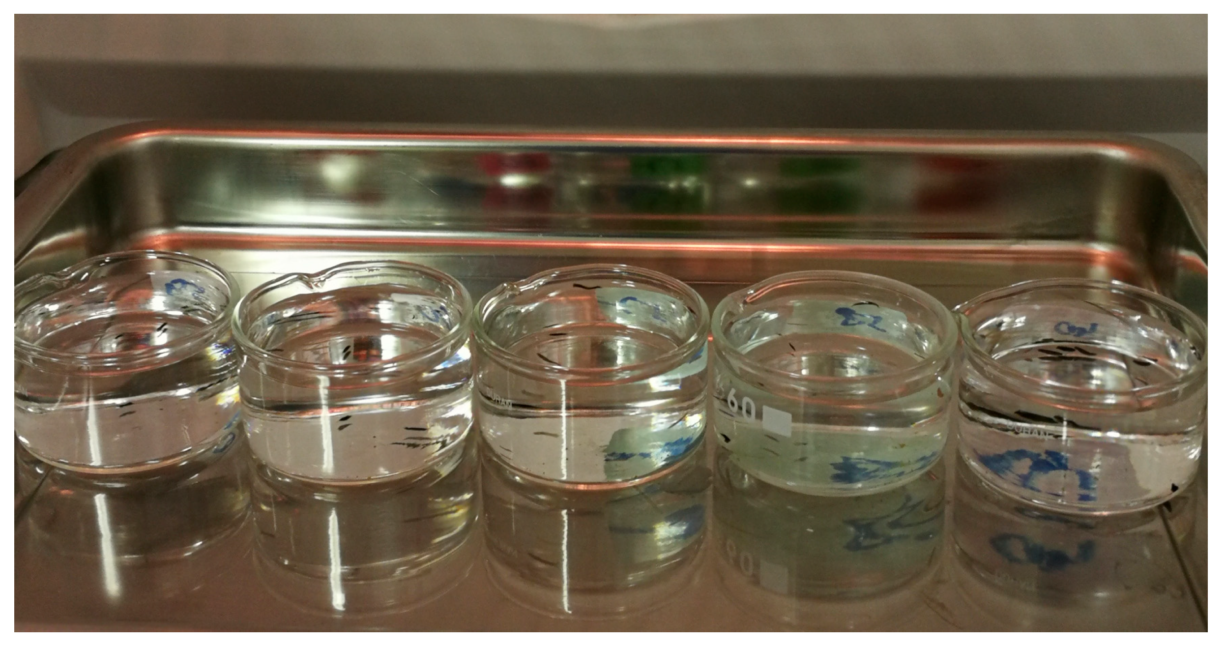
These experimental organisms were obtained from the breeding cultures of the Division of Zoology, Faculty of Science, University of Zagreb. The experiments were carried out with specific animal ratios and prey densities in microcosms in 60 mL crystallizing glass vessels (microcosms), 60 mm in diameter and 35 mm in height, each containing 50 mL of previously permanently aerated tap water. Each microcosm setup included 10 individuals of cladocerans as prey, while hydrozoans and turbellarians were added dropwise as predators in ratios of either 1:1 or 5:5 individuals. Additionally, separate dishes were prepared with hungry vs. satiated predators to compare their effects. Control groups were established for each species and relationship to ensure a rigorous experimental design. Each experiment setup was performed in five replicates (Figure 1). The interactions between the model organisms (Table 1) were recorded 1 h and 24 h after setting up the microcosms. The experiments were carried out in laboratory, and the experimental vessels were placed on trays and exposed to the experimental conditions. The aim was to investigate the interactions between model organisms (DM, HV, PF, and DG) under two temperature/light regimes: 25 °C with a photoperiod of an 8 h day and 16 h night, and 13.5 °C in the dark (in refrigerator). These two temperature conditions were selected to represent ecologically relevant scenarios: 25 °C simulates warm, summer-like conditions that increase metabolic and interaction rates, while 13.5 °C reflects cooler, low-light conditions typical of early spring, late autumn, or shaded aquatic habitats. This contrast allowed us to assess how seasonal or habitat-related thermal shifts influence species interactions and behavioral responses.
Figure 1. An overview of one experimental microcosm setup.
Table 1. Observed groups of model organisms in microcosms subjected to two experimental temperature/light regimes (25 °C with a photoperiod of an 8 h day and 16 h night, and 13.5 °C in the dark) with Daphnia magna (DM) as prey, and predators Hydra viridissima (HV), Polycelis felina (PF), and Dugesia gonocephala (DG), hungry vs. satiated predators.
The model organisms used in the experiments were constantly kept under controlled conditions prior to the experiments. HV and DG individuals were kept in permanently aerated aquarium water in 2 L glass containers at 21.5 °C and fed twice a week with shrimp larvae (Artemia salina). Similarly, PF individuals were kept in previously permanently aerated tap water in 1 L glass containers at 13.5 °C and fed with A. salina larvae once a week. The culture of DM was kept in aquaria at temperatures between 17 and 21 °C in 60 L aquaria with permanently aerated stale water, with a diet consisting of dry yeast, the alga Chlorella sp., and fish food once or twice a week. For the experiment, satiated animals were used directly from the cultures, while for the experiment with hungry animals, individuals were separated into separate glass containers with previously permanently aerated tap water, kept at 13.5 C, and deprived of food for at least three days prior to the beginning of the experiment. Previous laboratory studies/microcosms using Hydra as the predator and Daphnia [] or rotifers as prey [], or DG as the predator and nematodes as prey [], are known.
Changes were registered by visual observation and counting [,]. The last two figures in the manuscript were obtained using a stereomicroscope and digital camera, and Figure 1 was obtained using a digital camera.
Statistical analysis of the data was performed using Statistica 13.0, calculating means and standard deviations for continuous variables. Analysis of variance (ANOVA) and Tukey’s multiple comparisons post hoc test were used to assess possible differences between groups, with statistical significance set at p < 0.05.

3. Results

In the control setup of the experiment with DM, the individuals were alive at 25 °C and at 13.5 °C after 1 h of exposure. After an exposure time of 24 h, at 25 °C and 13.5 °C there was altogether one dead DM individual per temperature regime.
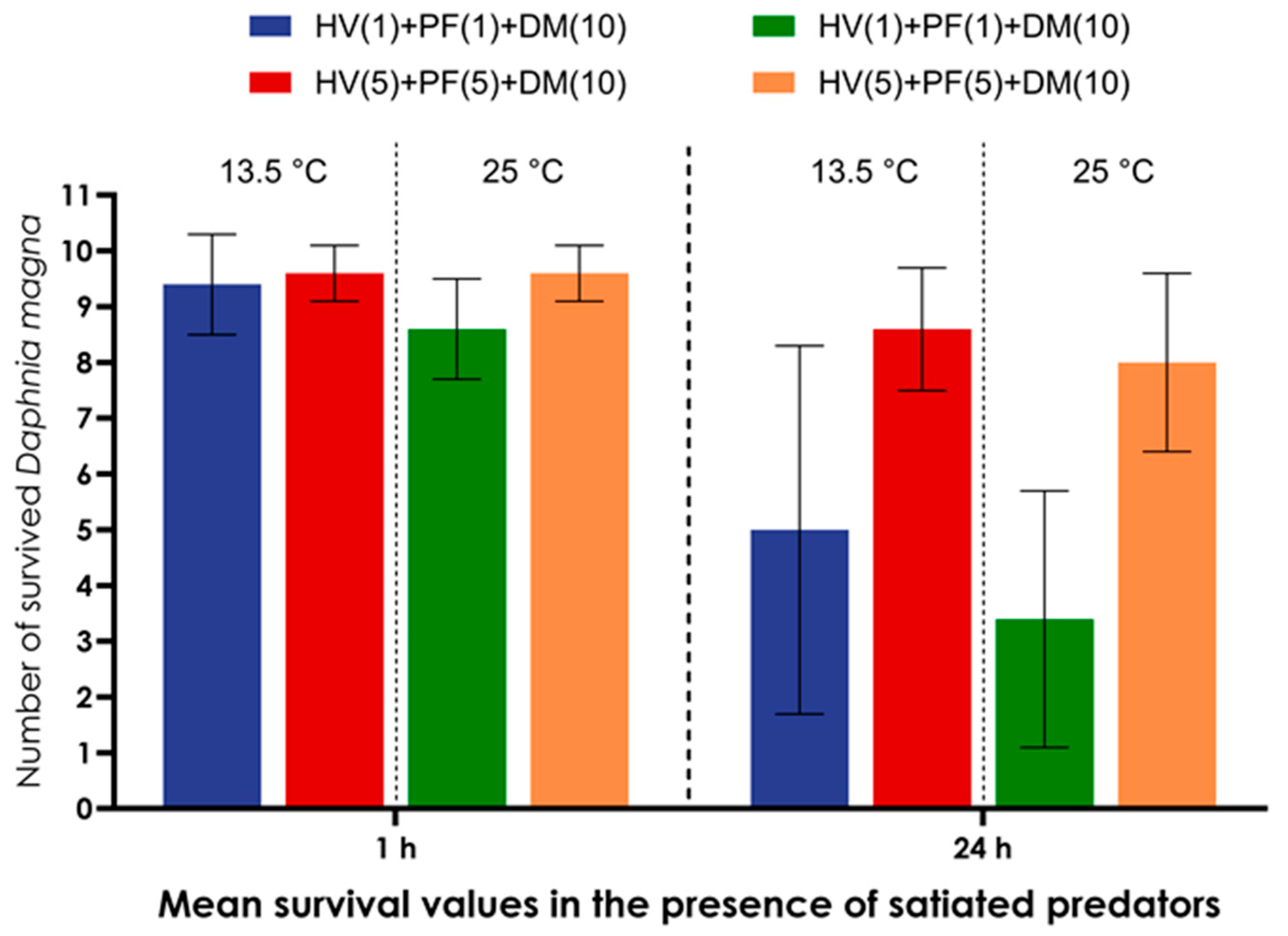
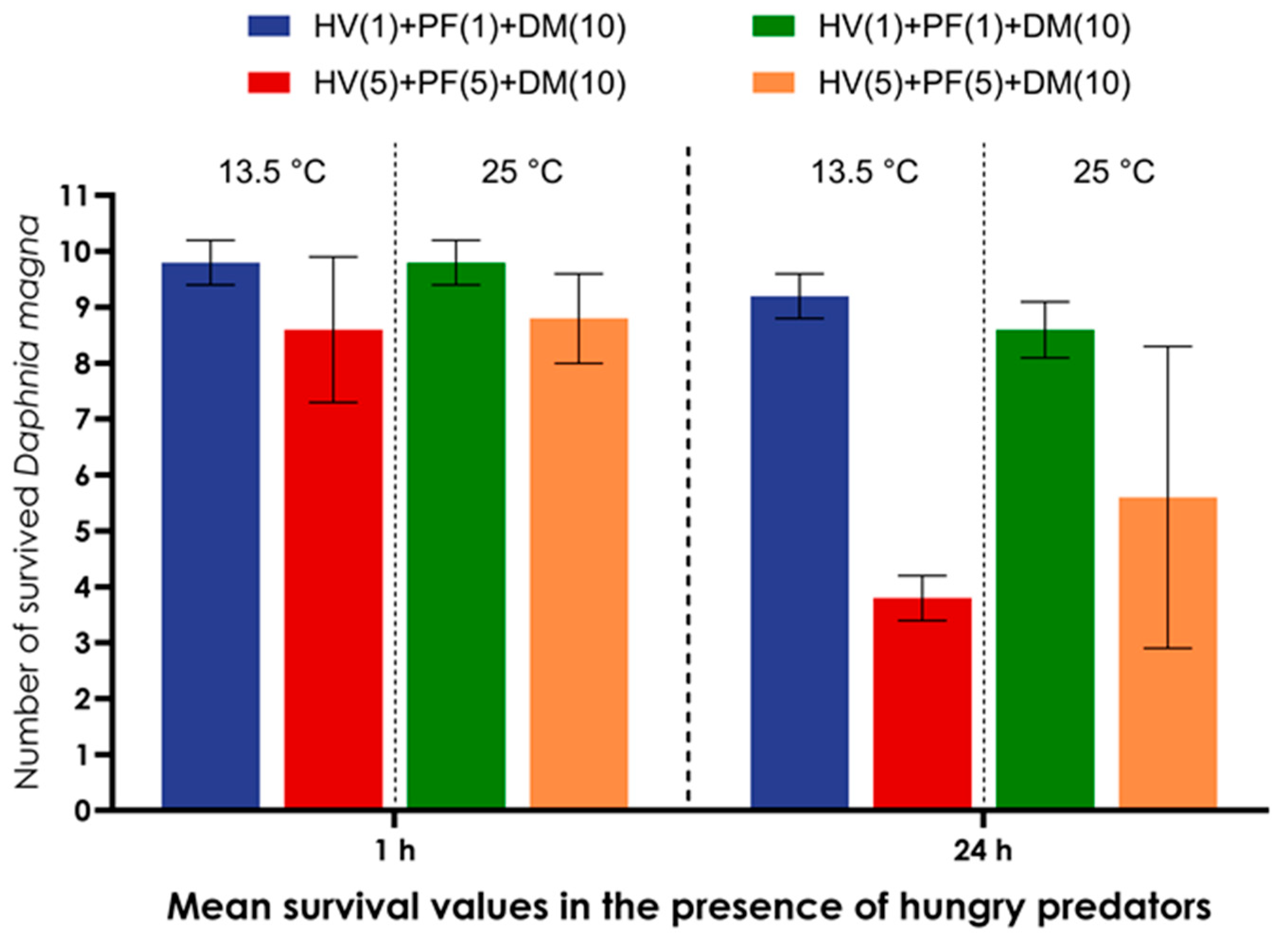
Experiment with HV, PF and DM. After 1 h exposure, the survival of DM in the presence of one satiated HV and PF individual was very high at both 13.5 °C (9.4 ± 0.9 individuals) and 25 °C (8.6 ± 0.9 individuals). After 24 h at 13.5 °C, a significant (p = 0.0206) decrease was observed compared to 1 h exposure (5.0 ± 3.3 individuals), and the lowest survival was recorded after 24 h at 25 °C (3.4 ± 2.3). In the presence of five satiated predators, HV and PF each, higher survival was observed at both temperatures and both exposure times, with a statistically significant difference between the two temperature regimes (13.5 °C vs. 25 °C) after 24 h of exposure (p = 0.0063; p = 0.0494). In an experiment with one hungry HV and PF each, predation was observed: HV kept one DM alive and moved with it. The survival of the DM in the presence of one hungry HV and PF was identical at both temperatures (9.8 ± 0.4 individuals). After 24 h at 13.5 °C, survival was significantly lower (p = 0.0451). A statistically significant difference in survival after 24 h was also observed at both temperatures (p = 0.0030). In an experiment with five hungry individuals of each species, survival was lower at both temperatures and both exposure times. A significant difference in survival at 13.5 °C between 1 h and 24 h was confirmed (p < 0.0001). The lowest survival was observed after 24 h at 25 °C (3.8 ± 0.4 individuals). The survival of DM was higher in the presence of hungry HV and PF at both temperatures and both exposure times, and this difference in survival between hungry and satiated individuals was also statistically significant after 24 h at 25 °C (p = 0.0017) and 13.5 °C (p = 0.0494) (Figure 2 and Figure 3).
Figure 2. Mean value and standard deviation (X ± SD) of surviving DM in microcosms with one satiated PF and one satiated HV, and five satiated PF and five satiated HV after 1 and 24 h at 13.5 °C and 25 °C.
Figure 3. Mean value and standard deviation (X ± SD) of surviving DM in microcosms with one hungry PF and one hungry HV, and five hungry PF and five hungry HV after 1 and 24 h at 13.5 °C and 25 °C.
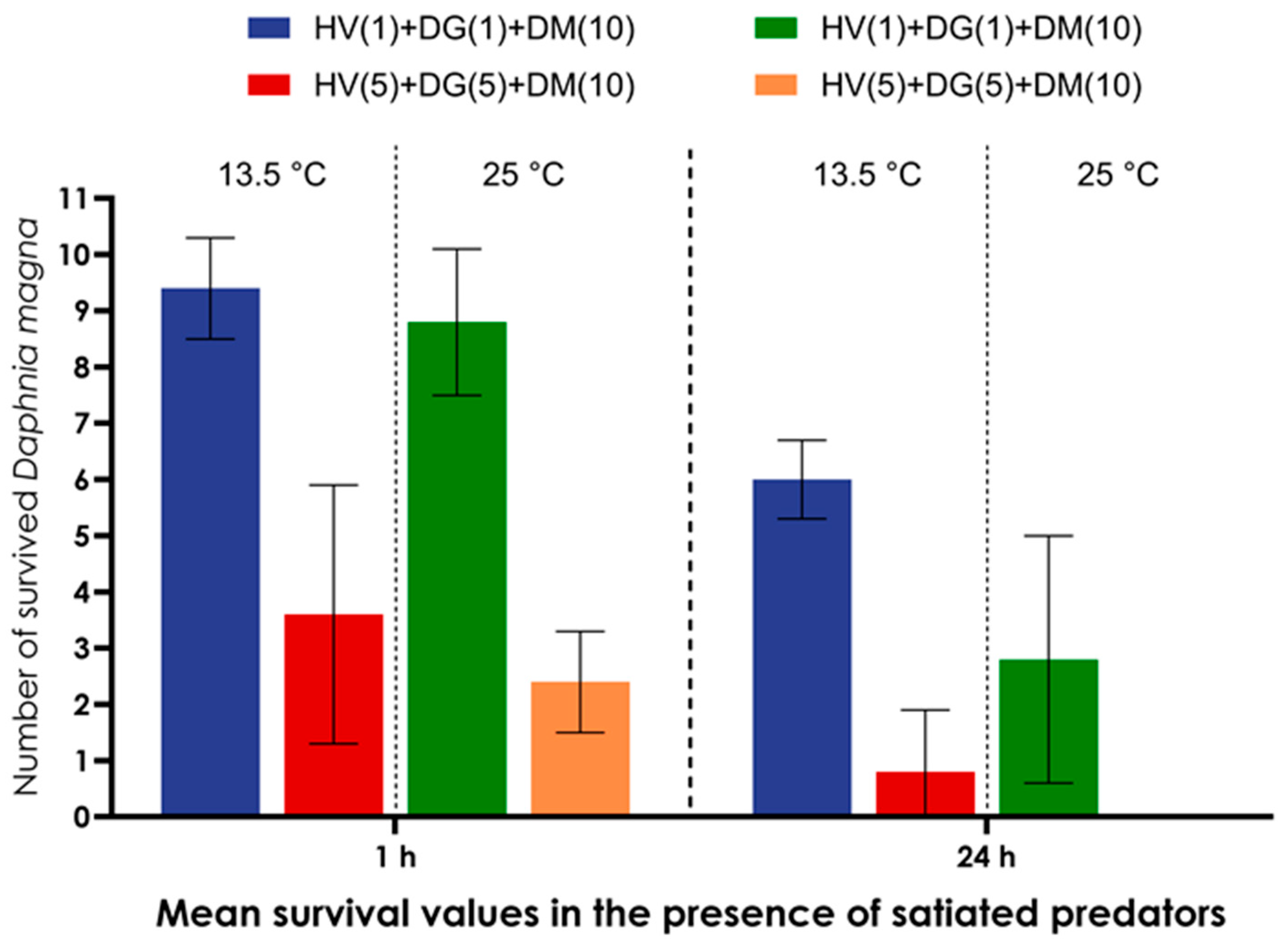
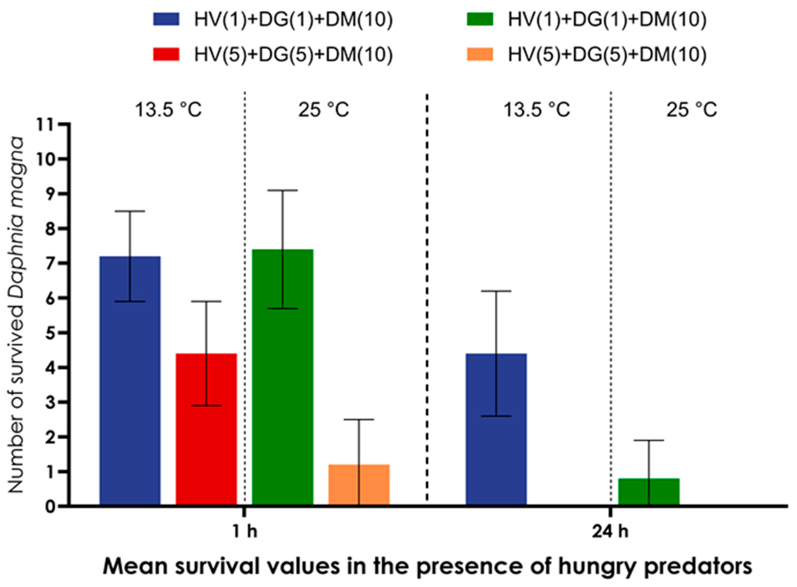
Experiment with HV, DG, and DM. The survival of DM in the experiment with one satiated HV and DG after 1 h at 13.5 °C was high (9.4 ± 0.9 individuals) and did not differ significantly from the survival at 25 °C (8.8 ± 1.3 individuals). However, 24 h exposure at 13.5 °C caused a significant (p = 0.0002) decrease in DM survival (6 ± 0.7 individuals) compared to 1 h exposure at the same temperature. A significantly lower survival was observed after 24 h at 25 °C (2.8 ± 2.2 individuals), compared to 24 h at 13.5 °C (p = 0.0147). A similar decline in survival was also confirmed after just 1 h at 25 °C (p = 0.0008). In the presence of five satiated DG and HV, the DM survival was significantly lower at both temperatures and both exposure times and amounted to 3.6 ± 2.3 individuals after 1 h at 13.5 °C and 2.4 ± 0.9 individuals at 25 °C. After 24 h at 13.5 °C, DM survival was 0.8 ± 1.1 individuals, which was significantly lower compared to 1 h at 13.5 °C (p = 0.0396). At 25 °C, none of the DM individuals survived after 24 h. The average survival of DM individuals in the presence of one hungry HV and DG after 1 h at 13.5 °C was 7.2 ± 1.3 individuals and at 25 °C was 7.4 ± 1.7 individuals. A significantly lower DM survival (4.4 ± 1.8 individuals) was observed after 24 h (p = 0.0225) compared to 1 h at 13.5 °C. After 24 h at 25 °C, the survival was 0.8 ± 1.1 individuals, which was significantly lower compared to 24 h exposure at 13.5 °C (p = 0.0051) and 1 h exposure at 25 °C (p = 0.0001). In the presence of five hungry HV and DG each, there were no surviving DM after 24 h exposure at each temperature, and after 1 h at 13.5 °C, there were 4.4 ± 1.5 individuals, with 1.2 ± 1.3 individuals at 25 °C. When comparing the survival of DM in the presence of one HV and one DG individual for both exposure times and both temperatures, the survival was higher in the presence of satiated HV and DG individuals. This difference was also statistically significant for the 24 h exposure at 25 °C (p = 0.0124).
In an experiment involving five HV and five DG individuals, no DM survived among the hungry HV and DG after 24 h at either temperature, nor among the satiated individuals at 25 °C. One-hour exposure at 13.5 °C resulted in a greater number of surviving DM individuals in the presence of hungry HV and DG individuals, while the opposite was observed at 25 °C (Figure 4 and Figure 5).
Figure 4. Mean value and standard deviation (X ± SD) of surviving DM in microcosms with one satiated HV and one satiated DG, and five satiated HV and five satiated DG after 1 and 24 h at 13.5 °C and 25 °C.
Figure 5. Mean value and standard deviation (X ± SD) of surviving DM in microcosms with one hungry HV and one hungry DG, and five hungry HV and five hungry DG after 1 and 24 h at 13.5 °C and 25 °C.
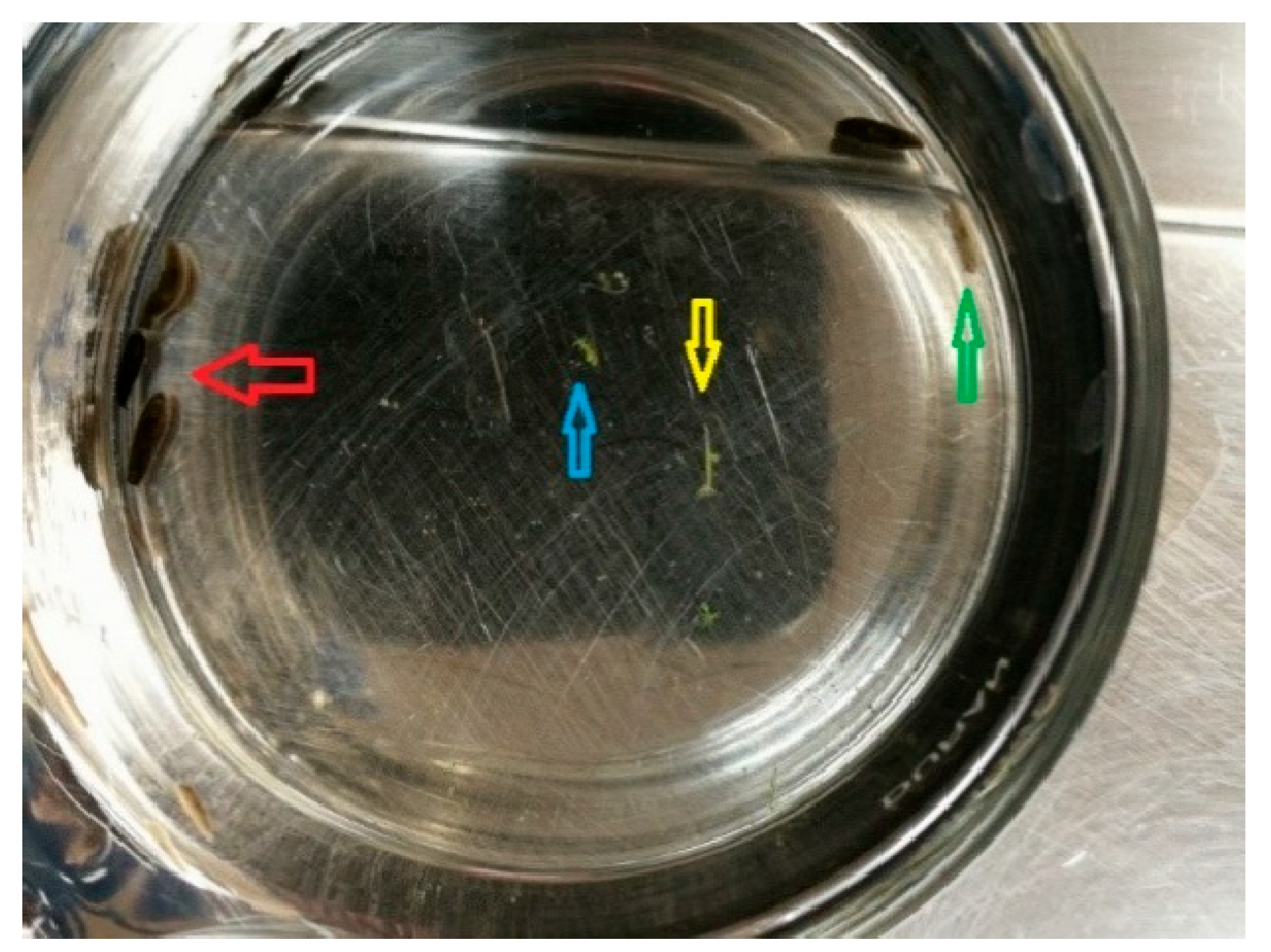
Experiment with HV, PF, and DG. After 24 h in the experiment at 25 °C in which one satiated individual of each species was present, predation of DG over PF was observed. The same was observed when five individuals of all predator species were present. DG was observed to take over the black pigmentation from PF and pieces of PF were observed in the experimental dishes. After 1 h at 25 °C, in an experiment with hungry individuals of all predator species, predation of DG over PF was observed. At 13.5 °C, predation was observed in the experiment with one individual of each predator species, and in the experiment with five individuals of each predator species too. DG was observed to take over the black pigmentation from PF, and pieces of PF were observed in the experimental dishes (Figure 6, Table 2).
Figure 6. Experimental setup with HV, PF, and DG. HV (yellow and blue arrow); visible grouping of PF (red arrow); DG took over pigmentation from PF (green arrow).
Table 2. Predation of turbellarians in microcosms containing one (1) or five (5) satiated (PFS) or hungry (PFH) PF, one (1) or five (1) satiated (DGS) or hungry (DGH) DG and one (1) or five (5) satiated (HVS) or hungry (HVH) HV after 1 and 24 h at 13.5 °C and 25 °C.
The experiment with PF and DG showed that DG behaved as the predator and PF as prey in the combination of one satiated PF and one satiated DG. When single individuals of the two turbellarian species were combined, no signs of predation were observed after 1 h exposure. After 24 h at 25 °C, DG took over the black pigmentation, with the PF individual completely eaten. When five satiated individuals of each turbellarian species were combined, after 1 h at 13.5 °C, DG individuals took over the black pigmentation from eaten PF individuals. At 25 °C, DG individuals took over the black pigmentation from eaten PF. After 24 h at 13.5 °C, black DG individuals were observed. After 24 h at 25 °C, black DG individuals were observed. In the combination of two hungry individuals, one PF and one DG, there were no signs of predation between the turbellarians during the short-term exposure (1 h) regardless of temperature, and the same was observed for individuals at 13.5 °C after 24 h, while after 24 h at 25 °C, damaged or partially eaten PF individuals were found, and consequently black DG individuals were found. In the combination of five hungry individuals from both species at short-term exposure (1 h), there were no signs of predation at any temperature, not even after 24 h at 13.5 °C. On the contrary, completely eaten individuals of PF were observed after 24 h at 25 °C, and most DG individuals exhibited black pigmentation (Table 3 and Table 4).
Table 3. Predation of turbellarians in microcosms containing one (1) or five (5) satiated (PFS) or hungry (PFH) PF and one (1) or five (1) satiated (DGS) or hungry (DGH) DG, kept for 1 and 24 h at 13.5 °C and 25 °C.
Table 4. Occurrence of PF grouping in experimental microcosms during the experiment. Each setup contained five individuals of represented predator (PF, DG, or HV).
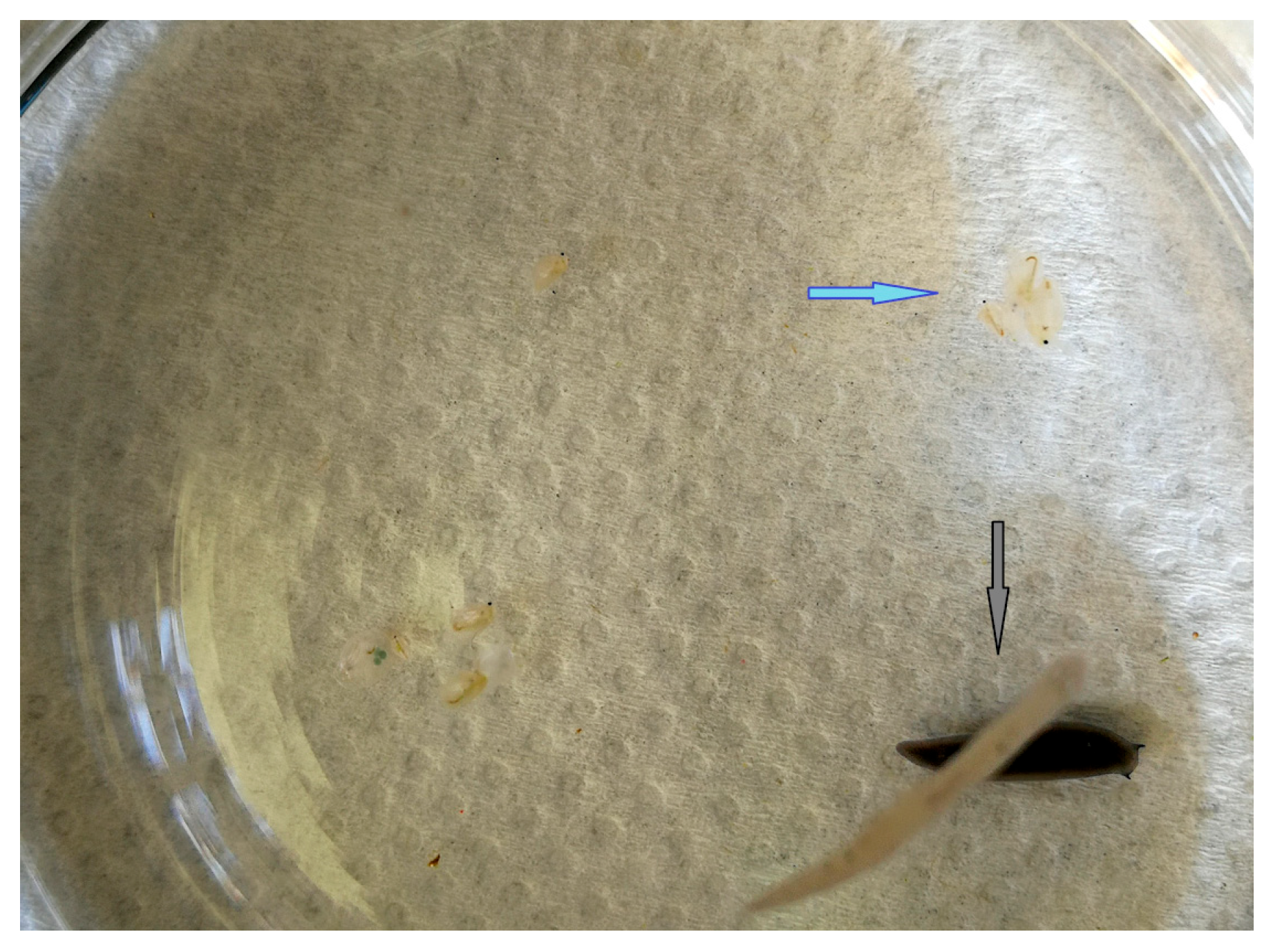
Experiment with PF, DG, and DM. After 1 h at 25 °C, pigment uptake from PF to DG occurred in the experiment with one satiated individual of each turbellarian species as well as in the experiment with five individuals. After 1 h exposure of DM individuals at 13.5 °C in the presence of one satiated individual of each of the two turbellarian species, the average number of surviving DM individuals was 8.2 ± 2.0, which is significantly higher than in the experiment at 25 °C (p = 0.0055), where the average number of surviving DM individuals was only 2.8 ± 2.5 individuals. After 24 h at 13.5 °C, the average number of surviving DM individuals was 2.0 ± 1.2 individuals, which was significantly lower than under the same temperature but short-term (1 h) exposure (p = 0.0003). In the experiment with five satiated turbellarian individuals, the number of surviving DM individuals was significantly lower for both temperatures and both exposure times compared to experiments with one individual each, and after 1 h at 13.5 °C, there were 2.6 ± 3.3 individuals, and at 25 °C, only 0.4 ± 0.9 individuals. After 24 h at 13.5 °C, there were no surviving DM, while at 25 °C, the number of surviving DM was 0.8 ± 1.3 individuals. In an experiment involving one hungry individual of each of two turbellarian species at 25 °C, the DG individual exhibited black pigmentation following the consumption of the PF individual, observed at both 1 h and 24 h. In an experiment involving five hungry individuals of each of two turbellarian species, black pigmentation was observed in DG individuals following 1 h exposure at 13.5 °C, as well as after 24 h exposures at both 13.5 °C and 25 °C. After a 1 h exposure at a temperature of 13.5 °C in the presence of one hungry individual of each of the two turbellarian species, the average DM survival was 7.0 ± 1.9 individuals, which is significantly higher (p = 0.0096) than for the same exposure time at 25 °C (2.6 ± 2.2). A significantly lower number (2.4 ± 1.5) of surviving DM individuals was observed after 24 h at 13.5 °C compared to 1 h (p = 0.0028). After 24 h at 25 °C, there were no surviving DM. In an experiment with five hungry turbellarians of each species, the number of surviving DM individuals after 1 h at 13.5 °C was twice as low as in the previous experiment, which was found to be statistically significant (p = 0.0197). At 25 °C after 1 h, the number of surviving DM individuals was twice as high (6.0 ± 1.0) as in the previous experiment, which was also statistically significant (p = 0.0137). After 24 h at 13.5 °C there were no surviving DM, and at 25 °C, the survival was only 0.8 ± 1.3 individuals. In the experiment with one individual of both turbellarian species, the number of surviving DM was higher in the presence of satiated individuals, except in the case of the 24 h exposure at 13.5 °C, where the survival was slightly higher in the presence of hungry turbellarians. None of these differences are statistically significant. An experiment with five individuals of both turbellarian species showed that for a short exposure time (1 h), regardless of temperature, the survival of DM was higher in the case of hungry individuals compared to the satiated ones, which was statistically significant at 25 °C (p < 0.0000). After 24 h at 13.5 °C, there were no surviving DM regardless of whether hungry or satiated turbellarians were present, and almost the same was observed at 25 °C (Figure 7, Table 4).
Figure 7. Experimental setup with PF and DG with DM as prey. At the bottom, predation of DG over PF (black arrow) and grouped individuals of DM (blue arrow) is visible.

4. Discussion

Consistent with our first hypothesis, predation intensity and behavioral responses varied among species, reflecting their distinct feeding mechanisms and ecological niches. The simultaneous presence of both flatworm species in the same dish resulted in DG preying on PF, highlighting predation between competing predator species and underscoring differences in their hunting strategies and ecological interactions. In general, changes in the behavior or morphology of Daphnia in the presence of predators are likely triggered by chemical cues released by the predators. These cues, known as kairomones, primarily consist of amino–fatty acid conjugates. Hungry Dugesia release these cues in significantly larger quantities, which in turn trigger a defensive response in water fleas [,]. The pronounced hunting instinct of Dugesia was described by []. In addition, flatworms excrete mucus pads that increase the viscosity of the water and reduce the ability of Daphnia to escape, making them easier targets []. Increased mucus excretion as a prey-hunting strategy has also been observed in the terrestrial flatworm Othelosoma impensum []. As already observed, one of the most prominent defense strategies against predators is group formation. In the group, they appear as a larger target for the predator’s attack, although this only has a significant effect when the predators are sighted []. Ref. [] reported a similar pattern of group formation of DM when Mesostoma cf. lingua was introduced into the system as a predator. Like other predators, hydras release attractants, which act as allelochemicals and can affect prey in different ways. For example, they can alter life history traits by shifting to an older age, thereby becoming sexually mature and reproducing earlier [,].
Two different turbellarian species with different hunting strategies were introduced into a DM population. PF and DG do not feed on/prey on DM in their natural habitat, but their predatory behavior towards DM in microcosm experimental setups in the laboratory has been recorded []. Although both species are predators and feed on smaller invertebrates, the simultaneous presence of PF and DG in the same dish resulted in DG feeding on PF. DG are more difficult to detect due to their pale color, which gives them a predatory advantage over PF when DM are considered prey. DG individuals that have fed on PF adopted their pigmentation. This helps them mimic PF and possibly catch them more easily. At the same time, PF start to act as prey and form groups themselves [].
Our results support the initial hypotheses that higher temperature and light exposure (25 °C, 8 h light/16 h dark) increase predator activity and predation compared to lower temperature and darkness (13.5 °C, dark) (hypothesis (ii)), and that hungry predators exhibit higher predation and reduced prey survival compared to satiated individuals (hypothesis (iii)). Predation was most intense after 24 h at 25 °C, indicating that elevated temperature stimulates predatory behavior regardless of feeding state. However, the observation that previously fed DG individuals attacked PF more frequently than starved ones suggests a more complex, non-linear relationship between hunger and predation, consistent with hypothesis (iii). This effect highlights that factors such as feeding state can influence predation dynamics in context-dependent ways. The observed pattern aligns with the idea that environmental factors such as temperature and light can modulate predator–prey dynamics by influencing metabolic rates and activity levels.
Temperature is a well-known driver of metabolic and behavioral changes in aquatic ectotherms, often increasing predator activity and feeding rates as temperature rises []. Our results show that increased temperature generally correlates with reduced prey survival, and the interaction with predator satiation suggests differential behavioral adaptations, such as increased hunger-driven predation at higher temperatures. Food availability further modulates feeding strategies, as satiated predators exhibit lower predation pressure compared to hungry ones, consistent with optimal foraging theory []. Overall, these findings emphasize the importance of considering both biotic factors, such as predator hunger, and abiotic factors, particularly temperature, to better predict ecological outcomes and resilience in freshwater ecosystems facing global change. Our results showed that among flatworms, DG has been shown to be a dominant predator that attacks PF regardless of the presence of other food sources.
In microcosms containing both flatworm species, DG consistently preyed on PF, acquiring the dark pigmentation of the consumed individuals. This pigmentation may provide DG with an adaptive advantage in natural ecosystems by enhancing camouflage within littoral habitats. In contrast, PF employed a grouping strategy as a defensive response to DG predation, indicating behavioral adaptation to avoid attack. DG proved to be the stronger predator, as PF did not prey on DG in any of the experiments. A similar pattern of predation and pigment transfer was observed when both turbellarian species were placed in a system with the cladoceran DM, suggesting competition between PF and DG. This behavior was also evident when both species coexisted with HV, where a clear spatial separation of predator species was observed, with flatworms and hydras occupying opposite poles of the microcosm. Such spatial distancing and interspecific predation can likely be attributed to chemical communication, which enables predators to avoid competition, minimize predation risk, and reduce unnecessary conflict []. Furthermore, previously fed DG individuals were more likely to attack PF than starved ones, a pattern consistent with context-dependent predatory behavior. Similar dominance of DG was also reported by [], who described DG as a highly competent predator capable of preying on Caenorhabditis elegans in microcosm environments. This finding aligns with the idea that feeding state can influence aggressive or predatory behavior, though the relationship is often non-linear and context-dependent. As highlighted in [], the effect of starvation on behavior varies with the type of interaction (e.g., predation, cannibalism, or competition), the cost–benefit balance of attacking, and the individual’s physical condition. Several studies (e.g., []) suggest a “hump-shaped” relationship, where mild food deprivation increases aggression due to heightened motivation, but severe starvation may reduce it as a result of diminished physiological capacity. In our case, the reduced aggression observed in starved DG individuals may indicate that extreme hunger impaired their ability or willingness to initiate attacks, supporting this non-linear model. Also, higher predation was present at a higher temperature (25 °C).
In our study HV was practically not preyed upon in either of the two experimental microcosms, regardless of the presence of other organisms or growth conditions such as temperature or number of individuals. This resistance can be explained by HV’s tentacles, which contain a large number of stinging cells, including cnidocytes. These cells are highly sensitive to contact due to a hair-like structure on their surface called the cnidocil. Upon contact, the cnidocytes eject an arrow-like filament containing neurotoxins and hemolytic proteins, providing both an efficient prey capture mechanism and a strong defense against potential predators [,,]. The defensive capability of these stinging cells helps explain why HV avoids predation. This is supported by studies such as [], who observed freshwater species interactions involving two Hydra species (H. salmacidis and H. viridissima). Their experiments demonstrated that hydras prey on small invertebrates such as Odonata nymphs, Chaoborus larvae, adult Copepoda, and small fish (Poecilia reticulata), but are not themselves attacked by any other predator species. Similarly, [] reported pronounced predatory behavior of HV towards the rotifer Euchlanis dilatata, positioning hydras near the top of the littoral food chain. Further emphasizing their defensive advantage, ref. [] showed that the nematocyst filaments’ toxins paralyze hydra prey, but do not aid in digestion, as evidenced by their experiments with crustaceans (A. salina), which remained paralyzed but mostly alive for 24 h without decomposing. Consistent with this, our study observed a decrease in the survival of DM in microcosms with HV after 24 h, but no signs of decomposition or detached body parts in the dish. Of all three predator species studied in the microcosm with DM as prey, HV demonstrated the most pronounced predatory efficiency. They not only captured their prey using their tentacles but also employed the basal disk during hunting, confirming their highly effective and versatile predation strategy. Additionally, as reported by [], HV possesses another evolutionary advantage: when prey of suitable size is scarce, its symbiotic algae synthesize and excrete maltose, maltotriose, and maltotetrose—precursors of glucose—or the Hydra can digest the algae within its own cells []. This adaptation further strengthens HV’s survival and resilience as a predator at the top of the freshwater food web.
In line with our fourth hypothesis, controlled microcosm experiments are expected to produce consistent and reproducible results that reflect general ecological patterns observed in natural freshwater systems. While microcosms offer valuable insights under controlled conditions, mesocosm experiments further enhance our understanding by simulating more complex environmental factors. For example, mesocosm studies [] have been instrumental in predicting how increases in temperature and salinity [], as well as eutrophication effects [,,,], impact freshwater lake ecosystems, particularly in the context of global change. Our results confirm that microcosm-based approaches can reliably capture key ecological processes and predator–prey dynamics, supporting their use as reproducible, scalable models for understanding and predicting patterns observed in natural freshwater environments.

5. Conclusions

Based on the results of our study and the literature data, we can conclude that HV is an extremely strong competitor and predator in the freshwater microcosm, while DG took over the camouflage coloration through predation. The results of this study indicate which predispositions enable some of these species (HV, PF, DG) to play a stronger role as competitors or predators. The findings reveal that survival of DM is significantly influenced by both predator satiation status and temperature, with higher predation pressure and lower survival at elevated temperatures (25 °C) and prolonged exposures (24 h).
Overall, the experiments revealed that hungry individuals generally exhibited higher predation and caused lower prey survival across treatments and temperatures, supporting the hypothesis that feeding state influences predatory intensity. However, this pattern was not entirely uniform. These results suggest that hunger amplifies predatory behavior, but its effect interacts with environmental factors such as temperature and exposure time, indicating a context-dependent relationship between feeding state and predation pressure. DG proved to be the dominant predator, preying on PF and exhibiting adaptive pigmentation changes, while PF relied on grouping as a defense strategy—highlighting clear interspecific differences in predatory strength and behavior.
With this study we showed the coexistence of predators in the same microcosm and their interrelationships as predators, demonstrated the effects of predation in a microcosm with three different invertebrate predator species, showed the capabilities of the hydra as a predator and competitor, and demonstrated the possible concept of camouflage of one planarian species when preying on another planarian species, by mimicking both the prey and the environment. To the best of our knowledge, there is no peer-reviewed study documenting a planarian predator employing both prey mimicry and environmental camouflage to facilitate predation. Therefore, our observation of planarian species performing such behavior (i.e., mimicking its prey and the environment) appears to represent a novel pattern in flatworm behavioral ecology.

Author Contributions

Conceptualization, G.K., P.T.L., D.P., M.S.P. and M.Š.; Methodology, G.K., P.T.L., D.P., D.Ž. and M.Š.; Validation, G.K., D.S., P.T.L. and D.P.; Formal Analysis, G.K., D.S., P.T.L., D.P., M.S.P., D.Ž., M.N. and M.Š.; Investigation, G.K., P.T.L. and D.P.; Resources, G.K., D.S., P.T.L., D.P., M.S.P., D.Ž., M.N. and M.Š.; Writing—Original Draft Preparation, G.K., D.S., P.T.L., D.P., M.S.P., D.Ž., M.N. and M.Š.; Writing—Review and Editing, G.K., P.T.L., D.P. and M.Š.; Visualization, G.K., D.S., P.T.L., D.P., M.S.P., D.Ž., M.N. and M.Š.; Supervision, G.K. and D.S.; Project Administration, G.K.; Funding Acquisition, G.K. and D.Ž. Author Davor Želježić passed away prior to the publication of this manuscript. All authors have read and agreed to the published version of the manuscript.

Funding

This research was funded by the University of Zagreb, institutional project number 106-F19-00056/20284116 and support number 20286544. The paper is part of the project of the Institute for Medical Research and Occupational Health “Evaluation of Efficacy and Toxicity of Biologically Active Substances WP5: Ecogenetic Research in Biomonitoring of Populations in vivo and in vitro” by the Institute for Medical Research and Occupational Health, support number 533-03-23-0006.

Data Availability Statement

The original contributions presented in this study are included in the article. Further inquiries can be directed at the corresponding author.

Conflicts of Interest

The authors declare no conflicts of interest.

References

  1. Walther, G.R.; Post, E.; Convey, P.; Menzel, A.; Parmesan, C.; Beebee, T.J.C.; Bairlein, F. Ecological responses to recent climate change. Nature 2002, 416, 389–395. [Google Scholar] [CrossRef]
  2. Root, T.L.; Price, J.T.; Hall, K.R.; Schneider, S.H.; Rosenzweig, C.; Pounds, J.A. Fingerprints of global warming on wild animals and plants. Nature 2003, 421, 57–60. [Google Scholar] [CrossRef]
  3. Thomas, C.D.; Cameron, A.; Green, R.E.; Bakkenes, M.; Beaumont, L.J.; Collingham, Y.C.; Erasmus, B.F.N.; Ferreira de Siqueira, M.; Grainger, A.; Hannah, L.; et al. Extinction risk from climate change. Nature 2004, 427, 145–148. [Google Scholar] [CrossRef] [PubMed]
  4. Parmesan, C. Ecological and Evolutionary Responses to Recent Climate Change. Annu. Rev. Ecol. Evol. Syst. 2006, 37, 637–669. [Google Scholar] [CrossRef]
  5. Meehan, M.L.; Turnbull, K.F.; Sinclair, B.J.; Lindo, Z. Predators minimize energy costs, rather than maximize energy gains under warming: Evidence from a microcosm feeding experiment. Funct. Ecol. 2022, 36, 2279–2288. [Google Scholar] [CrossRef]
  6. Sertić Perić, M.; Dražina, T.; Žutinić, P.; Rubinić, J.; Kuczyńska-Kippen, N.; Zhang, C.; Špoljar, M. Ecological Dynamics and Conservation Strategies for Mediterranean Salt Marshes: Insights from a Pilot Study of Biodiversity and Environmental Drivers in the Palud Marsh, Croatia. Sustainability 2024, 16, 10523. [Google Scholar] [CrossRef]
  7. Woodward, G.; Perkins, D.M.; Brown, L.E. Climate change and freshwater ecosystems: Impacts across multiple levels of organization. Philos. Trans. R. Soc. Lond. B Biol. Sci. 2010, 365, 2093–2106. [Google Scholar] [CrossRef]
  8. Palkovacs, E.P.; Wasserman, B.A.; Kinnison, M.T. Eco-evolutionary trophic dynamics: Loss of top predators drives trophic evolution and ecology of prey. PLoS ONE 2011, 6, e18879. [Google Scholar] [CrossRef]
  9. Price, E.L.; Sertić Perić, M.; Romero, G.Q.; Kratina, P. Land use alters trophic redundancy and resource flow through stream food webs. J. Anim. Ecol. 2019, 88, 677–689. [Google Scholar] [CrossRef]
  10. Lytle, D.A. Life-history and behavioural adaptations to flow regime in aquatic insects. In Aquatic Insects: Challenges to Populations; Royal Entomological Society: St Albans, UK, 2008; pp. 122–138. [Google Scholar] [CrossRef]
  11. Duarte, R.C.; Flores, A.A.V.; Stevens, M. Camouflage through colour change: Mechanisms, adaptive value and ecological significance. Phil. Trans. R. Soc. B 2017, 372, 20160342. [Google Scholar] [CrossRef]
  12. Blewett, T.A.; Binning, S.A.; Weinrauch, A.M.; Ivy, C.M.; Rossi, G.S.; Borowiec, B.G.; Lau, G.Y.; Overduin, S.L.; Aragao, I.; Norin, T. Physiological and behavioural strategies of aquatic animals living in fluctuating environments. J. Exp. Biol. 2022, 225, jeb242503. [Google Scholar] [CrossRef] [PubMed]
  13. Cáceres, C.E.; Tessier, A.J. To sink or swim: Variable diapause strategies among Daphnia species. Limnol. Oceanogr. 2004, 49, 1333–1340. [Google Scholar] [CrossRef]
  14. Heo, Y.M.; Oh, S.-Y.; Kim, K.; Han, S.-I.; Kwon, S.L.; Yoo, Y.; Kim, D.; Khim, J.S.; Kang, S.; Lee, H.; et al. Comparative Genomics and Transcriptomics Depict Marine Algicolous Arthrinium Species as Endosymbionts That Help Regulate Oxidative Stress in Brown Algae. Front. Mar. Sci. 2021, 8, 753222. [Google Scholar] [CrossRef]
  15. Vacant, S.; Benites, L.F.; Salmeron, C.; Intertaglia, L.; Norest, M.; Cadoudal, A.; Sanchez, F.; Caceres, C.; Piganeau, G. Long-Term Stability of Bacterial Associations in a Microcosm of Ostreococcus tauri (Chlorophyta, Mamiellophyceae). Front. Plant Sci. 2022, 13, 814386. [Google Scholar] [CrossRef] [PubMed]
  16. Van Doorslaer, W.; Stoks, R.; Duvivier, C.; Bednarska, A.; De Meester, L. Population dynamics determine genetic adaptation to temperature in daphnia. Evolution 2009, 63, 1867–1878. [Google Scholar] [CrossRef]
  17. Peck, L.S. Organisms and responses to environmental change. Mar. Genom. 2011, 4, 237–243. [Google Scholar] [CrossRef]
  18. Stoks, R.; Geerts, A.N.; De Meester, L. Evolutionary and plastic responses of freshwater invertebrates to climate change:realized patterns and future potential. Evol. Appl. 2014, 7, 42–55. [Google Scholar] [CrossRef]
  19. Abrams, P.A. The Evolution of Predator-Prey Interactions: Theory and Evidence. Annu. Rev. Ecol. Evol. Syst. 2000, 31, 79–105. [Google Scholar] [CrossRef]
  20. Ruxton, G.D.; Allen, W.L.; Sherratt, T.N.; Speed, M.P. Avoiding Attack: The Evolutionary Ecology of Crypsis, Warning Signals and Mimicry; Oxford University Press: Oxford, UK, 2004. [Google Scholar] [CrossRef]
  21. Minelli, A. Predation. In Encyclopedia of Ecology; Jørgensen, S.E., Fath, B.D., Eds.; Elsevier: Amsterdam, The Netherlands; Academic Press: Cambridge, MA, USA, 2008; pp. 2923–2929. [Google Scholar] [CrossRef]
  22. Burns, C.W. The relationship between body size of filter-feeding Cladocera and the maximum size of particle ingested. Limnol. Oceanogr. 1968, 13, 675–678. [Google Scholar] [CrossRef]
  23. Anderson, R.S. Predator relationships and predation rates for crustacean zooplankters from some lakes in western Canada. Can. J. Zool. 1970, 48, 1229–1240. [Google Scholar] [CrossRef]
  24. Allan, J.D. Competition and the relative abundance of two cladocerans. Ecology 1973, 54, 484–498. [Google Scholar] [CrossRef]
  25. Dodson, S.I. Zooplankton competition and predation: An experimental test of the size efficiency hypothesis. Ecology 1974, 55, 605–613. [Google Scholar] [CrossRef]
  26. Muscatine, L.; Lenhoff, H.M. Symbiosis of hydra and algae. II. Effects of limited food and starvation on growth of symbiotic and aposymbiotic hydra. Biol. Bull. 1965, 123, 316–328. [Google Scholar] [CrossRef]
  27. Weber, J.; Klug, M.; Tardent, P. Some physical and chemical properties of purified nematocysts of Hydra attenuata pall. (Hydrozoa, cnidaria). Comp. Biochem. Physiol. Part B Comp. Biochem. 1987, 88, 855–862. [Google Scholar] [CrossRef]
  28. Calevro, F.; Filippi, C.; Deri, P.; Albertosi, C.; Batistoni, R. Toxic effects of aluminium, chromium and cadmium in intact and regenerating freshwater planarians. Chemosphere 1998, 37, 651–659. [Google Scholar] [CrossRef]
  29. Kovačević, G.; Gregorović, G.; Kalafatić, M.; Jaklinović, I. The Effect of Aluminium on the Planarian Polycelis felina (Daly.). Water Air Soil Pollut. 2009, 196, 333–344. [Google Scholar] [CrossRef]
  30. Roca, J.R.; Ribas, M.; Baguñà, J. Distribution, ecology, mode of reproduction and karyology of freshwater planarians (Platyhelminthes; Turbellaria; Tricladida) in the springs of the central Pyrenees. Ecography 1992, 15, 373–384. [Google Scholar] [CrossRef]
  31. Beier, S.; Bolley, M.; Traunspurger, W. Predator-prey interactions between Dugesia gonocephala and free-living nematodes. Freshw. Biol. 2004, 49, 77–86. [Google Scholar] [CrossRef]
  32. Miner, B.E.; De Meester, L.; Pfrender, M.E.; Lampert, W.; Hairston, N.G. Linking genes to communities and ecosystems: Daphnia as an ecogenomic model. Proc. R. Soc. B Biol. Sci. 2012, 279, 1873–1882. [Google Scholar] [CrossRef]
  33. Lynch, M. The evolution of cladoceran life histories. Q. Rev. Biol. 1980, 55, 23–42. [Google Scholar] [CrossRef]
  34. Ten Berge, W.F. Breeding Daphnia magna. Hydrobiologia 1978, 59, 121–123. [Google Scholar] [CrossRef]
  35. Bettinetti, R.; Croce, V.; Noè, F.; Ponti, B.; Quadroni, S.; Galassi, S. Ecotoxicity of pp’DDE to Daphnia magna. Ecotoxicology 2013, 22, 1255–1263. [Google Scholar] [CrossRef]
  36. Nagel, A.H.; Cuss, C.W.; Goss, G.G.; Shotyk, W.; Glover, C.N. Chronic toxicity of waterborne thallium to Daphnia magna. Environ. Pollut. 2021, 268, 115776. [Google Scholar] [CrossRef]
  37. Rivera-De La Parra, L.; Sarma, S.S.S.; Nandini, S. Effects of predation by Hydra (Cnidaria) on cladocerans (Crustacea: Cladocera). J. Limnol. 2016, 75, 39–47. [Google Scholar] [CrossRef]
  38. Walsh, E.J. Habitat-specific predation susceptibilities of a littoral rotifer to two invertebrate predators. Hydrobiologia 1995, 313, 205–211. [Google Scholar] [CrossRef]
  39. Kovačević, G.; Petrinec, D.; Tramontana Ljubičić, P.; Reipert, S.; Sirovina, D.; Špoljar, M.; Peharec Štefanić, P.; Želježić, D. Formation of Microalgal Hunting Nets in Freshwater Microcosm Food Web: Microscopic Evidence. Water 2023, 15, 3448. [Google Scholar] [CrossRef]
  40. Kovačević, G.; Tramontana Ljubičić, P.; Petrinec, D.; Sirovina, D.; Novosel, M.; Želježić, D. How Daphnia magna Defends Itself against Predators: Mechanisms and Adaptations in a Freshwater Microcosm. Water 2024, 16, 398. [Google Scholar] [CrossRef]
  41. Wilden, B.; Majdi, N.; Kuhlicke, U.; Neu, T.R.; Traunspurger, W. Flatworm mucus as the base of a food web. BMC Ecol. 2019, 19, 15. [Google Scholar] [CrossRef]
  42. Klintworth, S.; von Elert, E. Inducible morphological defense in Daphnia pulex: Food quantity effects revised. Aquat. Ecol. 2021, 55, 47–57. [Google Scholar] [CrossRef]
  43. Blaustein, L.; Dumont, H.J. Typhloplanid flatworms (Mesostoma and related genera): Mechanisms of predation and evidence that they structure aquatic invertebrate communities. Hydrobiologia 1990, 198, 61–77. [Google Scholar] [CrossRef]
  44. Thielicke, M.; Sluys, R. Prey capture and feeding behaviour in an endemic land flatworm from São Tomé Island. J. Nat. Hist. 2019, 53, 1385–1393. [Google Scholar] [CrossRef]
  45. Dumont, H.J.; Carets, I. Flatworm predator (Mesostoma cf. lingua) releases a toxin to catch planktonic prey (Daphnia magna). Limnol. Oceanogr. 1987, 32, 699–702. [Google Scholar] [CrossRef]
  46. Lass, S.; Spaak, P. Chemically induced anti-predator defences in plankton: A review. Hydrobiologia 2003, 491, 221–239. [Google Scholar] [CrossRef]
  47. Chakri, K.; Touati, L.; Alfarhan, A.H.; Al-Rasheid, K.A.S.; Samraoui, B. Effect of vertebrate and invertebrate kairomones on the life history of Daphnia magna Straus (Crustacea: Branchiopoda). Comptes Rendus Biologies 2010, 333, 836–840. [Google Scholar] [CrossRef] [PubMed]
  48. Petrinec, D. Green Hydra (Hydra viridissima Pallas, 1766) as a Competitor in Freshwater Microcosm and the Possibility of Application in the Teaching Process (Original in Croatian). Master’s Thesis, University of Zagreb, Zagreb, Croatia, 2020. [Google Scholar]
  49. Archer, L.C.; Sohlström, E.H.; Gallo, B.; Jochum, M.; Woodward, G.; Kordas, R.L.; Rall, B.C.; O’Gorman, E.J. Consistent temperature dependence of functional response parameters and their use in predicting population abundance. J. Anim. Ecol. 2019, 88, 1670–1683. [Google Scholar] [CrossRef]
  50. Stephens, D.W.; Krebs, J.R. Foraging Theory; Series: Monographs in Behavior and Ecology; Princeton University Press: Princeton, NJ, USA, 1986; Volume 1. [Google Scholar] [CrossRef]
  51. Sura, S.A.; Mahon, H.K. Effects of competition and predation on the feeding rate of the freshwater snail, Helisoma trivolvis. Am. Midl. Nat. 2011, 166, 358–368. [Google Scholar] [CrossRef]
  52. Scharf, I. The multifaceted effects of starvation on arthropod behaviour. Anim. Behav. 2016, 119, 37–48. [Google Scholar] [CrossRef]
  53. Belenioti, M.; Chaniotakis, N. Aggressive Behaviour of Drosophila suzukii in Relation to Environmental and Social Factors. Sci. Rep. 2020, 10, 7898. [Google Scholar] [CrossRef]
  54. Klug, M.; Weber, J.; Tardent, P. Hemolytic and toxic properties of Hydra attenuata nematocysts. Toxicon 1989, 27, 325–339. [Google Scholar] [CrossRef]
  55. Sher, D.; Zlotkin, E. A hydra with many heads: Protein and polypeptide toxins from hydra and their biological roles. Toxicon 2009, 54, 1148–1161. [Google Scholar] [CrossRef] [PubMed]
  56. Habdija, I.; Primc Habdija, B.; Radanović, I.; Špoljar, M.; Matoničkin Kepčija, R.; Vujčić Karlo, S.; Miliša, M.; Ostojić, A.; Sertić Perić, M. Protista-Protozoa i Metazoa- Invertebrata. Strukture i funkcije; Alfa d.d.: Zagreb, Croatia, 2011; p. 584. [Google Scholar]
  57. Massaro, F.C.; Negreiros, N.F.; Rocha, O. A search for predators and food selectivity of two native species of Hydra (Cnidaria: Hydrozoa) from Brazil. Biota Neotrop. 2013, 13, 35–40. [Google Scholar] [CrossRef]
  58. Sher, D.; Fishman, Y.; Zhang, M.; Melamed-Book, N.; Zlotkin, E. Osmotically-driven prey disintegration in the gastro-vascular cavity of the green hydra by a pore-forming protein. FASEB J. 2008, 22, 207–214. [Google Scholar] [CrossRef]
  59. Mews, L.K.; Smith, D.C. The green hydra symbiosis. VI. What is the role of maltose transfer from alga to animal? Proc. R. Soc. London. Ser. B Biol. Sci. 1982, 216, 397–413. [Google Scholar] [CrossRef]
  60. Luiselli, L.; Pacini, N. Microcosms and Mesocosms: Small-Scale Experiments, Big Impacts for Tropical Ecology. Afr. J. Ecol. 2025, 63, e70076. [Google Scholar] [CrossRef]
  61. Özkan, K.; Korkmaz, M.; Amorim, C.A.; Yılmaz, G.; Koru, M.; Can, Y.; Pacheco, J.P.; Acar, V.; Çolak, M.A.; Yavuz, G.C.; et al. Mesocosm Design and Implementation of Two Synchronized Case Study Experiments to Determine the Impacts of Salinization and Climate Change on the Structure and Functioning of Shallow Lakes. Water 2023, 15, 2611. [Google Scholar] [CrossRef]
  62. Liboriussen, L.; Landkildehus, F.; Meerhof, M.; Bramm, M.E.; Sondegaard, M.; Christoffersen, K.; Richardson, K.; Sondegaard, M.; Lauridsen, T.L.; Jeppesen, E. Global warming: Design of a flow through shallow lake mesocosm climate experiment. Limnol. Oceanogr. Methods 2005, 3, 1–9. [Google Scholar] [CrossRef]
  63. Stewart, R.I.A.; Dossena, M.; Bohan, D.A.; Jeppesen, E.; Kordas, R.L.; Ledger, M.E.; Meerhoff, M.; Moss, B.; Mulder, C.; Shurin, J.B.; et al. Mesocosm Experiments as a Tool for Ecological Climate-Change Research. In Advances in Ecological Research; Woodward, G., O’Gorman, E.J., Eds.; Elsevier: Amsterdam, The Netherlands; Academic Press: Cambridge, MA, USA, 2013; Volume 48, pp. 71–181. ISBN 978-0-12-417199-2. [Google Scholar]
  64. Šorf, M.; Davidson, T.A.; Brucet, S.; Menezes, R.F.; Søndergaard, N.; Lauridsen, T.L.; Landkildehus, F.; Liboriussen, L.; Jeppesen, E. Zooplankton response to climate warming: A mesocosm experiment at contrasting temperatures and nutrient levels. Hydrobiologia 2015, 745, 185–203. [Google Scholar] [CrossRef]
  65. Ersoy, Z.; Scharfenberger, U.; Baho, D.L.; Bucak, T.; Feldmann, T.; Hejzlar, J.; Levi, E.E.; Mahdy, A.; Nõges, T.; Papastergiadou, E.; et al. Impact of nutrients and water level changes on submerged macrophytes along a temperature gradient: A pan-European mesocosm experiment. Glob. Chang. Biol. 2020, 26, 6831–6851. [Google Scholar] [CrossRef]
Disclaimer/Publisher’s Note: The statements, opinions and data contained in all publications are solely those of the individual author(s) and contributor(s) and not of MDPI and/or the editor(s). MDPI and/or the editor(s) disclaim responsibility for any injury to people or property resulting from any ideas, methods, instructions or products referred to in the content.

Article Metrics

Citations

Article Access Statistics

Multiple requests from the same IP address are counted as one view.