
Visible Light Photocatalytic Degradation of Environmental Pollutants Using Zn-Doped NiO Nanoparticles

 , and
, and 
Abstract
1. Introduction
2. Materials and Methods
2.1. Materials
2.2. Synthesis of Zn-Doped NiO NPs
2.3. Characterization of Synthesized NPs
2.4. Pollutants Photocatalytic Degradation Experiment
3. Results and Discussions
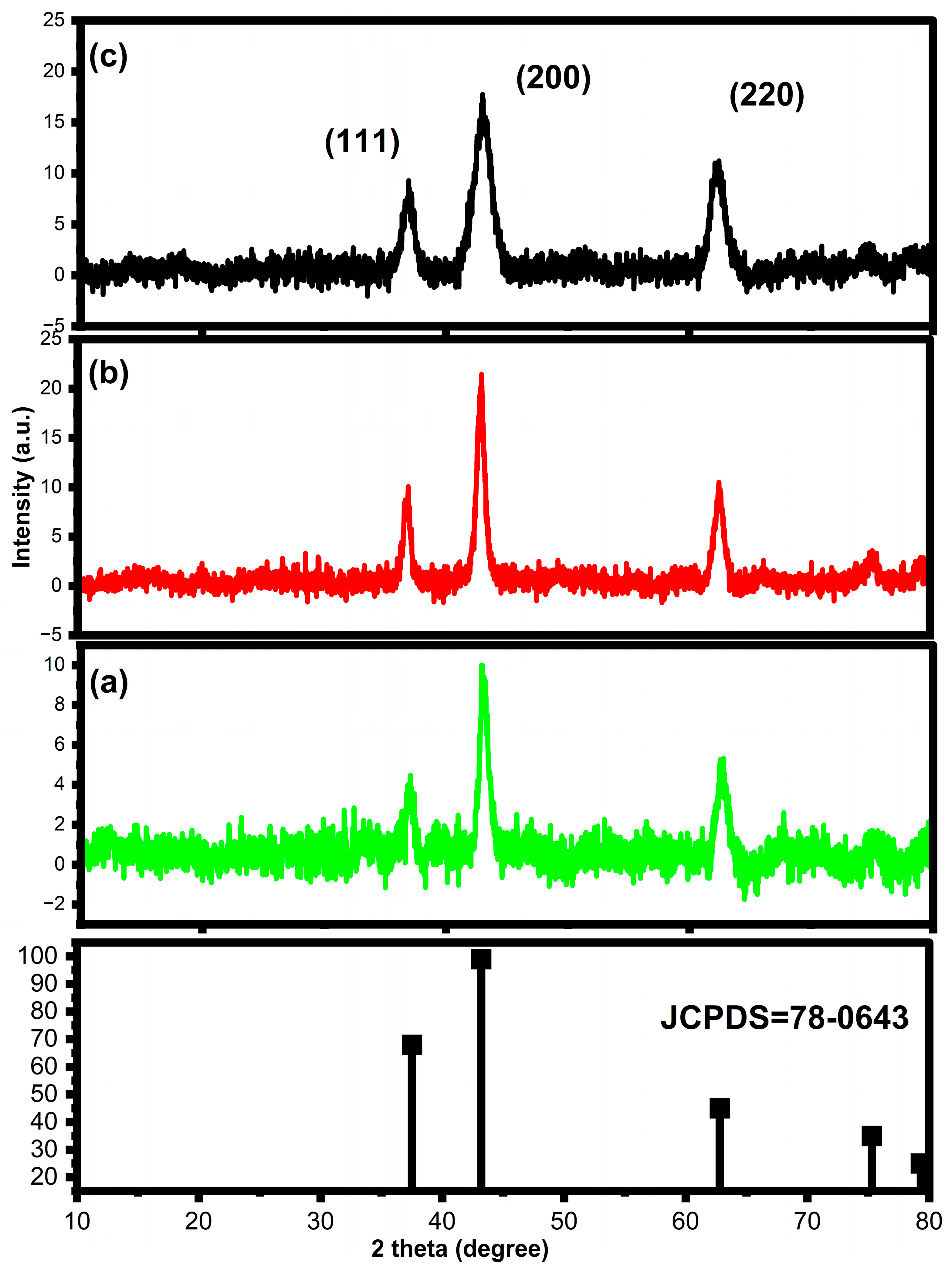
3.1. XRD Analysis
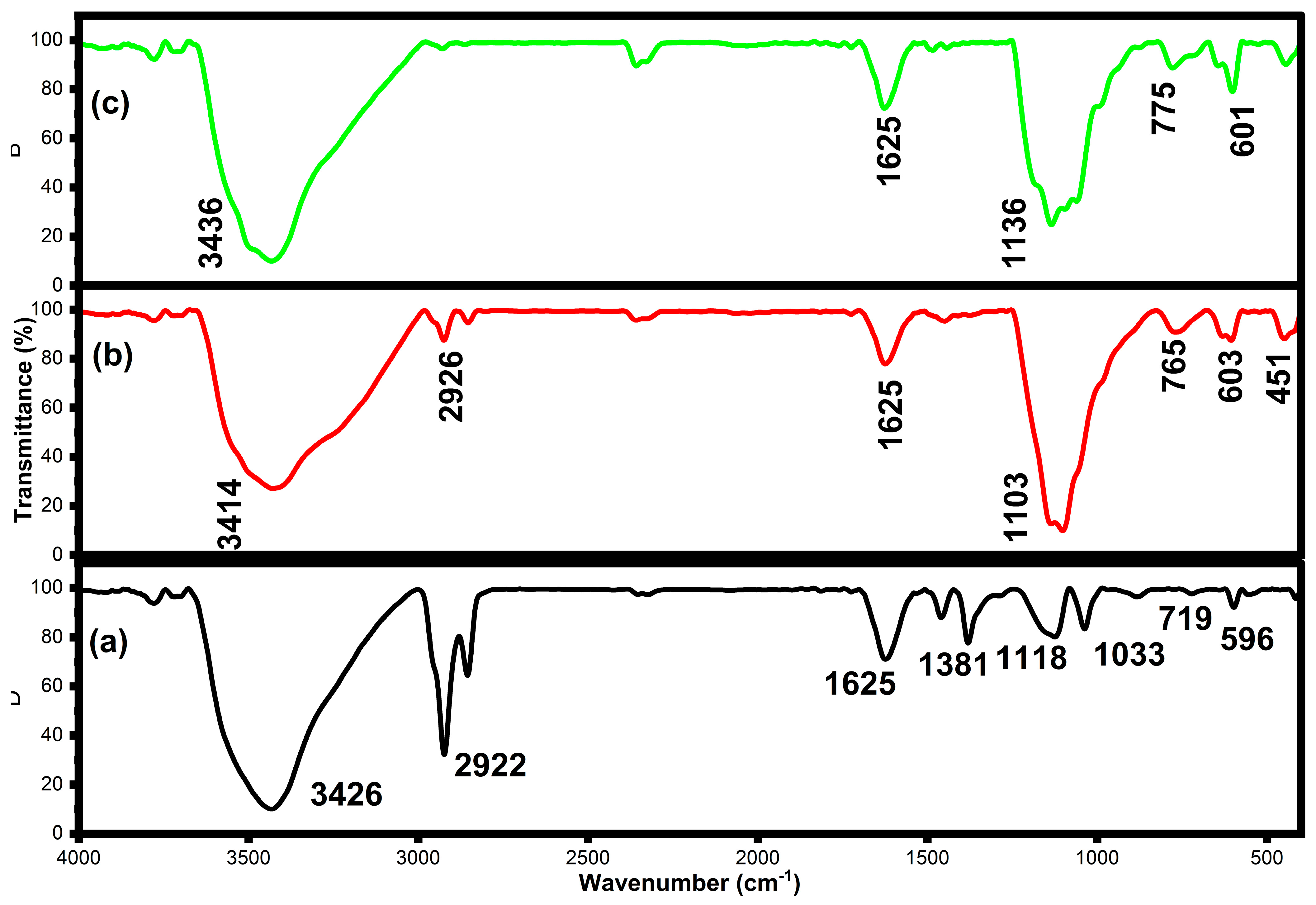
3.2. FTIR Analysis
3.3. UV-Visible Analysis
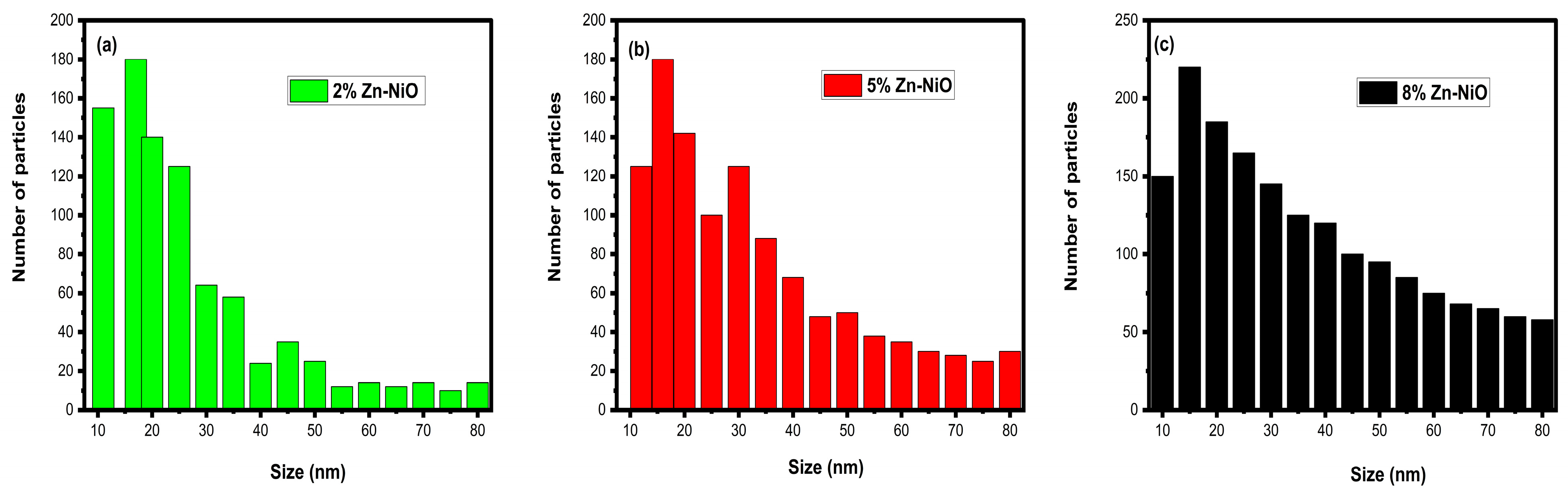
3.4. SEM with EDX Analysis
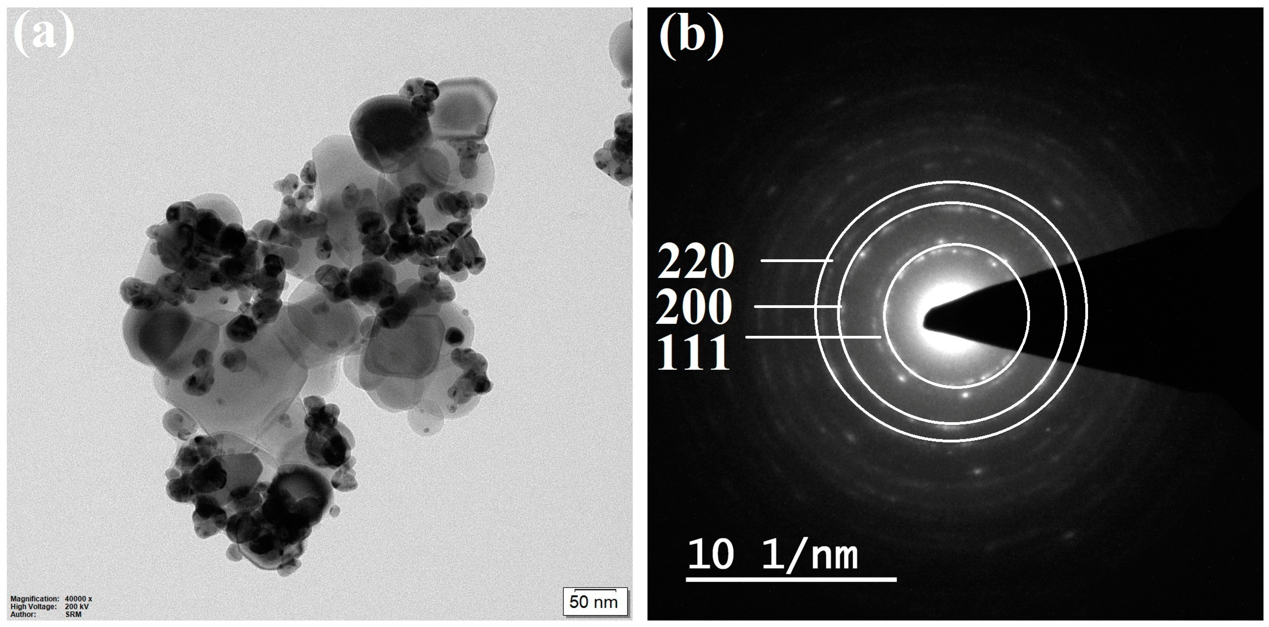
3.5. TEM with SAED Pattern Analysis
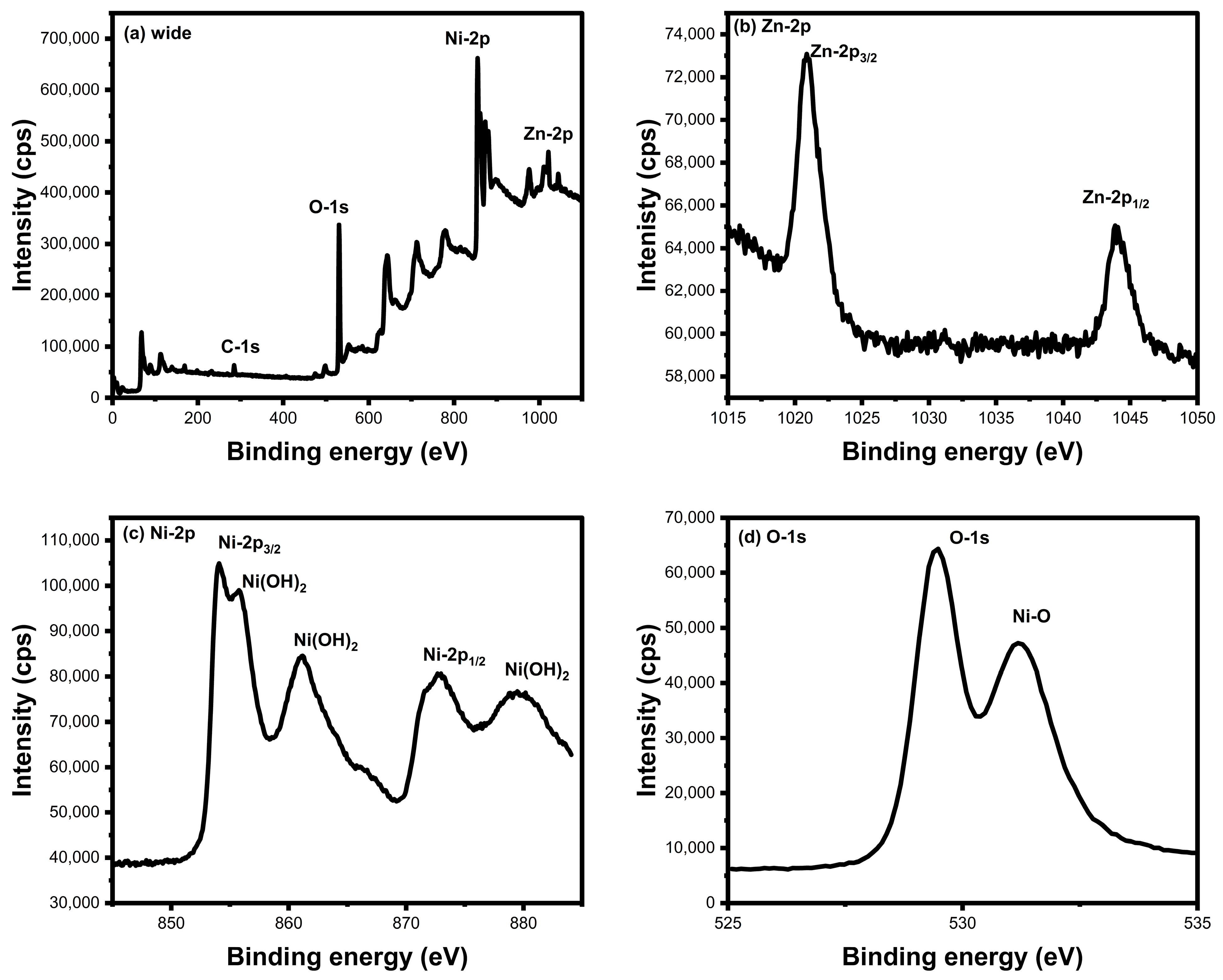
3.6. XPS Analysis
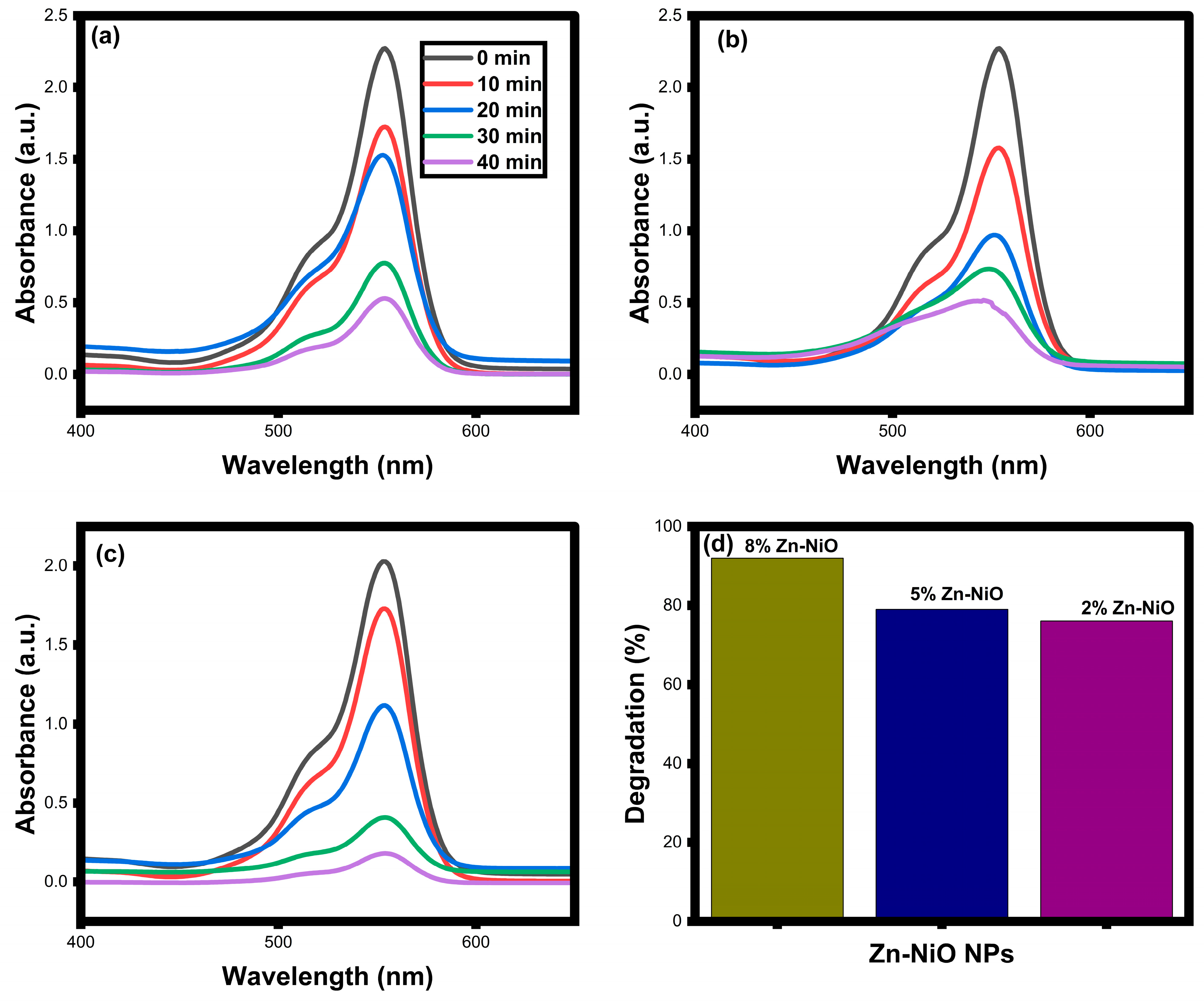
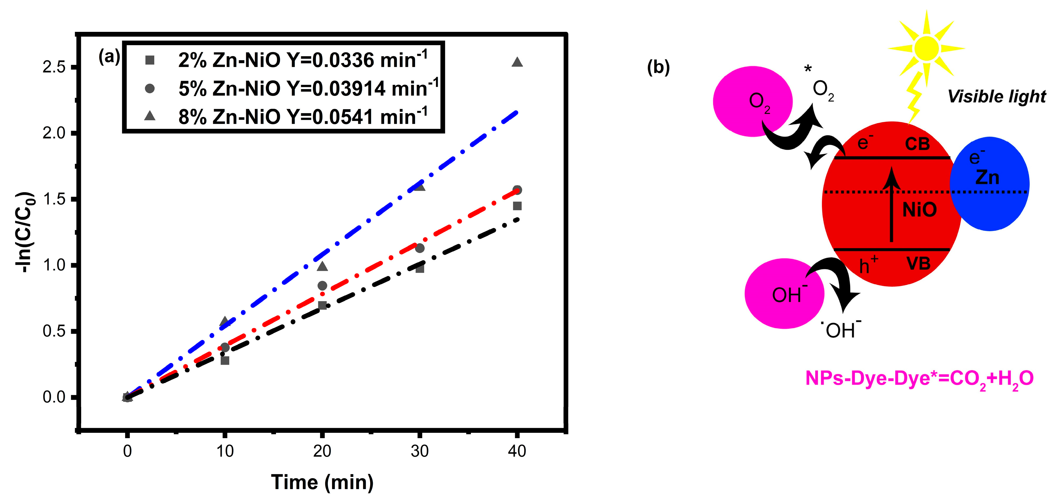
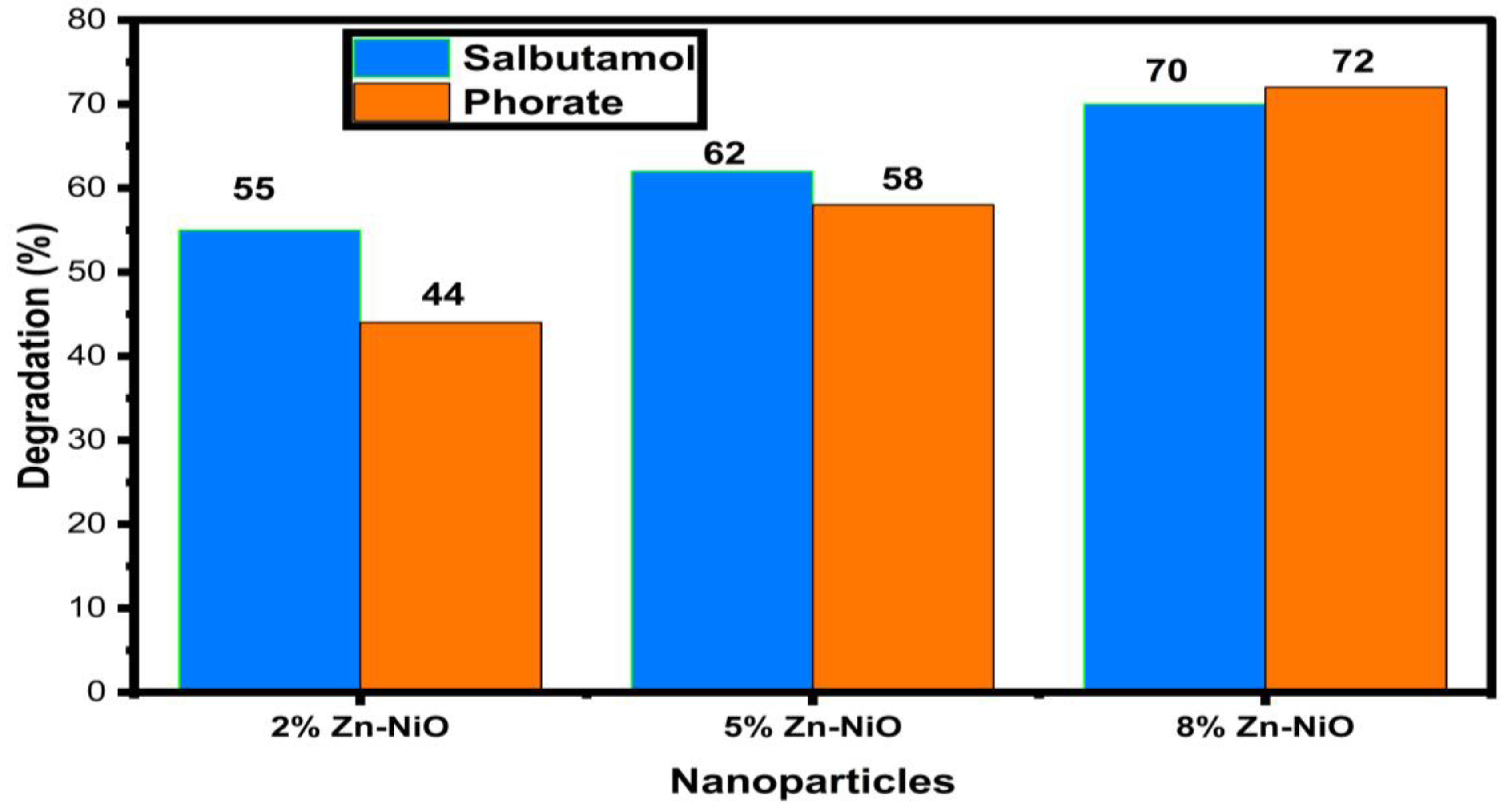
3.7. Pollutants Photocatalytic Degradation Analysis
4. Conclusions
Author Contributions
Funding
Data Availability Statement
Acknowledgments
Conflicts of Interest
References
- Sun, S.; Tang, Q.; Konar, M.; Fang, C.; Liu, H.; Liu, X.; Fu, G. Water transfer infrastructure buffers water scarcity risks to supply chains. Water Res. 2023, 229, 119442. [Google Scholar] [CrossRef]
- Liu, J.; Fu, Z.; Liu, W. Impacts of precipitation variations on agricultural water scarcity under historical and future climate change. J. Hydrol. 2023, 617, 128999. [Google Scholar] [CrossRef]
- Cai, B.; Jiang, L.; Liu, Y.; Zhang, W.; Zhang, Z. The impact of interregional trade on both quantity-and quality-related water scarcity of Beijing-Tianjin-Hebei urban agglomeration in China. Sustain. Cities Soc. 2023, 88, 104270. [Google Scholar] [CrossRef]
- Raj, R.S.; Krishnan, K.A. A comprehensive review on the impact of emerging organophosphorous pesticides and their remedial measures: Special focus on acephate. Environ. Nanotechnol. Monit. Manag. 2023, 20, 100813. [Google Scholar]
- Singh, H.; Pant, G. Phytoremediation: Low input-based ecological approach for sustainable environment. Appl. Water Sci. 2023, 13, 85. [Google Scholar] [CrossRef]
- Daripa, A.; Malav, L.C.; Yadav, D.K.; Chattaraj, S. Metal contamination in water resources due to various anthropogenic activities. In Metals in Water; Elsevier: Amsterdam, The Netherlands, 2023; pp. 111–127. [Google Scholar]
- Liu, P.; Dai, J.; Huang, K.; Yang, Z.; Zhang, Z.; Guo, X. Sources of micro (nano) plastics and interaction with co-existing pollutants in wastewater treatment plants. Crit. Rev. Environ. Sci. Technol. 2023, 53, 865–885. [Google Scholar] [CrossRef]
- Xu, M.; Deng, Y.; Li, S.; Zheng, J.; Liu, J.; Tremblay, P.L.; Zhang, T. Bacterial cellulose flakes loaded with Bi2MoO6 nanoparticles and quantum dots for the photodegradation of antibiotic and dye pollutants. Chemosphere 2023, 312, 137249. [Google Scholar] [CrossRef]
- Rajesh, G.; Kumar, P.S.; Akilandeswari, S.; Rangasamy, G.; Lohita, S.; Shankar, V.U.; Ramya, M.; Thirumalai, K. Preparation and characterization of a novel cobalt-substitution cadmium aluminate spinel for the photodegradation of azo dye pollutants. Chemosphere 2023, 323, 138232. [Google Scholar] [CrossRef]
- Reddy, C.V.; Kakarla, R.R.; Cheolho, B.; Shim, J.; Rezakazemi, M.; Aminabhavi, T.M. Highly efficient photodegradation of toxic organic pollutants using Cu-doped V2O5 nanosheets under visible light. Chemosphere 2023, 311, 137015. [Google Scholar] [CrossRef]
- Zahmatkesh, S.; Klemeš, J.J.; Bokhari, A.; Wang, C.; Sillanpaa, M.; Amesho, K.T.T.; Vithanage, M. Various advanced wastewater treatment methods to remove microplastics and prevent transmission of SARS-CoV-2 to airborne microplastics. Int. J. Environ. Sci. Technol. 2023, 20, 2229–2246. [Google Scholar] [CrossRef]
- Guerrero-Gualan, D.; Valdez-Castillo, E.; Crisanto-Perrazo, T.; Toulkeridis, T. Methods of Removal of Hormones in Wastewater. Water 2023, 15, 353. [Google Scholar] [CrossRef]
- Owodunni, A.A.; Ismail, S.; Kurniawan, S.B.; Ahmad, A.; Imron, M.F.; Abdullah, S.R.S. A review on revolutionary technique for phosphate removal in wastewater using green coagulant. J. Water Process Eng. 2023, 52, 103573. [Google Scholar] [CrossRef]
- Shakil, M.; Inayat, U.; Tanveer, M.; Nabi, G.; Gillani, S.S.A.; Rafique, M.; Tariq, N.H.; Shah, A.; Mahmood, A. NiO and Ag–Cd co-doped NiO nanoparticles: Study of photocatalytic degradation of rhodamine B dye for wastewater treatment. Int. J. Environ. Sci. Technol. 2023, 20, 2021–2036. [Google Scholar] [CrossRef]
- Gatou, M.A.; Lagopati, N.; Vagena, I.A.; Gazouli, M.; Pavlatou, E.A. ZnO Nanoparticles from Different Precursors and Their Photocatalytic Potential for Biomedical Use. Nanomaterials 2023, 13, 122. [Google Scholar] [CrossRef] [PubMed]
- Chandekar, K.V.; Palanivel, B.; Alkallas, F.H.; Trabelsi, A.B.G.; Khan, A.; Ashraf, I.M.; AlFaify, S.; Shkir, M. Photocatalytic activities of Mg doped NiO NPs for degradation of Methylene blue dye for harmful contaminants: A kinetics, mechanism and recyclability. J. Phys. Chem. Solids 2023, 178, 111345. [Google Scholar] [CrossRef]
- Sudhaik, A.; Hasija, V.; Selvasembian, R.; Ahamad, T.; Singh, A.; Khan, A.A.P.; Raizada, P.; Singh, P. Applications of graphitic carbon nitride-based S-scheme heterojunctions for environmental remediation and energy conversion. Nanofabrication 2023, 8, 1–36. [Google Scholar] [CrossRef]
- Jagan, K.S.G.; Surendhiran, S.; Savitha, S.; Balu, K.S.; Karthick, M.; Vidaarth, T.N.; Karthik, A.; Kalpana, B.; Senthilmurugan, R. Influence of different alkaline actuators in synthesis of NiO NPs: A comparative green approach on photocatalytic and in vitro biological activity. Inorg. Chem. Commun. 2023, 151, 110618. [Google Scholar] [CrossRef]
- Fatimah, I.; Citradewi, P.W.; Purwiandono, G.; Hidayat, H.; Sagadevan, S. Nickel oxide decorated reduced graphene oxide synthesized using single bioreductor of Pometia pinnata leaves extract as photocatalyst in tetracycline photooxidation and antibacterial agent. Inorg. Chem. Commun. 2023, 148, 110287. [Google Scholar] [CrossRef]
- Majeed, K.; Ambreen, J.; Khan, S.A.; Muhammad, S.; Shah, A.A.; Bhatti, M.A.; Batool, S.S.; Farooq, M.; Shah Bukhari, S.N.U.; Chandio, A.D.; et al. Effective Removal of Methylene Blue by Mn3O4/NiO Nanocomposite under Visible Light. Separations 2023, 10, 200. [Google Scholar] [CrossRef]
- Mancuso, A.; Blangetti, N.; Sacco, O.; Freyria, F.S.; Bonelli, B.; Esposito, S.; Sannino, D.; Vaiano, V. Photocatalytic Degradation of Crystal Violet Dye under Visible Light by Fe-Doped TiO2 Prepared by Reverse-Micelle Sol–Gel Method. Nanomaterials 2023, 13, 270. [Google Scholar] [CrossRef]
- Mancuso, A.; Sacco, O.; Mottola, S.; Pragliola, S.; Moretta, A.; Vaiano, V.; De Marco, I. Synthesis of Fe-doped ZnO by supercritical antisolvent precipitation for the degradation of azo dyes under visible light. Inorganica Chim. Acta 2023, 549, 121407. [Google Scholar] [CrossRef]
- Xiong, J.; Zeng, H.Y.; Peng, J.F.; Wang, L.H.; Peng, D.Y.; Liu, F.Y.; Xu, S.; Yang, Z.L. Fabrication of Cu2O/ZnTi-LDH pn heterostructure by grafting Cu2O NPs onto the LDH host layers from Cu-doped ZnTi-LDH and insight into the photocatalytic mechanism. Compos. Part B Eng. 2023, 250, 110447. [Google Scholar] [CrossRef]
- Hossain, M.K.; Hossain, M.M.; Akhtar, S. Studies on Synthesis, Characterization, and Photocatalytic Activity of TiO2 and Cr-Doped TiO2 for the Degradation of p-Chlorophenol. ACS Omega 2023, 8, 1979–1988. [Google Scholar] [CrossRef] [PubMed]
- Bansal, S.; Singh, A.; Poddar, D.; Jain, P. Fabrication and photocatalytic evaluation of functionalized chitosan decorated vanadium pentoxide nano-adsorbents for water remediation. Ceram. Int. 2023, 49, 8871–8885. [Google Scholar] [CrossRef]
- Perumal, K.; Shanavas, S.; Ahamad, T.; Karthigeyan, A.; Murugakoothan, P. Construction of Ag2CO3/BiOBr/CdS ternary composite photocatalyst with improved visible-light photocatalytic activity on tetracycline molecule degradation. J. Environ. Sci. 2023, 125, 47–60. [Google Scholar] [CrossRef] [PubMed]
- Zare, A.; Saadati, A.; Sheibani, S. Modification of a Z-scheme ZnO-CuO nanocomposite by Ag loading as a highly efficient visible light photocatalyst. Mater. Res. Bull. 2023, 158, 112048. [Google Scholar] [CrossRef]
- Saad, S.K.M.; Umar, A.A.; Rahman, M.Y.A.; Salleh, M.M. Porous Zn-doped TiO2 nanowall photoanode: Effect of Zn2+ concentration on the dye-sensitized solar cell performance. Appl. Surf. Sci. 2015, 353, 835–842. [Google Scholar] [CrossRef]
- Bano, N.; Musarrat, J. Isolation and characterization of phorate degrading soil bacteria of environmental and agronomic significance. Lett. Appl. Microbiol. 2003, 36, 349–353. [Google Scholar] [CrossRef]
- Alberti, S.; Rucco, M.; Di Carro, M.; Magi, E.; Ferretti, M.; Benedetti, B. A multidisciplinary approach to the environmental problem of emerging pollution: Synthesis and application of a novel composite photocatalyst and the case study of salbutamol degradation. J. Environ. Chem. Eng. 2023, 11, 110262. [Google Scholar] [CrossRef]
- Wojnarovits, L.; Tóth, T.; Takacs, E. Rate constants of carbonate radical anion reactions with molecules of environmental interest in aqueous solution: A review. Sci. Total Environ. 2020, 717, 137219. [Google Scholar] [CrossRef]
- Rahman, M.M.; Lee, D.J.; Jo, A.; Yun, S.H.; Eun, J.B.; Im, M.H.; Shim, J.H.; Abd El-Aty, A.M. Onsite/on-field analysis of pesticide and veterinary drug residues by a state-of-art technology: A review. J. Sep. Sci. 2021, 44, 2310–2327. [Google Scholar] [CrossRef] [PubMed]
- Vazhayail, A.; Thomas, J.; Thomas, N. Synergistic modulation of active site of NiO via cobalt doping by solution combustion for improving oxygen evolution reaction. Mater. Chem. Phys. 2023, 300, 127540. [Google Scholar] [CrossRef]
- Menon, P.S.; Kunjumon, J.; Bansal, M.; Nair, S.S.; Beryl, C.; Vinitha, G.; Maity, T.; Abraham, P.M.; Sajan, D.; Philip, R. Role of surface defects in the third order nonlinear optical properties of pristine NiO and Cr doped NiO nanostructures. Ceram. Int. 2023, 49, 5815–5827. [Google Scholar] [CrossRef]
- Sharmila, M.; Mani, R.J.; Parvathiraja, C.; Kader, S.M.A.; Siddiqui, M.R.; Wabaidur, S.M.; Islam, M.A.; Lai, W.C. Photocatalytic Dye Degradation and Bio-Insights of Honey-Produced α-Fe2O3 Nanoparticles. Water 2022, 14, 2301. [Google Scholar] [CrossRef]
- Parvathiraja, C.; Katheria, S.; Siddiqui, M.R.; Wabaidur, S.M.; Islam, M.A.; Lai, W.C. Activated Carbon-Loaded Titanium Dioxide Nanoparticles and Their Photocatalytic and Antibacterial Investigations. Catalysts 2022, 12, 834. [Google Scholar] [CrossRef]
- Chavan, A.U.; Jadhav, L.D.; Jamale, A.P.; Patil, S.P.; Bhosale, C.H.; Bharadwaj, S.R.; Patil, P.S. Effect of variation of NiO on properties of NiO/GDC (gadolinium doped ceria) nano-composites. Ceram. Int. 2012, 38, 3191–3196. [Google Scholar] [CrossRef]
- Chen, M.; Zhang, J.; Xia, X.; Qi, M.; Yin, J.; Chen, Q. Self-supported Ni decorated NiO nanoflake arrays as promising cathode materials of hybrid batteries. Mater. Res. Bull. 2016, 76, 113–117. [Google Scholar] [CrossRef]
- Mao, H.K.; Bassett, W.A.; Takahashi, T. Effect of pressure on crystal structure and lattice parameters of iron up to 300 kbar. J. Appl. Phys. 1967, 38, 272–276. [Google Scholar] [CrossRef]
- Spedding, F.H.; Daane, A.H.; Herrmann, K.W. The crystal structures and lattice parameters of high-purity scandium, yttrium and the rare earth metals. Acta Crystallogr. 1956, 9, 559–563. [Google Scholar] [CrossRef]
- Sharma, M.; Rani, S.; Pathak, D.K.; Bhatia, R.; Kumar, R.; Sameera, I. Temperature dependent Raman modes of reduced graphene oxide: Effect of anharmonicity, crystallite size and defects. Carbon 2021, 184, 437–444. [Google Scholar] [CrossRef]
- Lehmann, V.; Jobst, B.; Muschik, T.; Kux, A.; Petrova-Koch, V. Correlation between optical properties and crystallite size in porous silicon. Jpn. J. Appl. Phys. 1993, 32, 2095. [Google Scholar] [CrossRef]
- Noei, H.; Qiu, H.; Wang, Y.; Löffler, E.; Wöll, C.; Muhler, M. The identification of hydroxyl groups on ZnO nanoparticles by infrared spectroscopy. Phys. Chem. Chem. Phys. 2008, 10, 7092–7097. [Google Scholar] [CrossRef] [PubMed]
- Krishnakumar, T.; Pinna, N.; Kumari, K.P.; Perumal, K.; Jayaprakash, R. Microwave-assisted synthesis and characterization of tin oxide nanoparticles. Mater. Lett. 2008, 62, 3437–3440. [Google Scholar] [CrossRef]
- Morsi, R.E.; Elsalamony, R.A. Superabsorbent enhanced-catalytic core/shell nanocomposites hydrogels for efficient water decolorization. New J. Chem. 2016, 40, 2927–2934. [Google Scholar] [CrossRef]
- Kumar, P.R.; Maharajan, T.M.; Chinnasamy, M.; Pitchiah, A.; Prabu, J.; Kumar, K.S. Hydroxyl radical scavenging activity of La2O3 nanoparticles. Pharma Innov. 2019, 8, 759–763. [Google Scholar]
- Pershina, A.G.; Sazonov, A.E.; Ogorodova, L.M. Investigation of the interaction between DNA and cobalt ferrite nanoparticles by FTIR spectroscopy. Russ. J. Bioorganic Chem. 2009, 35, 607–613. [Google Scholar] [CrossRef] [PubMed]
- Ramakrishna, K.S.; Srinivas, C.; Prajapat, C.L.; Meena, S.S.; Mehar, M.V.K.; Potukuchi, D.M.; Sastry, D.L. Structural and magnetic investigations: Study of magnetocrystalline anisotropy and magnetic behavior of 0.1% Cu2+ substituted Ni–Zn ferrite nanoparticles. Ceram. Int. 2018, 44, 1193–1200. [Google Scholar] [CrossRef]
- Aslibeiki, B.; Eskandarzadeh, N.; Jalili, H.; Varzaneh, A.G.; Kameli, P.; Orue, I.; Chernenko, V.; Hajalilou, A.; Ferreira, L.P.; Cruz, M.M. Magnetic hyperthermia properties of CoFe2O4 nanoparticles: Effect of polymer coating and interparticle interactions. Ceram. Int. 2022, 48, 27995–28005. [Google Scholar] [CrossRef]
- Naldoni, A.; Allieta, M.; Santangelo, S.; Marelli, M.; Fabbri, F.; Cappelli, S.; Bianchi, C.L.; Psaro, R.; Dal Santo, V. Effect of nature and location of defects on bandgap narrowing in black TiO2 nanoparticles. J. Am. Chem. Soc. 2012, 134, 7600–7603. [Google Scholar] [CrossRef]
- Haq, S.; Dildar, S.; Ali, M.B.; Mezni, A.; Hedfi, A.; Shahzad, M.I.; Shahzad, N.; Shah, A. Antimicrobial and antioxidant properties of biosynthesized of NiO nanoparticles using Raphanus sativus (R. sativus) extract. Mater. Res. Express 2021, 8, 055006. [Google Scholar] [CrossRef]
- Rao, B.P.; Rao, P.S.; Rao, K.H. X-ray and magnetic studies of scandium substituted Ni-Zn ferrites. IEEE Trans. Magn. 1997, 33, 4454–4458. [Google Scholar]
- Almessiere, M.A.; Slimani, Y.; Güngüneş, H.; Kostishyn, V.G.; Trukhanov, S.V.; Trukhanov, A.V.; Baykal, A. Impact of Eu3+ ion substitution on structural, magnetic and microwave traits of Ni–Cu–Zn spinel ferrites. Ceram. Int. 2020, 46, 11124–11131. [Google Scholar] [CrossRef]
- Al-Sehemi, A.G.; Al-Shihri, A.S.; Kalam, A.; Du, G.; Ahmad, T. Microwave synthesis, optical properties and surface area studies of NiO nanoparticles. J. Mol. Struct. 2014, 1058, 56–61. [Google Scholar] [CrossRef]
- Wani, I.A.; Ganguly, A.; Ahmed, J.; Ahmad, T. Silver nanoparticles: Ultrasonic wave assisted synthesis, optical characterization and surface area studies. Mater. Lett. 2011, 65, 520–522. [Google Scholar] [CrossRef]
- Agostini, G.; Pellegrini, R.; Leofanti, G.; Bertinetti, L.; Bertarione, S.; Groppo, E.; Zecchina, A.; Lamberti, C. Determination of the particle size, available surface area, and nature of exposed sites for silica−alumina-supported Pd nanoparticles: A multitechnical approach. J. Phys. Chem. C 2009, 113, 10485–10492. [Google Scholar] [CrossRef]
- Hohl, L.; Röhl, S.; Stehl, D.; von Klitzing, R.; Kraume, M. Influence of nanoparticles and drop size distributions on the rheology of w/o Pickering emulsions. Chem. Ing. Tech. 2016, 88, 1815–1826. [Google Scholar] [CrossRef]
- Hasany, S.F.; Ahmed, I.; Rajan, J.; Rehman, A. Systematic review of the preparation techniques of iron oxide magnetic nanoparticles. Nanosci. Nanotechnol. 2012, 2, 148–158. [Google Scholar] [CrossRef]
- Ramimoghadam, D.; Bagheri, S.; Abd Hamid, S.B. Progress in electrochemical synthesis of magnetic iron oxide nanoparticles. J. Magn. Magn. Mater. 2014, 368, 207–229. [Google Scholar] [CrossRef]
- Djurišić, A.B.; He, Y.; Ng, A.M. Visible-light photocatalysts: Prospects and challenges. Apl Mater. 2020, 8, 030903. [Google Scholar] [CrossRef]
- Sen, T.; Biswas, A.; Rout, T.K.; Thangavel, R.; Nair, U.G. Comparative study of morphological, optical and conductive properties between low and heavily zinc doped nickel oxide thin films as hole transporting material. J. Alloys Compd. 2021, 889, 161613. [Google Scholar] [CrossRef]
- Jiang, X.; Wang, Z.; Deng, Q.; Zhang, F.; You, F.; Yao, C. Zinc-Doped Nickel Oxide Hollow Microspheres–Preparation Hydrothermal Synthesis and Electrochemical Properties. Eur. J. Inorg. Chem. 2018, 2018, 4345–4348. [Google Scholar] [CrossRef]
- Thiruchelvan, P.S.; Lai, C.C.; Tsai, C.H. Combustion processed nickel oxide and zinc doped nickel oxide thin films as a hole transport layer for perovskite solar cells. Coatings 2021, 11, 627. [Google Scholar] [CrossRef]
- Khatri, A.; Rana, P.S. Visible light assisted photocatalysis of Methylene Blue and Rose Bengal dyes by iron doped NiO nanoparticles prepared via chemical co-precipitation. Phys. B Condens. Matter 2020, 579, 411905. [Google Scholar] [CrossRef]
- Thambidurai, S.; Gowthaman, P.; Venkatachalam, M.; Suresh, S. Enhanced bactericidal performance of nickel oxide-zinc oxide nanocomposites synthesized by facile chemical co-precipitation method. J. Alloys Compd. 2020, 830, 154642. [Google Scholar] [CrossRef]
- Gultom, N.S.; Abdullah, H.; Kuo, D.H. Phase transformation of bimetal zinc nickel oxide to oxysulfide photocatalyst with its exceptional performance to evolve hydrogen. Appl. Catal. B Environ. 2020, 272, 118985. [Google Scholar] [CrossRef]
- Guan, B.; Yu, J.; Guo, S.; Yu, S.; Han, S. Porous nickel doped titanium dioxide nanoparticles with improved visible light photocatalytic activity. Nanoscale Adv. 2020, 2, 1352–1357. [Google Scholar] [CrossRef]
- Ezhilarasi, A.A.; Vijaya, J.J.; Kaviyarasu, K.; Zhang, X.; Kennedy, L.J. Green synthesis of nickel oxide nanoparticles using Solanum trilobatum extract for cytotoxicity, antibacterial and photocatalytic studies. Surf. Interfaces 2020, 20, 100553. [Google Scholar] [CrossRef]
- Malathy, P.; Vignesh, K.; Rajarajan, M.; Suganthi, A. Enhanced photocatalytic performance of transition metal doped Bi2O3 nanoparticles under visible light irradiation. Ceram. Int. 2014, 40, 101–107. [Google Scholar] [CrossRef]
- George, A.; Raj, D.M.A.; Venci, X.; Raj, A.D.; Irudayaraj, A.A.; Josephine, R.; Sundaram, S.J.; Al-Mohaimeed, A.M.; Al Farraj, D.A.; Chen, T.-W.; et al. Photocatalytic effect of CuO nanoparticles flower-like 3D nanostructures under visible light irradiation with the degradation of methylene blue (MB) dye for environmental application. Environ. Res. 2022, 203, 111880. [Google Scholar] [CrossRef]
- Ahmad, I. Catharanthus roseus leaf extract mediated Ag-MgO nanocatalyst for Photocatalytic degradation of congo red dye and their antibacterial activity. J. Mol. Struct. 2022, 1262, 133005. [Google Scholar]
- Alaizeri, Z.M.; Alhadlaq, H.A.; Aldawood, S.; Akhtar, M.J.; Amer, M.S.; Ahamed, M. Facile Synthesis, Characterization, Photocatalytic Activity, and Cytotoxicity of Ag-Doped MgO Nanoparticles. Nanomaterials 2021, 11, 2915. [Google Scholar] [CrossRef] [PubMed]
- Habib, I.Y.; Burhan, J.; Jaladi, F.; Lim, C.M.; Usman, A.; Kumara, N.; Tsang, S.C.E.; Mahadi, A.H. Effect of Cr doping in CeO2 nanostructures on photocatalysis and H2O2 assisted methylene blue dye degradation. Catal. Today 2021, 375, 506–513. [Google Scholar] [CrossRef]
- Khan, M.M.; Khan, W.; Khan, M.N.; Alhazaa, A.N. Enhanced visible light-driven photocatalytic performance of Zr doped CeO2 nanoparticles. J. Mater. Sci. Mater. Electron. 2019, 30, 8291–8300. [Google Scholar] [CrossRef]
- Kumar, S.; Singh, V.; Tanwar, A. Structural, morphological, optical and photocatalytic properties of Ag-doped ZnO nanoparticles. J. Mater. Sci. Mater. Electron. 2016, 27, 2166–2173. [Google Scholar] [CrossRef]
- Shahid, M.; Farrukh, M.A.; Umar, A.A.; Khaleeq-ur-Rahman, M. Solvent controlled synthesis of CaO-MgO nanocomposites and their application in the photodegradation of organic pollutants of industrial waste. Russ. J. Phys. Chem. A 2014, 88, 836–844. [Google Scholar] [CrossRef]
- Wu, R.J.; Chen, C.C.; Lu, C.S.; Hsu, P.Y.; Chen, M.H. Phorate degradation by TiO2 photocatalysis: Parameter and reaction pathway investigations. Desalination 2010, 250, 869–875. [Google Scholar] [CrossRef]
- Shifu, C.; Gengyu, C. Photocatalytic degradation of organophosphorus pesticides using floating photocatalyst TiO2·SiO2/beads by sunlight. Sol. Energy 2005, 79, 1–9. [Google Scholar] [CrossRef]
- Sakkas, V.A.; Calza, P.; Medana, C.; Villioti, A.E.; Baiocchi, C.; Pelizzetti, E.; Albanis, T. Heterogeneous photocatalytic degradation of the pharmaceutical agent salbutamol in aqueous titanium dioxide suspensions. Appl. Catal. B Environ. 2007, 77, 135–144. [Google Scholar] [CrossRef]
- Mingmongkol, Y.; Polnok, A.; Phuinthiang, P.; Channei, D.; Ratananikom, K.; Nakaruk, A.; Khanitchaidecha, W. Photocatalytic Degradation Mechanism of the Pharmaceutical Agent Salbutamol Using the Mn-Doped TiO2 Nanoparticles Under Visible Light Irradiation. ACS Omega 2023, 8, 17254–17263. [Google Scholar] [CrossRef]


| S.NO | Nanoparticles | Source | Time (min) | Degradation (%) | References |
|---|---|---|---|---|---|
| 1 | MgO | UV light | 120 | 52 | [71] |
| 2 | MgO | UV light | 180 | 52 | [72] |
| 3 | ZnO | Visible light | 50 | 46 | [73] |
| 4 | Cr/CeO2 | UV light | 100 | 56 | [74] |
| 5 | Ag/ZnO | UV light | 180 | 35 | [75] |
| 6 | Zr-CeO2 | Visible light | 150 | 75 | [76] |
| 7 | 2%Zn-NiO | Visible light | 40 | 76 | Present work |
| 8 | 5%Zn-NiO | Visible light | 40 | 79 | Present work |
| 9 | 8%Zn-NiO | Visible light | 40 | 92 | Present work |
Disclaimer/Publisher’s Note: The statements, opinions and data contained in all publications are solely those of the individual author(s) and contributor(s) and not of MDPI and/or the editor(s). MDPI and/or the editor(s) disclaim responsibility for any injury to people or property resulting from any ideas, methods, instructions or products referred to in the content. |
© 2024 by the authors. Licensee MDPI, Basel, Switzerland. This article is an open access article distributed under the terms and conditions of the Creative Commons Attribution (CC BY) license (https://creativecommons.org/licenses/by/4.0/).
Share and Cite
Minisha, S.; Johnson, J.; Mohammad, S.; Gupta, J.K.; Aftab, S.; Alothman, A.A.; Lai, W.-C. Visible Light Photocatalytic Degradation of Environmental Pollutants Using Zn-Doped NiO Nanoparticles. Water 2024, 16, 340. https://doi.org/10.3390/w16020340
Minisha S, Johnson J, Mohammad S, Gupta JK, Aftab S, Alothman AA, Lai W-C. Visible Light Photocatalytic Degradation of Environmental Pollutants Using Zn-Doped NiO Nanoparticles. Water. 2024; 16(2):340. https://doi.org/10.3390/w16020340
Chicago/Turabian StyleMinisha, S., J. Johnson, Saikh Mohammad, Jeetendra Kumar Gupta, Sikandar Aftab, Asma A. Alothman, and Wen-Cheng Lai. 2024. "Visible Light Photocatalytic Degradation of Environmental Pollutants Using Zn-Doped NiO Nanoparticles" Water 16, no. 2: 340. https://doi.org/10.3390/w16020340
APA StyleMinisha, S., Johnson, J., Mohammad, S., Gupta, J. K., Aftab, S., Alothman, A. A., & Lai, W.-C. (2024). Visible Light Photocatalytic Degradation of Environmental Pollutants Using Zn-Doped NiO Nanoparticles. Water, 16(2), 340. https://doi.org/10.3390/w16020340