Abstract
The presence of micro- and nano-plastics in the atmosphere has become evident, necessitating risk assessments for humans. Although submerged culture systems are often used to evaluate the safety of fine particles, some plastics float in culture media owing to their low density. Therefore, developing an air–liquid interface (ALI) system capable of assessing plastic exposure is essential. In this study, we developed a chamber for exposing nanoplastic aerosols to ALI cultures and evaluated their toxicological effects. A glass exposure chamber integrated with a donut-shaped culture plate was constructed. The aerosols were introduced through four upper inlets and discharged through five lower outlets. The culture temperature was controlled by circulating water through the inside space of the plate. A nano-polystyrene (PS) suspension was nebulized and introduced into the chamber. Exposure of co-culture of Calu-3 and U937 cells to nano-PS aerosols resulted in a spatial mass concentration-dependent increase in hydrogen peroxide concentration in the culture medium, elevated expression of inflammatory cytokines and chemokines (including IL-6 and IL-8) in Calu-3 cells and decreased trans-epithelial electrical resistance. These findings indicate that nano-PS aerosol exposure induces oxidative stress and inflammatory responses, leading to alveolar barrier dysfunction. Overall, the developed ALI exposure system provides a useful in vitro culture system for evaluating the safety of nanomaterials, including nanoplastics, and highlights the importance of aerosol-based approaches in assessing the toxicity of respirable particles.
1. Introduction
Microplastics (MPs), defined as plastic fragments smaller than 5 mm, have a major concern in marine environments. Airborne MPs were first reported in a study of atmospheric deposition (fallout) in Paris, France []. Zarus et al. estimated that humans inhale approximately 14 MPs per day outdoors and 81 MPs per day indoors []. In a study using human lung samples, Jenner et al. detected MPs in 11 of 13 tissues examined, with 39 MPs identified in total []. Moreover, higher concentrations were observed in the lower lung (3.12 ± 1.30 MPs/g) compared with the upper lung region (0.80 ± 0.96 MPs/g), suggesting that MPs can deposit deep into the lung.
Spectroscopic techniques for MP detection have particle size limit of approximately 10 μm by micro-FTIR or 2 μm for Raman techniques. Morioka et al. combined fractional collection using an Andersen sampler with pyrolysis gas chromatography–mass spectrometry to reveal nanoplastics (NPs) also exist in the atmosphere []. In Kyoto, Japan, the spatial mass concentration of NPs with diameters between 0.43 μm and 1.1 μm was approximately 250 ng/m3, based on targeted analysis of polyethylene (PE), polystyrene (PS), polyethylene terephthalate (PET) and polyamide 66. Levels of micro/nanoplastics (MNPs) in PM2.5 in Shenyang City utilizing pyrolysis gas chromatography–mass spectrometry were reportedly 28.92 μg/m3 at highest concentration, with an average concentration of 7.62 μg/m3, accounting for 12.33% of the total PM2.5 mass []. The deposition site of inhaled particles in the respiratory system was closely related to the aerodynamic diameter of the particles []. Particles with an aerodynamic diameter of 2–5 μm can pass through the trachea and bronchi and be deposited in the lungs. Furthermore, due to Brownian motion, particles with an aerodynamic diameter of less than 1 μm are likely to be retained in the gas exchange area and alveoli. Particles smaller than 0.5 μm can enter lymphatic vessels and the bloodstream, spreading to various tissues and organs []. Therefore, investigating the respiratory effects of submicron to nanosized plastic particles to clarify their risks.
The effects of inhaled MNPs on the respiratory system were examined through in vivo experiments using rodents and in vitro experiments using cultured cells/tissues. In in vivo experiments, microplastic suspensions were administered intranasally or intra-bronchially to examine their effects. Exposure to polystyrene MPs has been reported to contribute to respiratory injury elicited by toll-like receptor 2-mediated NF-κB signaling and subsequent inflammatory responses []. Polystyrene MPs also induce pulmonary fibrosis via the Wnt/β-catenin signaling pathway []. When we investigated the effects of sunlight-degraded PET MPs on pulmonary function, we found that they caused respiratory inflammation and injury []. This indicates that the respiratory toxicity of MPs changes depending on their behavior in the environment. Increasing evidence has shown that NPs also affect pulmonary function. Polystyrene NPs have been shown to impair purine and arachidonic acid metabolism in the lungs []. Polystyrene NPs exacerbate house dust-induced allergic airway inflammation through EGFR/ERK-dependent pulmonary epithelial barrier dysfunction []. In addition, it has been suggested that NPs act synergistically with ozone to exacerbate airway inflammation []. Taken together, MNP inhalation can induce lung inflammation and subsequent injury, depending on the size, amount, plastic species, and degradation rate.
While in vivo analysis is advantageous for examining pathophysiology, it is not suitable for analyzing the mechanism of action, including MNP-binding molecules and downstream signaling [,], and research on these points should be carried out using in vitro systems. Although many in vitro studies on MNPs have been conducted, there is an important concern that several MNPs float in the culture medium and are not exposed to cells because their density is low. For instance, PE and polypropylene have densities < 1 g/cm3, the density of PS is approximately 1 g/cm3, while some polymers, such as polyvinyl chloride or PET, are heavier. When the plastic density is lower than that of the culture medium in which plastics are suspended for toxicological assays (typically around 1 g/cm3), they will float at the medium surface and will not be able to establish contact with the cells adherent at the bottom of the culture dish []. In this regard, an aerosol exposure system is desirable to investigate the respiratory effects of MNPs. In this study, we improved on our previously published exposure system and investigated the effects of polystyrene NPs on the alveolar epithelium.
2. Materials and Methods
2.1. Air–Liquid Interface (ALI) Exposure System (Figure 1)
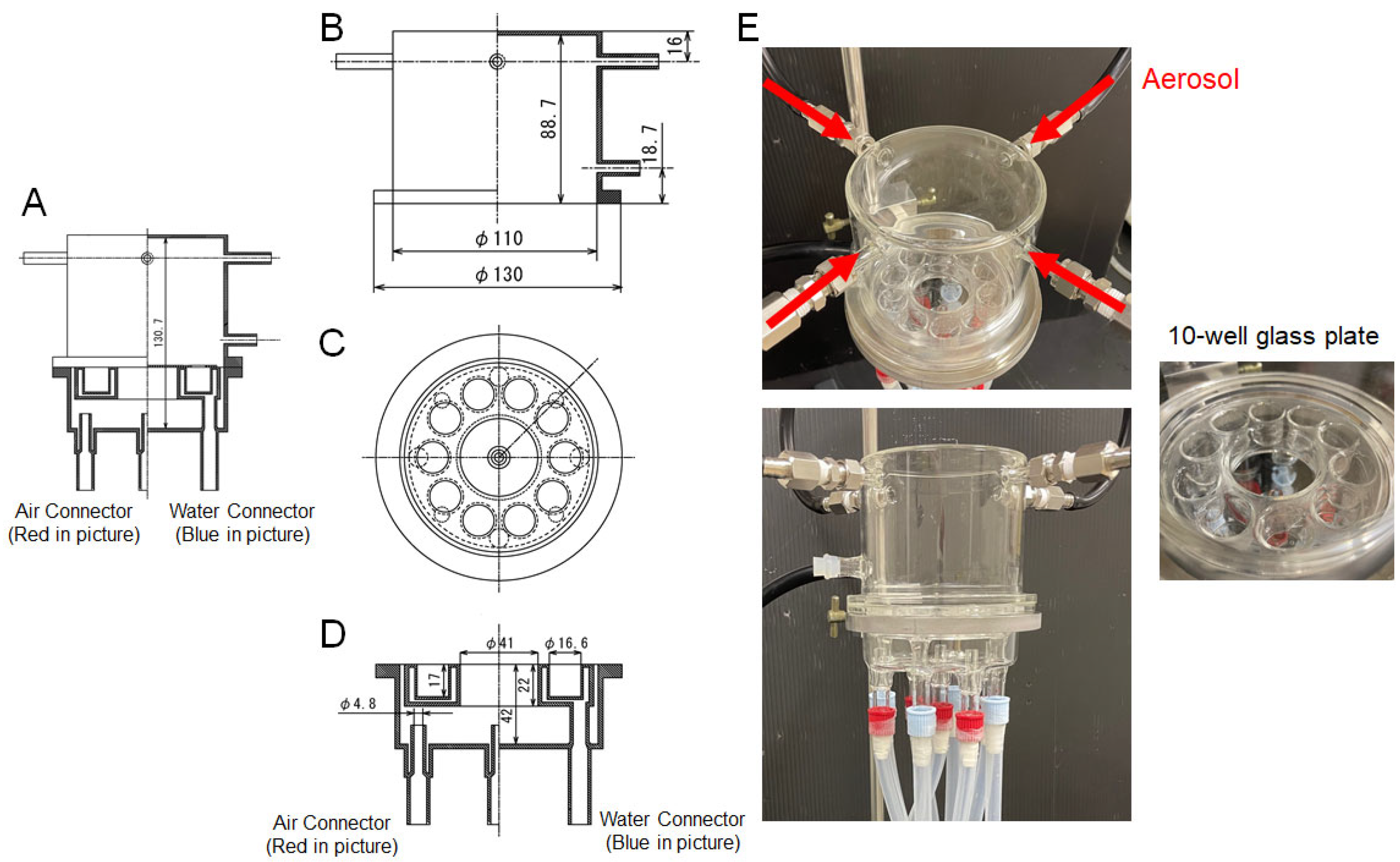
The air–liquid interface exposure system was constructed by modifying our previous system []. A suspension of PS NPs (0.51 μm, Polysciences, Warrington, PA, USA) in H2O was introduced into a mesh nebulizer head (NEB-CTRL18910; Emka Technologies, Paris, France) to transfer the PS NP mist. The suspension of 100 mL was nebulized for 1 h. A pump (MP-W5P; Sibata Scientific Technology, Tokyo, Japan) was operated at a rate of 4 L/min to direct the mist into the exposure chamber []. A particle counter (Model3889; KANOMAX, Osaka, Japan) was connected to the exposure chamber to monitor the PS mass concentration [].
A donut-shaped glass plate containing 10 wells was placed inside the exposure chamber, with water at 37 °C circulated through its interior via a chiller (Neslab RTE7; Thermo Fisher Scientific, Waltham, MA, USA). A flat, circular glass plate to fix the cell culture insert was placed on the plate to prevent aerosols from directly entering the medium. The bottom of the exposure chamber is covered with distilled water. Except for the glass plates, the system was maintained at room temperature (25 °C).
Figure 1.
A novel exposure chamber developed in this study. (A) Entire figure of exposure chamber. The numbers written in figures are the length (mm). Upper part (B), middle part (C) and lower part (D) of exposure chamber. (E) Picture of exposure chamber. The aerosol that enters into the chamber from 4 tubes (red arrows) is discharged from the chamber through 5 tubes at the bottom (red caps).
2.2. Calculation of Nano PS Mass Concentration
A particle counter (Model3889; KANOMAX, Osaka, Japan) was used to count particles with the diameters: 0.3–0.5 µm, 0.5–1 µm, as the PS NPs used in this study was approximately 0.5 µm. The total volume (VE) of the counted particles was calculated by approximating their shapes as spheres:
where Np-q is the number of particles with a diameter from p μm to q μm and Dx−y is the geometric mean of x and y.
VE (μm3) = 4π [(N0.3–0.5) · (D0.3–0.5)3 + (N0.5–1) · (D0.5–1)3]/3
A particle counter was used to count the number of particles in 0.283 L of air. Given that the density of PS is 1.05 g/cm3. Therefore, the mass concentration (M) of nano PS particles in the chamber was calculated using the following equation:
M (μg/m3) = (1.05 VE/0.283) · 1000
Spatial concentration measurements were repeated three times, and the average value was used for the above calculations.
2.3. Co-Culture of Calu-3 and U937
Human lung epithelial cells Calu-3 and human monocytes U937 were purchased from the American Type Culture Collection (HTB-55 and CRL-1593.2, respectively, Manassas, VA, USA) and were maintained using DMEM (Sigma-Aldrich, Burlington, MA, USA) with 10% fetal bovine serum (FBS, Nichirei Corporation, Tokyo, Japan) or RPMI-1640 (Sigma-Aldrich) with 10% FBS, respectively.
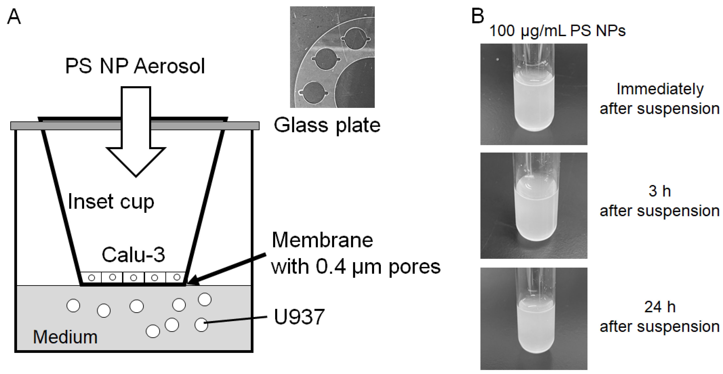
Calu-3 cells (2 × 10⁵ cells) were placed on Falcon cell culture insert (One Riverfront Plaza, Corning, NY, USA) and then cultured for 4 days under 5% CO2 at 37 °C until nano PS exposure. U937 cells cultured in RPMI-1640 buffered with 25 mM HEPES (pH 7.2) were added to the glass plate in the chamber (1 × 10⁵ cells in 500 μL medium), and then the cell culture insert with Calu-3 cells was moved for each well in the exposure chamber (Figure 2A). Calu-3 cells were covered with 50 μL medium. After aerosol PS NP exposure, both Calu-3 cells on a culture insert and U937 cells in RPMI media were moved to 24 well plate and subsequently cultured in a CO2 incubator at 37 °C for 23 h.
Figure 2.
Schematic diagram of co-culture of Calu-3 and U937 cells in the camber and PS NP suspension. (A) PS NP aerosol exposure to co-culture of Calu-3 and U937 cells. Note that a glass plate put on the well of culture plate to prevent PS NPs from directly entering the culture medium. (B) PS NPs were suspended in distilled water and then observed for up to 24 h.
2.4. Measurement of Trans-Epithelial Electrical Resistance (TEER)
Before moving the culture insert with Calu-3 to the exposure chamber, the medium in the culture insert was removed and then fresh culture medium was added into the culture insert. TEER was measured using a Millicell ERS-2 Voltohmmeter (Merck Millipore, Tokyo, Japan). After measurement, the medium was removed from the culture insert and 50 μL of fresh medium was added in it to start exposed to PS NP aerosol. After PS NP aerosol exposure and subsequent culture, the culture medium was added into the culture insert to measure TEER again.
2.5. Cell Viability Assessment
Cell viability was measured using a Lactate Assay Kit-WST (Dojindo Laboratories, Kumamoto, Japan) according to the manufacturer’s instructions. Calu-3 cells and U937 cells treated with 0.1% Triton X-100 (Sigma-Aldrich, Burlington, MA, USA) were used as controls to induce 100% cell death.
2.6. Determination of Reactive Species in the Medium
The hydrogen peroxide concentration in the culture medium was measured using Amplex Red (10-acetyl-3,7-dihydroxyphenoxazine, Cayman Chemical, Ann Arbor, MI, USA) as previously reported []. Nitrite in the culture medium was measured as an indicator of nitric oxide production using Griess assay []. Nitrite concentration in the samples was interpolated with a sodium nitrite standard curve ranging from 0 to 100 μΜ.
2.7. Total RNA Extraction and Real-Time PCR
mRNA levels were determined as previously described []. Primer sequences are listed in Table 1. The mRNA expression levels were normalized to GAPDH, and relative expression was calculated by dividing the values of treated samples by those of untreated controls.
Table 1.
Primers for real-time PCR.
2.8. Statistics
All data are presented as the mean ± standard deviation (S.D.). Statistical analyses were performed using the GraphPad Prism 10 software (GraphPad Software, San Diego, CA, USA). One-way analysis of variance (ANOVA) with Dunnett’s corrected multiple comparison test was used to determine significant differences between the means of the independent groups. Statistical significance was set at p < 0.05.
3. Results
3.1. Development of the Exposure Camber
We previously reported a PM2.5-aerosol exposure system for 3D epidermis and cornea []. This system can effectively produce PM2.5 of aerosol from a suspension of PM2.5, collected with a cyclone in powder form. However, because this system had a rectangular glass plate inside a rectangular exposure chamber and an aerosol was designed to pass through the rectangular chamber from the top left to the bottom right, there was concern about whether all wells would be equally exposed to aerosols. To address this limitation, we developed a novel exposure system, comprising a donut-shaped 10-well glass plate placed inside a cylindrical exposure chamber (Figure 1). Aerosol generated by a nebulizer was introduced into the cylindrical exposure chamber through the four upper tubes and discharged from the five lower exhaust pipes. The exhaust pipes were connected to a pump, and the aerosol was passed through the exposure chamber at a flow rate of 4 L/min. Because the PS NP mist flowed into the chamber from the nebulizer, and the bottom of the chamber was filled with water for humidification, the humidity inside the chamber was maintained at over 90%, indicating that this condition was sufficient for the culture of pulmonary epithelial cells at the air–liquid interface.
PS NPs were suspended in distilled water at concentrations of 0, 0.001, 0.01, 0.1 and 1 mg/mL, and each suspension was nebulized to generate PS NP-containing mist. The resulting mass concentrations of PS NPs in the chamber were 0, 0.18, 1.28, 10.5 and 23.6 μg/m3 generated by 0, 0.01, 0.01, 0.1 and 1 mg/mL PS NP suspensions, respectively. We used two concentrations of PS NP suspension, 0.1 and 1 mg/mL, to examine the effects of PS NP exposure on the pulmonary epithelium.
In this study, the human pulmonary epithelial cell line Calu-3 was cultured on the cell culture insert, and the human monocyte cell line U937 was cultured in the well. The structure of the co-culture mimicked the intravascular monocytes found beneath the alveolar epithelium. The cell culture inserts contained 0.4 μm pores, while the PS NPs used in this study had a diameter of 0.5 μm. Therefore, even if the Calu-3 cells were damaged, the exposed PS NPs would not reach the culture medium. Furthermore, the cell culture inserts fixed to a glass plate was placed on a glass culture plate to prevent PS NPs from directly entering the culture medium (Figure 2A).
PS NPs were suspended in water at a concentration of 100 μg/mL. No precipitation of PS NPs was observed even after leaving the suspension for 24 h (Figure 2B). Therefore, PS NPs are not suitable for submerged exposure, and because they are uniformly suspended, they are considered suitable for nebulization to generate uniform aerosols.
3.2. Effects of PS NP Exposure on Calu-3 Cells
Calu-3 cells were seeded onto a cell culture insert and cultured until they reached confluence. U937 cells were added to the wells of a glass culture plate, and a cell culture insert with Calu-3 cells was placed in the well. PS NP suspensions at concentrations of 0.1 mg/mL (10.5 μg/m3) and 1 mg/mL (23.6 μg/m3) were nebulized and exposed to the cells for 1 h. The cell culture inserts with Calu-3 and U937 cells were transferred to a culture plate with medium, and the plate was incubated in a CO2 incubator for 23 h. PS NP aerosol exposure did not affect cellular viability, as evaluated by LDH release (Figure 3A). TEER was decreased by PS NP aerosols in a mass concentration-dependent manner, indicating epithelial barrier dysfunction induced by PS NP aerosol (Figure 3B).
Figure 3.
Effects of PS NP aerosol exposure on epithelial barriers. The Calu-3 and U937 cells were cultured on glass plates in an exposure chamber. Cells were exposed to PS NP aerosol for 1 h and then cultured in an incubator with a CO2 atmosphere for 23 h. (A) Cell viability was measured by the LDH assay. The reported values are the mean ± S.D. (n = 6 in each group). (B) TEER was measured and TEER values before exposure set to 100% to calculate relative TEER values. The reported values are the mean ± S.D. (n = 6 in each group). Data were analyzed using one-way ANOVA [F(2, 15) = 15.20, p = 0.0002] with Dunnett’s corrected multiple comparison test.
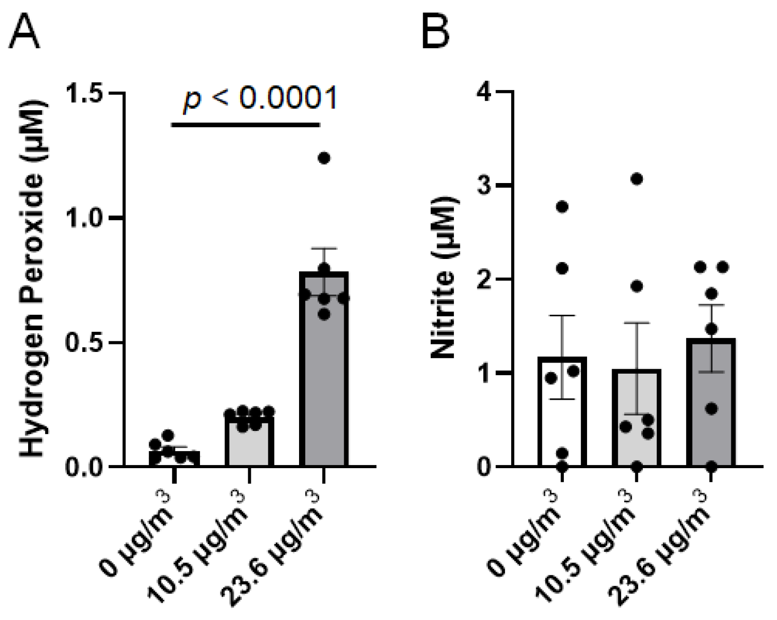
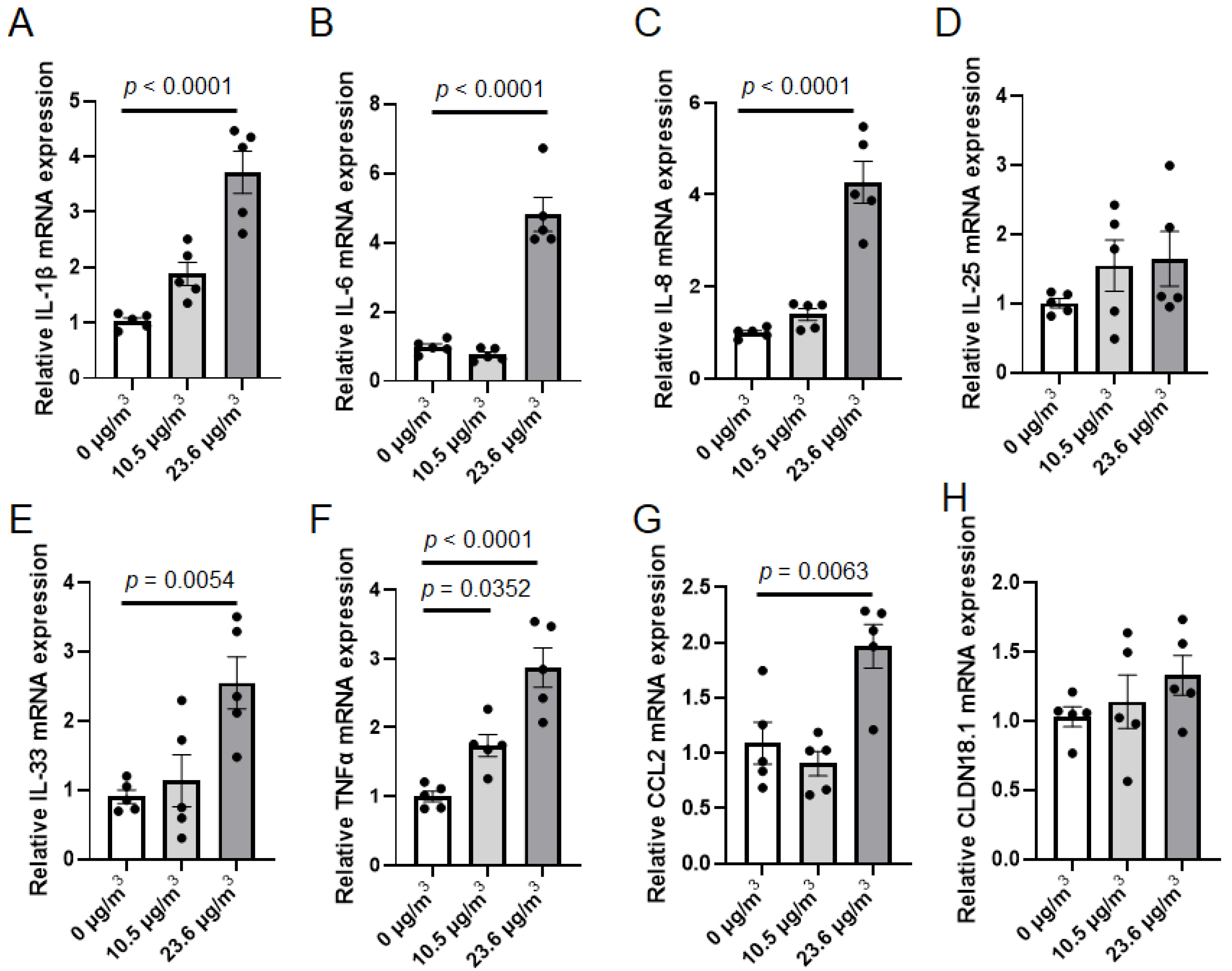
MPs have been reported to induce pulmonary damage, mainly through oxidative stress and inflammation []. Therefore, we examined oxidative stress and inflammation in the culture system after exposure to PS NP aerosols. The concentration of hydrogen peroxide in the culture media dose-dependently increased by PS NP exposure (Figure 4A). However, there was no difference in the nitrite levels in the culture media between the water- and PS NP nebulized groups (Figure 4B). Therefore, oxidative stress can be induced by PS NP aerosols but is insufficient to cause cell death. To assess inflammatory reactions, we selected epithelial pro-inflammatory molecules, interleukin-1β (IL-1β), IL-6, IL-8, IL-33, tumor necrosis factor α (TNFα) and CCL2 to be investigated. In addition, we measured the mRNA expression of IL-25 and claudin 18.1 (CLDN18.1) because the expression of these two mRNA was upregulated by urban aerosol exposure in our previous study []. mRNA levels of IL-1β, IL-6, IL-8, IL-33 and TNFα increased in Calu-3 cells by exposure to PS NP aerosol in a mass concentration-dependent manner (Figure 5A–C,E,F). Exposure to PS NP aerosols did not affect the expression of IL-25 and CLDN18.1 in Calu-3 cells. Taken together, PS NP aerosols are thought to induce oxidative stress and inflammatory reactions in epithelial cells, decreasing barrier function.
Figure 4.
Oxidative stress induced by PS NP aerosol exposure. The Calu-3 and U937 cells were cultured on glass plates in an exposure chamber. Cells were exposed to PS NP aerosols for 1 h and then cultured in an incubator with a CO2 atmosphere for 23 h. (A) Hydrogen peroxide in the culture media was measured using the Amplex Red assay. The reported values are the mean ± S.D. (n = 6 in each group). Data were analyzed using one-way ANOVA [F(2, 15) = 46.66, p < 0.0001] with Dunnett’s corrected multiple comparison test. (B) Nitric oxide generation was measured as an index of nitrite in the culture media. The reported values are the mean ± S.D. (n = 6 in each group).
Figure 5.
Inflammatory reaction in Calu-3 cells induced by PS NP aerosol exposure. The Calu-3 and U937 cells were cultured on glass plates in an exposure chamber. Cells were exposed to PS NP aerosol for 1 h, and then the cells were cultured in an incubator with a CO2 atmosphere for 23 h. Total RNA was extracted from Calu-3 cells and then real-time PCR was performed to determine mRNA levels of (A) IL-1β, (B) IL-6, (C) IL-8, (D) IL-25, (E) IL-33, (F) TNFα, (G) CCL2 and (H) CLDN18.1. The reported values are the mean ± S.D. (n = 5 in each group). Data were analyzed using one-way ANOVA [A: F(2, 12) = 29.25, p < 0.0001, B: F(2, 12) = 60.20, p < 0.0001, C: F(2, 12) = 42.45, p < 0.0001, D: F(2, 12) = 1.202, p = 0.3344, E: F(2, 12) = 8.133, p = 0.0059, F: F(2, 12) = 23.49, p < 0.0001, G: F(2, 12) = 11.08, p = 0.0019, H: F(2, 12) = 1.082, p = 0.3698] with Dunnett’s corrected multiple comparison test.
3.3. Effects of PS NP Exposure on U937 Cells Under Cell Culture Insert with Calu-3 Cells
In this system, U937 cells were cultured in wells under cell culture inserts with Calu-3 cells. The exposed PS NP aerosol did not reach the U937 cells because of the limited pore size of the cell culture insert and glass plate on the well (Figure 2). On the other hand, molecules released from Calu-3 cells can act on U937 cells that pass through the membrane under cell culture inserts.
Results of real-time PCR indicated that mRNA expression of IL-1β and TNFα decreased by PS NP aerosol exposure (Figure 6A,D). However, the mRNA expression of IL-8 and CCL2 was elevated by high mass concentrations of PS NP aerosol (Figure 6C,E). Levels of IL-6 mRNA did were not affected by PS NP aerosol (Figure 6B).
Figure 6.
Inflammatory reaction in U937 cells via PS NP aerosol exposure to Calu-3. The Calu-3 and U937 cells were cultured on glass plates in an exposure chamber. Cells were exposed to PS NP aerosol for 1 h, and then the cells were cultured in an incubator with a CO2 atmosphere for 23 h. Total RNA was extracted from U937 cells and then real-time PCR was performed to determine mRNA levels of (A) IL-1β, (B) IL-6, (C) IL-8, (D) TNFα and (E) CCL2. The reported values are the mean ± S.D. (n = 5 in each group). Data were analyzed using one-way ANOVA [A: F(2, 12) = 8.877, p = 0.0043, B: F(2, 12) = 1.739, p = 0.2172, C: F(2, 12) = 77.55, p < 0.0001, D: F(2, 12) = 18.53, p = 0.0002, E: F(2, 12) = 48.39, p < 0.0001] with Dunnett’s corrected multiple comparison test.
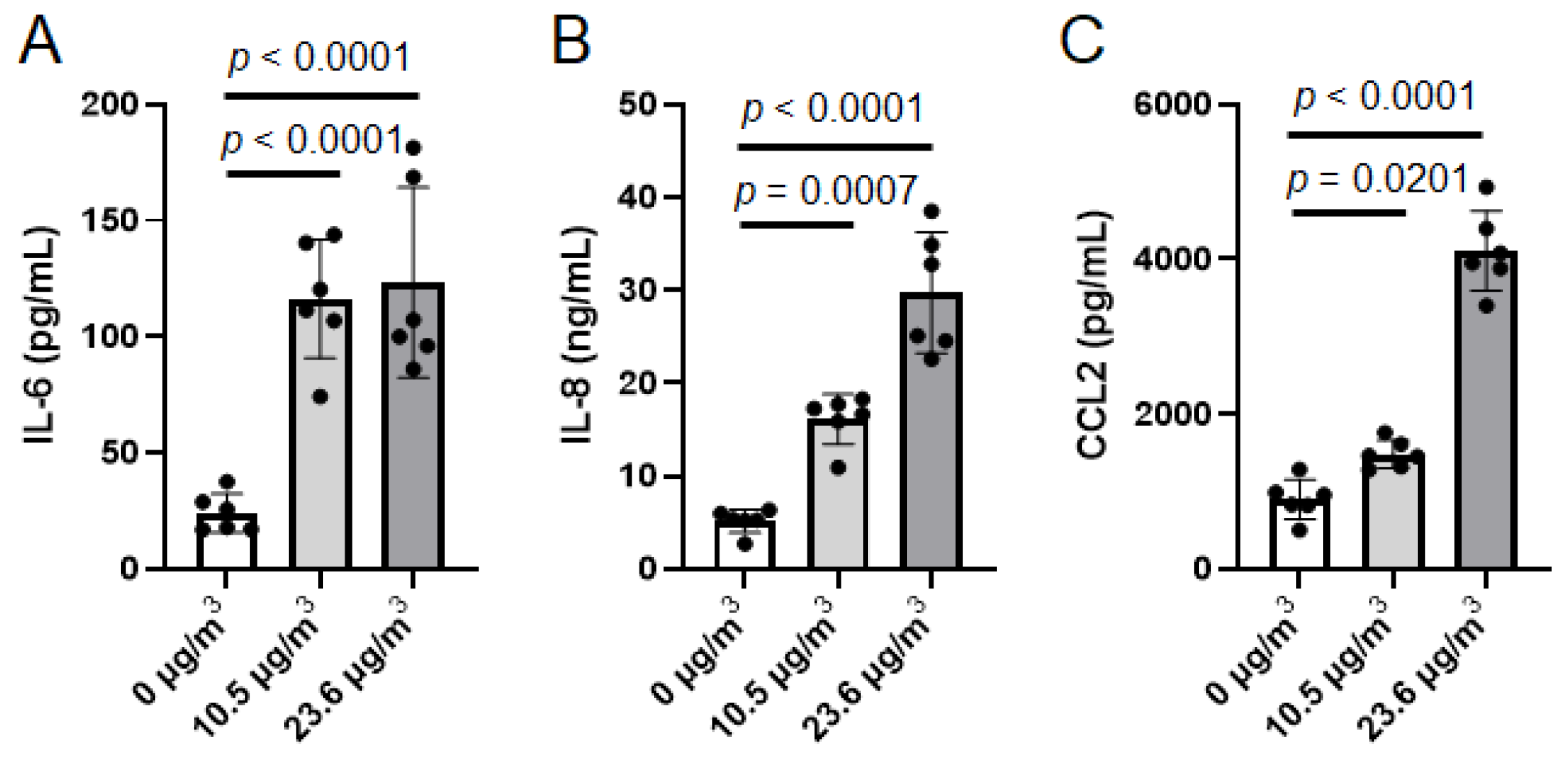
The culture medium was affected by both Calu-3 and U937 cells; the molecules released by the Calu-3 and U937 cells were mixed into the media. When we measured several cytokines/chemokines in the culture media using a PLEX assay, the levels of IL-6, IL-8, and CCL2 were increased by PS NP aerosol exposure (Figure 7). IL-1β and TNFα were below the detection limit in control and PS NP-exposed samples. In this regard, it is considered that Calu-3 cells released several cytokines/chemokines into the media in response to PS NP aerosols, whereas the contribution of U937 cells to the cytokine/chemokine levels in the media was relatively small.
Figure 7.
Release of inflammatory molecules to the culture medium. The Calu-3 and U937 cells were cultured on glass plates in an exposure chamber. Cells were exposed to the PS NP aerosol for 1 h and then cultured in an incubator under a CO2 atmosphere for 23 h. The culture medium was collected and (A) IL-6, (B) IL-8, and (C) CCL2 levels were determined using a multiplex assay. The reported values are the mean ± S.D. (n = 6 in each group). Data were analyzed using one-way ANOVA [A: F(2, 15) = 23.09, p < 0.0001; B: F(2, 15) = 53.44, p < 0.0001; C: F(2, 15) = 144.4, p < 0.0001] with Dunnett’s corrected multiple comparison test.
4. Discussion
In this study, we developed a simple ALI system for exposure to NP aerosols. The PS NP aerosol mass concentration in the exposure chamber was controlled by changing the concentration of the PS NP suspension. When the PS NP aerosol was exposed to a Calu-3-U937 co-culture system, cell death was not detected; however, oxidative stress and an inflammatory response were induced, resulting in a decrease in the TEER of the Calu-3 layer, indicating damage to the alveolar barrier.
Various ALI exposure systems have been used to investigate the respiratory effects of aerosols. As exposure of the ALI system to dry air causes cell death, a humidifier is used to increase humidity during exposure. Alternatively, some systems use atomizers or nebulizers to generate humidified aerosols. Commonly used models include the CULTEX system, in which the aerosol is humidified using a Naficon humidifier and exposed to culture dishes using insert cups [] and the VITROCELL system, in which a mist containing nanoparticles is exposed to culture dishes []. Recently, the VITROCELL Cloud System, which uses a nebulizer unit attached to the top of a chamber to expose the multi-well culture plates inside the chamber, was used []. In the DAVID system, dry aerosols are condensed in a water-condensation region around the aerosol nucleus, creating droplets that are then exposed to cells []. However, regulating the mass concentration of nanomaterial aerosols in these systems is difficult. Furthermore, if the mist is directly exposed to ALI, the cells become covered with water depending on the exposure duration. In our system, the mist generated by a nebulizer was drawn into the exposure chamber from four directions by a pump, ensuring uniform exposure of the aerosol to the cells in a doughnut-shaped culture plate. In addition, the chamber was humidified with water to ensure that the humidity levels exceeded 90% during exposure. However, the MatriGrid culture device appears to be capable of exposing ALI to dry aerosols directly from a diffusion dryer []. Thus, this system may not be useful for ALI culture of alveolar epithelium.
In addition to aqueous solutions, organic solvents have been used to generate aerosols. Silver nanoparticle aerosols are produced by dissolving silver nitrate in methanol, atomizing it, and then heating the flow path to volatilize the methanol []. Nanoparticles were generated by applying a high voltage between the electrodes using a spark generator, and their mass concentration was varied by changing the discharge frequency []. However, these methods limit the generation of aerosols from metals. The use of a heated path makes it impossible to produce aerosols from heat-sensitive polymers, such as plastics. Atomization of aqueous solutions or suspensions is highly versatile, as it can be used to produce aerosols of any substance.
Savi et al. [] developed a system in which aerosols were electrically charged using a particle charger, and electrodes were placed at the bottom of the ALI chamber, to deposit aerosols onto cultured cells by electrostatic force. In their report, although particle deposition into the wells was confirmed, exposure did not induce cell death or inflammatory responses, even when alveolar macrophages were used. In this study, a pump was used to generate an airflow from the top to the bottom of the exposure chamber to promote deposition. The inner diameter of the exposure chamber was 110 mm, and the pump flow rate was 4 L/min and thus, the flux was calculated as 7.0 mm/sec. Since the mass concentration in the chamber between nebulization of 1 mg/mL PS NPs was nebulized was 23.6 μg/m3, the deposition rate per hour was calculated as 59 ng/cm2/h. However, dry deposition is primarily governed by turbulence, and for submicron particles, the deposition coefficient is estimated to be approximately 0.1 mm/sec using the aerosol transport model []. Actual measurement of the deposition amount using electron microscopes or quartz crystal microbalances will be a challenge in the future.
Han et al. recently reported that PS NPs induced oxidative stress and inflammation in alveolar epithelial cells primarily through integrin α5β1-mediated endocytosis, indicating the internalization of polystyrene nanoparticles inside cells []. PS NPs elicited mitochondrial Ca2+ dysfunction and depolarization, leading to the production of ROS. ROS are well known to activate NF-κB transcription factor, which induces transcription of inflammatory molecules including cytokines and chemokines. Liu et al. mentioned more specific signaling: DNA damages induced by PS NPs activate the cyclic GMP-AMP synthase-stimulator of interferon genes-signaling pathway, which leads to NF-κB activation []. These signaling might be induced in our exposure experiments.
We previously reported that urban aerosol exposure in the Calu-3 ALI system increased the expression of IL-25 and CLDN18.1 []. However, in the present study, PS NP aerosol exposure did not alter the expression of IL-25 and CLDN18.1. Therefore, these markers were not used as markers of PS exposure. PS NP aerosols increases the expression and release of pro-inflammatory cytokines and chemokines in Calu-3 cells. However, these changes are not thought to be specific to PS NP because exposure to PM2.5 increases the expression of pro-inflammatory cytokines []. A comprehensive analysis is required to identify the markers specific to plastic exposure. Furthermore, while PS NP exposure increased inflammatory markers in Calu-3 cells, it decreased the expression of inflammatory cytokines such as IL-1β and TNFα in U937 cells. Although the alveolar epithelium is known to exhibit pro-inflammatory responses, as demonstrated in this study, it also possess anti-inflammatory properties. The aryl hydrocarbon receptor, expressed in alveolar epithelial cells, becomes activated during acute lung injury and suppress the expression of inflammatory cytokines []. Insulin-like growth factor 1 inhibits the activation of p38 mitogen-activated protein kinase in alveolar epithelial cells and subsequently reduces the expression of inflammatory molecules such as IL-1β, TNF-α and CCL2 []. Prostaglandin E2 and suppressor of cytokine signaling 3, both secreted by the alveolar epithelium and suppress inflammatory responses []. These inhibitory signals are thought to protect tissues by suppressing excessive inflammatory responses of immune cells such as alveolar macrophages. In this study, Calu-3-derived anti-inflammatory factors may have acted on U937 cells to suppress the expression of several pro-inflammatory cytokines.
Surfactants, mainly composed of lipids, are released from alveolar epithelial cells. Geiser et al. reported that alveolar surface tension affects fine particle retention []. Manmade vitreous fibers 10a were immersed in the aqueous subphase by approximately 50% at film surface tensions of 20–25 mJ/m2 and submerged at approximately 10 mJ/m2. This report suggests that surfactants play an important role in nanomaterial-induced toxicity. Recently, surfactants have been reported to inhibit nanoparticle uptake into the epithelium []. In this study, we added small amount (50 μL) of culture medium on a Calu-3 layer before PS NP exposure. These conditions are insufficient to reproduce the function of a surfactant, and experiments based on the composition of the surfactant components are necessary. Recently, novel ALI systems such as EpiAirway by MatTek and MucilAirTM by Epithelix Sarl have been developed. These systems may be useful for examining the effects of sustained exposure to nanomaterials on respiratory damage. One challenge is to make ALI culture more realistic and create a more accurate safety evaluation system.
In conclusion, we developed a simple and easy-to-use ALI system that can be used for aerosol exposure. PS NP aerosols induced oxidative stress, inflammatory responses, and barrier dysfunction in Calu-3 cells cultured in ALI with U937 cells. We believe that our system can be used for the safety assessment of nanomaterials including NPs.
Author Contributions
Conceptualization: M.F. and Y.I.; methodology: I.O. and Y.I.; investigation: I.O., Y.W., M.F. and Y.I.; visualization: I.O. and Y.I.; funding acquisition: Y.I.; project administration: M.F. and Y.I.; writing—original draft: I.O. and Y.I.; writing—review and editing: all authors. All authors have read and agreed to the published version of the manuscript.
Funding
This study was supported by a KAKENHI grant from the Japan Society for the Promotion of Science (grant number 24K03085 to Y.I.), the Environment Research and Technology Development Funds of the Environmental Restoration and Conservation Agency of Japan (JPMEERF20215003 and JPMEERF20245004 to Y.I.), the Steel Foundation for Environmental Protection Technology (to Y.I.), the Smoking Research Foundation (to Y.I.), and a Mandom International Research Grant (to Y.I.).
Institutional Review Board Statement
Not applicable.
Informed Consent Statement
Not applicable.
Data Availability Statement
All data are available upon request to the corresponding author.
Conflicts of Interest
The authors declare that they have no competing financial interests or personal relationships that may have influenced the work reported in this study.
References
- Dris, R.; Gasperi, J.; Rocher, V.; Saad, M.; Renault, N.; Tassin, B. Microplastic contamination in an urban area: A case study in Greater Paris. Environ. Chem. 2015, 12, 592–599. [Google Scholar] [CrossRef]
- Zarus, G.M.; Muianga, C.; Hunter, C.M.; Pappas, R.S. A review of data for quantifying human exposures to micro and nanoplastics and potential health risks. Sci. Total Environ. 2021, 756, 144010. [Google Scholar] [CrossRef]
- Jenner, L.C.; Rotchell, J.M.; Bennett, R.T.; Cowen, M.; Tentzeris, V.; Sadofsky, L.R. Detection of microplastics in human lung tissue using muFTIR spectroscopy. Sci. Total Environ. 2022, 831, 154907. [Google Scholar] [CrossRef]
- Morioka, T.; Tanaka, S.; Kohama-Inoue, A.; Watanabe, A. The quantification of the airborne plastic particles of 0.43-11 mum: Procedure development and application to atmospheric environment. Chemosphere 2024, 351, 141131. [Google Scholar] [CrossRef]
- Yuan, C.; Li, X.; Lu, C.; Sun, L.; Fan, C.; Fu, M.; Wang, H.; Duan, M.; Xia, S. Micro/nanoplastics in the Shenyang city atmosphere: Distribution and sources. Environ. Pollut. 2025, 372, 126027. [Google Scholar] [CrossRef]
- Luo, D.; Chu, X.; Wu, Y.; Wang, Z.; Liao, Z.; Ji, X.; Ju, J.; Yang, B.; Chen, Z.; Dahlgren, R.; et al. Micro- and nano-plastics in the atmosphere: A review of occurrence, properties and human health risks. J. Hazard. Mater. 2024, 465, 133412. [Google Scholar] [CrossRef]
- Cao, J.; Xu, R.; Geng, Y.; Xu, S.; Guo, M. Exposure to polystyrene microplastics triggers lung injury via targeting toll-like receptor 2 and activation of the NF-kappaB signal in mice. Environ. Pollut. 2023, 320, 121068. [Google Scholar] [CrossRef]
- Li, X.; Zhang, T.; Lv, W.; Wang, H.; Chen, H.; Xu, Q.; Cai, H.; Dai, J. Intratracheal administration of polystyrene microplastics induces pulmonary fibrosis by activating oxidative stress and Wnt/beta-catenin signaling pathway in mice. Ecotoxicol. Environ. Saf. 2022, 232, 113238. [Google Scholar] [CrossRef]
- Ishihara, Y.; Kajino, M.; Iwamoto, Y.; Nakane, T.; Nabetani, Y.; Okuda, T.; Kono, M.; Okochi, H. Impact of artificial sunlight aging on the respiratory effects of polyethylene terephthalate microplastics through degradation-mediated terephthalic acid release in male mice. Toxicol. Sci. 2025, 203, 242–252. [Google Scholar] [CrossRef]
- Wang, Z.; Wang, N.; Jiang, S.; Shi, R.; Wang, R.; Wu, W. Polystyrene nanoplastics induced lung injury in mice: Insights into lung metabolic disorders. Ecotoxicol. Environ. Saf. 2025, 303, 119060. [Google Scholar] [CrossRef] [PubMed]
- Wang, Q.; He, W.; Zhou, Y.; Feng, R.; Wang, Y.; Liu, L.; Yuan, Y.; Dai, J.; Liu, Y.; Zhang, X. Polystyrene nanoplastics aggravate house dust mite induced allergic airway inflammation through EGFR/ERK-dependent lung epithelial barrier dysfunction. Ecotoxicol. Environ. Saf. 2025, 298, 118329. [Google Scholar] [CrossRef]
- Jian, X.; Zhang, X.; Chang, S.; Xue, Y.; Shang, P.; Liu, Y.; Chen, H.; Zhou, X.; Wang, W.; Wang, P.; et al. Co-exposure of polystyrene nanoplastics and ozone synergistically induced airway inflammation: Evidence and biomarkers screening. Ecotoxicol. Environ. Saf. 2025, 302, 118643. [Google Scholar] [CrossRef]
- Brouwer, H.; Porbahaie, M.; Boeren, S.; Busch, M.; Bouwmeester, H. The in vitro gastrointestinal digestion-associated protein corona of polystyrene nano- and microplastics increases their uptake by human THP-1-derived macrophages. Part. Fibre Toxicol. 2024, 21, 4. [Google Scholar] [CrossRef] [PubMed]
- Ghosal, S.; Bag, S.; Bhowmik, S. Insights into the Binding Interactions between Microplastics and Human alpha-Synuclein Protein by Multispectroscopic Investigations and Amyloidogenic Oligomer Formation. J. Phys. Chem. Lett. 2024, 15, 6560–6567. [Google Scholar] [CrossRef] [PubMed]
- Forest, V.; Pourchez, J. Can the impact of micro- and nanoplastics on human health really be assessed using in vitro models? A review of methodological issues. Environ. Int. 2023, 178, 108115. [Google Scholar] [CrossRef] [PubMed]
- Kono, M.; Takaishi, M.; Okuda, T.; Fujihara, M.; Noguchi, S.; Ishihara, Y. A simple air-liquid interface exposure system for exposing cultured human 3D epidermis and cornea to PM2.5 collected through cyclonic separation. J. Toxicol. Sci. 2024, 49, 61–68. [Google Scholar] [CrossRef]
- Ishihara, Y.; Tsuji, M.; Kawamoto, T.; Yamazaki, T. Involvement of reactive oxygen species derived from mitochondria in neuronal injury elicited by methylmercury. J. Clin. Biochem. Nutr. 2016, 59, 182–190. [Google Scholar] [CrossRef]
- Green, L.C.; Wagner, D.A.; Glogowski, J.; Skipper, P.L.; Wishnok, J.S.; Tannenbaum, S.R. Analysis of nitrate, nitrite, and [15N] nitrate in biological fluids. Anal. Biochem. 1982, 126, 131–138. [Google Scholar] [CrossRef]
- Ishihara, Y.; Kawami, T.; Ishida, A.; Yamazaki, T. Tributyltin induces oxidative stress and neuronal injury by inhibiting glutathione S-transferase in rat organotypic hippocampal slice cultures. Neurochem. Int. 2012, 60, 782–790. [Google Scholar] [CrossRef]
- Huang, Y.; Shang, P.; Li, Y.; Wang, Y. Lung hazards of microplastics and their toxicological mechanisms. Environ. Pollut. 2025, 385, 127149. [Google Scholar] [CrossRef]
- Tanaka, Y.; Wada, Y.; Fujihara, M.; Ishihara, N.; Nakane, T.; Ishihara, Y. Development of a Novel Air-Liquid Interface Exposure System for Particle Exposure in Alveolar Epithelium. Altern. Anim. Test. Exp. 2025, 30, 41–49. [Google Scholar] [CrossRef]
- Buckley, A.; Guo, C.; Laycock, A.; Cui, X.; Belinga-Desaunay-Nault, M.F.; Valsami-Jones, E.; Leonard, M.; Smith, R. Aerosol exposure at air-liquid-interface (AE-ALI) in vitro toxicity system characterisation: Particle deposition and the importance of air control responses. Toxicol. Vitr. 2024, 100, 105889. [Google Scholar] [CrossRef] [PubMed]
- Steiner, S.; Majeed, S.; Kratzer, G.; Vuillaume, G.; Hoeng, J.; Frentzel, S. Characterization of the Vitrocell(R) 24/48 aerosol exposure system for its use in exposures to liquid aerosols. Toxicol. Vitr. 2017, 42, 263–272. [Google Scholar] [CrossRef] [PubMed]
- Leroux, M.M.; Hocquel, R.; Bourge, K.; Kokot, B.; Kokot, H.; Koklic, T.; Strancar, J.; Ding, Y.; Kumar, P.; Schmid, O.; et al. Aerosol-Cell Exposure System Applied to Semi-Adherent Cells for Aerosolization of Lung Surfactant and Nanoparticles Followed by High Quality RNA Extraction. Nanomaterials 2022, 12, 1362. [Google Scholar] [CrossRef]
- Tilly, T.B.; Ward, R.X.; Luthra, J.K.; Robinson, S.; Eiguren-Fernandez, A.; Lewis, G.S.; Salisbury, R.L.; Lednicky, J.A.; Sabo-Attwood, T.L.; Hussain, S.M.; et al. Condensational particle growth device for reliable cell exposure at the air-liquid interface to nanoparticles. Aerosol Sci. Technol. 2019, 53, 1415–1428. [Google Scholar] [CrossRef]
- Kustner, M.J.; Eckstein, D.; Brauer, D.; Mai, P.; Hampl, J.; Weise, F.; Schuhmann, B.; Hause, G.; Glahn, F.; Foth, H.; et al. Modular air-liquid interface aerosol exposure system (MALIES) to study toxicity of nanoparticle aerosols in 3D-cultured A549 cells in vitro. Arch. Toxicol. 2024, 98, 1061–1080. [Google Scholar] [CrossRef]
- Ihalainen, M.; Jalava, P.; Ihantola, T.; Kasurinen, S.; Uski, O.; Sippula, O.; Hartikainen, A.; Tissari, J.; Kuuspalo, K.; Lähde, A.; et al. Design and validation of an air-liquid interface (ALI) exposure device based on thermophoresis. Aerosol Sci. Technol. 2019, 53, 133–145. [Google Scholar] [CrossRef]
- Kapp, N.; Kreyling, W.; Schulz, H.; Im Hof, V.; Gehr, P.; Semmler, M.; Geiser, M. Electron energy loss spectroscopy for analysis of inhaled ultrafine particles in rat lungs. Microsc. Res. Tech. 2004, 63, 298–305. [Google Scholar] [CrossRef]
- Savi, M.; Kalberer, M.; Lang, D.; Ryser, M.; Fierz, M.; Gaschen, A.; Ricka, J.; Geiser, M. A novel exposure system for the efficient and controlled deposition of aerosol particles onto cell cultures. Environ. Sci. Technol. 2008, 42, 5667–5674. [Google Scholar] [CrossRef]
- Petroff, A.; Zhang, L. Development and validation of a size-resolved particle dry deposition scheme for application in aerosol transport models. Geosci. Model. Dev. 2010, 3, 753–769. [Google Scholar] [CrossRef]
- Han, M.; Liang, J.; Wang, K.; Si, Q.; Zhu, C.; Zhao, Y.; Khan, N.A.K.; Abdullah, A.L.B.; Shau-Hwai, A.T.; Li, Y.M.; et al. Integrin A5B1-mediated endocytosis of polystyrene nanoplastics: Implications for human lung disease and therapeutic targets. Sci. Total Environ. 2024, 953, 176017. [Google Scholar] [CrossRef]
- Liu, J.; Xu, F.; Guo, M.; Gao, D.; Song, Y. Nasal instillation of polystyrene nanoplastics induce lung injury via mitochondrial DNA release and activation of the cyclic GMP-AMP synthase-stimulator of interferon genes-signaling cascade. Sci. Total Environ. 2024, 948, 174674. [Google Scholar] [CrossRef]
- Goksel, O.; Sipahi, M.I.; Yanasik, S.; Saglam-Metiner, P.; Benzer, S.; Sabour-Takanlou, L.; Sabour-Takanlou, M.; Biray-Avci, C.; Yesil-Celiktas, O. Comprehensive analysis of resilience of human airway epithelial barrier against short-term PM2.5 inorganic dust exposure using in vitro microfluidic chip and ex vivo human airway models. Allergy 2024, 79, 2953–2965. [Google Scholar] [CrossRef]
- Zimmerman, E.; Sturrock, A.; Reilly, C.A.; Burrell-Gerbers, K.L.; Warren, K.; Mir-Kasimov, M.; Zhang, M.A.; Pierce, M.S.; Helms, M.N.; Paine, R., 3rd. Aryl Hydrocarbon Receptor Activation in Pulmonary Alveolar Epithelial Cells Limits Inflammation and Preserves Lung Epithelial Cell Integrity. J. Immunol. 2024, 213, 600–611. [Google Scholar] [CrossRef]
- Mu, M.; Gao, P.; Yang, Q.; He, J.; Wu, F.; Han, X.; Guo, S.; Qian, Z.; Song, C. Alveolar Epithelial Cells Promote IGF-1 Production by Alveolar Macrophages Through TGF-beta to Suppress Endogenous Inflammatory Signals. Front. Immunol. 2020, 11, 1585. [Google Scholar] [CrossRef]
- Speth, J.M.; Bourdonnay, E.; Penke, L.R.; Mancuso, P.; Moore, B.B.; Weinberg, J.B.; Peters-Golden, M. Alveolar Epithelial Cell-Derived Prostaglandin E2 Serves as a Request Signal for Macrophage Secretion of Suppressor of Cytokine Signaling 3 during Innate Inflammation. J. Immunol. 2016, 196, 5112–5120. [Google Scholar] [CrossRef] [PubMed]
- Geiser, M.; Matter, M.; Maye, I.; Im Hof, V.; Gehr, P.; Schurch, S. Influence of airspace geometry and surfactant on the retention of man-made vitreous fibers (MMVF 10a). Environ. Health Perspect. 2003, 111, 895–901. [Google Scholar] [CrossRef]
- Radiom, M.; Sarkis, M.; Brookes, O.; Oikonomou, E.K.; Baeza-Squiban, A.; Berret, J.F. Pulmonary surfactant inhibition of nanoparticle uptake by alveolar epithelial cells. Sci. Rep. 2020, 10, 19436. [Google Scholar] [CrossRef]
Disclaimer/Publisher’s Note: The statements, opinions and data contained in all publications are solely those of the individual author(s) and contributor(s) and not of MDPI and/or the editor(s). MDPI and/or the editor(s) disclaim responsibility for any injury to people or property resulting from any ideas, methods, instructions or products referred to in the content. |
© 2025 by the authors. Licensee MDPI, Basel, Switzerland. This article is an open access article distributed under the terms and conditions of the Creative Commons Attribution (CC BY) license (https://creativecommons.org/licenses/by/4.0/).