Next Issue
Volume 6, June
Previous Issue
Volume 5, December


Cells, Volume 6, Issue 1 (March 2017) – 8 articles

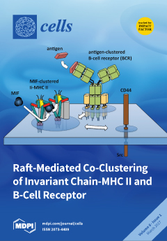
Cover Story (view full-size image): Invariant chain trimers on B-lymphocytes are often associated with less than par amounts of MHC II and, therefore, provide binding sites for the cytokine MIF. Under sub-saturation conditions, trimeric MIF is expected to engage other invariant chain-MHC II complexes, resulting in clustering and membrane raft formation. Antigen-clustered B-cell receptors form similar membrane rafts that coalesce with rafts containing MIF-invariant chain-MHC II. This coalescence fosters signaling and co-endocytosis, and thus may strengthen B-cell receptor-guided antigen presentation. View this paper
  • Issues are regarded as officially published after their release is announced to the table of contents alert mailing list.
  • You may sign up for e-mail alerts to receive table of contents of newly released issues.
  • PDF is the official format for papers published in both, html and pdf forms. To view the papers in pdf format, click on the "PDF Full-text" link, and use the free Adobe Reader to open them.
Order results
Result details
Section
Select all
Export citation of selected articles as:
8465 KiB  
Article
KDM2 Family Members are Regulated by HIF-1 in Hypoxia
by Michael Batie, Jimena Druker, Laura D’Ignazio and Sonia Rocha
Cells 2017, 6(1), 8; https://doi.org/10.3390/cells6010008 - 17 Mar 2017
Cited by 32 | Viewed by 7985
Abstract
Hypoxia is not only a developmental cue but also a stress and pathological stimulus in many human diseases. The response to hypoxia at the cellular level relies on the activity of the transcription factor family, hypoxia inducible factor (HIF). HIF-1 is responsible for [...] Read more.
Hypoxia is not only a developmental cue but also a stress and pathological stimulus in many human diseases. The response to hypoxia at the cellular level relies on the activity of the transcription factor family, hypoxia inducible factor (HIF). HIF-1 is responsible for the acute response and transactivates a variety of genes involved in cellular metabolism, cell death, and cell growth. Here, we show that hypoxia results in increased mRNA levels for human lysine (K)-specific demethylase 2 (KDM2) family members, KDM2A and KDM2B, and also for Drosophila melanogaster KDM2, a histone and protein demethylase. In human cells, KDM2 family member’s mRNA levels are regulated by HIF-1 but not HIF-2 in hypoxia. Interestingly, only KDM2A protein levels are significantly induced in a HIF-1-dependent manner, while KDM2B protein changes in a cell type-dependent manner. Importantly, we demonstrate that in human cells, KDM2A regulation by hypoxia and HIF-1 occurs at the level of promoter, with HIF-1 binding to the KDM2A promoter being required for RNA polymerase II recruitment. Taken together, these results demonstrate that KDM2 is a novel HIF target that can help coordinate the cellular response to hypoxia. In addition, these results might explain why KDM2 levels are often deregulated in human cancers. Full article
(This article belongs to the Section Cell Nuclei: Function, Transport and Receptors)
Show Figures

Figure 1

1213 KiB  
Review
Application of Induced Pluripotent Stem Cell Technology to the Study of Hematological Diseases
by Mailin Li, Pasquale Cascino, Simone Ummarino and Annalisa Di Ruscio
Cells 2017, 6(1), 7; https://doi.org/10.3390/cells6010007 - 8 Mar 2017
Cited by 12 | Viewed by 15803
Abstract
The burst of reprogramming technology in recent years has revolutionized the field of stem cell biology, offering new opportunities for personalized, regenerative therapies. The direct reprogramming of somatic cells to induced pluripotent stem cells (iPSCs) has provided an invaluable tool to study and [...] Read more.
The burst of reprogramming technology in recent years has revolutionized the field of stem cell biology, offering new opportunities for personalized, regenerative therapies. The direct reprogramming of somatic cells to induced pluripotent stem cells (iPSCs) has provided an invaluable tool to study and model a wide range of human diseases. Here, we review the transforming potential of such a strategy in research and in therapies applicable to the hematology field. Full article
(This article belongs to the Special Issue Ten Years of iPSCs: Current Status and Future Perspectives)
Show Figures

Figure 1

4420 KiB  
Review
Invariant Chain Complexes and Clusters as Platforms for MIF Signaling
by Robert Lindner
Cells 2017, 6(1), 6; https://doi.org/10.3390/cells6010006 - 10 Feb 2017
Cited by 18 | Viewed by 9387
Abstract
Invariant chain (Ii/CD74) has been identified as a surface receptor for migration inhibitory factor (MIF). Most cells that express Ii also synthesize major histocompatibility complex class II (MHC II) molecules, which depend on Ii as a chaperone and a targeting factor. The assembly [...] Read more.
Invariant chain (Ii/CD74) has been identified as a surface receptor for migration inhibitory factor (MIF). Most cells that express Ii also synthesize major histocompatibility complex class II (MHC II) molecules, which depend on Ii as a chaperone and a targeting factor. The assembly of nonameric complexes consisting of one Ii trimer and three MHC II molecules (each of which is a heterodimer) has been regarded as a prerequisite for efficient delivery to the cell surface. Due to rapid endocytosis, however, only low levels of Ii-MHC II complexes are displayed on the cell surface of professional antigen presenting cells and very little free Ii trimers. The association of Ii and MHC II has been reported to block the interaction with MIF, thus questioning the role of surface Ii as a receptor for MIF on MHC II-expressing cells. Recent work offers a potential solution to this conundrum: Many Ii-complexes at the cell surface appear to be under-saturated with MHC II, leaving unoccupied Ii subunits as potential binding sites for MIF. Some of this work also sheds light on novel aspects of signal transduction by Ii-bound MIF in B-lymphocytes: membrane raft association of Ii-MHC II complexes enables MIF to target Ii-MHC II to antigen-clustered B-cell-receptors (BCR) and to foster BCR-driven signaling and intracellular trafficking. Full article
(This article belongs to the Special Issue Signal Transduction 2016)
Show Figures

Figure 1

655 KiB  
Review
May I Cut in? Gene Editing Approaches in Human Induced Pluripotent Stem Cells
by Nicholas Brookhouser, Sreedevi Raman, Christopher Potts and David. A. Brafman
Cells 2017, 6(1), 5; https://doi.org/10.3390/cells6010005 - 6 Feb 2017
Cited by 37 | Viewed by 18719
Abstract
In the decade since Yamanaka and colleagues described methods to reprogram somatic cells into a pluripotent state, human induced pluripotent stem cells (hiPSCs) have demonstrated tremendous promise in numerous disease modeling, drug discovery, and regenerative medicine applications. More recently, the development and refinement [...] Read more.
In the decade since Yamanaka and colleagues described methods to reprogram somatic cells into a pluripotent state, human induced pluripotent stem cells (hiPSCs) have demonstrated tremendous promise in numerous disease modeling, drug discovery, and regenerative medicine applications. More recently, the development and refinement of advanced gene transduction and editing technologies have further accelerated the potential of hiPSCs. In this review, we discuss the various gene editing technologies that are being implemented with hiPSCs. Specifically, we describe the emergence of technologies including zinc-finger nuclease (ZFN), transcription activator-like effector nuclease (TALEN), and clustered regularly interspaced short palindromic repeats (CRISPR)/Cas9 that can be used to edit the genome at precise locations, and discuss the strengths and weaknesses of each of these technologies. In addition, we present the current applications of these technologies in elucidating the mechanisms of human development and disease, developing novel and effective therapeutic molecules, and engineering cell-based therapies. Finally, we discuss the emerging technological advances in targeted gene editing methods. Full article
(This article belongs to the Special Issue Ten Years of iPSCs: Current Status and Future Perspectives)
Show Figures

Figure 1

203 KiB  
Review
Stem Cell Therapies in Retinal Disorders
by Aakriti Garg, Jin Yang, Winston Lee and Stephen H. Tsang
Cells 2017, 6(1), 4; https://doi.org/10.3390/cells6010004 - 2 Feb 2017
Cited by 34 | Viewed by 10765
Abstract
Stem cell therapy has long been considered a promising mode of treatment for retinal conditions. While human embryonic stem cells (ESCs) have provided the precedent for regenerative medicine, the development of induced pluripotent stem cells (iPSCs) revolutionized this field. iPSCs allow for the [...] Read more.
Stem cell therapy has long been considered a promising mode of treatment for retinal conditions. While human embryonic stem cells (ESCs) have provided the precedent for regenerative medicine, the development of induced pluripotent stem cells (iPSCs) revolutionized this field. iPSCs allow for the development of many types of retinal cells, including those of the retinal pigment epithelium, photoreceptors, and ganglion cells, and can model polygenic diseases such as age-related macular degeneration. Cellular programming and reprogramming technology is especially useful in retinal diseases, as it allows for the study of living cells that have genetic variants that are specific to patients’ diseases. Since iPSCs are a self-renewing resource, scientists can experiment with an unlimited number of pluripotent cells to perfect the process of targeted differentiation, transplantation, and more, for personalized medicine. Challenges in the use of stem cells are present from the scientific, ethical, and political realms. These include transplant complications leading to anatomically incorrect placement, concern for tumorigenesis, and incomplete targeting of differentiation leading to contamination by different types of cells. Despite these limitations, human ESCs and iPSCs specific to individual patients can revolutionize the study of retinal disease and may be effective therapies for conditions currently considered incurable. Full article
(This article belongs to the Special Issue Stem Cells and Regenerative Medicine)
864 KiB  
Review
Implications for Diverse Functions of the LINC Complexes Based on the Structure
by Miki Hieda
Cells 2017, 6(1), 3; https://doi.org/10.3390/cells6010003 - 26 Jan 2017
Cited by 24 | Viewed by 7604
Abstract
The linker of nucleoskeleton and cytoskeleton (LINC) complex is composed of the outer and inner nuclear membrane protein families Klarsicht, Anc-1, and Syne homology (KASH), and Sad1 and UNC-84 (SUN) homology domain proteins. Increasing evidence has pointed to diverse functions of the LINC [...] Read more.
The linker of nucleoskeleton and cytoskeleton (LINC) complex is composed of the outer and inner nuclear membrane protein families Klarsicht, Anc-1, and Syne homology (KASH), and Sad1 and UNC-84 (SUN) homology domain proteins. Increasing evidence has pointed to diverse functions of the LINC complex, such as in nuclear migration, nuclear integrity, chromosome movement and pairing during meiosis, and mechanotransduction to the genome. In metazoan cells, the nuclear envelope possesses the nuclear lamina, which is a thin meshwork of intermediate filaments known as A-type and B-type lamins and lamin binding proteins. Both of lamins physically interact with the inner nuclear membrane spanning SUN proteins. The nuclear lamina has also been implicated in various functions, including maintenance of nuclear integrity, mechanotransduction, cellular signalling, and heterochromatin dynamics. Thus, it is clear that the LINC complex and nuclear lamins perform diverse but related functions. However, it is unknown whether the LINC complex–lamins interactions are involved in these diverse functions, and their regulation mechanism has thus far been elusive. Recent structural analysis suggested a dynamic nature of the LINC complex component, thus providing an explanation for LINC complex organization. This review, elaborating on the integration of crystallographic and biochemical data, helps to integrate this research to gain a better understanding of the diverse functions of the LINC complex. Full article
(This article belongs to the Collection Lamins and Laminopathies)
Show Figures

Figure 1

3271 KiB  
Article
Vimentin Levels and Serine 71 Phosphorylation in the Control of Cell-Matrix Adhesions, Migration Speed, and Shape of Transformed Human Fibroblasts
by Emmanuel Terriac, Giovanna Coceano, Zahra Mavajian, Tijmen A. G. Hageman, Andreas F. Christ, Ilaria Testa, Franziska Lautenschläger and Annica K. B. Gad
Cells 2017, 6(1), 2; https://doi.org/10.3390/cells6010002 - 22 Jan 2017
Cited by 41 | Viewed by 8197
Abstract
Metastasizing tumor cells show increased expression of the intermediate filament (IF) protein vimentin, which has been used to diagnose invasive tumors for decades. Recent observations indicate that vimentin is not only a passive marker for carcinoma, but may also induce tumor cell invasion. [...] Read more.
Metastasizing tumor cells show increased expression of the intermediate filament (IF) protein vimentin, which has been used to diagnose invasive tumors for decades. Recent observations indicate that vimentin is not only a passive marker for carcinoma, but may also induce tumor cell invasion. To clarify how vimentin IFs control cell adhesions and migration, we analyzed the nanoscale (30–50 nm) spatial organization of vimentin IFs and cell-matrix adhesions in metastatic fibroblast cells, using three-color stimulated emission depletion (STED) microscopy. We also studied whether wild-type and phospho-deficient or -mimicking mutants of vimentin changed the size and lifetime of focal adhesions (FAs), cell shape, and cell migration, using live-cell total internal reflection imaging and confocal microscopy. We observed that vimentin exists in fragments of different lengths. Short fragments were mostly the size of a unit-length filament and were mainly localized close to small cell-matrix adhesions. Long vimentin filaments were found in the proximity of large FAs. Vimentin expression in these cells caused a reduction in FAs size and an elongated cell shape, but did not affect FA lifetime, or the speed or directionality of cell migration. Expression of a phospho-mimicking mutant (S71D) of vimentin increased the speed of cell migration. Taken together, our results suggest that in highly migratory, transformed mesenchymal cells, vimentin levels control the cell shape and FA size, but not cell migration, which instead is linked to the phosphorylation status of S71 vimentin. These observations are consistent with the possibility that not only levels, but also the assembly status of vimentin control cell migration. Full article
(This article belongs to the Special Issue Beyond Cell Mechanics: Novel Functions of Intermediate Filaments)
Show Figures

Figure 1

156 KiB  
Editorial
Acknowledgement to Reviewers of Cells in 2016
by Cells Editorial Office
Cells 2017, 6(1), 1; https://doi.org/10.3390/cells6010001 - 11 Jan 2017
Viewed by 3149
Abstract
The editors of Cells would like to express their sincere gratitude to the following reviewers for assessing manuscripts in 2016.[...] Full article
Previous Issue
Next Issue
Back to TopTop