You are currently viewing a new version of our website. To view the old version click .
Cells
  • Review
  • Open Access

16 November 2025

Retinal Neurovascular Coupling: From Mechanisms to a Diagnostic Window into Brain Disorders

Department of Biomedical Science, Charles E Schmidt College of Medicine, Florida Atlantic University, Boca Raton, FL 33431, USA
Cells2025, 14(22), 1798;https://doi.org/10.3390/cells14221798 
(registering DOI)
This article belongs to the Special Issue Cell Stress and Intervention in Neurological Disease: In Honor of Dr. Jang-Yen Wu for His Long and Distinguished Career in Neuroscience

Abstract

Retinal neurovascular coupling reflects the precise coordination between neuronal activity, glial support, and vascular responses, mirroring key neurovascular mechanisms in the brain. This review emphasizes the cellular and molecular processes underlying retinal neurovascular coupling and positions the retina as a sensitive and accessible model for investigating neurovascular function in the brain. It highlights how parallel neurovascular degeneration in the brain and retina provides critical insights into the pathophysiology of neurodegenerative and vascular disorders. Advances in retinal imaging, including functional optical coherence tomography (fOCT), OCT angiography (OCTA), and functional electrophysiology, offer unprecedented opportunities to detect early neuronal and vascular dysfunction, establishing the retina as a non-invasive biomarker for early detection, disease monitoring, and therapeutic evaluation in Alzheimer’s, Parkinson’s and Huntington’s disease, and stroke. By integrating structural, functional, and mechanistic approaches, the review emphasizes the retina’s potential as a translational platform bridging basic science and clinical applications in neurovascular research.

1. Introduction

The retina shares core neurovascular features with the brain, yet remains optically accessible, which offers a unique advantage for studying neurovascular coupling in real time. Neurovascular coupling, the mechanism that matches neural activity with proportional blood flow, is orchestrated by the neurovascular unit, a coordinated ensemble of neurons, glia, pericytes, and endothelial cells []. In the retina, this unit not only mediates activity-dependent vascular responses but also governs endothelial tight junctions that compose the inner blood–retina barrier (iBRB). Similar to the brain’s blood–brain barrier (BBB), the iBRB tightly regulates vascular permeability to maintain neural homeostasis. The retinal vasculature is organized into two distinct barrier systems: the outer blood–retina barrier (oBRB), formed by the retinal pigment epithelium and choroidal vasculature, and the iBRB, maintained by the retinal neurovascular unit (Figure 1) [,]. Serving as both a specialized sensory organ and an accessible model system, the retina integrates photoreceptor activity with downstream neural processing to generate visual signals, relying on tightly regulated neurovascular coupling to meet metabolic demands.
Figure 1. Retinal layers and vasculature organization. The inner retinal circulation consists of the superficial (SVP), intermediate (IVP), and deep vascular plexuses (DVP), while the outer retina, including photoreceptors and retinal pigment epithelium (RPE), is supplied by the choroidal vasculature (CV). RGCs, retinal ganglion cells; IPL, inner plexiform layer; INL, inner nuclear layer; OPL, outer plexiform layer.
While the retina and brain share many structural and cellular features, their vascular architectures vary to meet distinct metabolic and signaling demands. The inner retinal vasculature exhibits a laminar vascular organization composed of three distinct layers: the superficial-, intermediate-, and deep-vascular plexus, each with unique structural and functional roles, whereas the brain’s vasculature forms a complex, interconnected network of capillaries branching at various angles to support diverse neuronal circuits [,]. Despite these architectural differences, the inner retinal neurovascular interactions and the iBRB preserve the fundamental molecular and cellular signaling pathways and vascular organization of the brain [,]. Moreover, the three-plexus vascular network of the retina provides an ideal platform for investigating neurovascular coupling in both health and disease. In recent years, increasing evidence has shown that neurovascular coupling dysfunction is a unifying feature in a range of central nervous system (CNS) disorders, from Alzheimer’s, Parkinson’s, Huntington’s disease, to stroke, and that similar pathological changes are detectable in the retina [,,,,]. Accordingly, multiple advanced imaging techniques and computational analytical approaches have been employed for retinal assessment, including dynamic vessel analysis (DVA), adaptive optics ophthalmoscopy, functional optical coherence tomography (fOCT), OCT angiography (OCTA), and integrated electrophysiology [,,,]. As a result, these technological advances have deepened our understanding of neurovascular coupling mechanisms in the retina, thereby providing insights directly translatable to our understanding of neurovascular coupling and its dysregulation in health and diseased brains.
Taken together, this review emphasizes fundamental signaling pathways of neurovascular coupling, recent discoveries, methodological frontiers, disease-relevant insights, and translational opportunities. By positioning the retina as both a sensory organ and a model system, it offers a unique perspective on how neuronal activity, blood flow, and glial/astrocytes are integrated to regulate brain function.

2. Retinal Neurovascular Unit: Mechanism and Diversity

2.1. Cellular Components of Neurovascular Unit

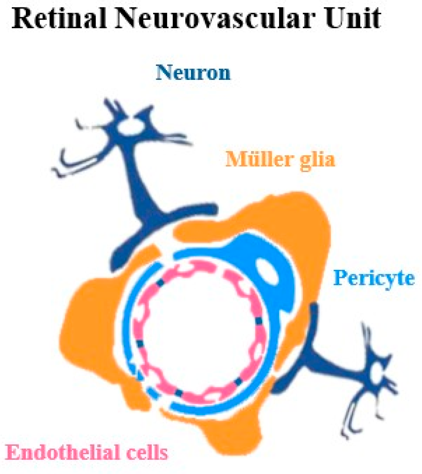
The retinal neurovascular unit is a highly integrated system composed of neurons, glial cells (including astrocytes, Müller cells, and microglia), and vascular elements (endothelial cells, pericytes, and vascular smooth muscle cells), which operate synergistically to preserve retinal homeostasis and sustain optimal visual function (Figure 2). In addition, the extracellular matrix (ECM) supports and regulates interactions between cells and blood vessels, helping the unit function normally. Retinal neurovascular coupling occurs within this unit: photoreceptors, bipolar cells, and ganglion cells initiate signaling by releasing glutamate, activating Müller glia and astrocytes, and interneurons. These, in turn, release vascular mediators including adenosine triphosphate (ATP) and nitric oxide (NO) that regulate vascular tone [,]. Glia and astrocytes in the unit act as pivotal intermediaries, sensing neural activity and releasing vasoactive factors that adjust vessel diameter to ensure blood flow meets the metabolic demands of active neurons [,,,]. Beyond this, glial cells, in particular, provide metabolic support, buffer extracellular potassium and other ions, and remove excess neurotransmitters, thereby maintaining homeostasis within the retinal microenvironment [,]. Simultaneously, the vascular elements of the unit, endothelial cells, pericytes, and vascular smooth muscle cells, along with the basement membrane, respond to these neuronal and glial signals in a coordinated manner []. Endothelial cells adjust iBRB permeability and mediate transcellular and paracellular transport, while pericytes regulate capillary diameter and stabilize vessel structure. Vascular smooth muscle cells modulate arteriolar tone to control blood flow []. Overall, this tightly coordinated system ensures that retinal blood flow dynamically adapts to neuronal activity, delivering adequate oxygen and nutrients while preventing harmful fluctuations in the retinal microenvironment [,].
Figure 2. Composition of the retinal neurovascular unit in the inner retinal circuitry.

2.2. Heterogeneity of Neurovascular Unit

Extensive research indicates that the mechanisms underlying retinal neurovascular coupling closely parallel those in the brain, reflecting the conserved organization of the neurovascular unit across the CNS []. Yet, despite this shared framework, the neurovascular unit exhibits considerable regional heterogeneity driven by differences in cellular composition, structural organization, and metabolic demands. Within the eye, retinal capillaries form superficial, intermediate, and deep plexuses that supply the inner retinal neurons, while the choroid contains a fenestrated, high-flow vascular network that nourishes photoreceptors. The iris–ciliary body vasculature, in turn, supports aqueous humor production. These vascular compartments differ in endothelial features, pericyte coverage, basement membrane composition, and barrier properties, highlighting the specialization of the ocular neurovascular unit and its variable susceptibility to disease [,]. Importantly, the distinctions extend beyond ocular tissues to the broader CNS. In the retina, Müller cells, radial glia spanning the retinal layers, coordinate neuronal support, metabolism, and blood flow, whereas in the brain, astrocytes fulfill similar roles through perivascular end-feet that regulate BBB, vascular tone, and metabolic exchange, thereby ensuring adequate oxygen and nutrient delivery [,].
Additional distinctions in the neurovascular unit include variations in astrocytic end-foot density and morphology, pericyte density, endothelial tight junction complexity, and basement membrane composition across cortical gray matter, white matter, brainstem nuclei, and retinal layers []. These structural differences translate into regional variation in neurovascular coupling efficiency, barrier permeability, and susceptibility to inflammatory or ischemic injury. Such heterogeneity not only allows local adaptation to specific energy demands and signaling environments, but it also contributes to selective vulnerability in disease, for example, the selective retinal layer injury in diabetic retinopathy or preferential involvement of white matter in cerebral small-vessel disease [,,]. Recent single-cell transcriptomic studies further highlight the diversity of endothelial cells, revealing distinct molecular signatures for arteriolar, capillary, and venular segments, along with differential responses to pericyte loss [,]. These include segment-specific pericyte depletion, altered tight junction protein expression, and shifts in inflammatory and metabolic gene profiles, underlining the variable vulnerability and adaptive capacity of neurovascular components across the vascular tree [,]. Recognizing this heterogeneity is therefore essential for developing targeted therapies that address region-specific neurovascular pathophysiology.

3. Signal Pathways of Neurovascular Coupling

The regulation of retinal blood flow relies on tightly orchestrated signaling pathways that link neuronal activity to vascular responses in neurovascular coupling. Multiple mechanisms, including NO production, purinergic signaling, ion/metabolic pathways, and vascular endothelial growth factor (VEGF), act in concert within the retinal neurovascular unit to translate synaptic activity into dynamic changes in vascular tone. Together, these pathways ensure precise matching of blood supply to the metabolic demands of visual processing and safeguard retinal homeostasis.

3.1. NO Signaling Pathways

During visual activity, glutamatergic input activates N-methyl-D-aspartate (NMDA) receptors in bipolar, amacrine, and ganglion cells. These neurons, which express the neuronal isoform of nitric oxide synthase (nNOS), produce NO upon depolarization [,,]. Once generated, NO readily diffuses to the smooth muscle cells of adjacent arterioles, where it modulates vascular tone [,]. Because of this direct action, NO production is regarded as a central mechanism in neurovascular coupling []. Furthermore, Müller glia expressing inducible nitric oxide synthase (iNOS) can generate NO under conditions of intensified neuronal activity or metabolic stress, thereby providing further support for vascular regulation [,]. These neuronal and glial cues guide endothelial cells and vascular smooth muscle cells to execute precise vascular adjustments during neurovascular coupling, with NO acting as a key mediator of vasodilation. NO is synthesized not only by neurons and glia but also by endothelial cells through the activation of endothelial nitric oxide synthase (eNOS) []. Upon release, NO diffuses into adjacent vascular smooth muscle cells, where it activates soluble guanylate cyclase (sGC), increasing cyclic GMP (cGMP) levels []. Elevated cGMP activates protein kinase G (PKG), which phosphorylates multiple targets, including myosin light chain phosphatase and calcium-handling proteins, leading to reduced intracellular calcium and dephosphorylation of myosin light chains. These changes relax smooth muscle fibers, dilate vessels, and increase local blood flow to meet metabolic demand [,].
In both the brain and retina, neuronal nNOS and glial iNOS generate NO in response to activity, while endothelial eNOS contributes additional NO. This NO diffuses to nearby vascular smooth muscle cells, activating sGC → cGMP → PKG signaling to relax vessels and increase blood flow. In the retina, this pathway is further stratified across the superficial, intermediate, and deep plexuses, supporting different neuronal layers and metabolic demands. For example, the deep capillary plexus supplies the inner segment/outer segment region of photoreceptors, while the superficial plexus serves the ganglion cell layer. This stratified microvascular architecture allows finely tuned, layer-specific blood flow regulation: NO-driven vasodilation may predominate in the superficial plexus during ganglion cell activity [], while other mediators may complement NO signaling in deeper layers to support photoreceptor metabolism.

3.2. Signaling Pathways for ATP in Vasodilation and Constriction

Alongside this NO-dependent pathway, purinergic signaling provides another important layer of control over neurovascular responses. Müller cells release ATP upon activation of metabotropic glutamate receptors, and ATP itself can act as a feed-forward activator in the retina []. During flickering light stimulation, amacrine and ganglion cells also release ATP, which activates P2X and P2Y receptors on the endfeet of Müller glia and astrocytes that wrap around retinal blood vessels. Activation of P2X receptors promotes calcium influx, whereas stimulation of P2Y receptors engages the phospholipase C (PLC)—inositol triphosphate (IP3) pathway, leading to calcium release from intracellular stores [,,]. These calcium signals trigger the release of vasoactive metabolites, including arachidonic acid derivatives, such as prostaglandin E2 (PGE2) and epoxyeicosatrienoic acids (EETs), which increase blood flow. PGE2 acts by binding to specific G-protein-coupled E-prostanoid (EP) receptors on endothelial cells and smooth muscle cells, activating adenylyl cyclase (AC), raising intracellular cyclic adenosine monophosphate (cAMP) levels, and activating protein kinase A (PKA), ultimately promoting vasodilation [,]. EETs, in turn, increase the activity of calcium-activated potassium (BKCa) channels in vascular smooth muscle cells and endothelial cells, causing hyperpolarization, relaxation, and vasodilation, although their precise receptor remains incompletely defined [,,]. In contrast to these vasodilatory mechanisms, vasoconstriction is mediated in part by Müller cells and astrocytes releasing 20-HETE, an arachidonic acid derivative []. A key aspect of its action involves the inhibition of BKCa channels in vascular smooth muscle cells through signaling pathways that include activation of protein kinase C (PKC), mitogen-activated protein kinase (MAPK), and Src-type tyrosine kinases (c-Src) []. By blocking potassium efflux required for smooth muscle relaxation, 20-HETE promotes vasoconstriction, providing a complementary mechanism that balances retinal blood flow alongside NO- and ATP-mediated vasodilation. In addition to its activity-dependent regulation, ATP also contributes to baseline vascular tone: tonic release from astrocytes can induce mild vasoconstriction under resting conditions, which is subsequently overridden by vasodilation during neuronal activity []. Finally, the breakdown of ATP to adenosine further refines vascular tone through activation of A1 and A2 adenosine receptors [,,]. Adenosine receptors (A2A, A2B) further enhance vasodilation through cAMP-dependent pathways [,]. These are well-characterized vasodilation mechanisms playing central roles in controlling vascular tone. In the retina, the stratified microvascular organization enables finely tuned regulation of blood flow: ATP/adenosine signaling appears to be particularly important in the deep capillary plexus, where high synaptic activity triggers ATP release from neurons and Müller glia, which in turn modulates pericyte and endothelial function to adjust capillary diameter []. In contrast, NO signaling predominates in the superficial plexus, where ganglion cell activity and astrocyte-neuron interactions drive rapid vasodilation. This division of labor enables the retina to match blood supply to local metabolic demand across its distinct vascular layers.

3.3. Ions in Regulation of Blood Flow

In addition to these multifaceted regulators, ion and metabolic coupling mechanisms are also critical for fine-tuning blood flow and ensuring adequate energy supply during neuronal activity. In both the brain and retina, K+ efflux from active neurons and glia alters extracellular potassium levels, thereby modulating vascular tone [,]. The inward rectifying K+ channel, Kir4.1 channels, abundantly expressed in Müller cells of the retina and astrocytes in the brain, play a central role in buffering extracellular potassium and relaying activity-dependent signals to vascular smooth muscle cells and pericytes [,]. Complementing this, bicarbonate transporters (e.g., Na+/HCO3 cotransporters) and carbonic anhydrases regulate extracellular pH and CO2, further influencing vasodilation and constriction [,]. In parallel, local elevations in extracellular K+ can trigger vasodilation via inward rectifier K+ channels in vascular cells, while Ca2+ signals within neurons and Müller glia coordinate the release of vasoactive mediators such as NO, ATP, and prostaglandins. Together, these interconnected signaling mechanisms integrate neuronal, glial, and astrocytic activity to precisely regulate neurovascular responses.

3.4. VEGF Signaling Pathways

Another key regulator is VEGF, which promotes endothelial survival, barrier integrity, and angiogenic potential. VEGF also enhances NO production by activating eNOS through VEGF receptor-2 (VEGFR2) signaling, with the resulting NO release contributing to vasodilation and improved perfusion []. VEGF also stimulates vascular permeability by reorganizing tight and adherens junctions, allowing dynamic exchange between the circulation and surrounding tissue []. In addition, VEGF drives retinal angiogenesis by binding to its receptors on endothelial cells, stimulating their sprouting, migration, and new capillary formation, thereby adapting blood supply to local metabolic demands to improve neovascularization []. However, when overactivated, as in diabetic retinopathy, ischemia, or inflammation, excessive VEGF disrupts tight junctions of the iBRB and promotes abnormal neovascularization []. Studies in the brain further show that following injury, such as stroke or brain tumors, increased VEGF expression enhances angiogenesis and promotes BBB, which is thought to represent an attempt to repair damaged tissue by enhancing blood supply and nutrient delivery to the affected area []. Thus, VEGF functions as both a vital physiological mediator of blood flow and a pathological driver of vascular dysregulation and dysfunction.

3.5. Signal Pathways of Pericytes

Within the neurovascular unit, pericytes work with endothelial cells to fine-tune capillary blood flow through contractile regulation. These specialized mural cells, embedded within the basement membrane and in close contact with endothelial cells, sense neuronal and glial signals through diverse receptor pathways, including purinergic (P2X/P2Y), adrenergic (A1, A2), and endothelin receptor B (ETRB) []. Pericytes respond to vasodilatory mediators such as NO, PGE2, and EETs with relaxation, thereby promoting capillary dilation, whereas vasoconstrictors, such as thromboxane A2 or endothelin-1, acting through ETRA, induce vasoconstriction [,]. They also contribute to extracellular K+ sensing, where elevated extracellular K+ induced membrane hyperpolarization and subsequent capillary relaxation. Beyond dynamic regulation of vascular tone, pericytes are essential for maintaining iBRB/BBB integrity through stabilization of tight junctions, controlling endothelial proliferation, and modulating angiogenic signaling [,]. These functions rely on intercellular communication between endothelial cells and pericytes, mediated by multiple pathways, including transforming growth factor-β (TGF-β), angiopoietins, platelet-derived growth factor-B (PDGF-B), sphingosine-1-phosphate (S1P), and Notch signaling. Moreover, pericytes exhibit multipotent differentiation capacity, with the ability to generate diverse cell types, positioning them as a key cellular target for tissue repair and regenerative therapies [,]. Conversely, the loss or dysfunction of pericytes, as seen in diabetic retinopathy, disrupts these signaling networks and leads to impaired neurovascular coupling, capillary nonperfusion, and iBRB breakdown, underscoring their critical role in fine-tuning retinal blood flow and maintaining vascular stability [,].
In summary, these signaling pathways link neuronal activity to vascular responses, ensuring precise neurovascular coupling and maintaining retinal homeostasis. While the retina and brain share core mechanisms such as NO-mediated vasodilation, ATP- and ion-dependent modulation, VEGF signaling, and pericyte regulation, the retina exhibits distinct adaptations: ATP/adenosine signaling is particularly prominent in the deep capillary plexus, NO signaling predominates in the superficial plexus, ionic (K+ and Ca2+) dynamics are tightly spatially restricted, VEGF primarily supports retinal vascular maintenance rather than angiogenesis under normal conditions, and pericytes display specialized roles in regulating capillary flow across stratified retinal layers (Table 1). When disrupted, they lead to mismatches between metabolic demand and blood supply, resulting in hypoxia, oxidative stress, and barrier breakdown that contribute to retinal diseases such as diabetic retinopathy, glaucoma, and age-related macular degeneration (AMD) and other neuro-dysfunctions.
Table 1. Summarizing shared and retina-distinct signaling pathways in neurovascular coupling for NO, ATP, ions, VEGF, and pericytes.

4. Neurovascular Coupling Dysfunction in Disease

Over the past decades, extensive research has explored retinal neurovascular coupling dysfunction in retinal neurodegenerative diseases such as diabetic retinopathy, AMD, and glaucoma. The classical mechanisms underlying neurovascular coupling deficits in these conditions have been comprehensively reviewed in the previous literatures [,,,]. Building on this foundation, recent investigations have focused on neurovascular coupling impairments in major brain disorders, including Alzheimer’s disease, Parkinson’s disease, Huntington’s disease, and stroke. Given the structural and functional parallels between the retina and brain, noninvasive imaging of retinal neurovascular coupling provides a unique window into early cerebrovascular and neuronal dysfunction. Advanced modalities such as fOCT, OCT and OCTA enable high-resolution, label-free assessment of activity-dependent vascular responses and microvascular alterations in vivo. By detecting subtle changes in retinal perfusion and neural activity, these techniques hold great promise as non-invasive, early diagnostic and monitoring tools for neurovascular deficits associated with brain diseases.

4.1. Retinal Structure and Neurovascular Alterations in Alzheimer’s Disease

Although Alzheimer’s disease has traditionally been regarded as a brain disorder, growing evidence indicates that it also manifests in the eye, with detectable biomarkers in the retina, vitreous, cornea, and other ocular structures. One key pathological feature is impaired neurovascular coupling, well documented in the brain and also observed in the retina, reflecting their shared neurovascular pathophysiology [,,]. Functional imaging and electrophysiological studies demonstrate reduced retinal blood flow responses to visual stimulation, indicating deficits in activity-dependent vascular changes in Alzheimer patients. These functional impairments are accompanied by structural and molecular changes within the retinal neurovascular unit, including pericyte loss or dysfunction, disruption of endothelial tight junctions, decreased expression of vasoactive mediators such as NOS, and altered gliotic responses of Müller cells and astrocytes [,]. Retinal vascular imaging in Alzheimer’s disease patients and animal models has revealed vessel narrowing, decreased vessel density, and altered capillary hemodynamics. fOCT and angiography (OCTA) and dynamic vessel analysis (DVA) further show diminished flicker light–induced vasodilation []. These findings support the use of multimodal retinal assessments as noninvasive approaches to detect neurovascular pathology in Alzheimer’s disease. In recent years, increasing attention has been given to tauopathy and amyloidosis within the retinal neurovascular unit, highlighting the retina as a potential window into early neurodegenerative processes. For example, tau accumulation in the retina promotes early neuronal dysfunction and precedes brain pathology in a mouse model of Alzheimer’s disease [,], demonstrating that retinal tau aggregation leads to synaptic and functional impairments before overt cortical pathology. This suggests that retinal tauopathy may serve as an early biomarker of central tau-related neurodegeneration. Similarly, amyloid-β (Aβ) deposition has been detected in the inner retinal layers and vascular walls of both Alzheimer’s disease patients and transgenic mouse models [,,], implicating retinal amyloidosis in vascular dysregulation and neuronal loss. Studies have further shown that Aβ accumulation can disrupt the integrity of the iBRB and impair neurovascular coupling [,,], which leads to altered retinal perfusion and neuronal dysfunction. Together, these findings indicate that tau and Aβ pathologies converge within the retinal neurovascular unit, contributing to early vascular and synaptic deficits that parallel those observed in the brain.
Beyond vascular alterations, retinal changes include microgliosis, Müller cell degeneration, thinning of the retinal nerve fiber layer, and ganglion cell loss, all of which have been proposed as potential biomarkers for early disease detection [,]. Building on these observations, OCT has gained attention as a promising tool for both early diagnosis and longitudinal monitoring of Alzheimer’s disease. Expanding on this potential, researchers are increasingly exploring retinal imaging as a noninvasive window into Alzheimer’s and dementia pathology. By leveraging high-resolution techniques such as OCT, OCTA, transmission electron microscopy (TEM), and ultra-spectral imaging, it is possible to detect subtle retinal changes—thinning of the nerve fiber layer, microvascular rarefaction, altered blood flow, and Aβ accumulation—that mirror in brain pathology [,]. These biomarkers may allow early identification of individuals at risk for Alzheimer’s, enabling timely interventions from lifestyle modifications to pharmacologic treatments that could delay onset, slow progression, and improve long-term outcomes.
Challenges and future directions in this research area have been increasingly recognized. While retinal imaging holds significant promise for detecting Alzheimer’s disease, further studies are needed to validate these biomarkers across larger cohorts and at different stages of disease progression. Therefore, standardizing imaging protocols, analytical algorithms, and diagnostic thresholds will be essential to ensure reproducibility and comparability across studies [,]. Moreover, understanding the exact cellular and molecular mechanisms underlying retinal manifestations of Alzheimer’s pathology, such as Aβ deposition, tau aggregation, microvascular dysfunction, and neuroinflammation, will be critical for identifying reliable therapeutic targets. Integrating retinal imaging with other biomarkers, including cerebrospinal fluid analysis, plasma assays, and neuroimaging, may also improve early detection and risk stratification [,]. Ultimately, translating these advances into accessible and cost-effective screening tools will require interdisciplinary collaboration among neuroscientists, ophthalmologists, engineers, and clinicians.

4.2. Using Biomarkers to Detect Early Signs of Parkinson’s Disease and Dementia

In Parkinson’s disease, hallmark structural and molecular changes include the loss of dopaminergic neurons and the accumulation of misfolded α-synuclein (α-syn) Lewy bodies in the substantia nigra pars compacta, both of which have also been reported in the retina [,]. Retinal neurovascular coupling dysfunction can be detected before significant structural changes appear, making it a potential early marker for Parkinson’s disease [,]. Indeed, reductions in superficial and deep capillary plexus densities have been observed in Parkinson’s disease patients, correlating with disease severity and retinal ganglion cell loss, consequently, leading to impair neurovascular coupling and attenuate visual stimulation-evoked blood flow [,]. Specific retinal alterations include the accumulation of misfolded α-syn aggregates in neurons and glia []. Mechanistically, α-syn aggregation may disrupt neurovascular signaling by impairing the release of NO and other vasoactive metabolites, thereby reducing vascular responsiveness. Additional contributors include pericyte loss, endothelial dysfunction, altered glial calcium signaling, and compromised neuronal metabolism, which together impair energy-dependent vascular regulation and neurovascular coupling [,]. Furthermore, a reduction in dopaminergic amacrine cells, which play critical roles in retinal signal processing and contrast sensitivity, has also been reported. Such alterations may contribute to impair visual function, including deficits in motion perception and contrast detection, which are commonly reported in Parkinson’s disease patients []. Retinal structural alterations, particularly the loss of amacrine and ganglion cells and thinning of the inner plexiform layer and peripapillary retinal nerve fiber layer, correlate with dopaminergic neuron loss in the substantia nigra, highlighting shared neurodegenerative mechanisms between the retina and brain []. This thinning can be quantitatively measured using OCT, a non-invasive, high-resolution imaging technique capable of detecting early, subclinical retinal changes, making it a promising biomarker for Parkinson’s disease progression and severity []. Importantly, these structural alterations often parallel functional deficits detected by electrophysiological methods such as electroretinography (ERG) or psychophysical tests assessing visual acuity, contrast sensitivity, and color discrimination []. In Parkinson’s disease, pattern ERG (PERG) recordings frequently reveal reduced amplitudes of P50- and N95-waves, reflecting impaired macular pathway function and ganglion cell activity, while full-field ERG may show altered oscillatory potentials linked to dopaminergic amacrine cell dysfunction []. Notably, some studies have reported partial recovery of ERG responses following dopaminergic therapy, suggesting that these measures may also serve as functional indicators of treatment efficacy []. Furthermore, since the loss of dopaminergic signaling in the retina can disrupt neurovascular coupling, dopamine plays a modulatory role in regulating retinal blood flow in response to visual stimuli []. This links Parkinson’s disease-associated neuronal changes to vascular dysregulation.
The retina offers a unique and accessible site to detect neurodegenerative changes that may precede motor symptoms in Parkinson’s disease. Emerging evidence suggests that retinal alterations, including structural thinning of the ganglion cell and inner plexiform layers, loss of dopaminergic amacrine cells, and accumulation of α-syn, may precede the clinical diagnosis [,]. While OCT-based structural changes are relatively well documented, evidence for dopaminergic cell loss and α-syn deposition in the prodromal stage remains preliminary and requires further longitudinal validation. Nevertheless, the early changes in the retina are being actively investigated as biomarkers for Parkinson’s disease, with the potential to enable earlier intervention and improved disease management []. Non-invasive imaging modalities such as OCT and OCTA provide rapid, cost-effective structural assessments, while complementary approaches, including functional imaging and electrophysiological tests (e.g., PERG, flicker ERG), can detect subtle deficits in visual processing and neurovascular coupling [,,]. Future studies incorporating artificial intelligence (AI)-driven analysis and large OCT cohorts across different disease stages have the potential to provide a more comprehensive view of the disease progression. Moving forward, progress in this area will depend on standardizing imaging protocols, integrating multimodal structural and functional readouts, and validating their predictive value in longitudinal cohorts. Such advances could establish retinal imaging as a practical and scalable tool for early Parkinson’s disease detection and monitoring.

4.3. Detecting Huntington’s Disease Through the Retina

Huntington’s disease is classically defined as a neurodegenerative disorder of the striatum and cerebral cortex, caused by an expanded CAG repeat in the huntingtin gene (HTT) that encodes a polyglutamine (polyQ) tract in the huntingtin protein. Consequently, the mutant huntingtin (mHTT) with an extended polyQ sequence forms aggregates in cortical and striatal neurons, leading to cellular damage and death []. Increasing evidence indicates that the retina is also affected, providing a potential window into disease mechanisms and progression [,,]. Both human studies and the transgenic Huntington’s disease mouse models (e.g., R6/2, YAC128, and zQ175) have demonstrated retinal structural and functional alterations, reinforcing the retina as a sensitive site of Huntington’s disease-associated neurodegeneration. These changes include thinning of the inner retinal layers, disrupted synaptic organization, and progressive loss of ganglion and photoreceptor cells. Specifically, photoreceptor involvement is evidenced by thinning of the outer nuclear layer and loss of nuclei in the zQ175 mice, as well as degeneration observed in the R6 models. Correspondingly, several OCT studies in humans report thinning of the inner retinal ganglion cell complex, mirroring the cellular alterations seen in mouse models [,]. Furthermore, retinal changes in the R6/1 mice emerge earlier in the outer retina than in the inner retina, beginning with cone opsin loss and progressing to widespread structural and functional deficits by 6 months []. Electrophysiological recordings, such as ERG, demonstrate deficits in both photopic and scotopic responses, suggesting dysfunction across multiple retinal cell types. The studies also localize this damage to the outer plexiform layer and link it to loss of synaptic connectivity between the outer nuclear layer and inner nuclear layer, consistent with the observed ERG b-wave abnormalities. However, since these alterations do not parallel the temporal spread of mutant huntingtin aggregates, the R6/1 retina may be more useful for investigating Huntington’s disease mechanisms and therapeutic strategies rather than serving as an early diagnostic marker [,]. Additionally, molecular studies have detected mutant huntingtin aggregates in retinal neurons and glia, accompanied by altered expression of neurotransmission-related genes, inflammatory markers, and components of the neurovascular unit [].
Vascular abnormalities have emerged as a significant feature of Huntington’s disease, extending beyond neuronal degeneration to include microvascular compromise in the retina. OCTA studies in humans have revealed decreased capillary density, choroidal thinning, and changes in foveal perfusion in patients with Huntington’s disease, highlighting the retina’s sensitivity to vascular pathology in both the central nervous system and systemic circulation [,]. Complementary evidence from Huntington’s disease mouse models, including R6/1 and R6/2 strains, demonstrates early iBRB disruption, retinal gliosis, and progressive retinal structural and functional changes, suggesting that barrier integrity and microvascular regulation are impaired alongside neuronal degeneration [,]. However, some reports in human patients have been more controversial, showing only minimal abnormalities in the temporal retinal nerve fiber layer and no other detectable structural or functional retinal changes. This discrepancy suggests that current OCT and color vision tests may not yet serve as reliable biomarkers for Huntington’s disease [].
Future studies should integrate fOCT, OCTA, and electrophysiology with AI-based analysis to detect early retinal changes in the disease. Large-scale longitudinal imaging in experimental and clinical settings should be prioritized to define retinal changes as reliable biomarkers for early disease detection and monitoring. Mechanistic studies in animal models exploring cellular drivers such as gliosis, vascular dysfunction, and neuronal loss will be critical for linking these imaging findings to underlying pathology.

4.4. Retinal Structure Changes in Stroke

Stroke induces both functional and structural alterations in the retina that mirror central neurovascular pathology []. In particular, retinal neurovascular coupling is disrupted, as shown by attenuated vessel dilation and delayed hemodynamic responses to visual or flicker stimulation, which reflect impaired communication between neurons, glia, and vascular cells. These abnormalities have been observed in stroke patients. To explore these mechanisms, several well-established animal models of stroke have been employed to examine how cerebrovascular injury impacts the retina, thereby providing a unique window into neurovascular dysfunction [,,]. Among them, the middle cerebral artery occlusion (MCAO) model is the most widely used; it induces focal ischemia and leads to thinning of inner retinal layers, retinal ganglion cell death, and impaired ERG responses, including delayed or diminished a- and b-wave amplitudes. Notably, the severity of these ERG deficits correlates with infarct size, behavioral impairments, and ganglion cell loss, mirroring the extent of cortical damage []. In addition, MCAO triggers inflammatory responses in the retina, characterized by elevated cytokine levels and microglial activation following reperfusion []. The bilateral common carotid artery occlusion (BCCAO) model, which produces chronic cerebral hypoperfusion, results in progressive inner retinal degeneration, vascular remodeling, and glial activation, closely resembling features of small vessel disease and vascular cognitive impairment [,]. Similarly, unilateral common carotid artery occlusion (UCCAO) in mice induces retinal hypoxia, marked by HIF-1α stabilization, gliosis, and ERG deficits that emerge within days of occlusion []. Finally, photothrombotic stroke models, which allow spatially controlled vascular occlusion, have provided additional insight by revealing localized disruptions in retinal blood flow and neurovascular coupling [,].
To better understand how stroke-induced cerebrovascular events affect the retina, researchers have employed a wide range of imaging and experimental techniques to examine structural alterations, providing insights into the consequences of cerebral ischemia on retinal tissue [,]. Among these, OCT has been widely used to provide precise measurement of retinal layer thickness, particularly the retinal nerve fiber layer, ganglion cell complex, and inner retina, which reflect neuronal loss and often parallel cortical degeneration []. Building on this structural information, OCTA enables visualization of the retinal microvasculature without dye injection, revealing reduced vessel density, perfusion deficits, and remodeling of the superficial and deep vascular plexuses after ischemic events []. Fundus photography (FP) and fluorescein angiography (FA) continue to play a role in documenting gross retinal vascular abnormalities, such as hemorrhages or ischemic regions, though they provide less quantitative data than OCT-based approaches []. Complementing these imaging modalities, retinal vessel analysis (RVA) offers dynamic assessment of vascular reactivity to flicker light stimulation, highlighting impaired neurovascular coupling as a hallmark of stroke-induced dysfunction []. Dynamic RVA can detect reduced vessel diameter and blunted neurovascular responses in the acute phase, some of which partially recover over time, and these observations hold promise as potential biomarkers for early detection of stroke-related brain injury. Additionally, in experimental animal models, histological and immunohistochemical techniques provide cellular-level resolution, demonstrating neuronal degeneration, synaptic alterations, microglial activation, and vascular pathology in the retina post-stroke [,,,]. Complementary to these, ERG, while primarily functional, also supports structural interpretations by correlating inner retinal damage with reduced a- and b-wave amplitudes. Together, these methodologies, spanning in vivo imaging, vascular reactivity testing, and postmortem tissue analysis, form a robust toolkit for linking retinal structural changes to stroke pathophysiology, with growing potential for translation into clinical biomarkers.
The retina provides a non-invasive, accessible window to monitor stroke-related neurovascular and structural changes. Advances in OCT, OCTA, and ERG enable detailed assessment of retinal microvascular alterations and neuronal degeneration, supporting early detection and monitoring of stroke progression. Future research should prioritize standardizing retinal imaging protocols in stroke patients and evaluating the potential of retinal biomarkers to predict outcomes and guide therapeutic interventions.
In summary, studying Alzheimer’s disease, Parkinson’s disease, Huntington’s disease, and stroke through the retina offers a powerful window into neurovascular and neurodegenerative mechanisms shared with the brain. The retina, as an accessible extension of the central nervous system, exhibits early and measurable alterations in neuronal, glial, and vascular function that are parallel to central pathology. Retinal imaging and electrophysiological analyses reveal that Alzheimer’s disease is characterized by Aβ accumulation, vascular attenuation, and impaired neurovascular coupling; Parkinson’s disease shows dopaminergic dysfunction, mitochondrial stress, and microvascular rarefaction; Huntington’s disease demonstrates early synaptic and vascular deficits; and stroke leads to ischemic damage and blood-retina barrier disruption. Comparative analysis highlights both shared neurovascular unit dysfunction and disease-specific signatures in retinal neurons, glia, and microvessels, underscoring the retina’s potential as a noninvasive biomarker for CNS disease progression (Table 2).
Table 2. Comparative summary of retinal neurovascular dysfunction across AD, PD, HD, and stroke.

5. Conclusions

The retina, as an extension of the central nervous system, offers a unique, accessible window into neurovascular health and disease. Retinal neurovascular coupling represents a highly coordinated interplay between neurons, glia, and vascular cells, ensuring that local blood flow dynamically matches metabolic demand [,]. Mechanistically, this process involves activity-dependent signaling pathways, including NO, ATP, arachidonic acid metabolites, VEGF and ions-mediated vascular responses, all of which are tightly regulated through neuron–glia–vascular interactions [,]. Disruption of these pathways is increasingly recognized in neurodegenerative and vascular disorders, with retinal neurovascular dysfunction reflecting parallel alterations in the brain []. Owing to its accessibility and structural homology with the brain, the retina offers a unique and non-invasive platform to monitor neurovascular health and identify biomarkers for early detection, disease progression, and therapeutic evaluation.
While fOCT, OCT, and OCTA provide powerful means to assess retinal microvascular structure and perfusion, these modalities primarily capture vascular parameters and do not directly measure neuronal or glial activity. Consequently, they yield only indirect information about neurovascular coupling, limiting mechanistic insight into how neural and glial signals regulate vascular responses. This is particularly significant because impaired neuron–vascular and glia–vascular communication occurs early in diseases such as Alzheimer’s, Parkinson’s, and stroke. Complementary approaches, including calcium or voltage imaging for neuronal activity and fluorescence-based or two-photon microscopy for astrocytic signaling, are therefore essential in basic and preclinical studies. Integrating these methods with fOCT and OCTA could provide a more comprehensive framework for understanding neurovascular coupling and its disruption in disease. Therefore, multidisciplinary approaches that integrate advanced imaging, electrophysiology, and molecular profiling will be vital to clarify neurovascular interaction mechanisms and bridge retinal findings to brain pathology.
Advancing our understanding of retinal neurovascular coupling holds significant promise for both basic neuroscience and clinical translation. Future studies should aim to dissect the molecular and cellular pathways that mediate neurovascular coupling, including neuron-to-glia and glia-to-vessel signaling, as well as the contributions of pericytes and endothelial heterogeneity [,]. The integration of high-resolution functional imaging, adaptive optics, and electrophysiological techniques will allow more precise mapping of activity-dependent vascular responses across retinal layers []. Clinically, longitudinal studies leveraging retinal imaging may provide sensitive biomarkers for early detection and progression monitoring in neurodegenerative and cerebrovascular diseases, complementing brain imaging and fluid-based diagnostics. Furthermore, exploring pharmacological or genetic modulation of retinal neurovascular coupling may reveal novel therapeutic avenues to protect neurovascular integrity, not only in the retina but across the CNS [,]. Ultimately, combining mechanistic insights with translational applications will establish the retina as a robust model for probing neurovascular health and dysfunction in human brain disease.

Funding

The National Science Foundation provides funding to W.S. (IOS2126141).

Data Availability Statement

Not applicable.

Conflicts of Interest

The author declares no conflicts of interest.

Abbreviations

The following abbreviations are used in this manuscript:
NVUNeurovascular unit
RGCsRetinal ganglion cells
iBRBinner blood–retina barrier
oBRBouter blood–retina barrier
BBBBlood–brain barrier
OCTOptical coherence tomography
OCTAOptical coherence tomography
NONitric oxide
NOSNitric oxide synthase
ATPAdenosine triphosphate
cAMPAdenosine monophosphate
PKAProtein kinase A
PKCProtein kinase C
MAPKMitogen-activated protein kinase
PLCPhospholipase C
PGE2prostaglandin E2
EETsepoxyeicosatrienoic acids

References

  1. Alkayed, N.J.; Cipolla, M.J. The Ever-Evolving Concept of the Neurovascular Unit. Stroke 2023, 54, 2178–2180. [Google Scholar] [CrossRef]
  2. Grimes, W.N.; Berson, D.M.; Sabnis, A.; Hoon, M.; Sinha, R.; Tian, H.; Diamond, J.S. The retina’s neurovascular unit: Müller glial sheaths and neuronal contacts. bioRxiv 2024. [Google Scholar] [CrossRef] [PubMed]
  3. Cholkar, K.; Dasari, S.R.; Pal, D.; Mitra, A.K. Eye: Anatomy, physiology and barriers to drug delivery. In Ocular Transporters and Receptors: Their Role in Drug Delivery; Elsevier: Amsterdam, The Netherlands, 2013; pp. 1–36. [Google Scholar] [CrossRef]
  4. Agarwal, N.; Carare, R.O. Cerebral Vessels: An Overview of Anatomy, Physiology, and Role in the Drainage of Fluids and Solutes. Front. Neurol. 2021, 11, 611485. [Google Scholar] [CrossRef] [PubMed]
  5. Grimes, W.N.; Berson, D.M.; Sabnis, A.; Hoon, M.; Sinha, R.; Tian, H.; Diamond, J.S. Layer-specific anatomical and physiological features of the retina’s neurovascular unit. Curr. Biol. 2024, 35, 109–120.e4. [Google Scholar] [CrossRef]
  6. Díaz-Coránguez, M.; Ramos, C.; Antonetti, D.A. The inner blood-retinal barrier: Cellular basis and development. Vis. Res. 2017, 139, 123–137. [Google Scholar] [CrossRef]
  7. Abbott, N.J.; Patabendige, A.A.K.; Dolman, D.E.M.; Yusof, S.R.; Begley, D.J. Structure and function of the blood–brain barrier. Neurobiol. Dis. 2010, 37, 13–25. [Google Scholar] [CrossRef]
  8. Hussain, A.; Sheikh, Z.; Subramanian, M. The Eye as a Diagnostic Tool for Alzheimer’s Disease. Life 2023, 13, 726. [Google Scholar] [CrossRef]
  9. Subramaniam, M.D.; Aishwarya Janaki, P.; Abishek Kumar, B.; Gopalarethinam, J.; Nair, A.P.; Mahalaxmi, I.; Vellingiri, B. Retinal Changes in Parkinson’s Disease: A Non-invasive Biomarker for Early Diagnosis. Cell. Mol. Neurobiol. 2023, 43, 3983–3996. [Google Scholar] [CrossRef]
  10. Amini, E.; Moghaddasi, M.; Habibi, S.A.H.; Azad, Z.; Miri, S.; Nilforushan, N.; Mirshahi, R.; Cubo, E.; Mohammadzadeh, N.; Rohani, M. Huntington’s disease and neurovascular structure of retina. Neurol. Sci. 2022, 43, 5933–5941. [Google Scholar] [CrossRef]
  11. Cheung, C.Y.L.; Ikram, M.K.; Chen, C.; Wong, T.Y. Imaging retina to study dementia and stroke. Prog. Retin. Eye Res. 2017, 57, 89–107. [Google Scholar] [CrossRef]
  12. de Carlo, T.E.; Romano, A.; Waheed, N.K.; Duker, J.S. A review of optical coherence tomography angiography (OCTA). Int. J. Retin. Vitr. 2015, 1, 5. [Google Scholar] [CrossRef] [PubMed]
  13. Williams, D.R.; Burns, S.A.; Miller, D.T.; Roorda, A. Evolution of adaptive optics retinal imaging [Invited]. Biomed. Opt. Express 2023, 14, 1307. [Google Scholar] [CrossRef] [PubMed]
  14. Peterfi, A.; Pinaffi-Langley, A.C.d.C.; Szarvas, Z.; Muranyi, M.; Kaposzta, Z.; Adams, C.; Pinto, C.B.; Mukli, P.; Kotliar, K.; Yabluchanshiy, A. Dynamic retinal vessel analysis: Flickering a light into the brain. Front. Aging Neurosci. 2024, 16, 1517368. [Google Scholar] [CrossRef] [PubMed]
  15. Mahroo, O.A. Visual electrophysiology and “the potential of the potentials”. Eye 2023, 37, 2399–2408. [Google Scholar] [CrossRef]
  16. Newman, E.A. Glial cell regulation of neuronal activity and blood flow in the retina by release of gliotransmitters. Philos. Trans. R. Soc. B Biol. Sci. 2015, 370, 20140195. [Google Scholar] [CrossRef]
  17. Yoshioka, T.; Nagaoka, T.; Song, Y.; Yokota, H.; Tani, T.; Yoshida, A. Role of Neuronal Nitric Oxide Synthase in Regulating Retinal Blood Flow During Flicker-Induced Hyperemia in Cats. Investig. Ophthalmol. Vis. Sci. 2015, 56, 3113–3120. [Google Scholar] [CrossRef]
  18. Macvicar, B.A.; Newman, E.A. Astrocyte Regulation of Blood Flow in the Brain. Cold Spring Harb. Perspect. Biol. 2015, 7, a020388. [Google Scholar] [CrossRef]
  19. Kugler, E.C.; Greenwood, J.; MacDonald, R.B. The “Neuro-Glial-Vascular” Unit: The Role of Glia in Neurovascular Unit Formation and Dysfunction. Front. Cell Dev. Biol. 2021, 9, 732820. [Google Scholar] [CrossRef]
  20. Newman, E.A. Glial Cell Inhibition of Neurons by Release of ATP. J. Neurosci. 2003, 23, 1659. [Google Scholar] [CrossRef]
  21. Kur, J.; Newman, E.A.; Kur, J.; Physiol, J. Purinergic control of vascular tone in the retina. J. Physiol. C 2014, 592, 491–504. [Google Scholar] [CrossRef]
  22. Karwoski, C.J.; Lu, H.-K.; Newman, E.A.; Karwoski, J.; Lu, H.-K.; Newman, E.A. Spatial buffering of light-evoked potassium increases by retinal Müller (glial) cells. Science 1989, 244, 578–580. [Google Scholar] [CrossRef]
  23. Stratman, A.N.; Davis, G.E. Endothelial cell-pericyte interactions stimulate basement membrane matrix assembly: Influence on vascular tube remodeling, maturation, and stabilization. Microsc. Microanal. 2012, 18, 68–80. [Google Scholar] [CrossRef]
  24. Hill, M.A.; Zou, H.; Potocnik, S.J.; Meininger, G.A.; Davis, M.J. Invited Review: Arteriolar smooth muscle mechanotransduction: Ca2+ signaling pathways underlying myogenic reactivity. J. Appl. Physiol. 2001, 91, 973–985. [Google Scholar] [CrossRef] [PubMed]
  25. van der Heide, F.C.T.; van Sloten, T.T.; Willekens, N.; Stehouwer, C.D.A. Neurovascular coupling unit dysfunction and dementia: Retinal measurements as tools to move towards population-based evidence. Front. Endocrinol. 2022, 13, 1014287. [Google Scholar] [CrossRef] [PubMed]
  26. Kim, S.J.; Lim, J.S.; Park, J.H.; Lee, J. Molecular, Cellular, and Functional Heterogeneity of Retinal and Choroidal Endothelial Cells. Investig. Ophthalmol. Vis. Sci. 2023, 64, 35. [Google Scholar] [CrossRef] [PubMed]
  27. Bernier, L.P.; Brunner, C.; Cottarelli, A.; Balbi, M. Location Matters: Navigating Regional Heterogeneity of the Neurovascular Unit. Front. Cell. Neurosci. 2021, 15, 696540. [Google Scholar] [CrossRef]
  28. Yang, S.; Webb, A.J.S. Associations between neurovascular coupling and cerebral small vessel disease: A systematic review and meta-analysis. Eur. Stroke J. 2023, 8, 895. [Google Scholar] [CrossRef]
  29. Duh, E.J.; Sun, J.K.; Stitt, A.W. Diabetic retinopathy: Current understanding, mechanisms, and treatment strategies. JCI Insight 2017, 2, e93751. [Google Scholar] [CrossRef]
  30. Mäe, M.A.; He, L.; Nordling, S.; Vazquez-Liebanas, E.; Nahar, K.; Jung, B.; Li, X.; Tan, B.C.; Chin Foo, J.; Cazenave-Gassiot, A.; et al. Single-Cell Analysis of Blood-Brain Barrier Response to Pericyte Loss. Circ. Res. 2021, 128, E46–E62. [Google Scholar] [CrossRef]
  31. Niu, R.Z.; Xu, H.Y.; Tian, H.; Zhang, D.; He, C.Y.; Li, X.L.; Li, Y.Y.; He, J. Single-cell transcriptome unveils unique transcriptomic signatures of human organ-specific endothelial cells. Basic Res. Cardiol. 2024, 119, 973–999. [Google Scholar] [CrossRef]
  32. Blom, J.; Giove, T.; Deshpande, M.; Eldred, W.D. Characterization of nitric oxide signaling pathways in the mouse retina. J. Comp. Neurol. 2012, 520, 4204–4217. [Google Scholar] [CrossRef]
  33. Agurto, A.; Vielma, A.H.; Cadiz, B.; Couve, E.; Schmachtenberg, O. NO signaling in retinal bipolar cells. Exp. Eye Res. 2017, 161, 30–35. [Google Scholar] [CrossRef]
  34. Goldstein, I.M.; Ostwald, P.; Roth, S. Nitric Oxide: A Review of Its Role in Retinal Function and Disease. Vis. Res. 1996, 36, 2979–2994. [Google Scholar] [CrossRef]
  35. Lourenço, C.F.; Laranjinha, J. Nitric Oxide Pathways in Neurovascular Coupling Under Normal and Stress Conditions in the Brain: Strategies to Rescue Aberrant Coupling and Improve Cerebral Blood Flow. Front. Physiol. 2021, 12, 729201. [Google Scholar] [CrossRef]
  36. Pournaras, C.J.; Rungger-Brändle, E.; Riva, C.E.; Hardarson, S.H.; Stefansson, E. Regulation of retinal blood flow in health and disease. Prog. Retin. Eye Res. 2008, 27, 284–330. [Google Scholar] [CrossRef]
  37. Noonan, J.E.; Lamoureux, E.L.; Sarossy, M. Neuronal activity-dependent regulation of retinal blood flow. Clin. Exp. Ophthalmol. 2015, 43, 673–682. [Google Scholar] [CrossRef]
  38. Kuhlencordt, P.J.; Rosel, E.; Gerszten, R.E.; Morales-Ruiz, M.; Dombkowski, D.; Aikinson, W.J.; Han, F.; Preffer, F.; Rosenzweig, A.; Sessa, W.C.; et al. Role of endothelial nitric oxide synthase in endothelial activation: Insights from eNOS knockout endothelial cells. Am. J. Physiol. Cell Physiol. 2004, 286, 1195–1202. [Google Scholar] [CrossRef]
  39. Stasch, J.P.; Pacher, P.; Evgenov, O.V. Soluble Guanylate Cyclase as an Emerging Therapeutic Target in Cardiopulmonary Disease. Circulation 2011, 123, 2263. [Google Scholar] [CrossRef] [PubMed]
  40. Jacoby, J.; Nath, A.; Jessen, Z.F.; Schwartz, G.W. A Self-Regulating Gap Junction Network of Amacrine Cells Controls Nitric Oxide Release in the Retina. Neuron 2018, 100, 1149–1162.e5. [Google Scholar] [CrossRef] [PubMed]
  41. Burnstock, G. Purinergic regulation of vascular tone and remodelling. Auton. Autacoid Pharmacol. 2009, 29, 63–72. [Google Scholar] [CrossRef] [PubMed]
  42. Wurm, A.; Pannicke, T.; Iandiev, I.; Francke, M.; Hollborn, M.; Wiedemann, P.; Reichenbach, A.; Osborne, N.N.; Bringmann, A. Purinergic signaling involved in Müller cell function in the mammalian retina. Prog. Retin. Eye Res. 2011, 30, 324–342. [Google Scholar] [CrossRef] [PubMed]
  43. Omori, K.; Kida, T.; Hori, M.; Ozaki, H.; Murata, T. Multiple roles of the PGE2-EP receptor signal in vascular permeability. Br. J. Pharmacol. 2014, 171, 4879. [Google Scholar] [CrossRef] [PubMed]
  44. Someya, E.; Akagawa, M.; Mori, A.; Morita, A.; Yui, N.; Asano, D.; Sakamoto, K.; Nakahara, T. Role of Neuron–Glia Signaling in Regulation of Retinal Vascular Tone in Rats. Int. J. Mol. Sci. 2019, 20, 1952. [Google Scholar] [CrossRef] [PubMed]
  45. Metea, M.R.; Newman, E.A. Glial Cells Dilate and Constrict Blood Vessels: A Mechanism of Neurovascular Coupling. J. Neurosci. 2006, 26, 2862–2870. [Google Scholar] [CrossRef]
  46. Rocic, P.; Schwartzman, M.L. 20-HETE in the regulation of vascular and cardiac function. Pharmacol. Ther. 2018, 192, 74. [Google Scholar] [CrossRef]
  47. Faria, R.X.; Freitas, H.R.; Reis, R.A.M. P2X7 receptor large pore signaling in avian Müller glial cells. J. Bioenerg. Biomembr. 2017, 49, 215–229. [Google Scholar] [CrossRef]
  48. Filosa, J.A.; Morrison, H.W.; Iddings, J.A.; Du, W.; Kim, K.J. Beyond neurovascular coupling, role of astrocytes in the regulation of vascular tone. Neuroscience 2016, 323, 96–109. [Google Scholar] [CrossRef]
  49. Mishra, A.; Reynolds, J.P.; Chen, Y.; Gourine, A.V.; Rusakov, D.A.; Attwell, D. Astrocytes mediate neurovascular signaling to capillary pericytes but not to arterioles. Nat. Neurosci. 2016, 19, 1619–1627. [Google Scholar] [CrossRef]
  50. Grant, M.B.; Tarnuzzer, R.W.; Caballero, S.; Ozeck, M.J.; Davis, M.I.; Spoerri, P.E.; Feoktistov, I.; Biaggioni, I.; Shryock, J.C.; Belardinelli, L. Adenosine receptor activation induces vascular endothelial growth factor in human retinal endothelial cells. Circ. Res. 1999, 85, 699–706. [Google Scholar] [CrossRef]
  51. Riis-Vestergaard, M.J.; Misfeldt, M.W.; Bek, T. Dual effects of adenosine on the tone of porcine retinal arterioles in vitro. Investig. Ophthalmol. Vis. Sci. 2014, 55, 1630–1636. [Google Scholar] [CrossRef]
  52. Li, Q.; Puro, D.G. Adenosine activates ATP-sensitive K+ currents in pericytes of rat retinal microvessels: Role of A1 and A2a receptors. Brain Res. 2001, 907, 93–99. [Google Scholar] [CrossRef]
  53. Filosa, J.A.; Bonev, A.D.; Straub, S.V.; Meredith, A.L.; Wilkerson, M.K.; Aldrich, R.W.; Nelson, M.T. Local potassium signaling couples neuronal activity to vasodilation in the brain. Nat. Neurosci. 2006, 9, 1397–1403. [Google Scholar] [CrossRef]
  54. Metea, M.R.; Newman, E.A. Signalling within the neurovascular unit in the mammalian retina. Exp. Physiol. 2007, 92, 635. [Google Scholar] [CrossRef]
  55. Jiang, S.; Wang, X.; Wei, J.; Zhang, G.; Zhang, J.; Xie, P.; Xu, L.; Wang, L.; Zhao, L.; Li, L.; et al. NaHCO3 dilates mouse afferent arteriole via Na+/HCO3 cotransporters NBCs. Hypertension 2019, 74, 1104. [Google Scholar] [CrossRef]
  56. García-Llorca, A.; Carta, F.; Supuran, C.T.; Eysteinsson, T. Carbonic anhydrase, its inhibitors and vascular function. Front. Mol. Biosci. 2024, 11, 1338528. [Google Scholar] [CrossRef] [PubMed]
  57. Pandey, A.K.; Singhi, E.K.; Arroyo, J.P.; Ikizler, T.A.; Gould, E.R.; Brown, J.; Beckman, J.A.; Harrison, D.G.; Moslehi, J. Mechanisms of VEGF (Vascular Endothelial Growth Factor) Inhibitor–Associated Hypertension and Vascular Disease. Hypertension 2018, 71, E1–E8. [Google Scholar] [CrossRef] [PubMed]
  58. Wang, W.; Dentler, W.L.; Borchardt, R.T. VEGF increases BMEC monolayer permeability by affecting occludin expression and tight junction assembly. Am. J. Physiol. Heart Circ. Physiol. 2001, 280, H434–H440. [Google Scholar] [CrossRef] [PubMed]
  59. Watanabe, D.; Suzuma, K.; Matsui, S.; Kurimoto, M.; Kiryu, J.; Kita, M.; Suzuma, I.; Ohashi, H.; Ojima, T.; Murakami, T.; et al. Erythropoietin as a Retinal Angiogenic Factor in Proliferative Diabetic Retinopathy. Nippon. Ganka Gakkai Zasshi 2005, 111, 892–898. [Google Scholar] [CrossRef]
  60. Callan, A.; Jha, S.; Valdez, L.; Tsin, A. Cellular and Molecular Mechanisms of Neuronal Degeneration in Early-Stage Diabetic Retinopathy. Curr. Vasc. Pharmacol. 2024, 22, 301–315. [Google Scholar] [CrossRef]
  61. Zhang, Z.G.; Zhang, L.; Jiang, Q.; Zhang, R.; Davies, K.; Powers, C.; Van Bruggen, N.; Chopp, M. VEGF enhances angiogenesis and promotes blood-brain barrier leakage in the ischemic brain. J. Clin. Investig. 2000, 106, 829. [Google Scholar] [CrossRef]
  62. Winkler, E.A.; Bell, R.D.; Zlokovic, B.V. Central nervous system pericytes in health and disease. Nat. Neurosci. 2011, 14, 1398–1405. [Google Scholar] [CrossRef]
  63. Hartmann, D.A.; Coelho-Santos, V.; Shih, A.Y. Pericyte Control of Blood Flow across Microvascular Zones in the Central Nervous System. Annu. Rev. Physiol. 2021, 84, 331. [Google Scholar] [CrossRef]
  64. Haefiiger, I.O.; Zschauer, A.; Anderson, D.R. Relaxation of retinal pericyte contractile tone through the nitric oxide-cyclic guanosine monophosphate pathway. Investig. Ophthalmol. Vis. Sci. 1994, 35, 991–997. [Google Scholar]
  65. Daneman, R.; Zhou, L.; Kebede, A.A.; Barres, B.A. Pericytes are required for blood-brain barrier integrity during embryogenesis. Nature 2010, 468, 562–566. [Google Scholar] [CrossRef] [PubMed]
  66. Santos, G.S.P.; Prazeres, P.H.D.M.; Mintz, A.; Birbrair, A. Role of pericytes in the retina. Eye 2018, 32, 483–486. [Google Scholar] [CrossRef] [PubMed]
  67. Armulik, A.; Abramsson, A.; Betsholtz, C. Endothelial/pericyte interactions. Circ. Res. 2005, 97, 512–523. [Google Scholar] [CrossRef]
  68. Rangasamy, S.; Monickaraj, F.; Legendre, C.; Cabrera, A.P.; Llaci, L.; Bilagody, C.; McGuire, P.; Das, A. Transcriptomics analysis of pericytes from retinas of diabetic animals reveals novel genes and molecular pathways relevant to blood-retinal barrier alterations in diabetic retinopathy. Exp. Eye Res. 2020, 195, 108043. [Google Scholar] [CrossRef]
  69. Joyal, J.S.; Gantner, M.L.; Smith, L.E.H. Retinal energy demands control vascular supply of the retina in development and disease: The role of neuronal lipid and glucose metabolism. Prog. Retin. Eye Res. 2017, 64, 131. [Google Scholar] [CrossRef]
  70. Weiner, G.A.; Shah, S.H.; Angelopoulos, C.M.; Bartakova, A.B.; Pulido, R.S.; Murphy, A.; Nudleman, E.; Daneman, R.; Goldberg, J.L. Cholinergic neural activity directs retinal layer-specific angiogenesis and blood retinal barrier formation. Nat. Commun. 2019, 10, 2477. [Google Scholar] [CrossRef]
  71. Hammes, H.P.; Lin, J.; Wagner, P.; Feng, Y.; Vom Hagen, F.; Krzizok, T.; Renner, O.; Breier, G.; Brownlee, M.; Deutsch, U. Angiopoietin-2 causes pericyte dropout in the normal retina: Evidence for involvement in diabetic retinopathy. Diabetes 2004, 53, 1104–1110. [Google Scholar] [CrossRef]
  72. Chan, K.K.W.; Tang, F.; Tham, C.C.Y.; Young, A.L.; Cheung, C.Y. Retinal vasculature in glaucoma: A review. BMJ Open Ophthalmol. 2017, 1, e000032. [Google Scholar] [CrossRef]
  73. Fleckenstein, M.; Keenan, T.D.L.; Guymer, R.H.; Chakravarthy, U.; Schmitz-Valckenberg, S.; Klaver, C.C.; Wong, W.T.; Chew, E.Y. Age-related macular degeneration. Nat. Rev. Dis. Primers 2021, 7, 1–25. [Google Scholar] [CrossRef] [PubMed]
  74. Sinclair, S.H.; Schwartz, S. Diabetic retinopathy: New concepts of screening, monitoring, and interventions. Surv. Ophthalmol. 2024, 69, 882–892. [Google Scholar] [CrossRef] [PubMed]
  75. Venkata, K.; Teja, R.; Berendschot, T.; Steinbusch, H.; Webers, C.; Murthy, P.; Ps, M. Cerebral and Retinal Neurovascular Changes: A Biomarker for Alzheimer’s Disease. J. Gerontol. Geriatr. Res. 2017, 6, 447. [Google Scholar]
  76. Shi, H.; Koronyo, Y.; Rentsendorj, A.; Fuchs, D.-T.; Sheyn, J.; Black, K.L.; Mirzaei, N.; Koronyo-Hamaoui, M. Retinal Vasculopathy in Alzheimer’s Disease. Front. Neurosci. 2021, 15, 731614. [Google Scholar] [CrossRef]
  77. Cao, Q.; Yang, S.; Wang, X.; Sun, H.; Chen, W.; Wang, Y.; Gao, J.; Wu, Y.; Yang, Q.; Chen, X.; et al. Transport of β-amyloid from brain to eye causes retinal degeneration in Alzheimer’s disease. J. Exp. Med. 2024, 221, e20240386. [Google Scholar] [CrossRef]
  78. Hamilton, N.B. Pericyte-mediated regulation of capillary diameter: A component of neurovascular coupling in health and disease. Front. Neuroenergetics 2010, 2, 1453. [Google Scholar] [CrossRef]
  79. Snyder, P.J.; Alber, J.; Alt, C.; Bain, L.J.; Bouma, B.E.; Bouwman, F.H.; DeBuc, D.C.; Campbell, M.C.W.; Carrillo, M.C.; Chew, E.Y.; et al. Retinal imaging in Alzheimer’s and neurodegenerative diseases. Alzheimer’s Dement. 2020, 17, 103–111. [Google Scholar] [CrossRef]
  80. Chiasseu, M.; Alarcon-Martinez, L.; Belforte, N.; Quintero, H.; Dotigny, F.; Destroismaisons, L.; Vande Velde, C.; Panayi, F.; Louis, C.; Di Polo, A. Tau accumulation in the retina promotes early neuronal dysfunction and precedes brain pathology in a mouse model of Alzheimer’s disease. Mol. Neurodegener. 2017, 12, 58. [Google Scholar] [CrossRef]
  81. Xia, F.; Ha, Y.; Shi, S.; Li, Y.; Li, S.; Luisi, J.; Kayed, R.; Motamedi, M.; Liu, H.; Zhang, W. Early alterations of neurovascular unit in the retina in mouse models of tauopathy. Acta Neuropathol. Commun. 2021, 9, 51. [Google Scholar] [CrossRef]
  82. Koronyo-Hamaoui, M.; Koronyo, Y.; Ljubimov, A.V.; Miller, C.A.; Ko, M.H.K.; Black, K.L.; Schwartz, M.; Farkas, D.L. Identification of amyloid plaques in retinas from Alzheimer’s patients and noninvasive in vivo optical imaging of retinal plaques in a mouse model. Neuroimage 2011, 54, S204–S217. [Google Scholar] [CrossRef]
  83. Yuan, A.; Lee, C.S. Retinal Biomarkers for Alzheimer’s Disease: The Facts and the Future. Asia Pac. J. Ophthalmol. 2022, 11, 140. [Google Scholar] [CrossRef] [PubMed]
  84. Ning, A.; Cui, J.; To, E.; Ashe, K.H.; Matsubara, J. Amyloid-β Deposits Lead to Retinal Degeneration in a Mouse Model of Alzheimer Disease. Investig. Ophthalmol. Vis. Sci. 2008, 49, 5136–5143. [Google Scholar] [CrossRef] [PubMed]
  85. Solis, E.; Hascup, K.N.; Hascup, E.R. Alzheimer’s Disease: The Link Between Amyloid-β and Neurovascular Dysfunction. J. Alzheimers Dis. 2020, 76, 1179. [Google Scholar] [CrossRef] [PubMed]
  86. Shi, H.; Koronyo, Y.; Rentsendorj, A.; Regis, G.C.; Sheyn, J.; Fuchs, D.T.; Kramerov, A.A.; Ljubimov, A.V.; Dumitrascu, O.M.; Rodriguez, A.R.; et al. Identification of early pericyte loss and vascular amyloidosis in Alzheimer’s disease retina. Acta Neuropathol. 2020, 139, 813–836. [Google Scholar] [CrossRef]
  87. Xu, Q.A.; Boerkoel, P.; Hirsch-Reinshagen, V.; Mackenzie, I.R.; Hsiung, G.Y.R.; Charm, G.; To, E.F.; Liu, A.Q.; Schwab, K.; Jiang, K.; et al. Müller cell degeneration and microglial dysfunction in the Alzheimer’s retina. Acta Neuropathol. Commun. 2022, 10, 145. [Google Scholar] [CrossRef]
  88. López-de-Eguileta, A.; López-García, S.; Lage, C.; Pozueta, A.; García-Martínez, M.; Kazimierczak, M.; Bravo, M.; Irure, J.; López-Hoyos, M.; Muñoz-Cacho, P.; et al. The retinal ganglion cell layer reflects neurodegenerative changes in cognitively unimpaired individuals. Alzheimer’s Res. Ther. 2022, 14, 57. [Google Scholar] [CrossRef]
  89. Czakó, C.; Ungvari, Z.; Csiszar, A.; Yabluchanskiy, A.; Conley, S.; Csipo, T.; Lipecz, A.; Horváth, H.; Sándor, G.L.; Kovács, T.; et al. Retinal biomarkers for Alzheimer’s disease and vascular cognitive impairment and dementia (VCID): Implication for early diagnosis and prognosis. Geroscience 2020, 42, 1499–1525. [Google Scholar] [CrossRef]
  90. Mirzaei, N.; Shi, H.; Oviatt, M.; Doustar, J.; Rentsendorj, A.; Fuchs, D.T.; Sheyn, J.; Black, K.L.; Koronyo, Y.; Koronyo-Hamaoui, M. Alzheimer’s Retinopathy: Seeing Disease in the Eyes. Front. Neurosci. 2020, 14, 921. [Google Scholar] [CrossRef]
  91. Alber, J.; Bouwman, F.; den Haan, J.; Rissman, R.A.; De Groef, L.; Koronyo-Hamaoui, M.; Lengyel, I.; Thal, D.R. Retina pathology as a target for biomarkers for Alzheimer’s disease: Current status, ophthalmopathological background, challenges, and future directions. Alzheimer’s Dement. 2024, 20, 728–740. [Google Scholar] [CrossRef]
  92. Gaire, B.P.; Koronyo, Y.; Fuchs, D.T.; Shi, H.; Rentsendorj, A.; Danziger, R.; Vit, J.P.; Mirzaei, N.; Doustar, J.; Sheyn, J.; et al. Alzheimer’s disease pathophysiology in the Retina. Prog. Retin. Eye Res. 2024, 101, 101273. [Google Scholar] [CrossRef]
  93. Kaštelan, S.; Gverović Antunica, A.; Puzović, V.; Didović Pavičić, A.; Čanović, S.; Kovačević, P.; Vučemilović, P.A.F.; Konjevoda, S. Non-Invasive Retinal Biomarkers for Early Diagnosis of Alzheimer’s Disease. Biomedicines 2025, 13, 283. [Google Scholar] [CrossRef]
  94. Ashraf, G.; McGuinness, M.; Khan, M.A.; Obtinalla, C.; Hadoux, X.; van Wijngaarden, P. Retinal imaging biomarkers of Alzheimer’s disease: A systematic review and meta-analysis of studies using brain amyloid beta status for case definition. Alzheimer’s Dementia Diagn. Assess. Dis. Monit. 2023, 15, e12421. [Google Scholar] [CrossRef] [PubMed]
  95. Hart de Ruyter, F.J.; Morrema, T.H.J.; den Haan, J.; Gase, G.; Twisk, J.W.R.; de Boer, J.F.; Scheltens, P.; Bouwman, F.H.; Verbraak, F.D.; Rozemuller, A.J.M.; et al. α-Synuclein pathology in post-mortem retina and optic nerve is specific for α-synucleinopathies. NPJ Parkinsons Dis. 2023, 9, 124. [Google Scholar] [CrossRef]
  96. Deng, Y.; Jie, C.; Wang, J.; Liu, Z.; Li, Y.; Hou, X. Evaluation of retina and microvascular changes in the patient with Parkinson’s disease: A systematic review and meta-analysis. Front. Med. 2022, 9, 957700. [Google Scholar] [CrossRef] [PubMed]
  97. Liu, K.; Wang, R.; Huang, L.; Zhang, H.; Gao, M.; Sun, B.; Tan, Y.; Ye, J.; Ding, Z.; Gu, Y.; et al. Transocular detection of premotor Parkinson’s disease via retinal capillary neurovascular coupling through functional OCT angiography. bioRxiv 2024. [Google Scholar] [CrossRef]
  98. Kivrak, U.; Özen Barut, B.; Sungu, İ.; Güçlü Altun, İ.; Telek, B.; Şimşek, Ş. Neurovascular Changes in the Retina of Parkinson’s Disease Patients: A Comprehensive Study on Disease Severity, Levodopa Dosage, and Stroke Risk. Noro Psikiyatr. Ars 2025, 62, 241–248. [Google Scholar] [CrossRef]
  99. Li, Y.; Wang, X.; Zhang, Y.; Zhang, P.; He, C.; Li, R.; Wang, L.; Zhang, H.; Zhang, Y. Retinal microvascular impairment in Parkinson’s disease with cognitive dysfunction. Park. Relat. Disord. 2022, 98, 27–31. [Google Scholar] [CrossRef]
  100. Tran, K.K.N.; Wong, V.H.Y.; Hoang, A.; Finkelstein, D.I.; Bui, B.V.; Nguyen, C.T.O. Retinal alpha-synuclein accumulation correlates with retinal dysfunction and structural thinning in the A53T mouse model of Parkinson’s disease. Front. Neurosci. 2023, 17, 1146979. [Google Scholar] [CrossRef]
  101. Weil, R.S.; Schrag, A.E.; Warren, J.D.; Crutch, S.J.; Lees, A.J.; Morris, H.R. Visual dysfunction in Parkinson’s disease. Brain 2016, 139, 2827–2843. [Google Scholar] [CrossRef]
  102. Ahn, J.; Lee, J.Y.; Kim, T.W.; Yoon, E.J.; Oh, S.; Kim, Y.K.; Kim, J.M.; Woo, S.J.; Kim, K.W.; Jeon, B. Retinal thinning associates with nigral dopaminergic loss in de novo Parkinson disease. Neurology 2018, 91, e1003–e1012. [Google Scholar] [CrossRef]
  103. Wagner, S.K.; Romero-Bascones, D.; Cortina-Borja, M.; Williamson, D.J.; Struyven, R.R.; Zhou, Y.; Patel, S.; Weil, R.S.; Antoniades, C.A.; Topol, E.J.; et al. Retinal Optical Coherence Tomography Features Associated with Incident and Prevalent Parkinson Disease. Neurology 2023, 101, E1581–E1593. [Google Scholar] [CrossRef]
  104. Cubo, E.; López Peña, M.J.; Diez-Feijo Varela, E.; Pérez Gil, O.; Garcia Gutierrez, P.; Araus González, E.; Prieto Tedejo, R.; Mariscal Pérez, N.; Armesto, D. Lack of association of morphologic and functional retinal changes with motor and non-motor symptoms severity in Parkinson’s disease. J. Neural. Transm. 2014, 121, 139–145. [Google Scholar] [CrossRef] [PubMed]
  105. Tran, K.K.N.; Wong, V.H.Y.; Vessey, K.A.; Finkelstein, D.I.; Bui, B.V.; Nguyen, C.T.O. Levodopa Rescues Retinal Function in the Transgenic A53T Alpha-Synuclein Model of Parkinson’s Disease. Biomedicines 2024, 12, 130. [Google Scholar] [CrossRef] [PubMed]
  106. Huemer, K.H.; Zawinka, C.; Garhöfer, G.; Golestani, E.; Litschauer, B.; Dorner, G.T.; Schmetterer, L. Effects of dopamine on retinal and choroidal blood flow parameters in humans. Br. J. Ophthalmol. 2007, 91, 1194. [Google Scholar] [CrossRef] [PubMed]
  107. Murueta-Goyena, A.; del Pino, R.; Reyero, P.; Galdós, M.; Arana, B.; Lucas-Jiménez, O.; Acera, M.; Tijero, B.; Ibarretxe-Bilbao, N.; Ojeda, N.; et al. Parafoveal thinning of inner retina is associated with visual dysfunction in Lewy body diseases. Mov. Disord. 2019, 34, 1315–1324. [Google Scholar] [CrossRef]
  108. Marrocco, E.; Indrieri, A.; Esposito, F.; Tarallo, V.; Carboncino, A.; Alvino, F.G.; De Falco, S.; Franco, B.; De Risi, M.; De Leonibus, E. α-synuclein overexpression in the retina leads to vision impairment and degeneration of dopaminergic amacrine cells. Sci. Rep. 2020, 10, 9619. [Google Scholar] [CrossRef]
  109. Dhaliwal, K.K.; Imani, E.; Hudson, C.; Wright, T.; Ballios, B.; Bizheva, K.K. Visually-evoked changes in human retinal blood flow measured with OCT. Investig. Ophthalmol. Vis. Sci. 2023, 64, 2537. [Google Scholar]
  110. Pescosolido, N.; Barbato, A.; Stefanucci, A.; Buomprisco, G. Role of Electrophysiology in the Early Diagnosis and Follow-Up of Diabetic Retinopathy. J. Diabetes Res. 2015, 2015, 319692. [Google Scholar] [CrossRef]
  111. Tagawa, K.; Hoshino, M.; Okuda, T.; Ueda, H.; Hayashi, H.; Engemann, S.; Okado, H.; Ichikawa, M.; Wanker, E.E.; Okazawa, H. Distinct aggregation and cell death patterns among different types of primary neurons induced by mutant huntingtin protein. J. Neurochem. 2004, 89, 974–987. [Google Scholar] [CrossRef]
  112. Dhalla, A.; Pallikadavath, S.; Hutchinson, C.V. Visual Dysfunction in Huntington’s Disease: A Systematic Review. J. Huntingtons Dis. 2019, 8, 233–242. [Google Scholar] [CrossRef]
  113. Murueta-Goyena, A.; Del Pino, R.; Acera, M.; Teijeira-Portas, S.; Romero, D.; Ayala, U.; Fernández-Valle, T.; Tijero, B.; Gabilondo, I.; Gómez Esteban, J.C. Retinal thickness as a biomarker of cognitive impairment in manifest Huntington’s disease. J. Neurol. 2023, 270, 3821–3829. [Google Scholar] [CrossRef]
  114. Xu, H.; Ajayan, A.; Langen, R.; Chen, J. Pleiotropic effects of mutant huntingtin on retinopathy in two mouse models of Huntington’s disease. Neurobiol. Dis. 2025, 205, 106780. [Google Scholar] [CrossRef] [PubMed]
  115. Cano-Cano, F.; Martín-Loro, F.; Gallardo-Orihuela, A.; González-Montelongo, M.d.C.; Ortuño-Miquel, S.; Hervás-Corpión, I.; de la Villa, P.; Ramón-Marco, L.; Navarro-Calvo, J.; Gómez-Jaramillo, L.; et al. Retinal dysfunction in Huntington’s disease mouse models concurs with local gliosis and microglia activation. Sci. Rep. 2024, 14, 4176. [Google Scholar] [CrossRef] [PubMed]
  116. Yang, D.; Huang, C.; Guo, X.; Li, Y.; Wu, J.; Zhang, Z.; Yan, S.; Xu, Y. Abnormal outer and inner retina in a mouse model of Huntington’s disease with age. Front. Aging Neurosci. 2024, 16, 1434551. [Google Scholar] [CrossRef]
  117. Ragauskas, S.; Leinonen, H.; Puranen, J.; Rö Nkkö, S.; Nymark, S.; Gurevicius, K.; Lipponen, A.; Kontkanen, O.; Puoliväli, J.; Tanila, H.; et al. Early Retinal Function Deficit without Prominent Morphological Changes in the R6/2 Mouse Model of Huntington’s Disease. PLoS ONE 2014, 9, e113317. [Google Scholar] [CrossRef]
  118. Helmlinger, D.; Yvert, G.; Picaud, S.; Merienne, K.; Sahel, J.; Mandel, J.L.; Devys, D. Progressive retinal degeneration and dysfunction in R6 Huntington’s disease mice. Human Mol. Genet. 2002, 11, 3351–3359. [Google Scholar] [CrossRef]
  119. Di Maio, L.G.; Montorio, D.; Peluso, S.; Dolce, P.; Salvatore, E.; De Michele, G.; Cennamo, G. Optical coherence tomography angiography findings in Huntington’s disease. Neurol. Sci. 2021, 42, 995–1001. [Google Scholar] [CrossRef]
  120. Shah, A.; Fuller, S.; Criswell, S.; Apte, R.S. Dark Adaptometry and Optical Coherence Tomography Angiography in Huntington Disease. J. Ophthalmic Vis. Res. 2024, 19, 18. [Google Scholar] [CrossRef]
  121. Dusek, P.; Kopal, A.; Brichova, M.; Roth, J.; Ulmanova, O.; Klempir, J.; Preiningerova, J.L. Is retina affected in Huntington’s disease? Is optical coherence tomography a good biomarker? PLoS ONE 2023, 18, e0282175. [Google Scholar] [CrossRef]
  122. Kumar, V.; Kumar, V. Understanding Retinal Changes after Stroke. Open J. Ophthalmol. 2017, 7, 281–292. [Google Scholar] [CrossRef]
  123. Lee, D.; Kopal, A.; Brichova, M.; Roth, J.; Ulmanova, O.; Klempir, J.; Preiningerova, J.L. Retinal Degeneration in a Murine Model of Retinal Ischemia by Unilateral Common Carotid Artery Occlusion. BioMed Res. Int. 2021, 2021, 7727648. [Google Scholar] [CrossRef]
  124. Ritzel, R.M.; Pan, S.J.; Verma, R.; Wizeman, J.; Crapser, J.; Patel, A.R.; Lieberman, R.; Mohan, R.; McCullough, L.D. Early retinal inflammatory biomarkers in the middle cerebral artery occlusion model of ischemic stroke. Mol. Vis. 2016, 22, 575. [Google Scholar] [PubMed]
  125. Conti, E.; Carlini, N.; Piccardi, B.; Mascaro, A.L.A.; Pavone, F.S. Photothrombotic Middle Cerebral Artery Occlusion in Mice: A Novel Model of Ischemic Stroke. eNeuro 2023, 10. [Google Scholar] [CrossRef] [PubMed]
  126. Allen, R.S.; Sayeed, I.; Cale, H.A.; Morrison, K.C.; Boatright, J.H.; Pardue, M.T.; Stein, D.G. Severity of middle cerebral artery occlusion determines retinal deficits in rats. Exp. Neurol. 2014, 254, 206. [Google Scholar] [CrossRef] [PubMed]
  127. Yamamoto, H.; Schmidt-Kastner, R.; Hamasaki, D.I.; Yamamoto, H.; Parel, J.M. Complex neurodegeneration in retina following moderate ischemia induced by bilateral common carotid artery occlusion in Wistar rats. Exp. Eye Res. 2006, 82, 767–779. [Google Scholar] [CrossRef]
  128. Qin, Y.; Ji, M.; Deng, T.; Luo, D.; Zi, Y.; Pan, L.; Wang, Z.; Jin, M. Functional and morphologic study of retinal hypoperfusion injury induced by bilateral common carotid artery occlusion in rats OPEN. Sci. Rep. 2019, 9, 80. [Google Scholar] [CrossRef]
  129. Lee, D.; Kang, H.; Yoon, K.Y.; Chang, Y.Y.; Song, H.B. A mouse model of retinal hypoperfusion injury induced by unilateral common carotid artery occlusion. Exp. Eye Res. 2020, 201, 108275. [Google Scholar] [CrossRef]
  130. Mosinger, J.L.; Olney, J.W. Photothrombosis-Induced Lschemic Neuronal Degeneration in the Rat Retina. Exp. Neurol. 1989, 105, 110–113. [Google Scholar] [CrossRef]
  131. Liang, Y.; Liu, B.; Xiao, Y.; Zeng, X.; Wu, G.; Du, Z.; Fang, Y.; Hu, Y.; Yang, X.; Yu, H. Retinal Neurovascular Changes in Patients With Ischemic Stroke Investigated by Optical Coherence Tomography Angiography. Front. Aging Neurosci. 2022, 14, 834560. [Google Scholar] [CrossRef]
  132. Kapupara, K.; Huang, T.L.; Wen, Y.T.; Huang, S.P.; Tsai, R.K. Optic nerve head width and retinal nerve fiber layer changes are proper indexes for validating the successful induction of experimental anterior ischemic optic neuropathy. Exp. Eye Res. 2019, 181, 105–111. [Google Scholar] [CrossRef]
  133. Kim, B.J.; Braun, T.A.; Wordinger, R.J.; Clark, A.F. Progressive morphological changes and impaired retinal function associated with temporal regulation of gene expression after retinal ischemia/reperfusion injury in mice. Mol. Neurodegener. 2013, 8, 21. [Google Scholar] [CrossRef] [PubMed]
  134. Silverman, S.M.; Kim, B.J.; Howell, G.R.; Miller, J.; John, S.W.M.; Wordinger, R.J.; Clark, A.F. C1q propagates microglial activation and neurodegeneration in the visual axis following retinal ischemia/reperfusion injury. Mol. Neurodegener. 2016, 11, 24. [Google Scholar] [CrossRef] [PubMed]
  135. Tian, H.; Wang, L.; Cai, R.; Zheng, L.; Guo, L. Identification of Protein Network Alterations upon Retinal Ischemia-Reperfusion Injury by Quantitative Proteomics Using a Rattus norvegicus Model. PLoS ONE 2014, 9, e116453. [Google Scholar] [CrossRef] [PubMed]
  136. Newman, E.A. Functional hyperemia and mechanisms of neurovascular coupling in the retinal vasculature. J. Cereb. Blood Flow Metab. 2013, 33, 1685–1695. [Google Scholar] [CrossRef]
  137. Kern, T.S. Interrelationships between the Retinal Neuroglia and Vasculature in Diabetes. Diabetes Metab. J. 2014, 38, 163. [Google Scholar] [CrossRef]
  138. Nian, S.; Lo, A.C.Y.; Mi, Y.; Ren, K.; Yang, D. Neurovascular unit in diabetic retinopathy: Pathophysiological roles and potential therapeutical targets. Eye Vis. 2021, 8, 15. [Google Scholar] [CrossRef]
  139. Garhöfer, G.; Chua, J.; Tan, B.; Wong, D.; Schmidl, D.; Schmetterer, L. Retinal Neurovascular Coupling in Diabetes. J. Clin. Med. 2020, 9, 2829. [Google Scholar] [CrossRef]
  140. London, A.; Benhar, I.; Schwartz, M. The retina as a window to the brain—From eye research to CNS disorders. Nat. Rev. Neurol. 2012, 9, 44–53. [Google Scholar] [CrossRef]
  141. Frost, S.; Kanagasingam, Y.; Sohrabi, H.; Vignarajan, J.; Bourgeat, P.; Salvado, O.; Villemagne, V.; Rowe, C.C.; Lance MacAulay, S.; Szoeke, C.; et al. Retinal vascular biomarkers for early detection and monitoring of Alzheimer’s disease. Transl. Psychiatry 2013, 3, e233. [Google Scholar] [CrossRef]
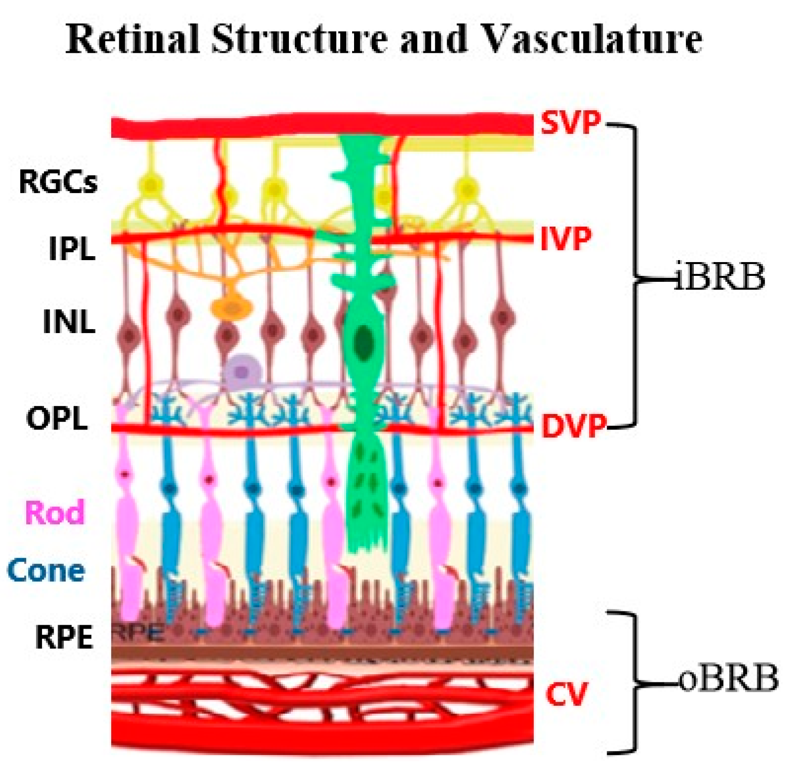
Disclaimer/Publisher’s Note: The statements, opinions and data contained in all publications are solely those of the individual author(s) and contributor(s) and not of MDPI and/or the editor(s). MDPI and/or the editor(s) disclaim responsibility for any injury to people or property resulting from any ideas, methods, instructions or products referred to in the content.

Article Metrics

Citations

Article Access Statistics

Article metric data becomes available approximately 24 hours after publication online.