Article

Identification, Pathogenicity and Fungicide Sensitivity of Colletotrichum Species Causing Anthracnose on Polygonatum cyrtonema Hua

1
Pharmacy Faculty, Hubei University of Chinese Medicine, Wuhan 430065, China
2
Hubei Shizhen Laboratory, Hubei University of Chinese Medicine, Wuhan 430065, China
3
Hubei Monyan Pharmaceutical Co., Ltd., Jingmen 448124, China
*
Authors to whom correspondence should be addressed.
These authors contributed equally to this work.
Agronomy 2025, 15(5), 1215; https://doi.org/10.3390/agronomy15051215
Submission received: 1 April 2025 / Revised: 14 May 2025 / Accepted: 15 May 2025 / Published: 16 May 2025
(This article belongs to the Section Pest and Disease Management)

Abstract

:
Anthracnose significantly threatens the cultivation of Polygonatum cyrtonema, severely impacting its quality and yield. Between 2022 and 2023, 50 Colletotrichum isolates were obtained from diseased leaves collected in three P. cyrtonema production areas within the Two Lakes region of China (Hubei and Hunan provinces). Morphological and molecular analyses identified six Colletotrichum species as the causative agents of anthracnose: C. aenigma, C. siamense, C. gloeosporioides, C. spaethianum, C. fructicola, and C. karsti. Among these pathogens, C. fructicola and C. spaethianum were predominant (82%), while C. siamense and C. fructicola exhibited the highest aggressiveness. Physiological investigations revealed that the optimal temperature range for all six pathogens was 25–28 °C. C. spaethianum thrived under acidic conditions, whereas C. aenigma, C. siamense, and C. gloeosporioides preferred alkaline environments. In contrast, C. fructicola and C. karsti showed no significant response to pH variations. Fungicide screening demonstrated that pyraclostrobin, prochloraz, and carbendazim effectively inhibited the growth of Colletotrichum species. These findings elucidate the epidemiological factors, primary pathogens, and effective control agents for P. cyrtonema anthracnose in the Two Lakes region, providing a basis for developing targeted prevention and control strategies.

1. Introduction

Polygonatum cyrtonema Hua, commonly known as Solomon’s seal, a perennial medicinal plant belonging to the Asparagaceae family, is predominantly found in the central and southern regions of China. The rhizomes of P. cyrtonema possess significant medicinal value and have been utilized clinically in traditional Chinese medicine for over two millennia [1]. This species is commonly used to treat various diseases, including cardiovascular diseases, hyperlipidemia, diabetes, hypertension, chronic bronchitis, and ischemic stroke [2,3]. Extensive research has identified multiple chemical constituents within P. cyrtonema, such as polysaccharides, saponins, and flavonoids, which exhibit a range of pharmacological activities [4]. These include blood sugar regulation, immune system enhancement, memory improvement, anti-ageing, anti-inflammatory, antiviral, and antitumor effects [5]. The increasing interest in the medicinal and health-promoting properties of P. cyrtonema has led to greater market demand, resulting in a significant decline in wild populations. This resource scarcity poses a challenge to the development of the P. cyrtonema industry [6].
Driven by market demand, the cultivation of P. cyrtonema has begun in several regions. Currently, the national cultivation area of P. cyrtonema encompasses approximately 330 acres, generating a comprehensive output value exceeding 1 billion USD. Provinces such as Hunan, Hubei, Anhui, Jiangxi, and Guizhou have established substantial P. cyrtonema cultivation bases [7]. However, the intensive cultivation of P. cyrtonema has precipitated outbreaks of pests and diseases, significantly impacting both the yield and quality. Common diseases affecting P. cyrtonema include anthracnose, leaf blight, leaf spot, black spot, stem rot, and root rot [8,9], with anthracnose being the principal disease responsible for substantial production losses. Anthracnose is induced by pathogens of the Colletotrichum spp. and is characterized by sunken necrotic lesions on leaves, stems, flowers, and fruit, as well as crown and stem rots and seedling blight [9].
Colletotrichum represents a highly intricate fungal genus, distinguished by its extensive species diversity, rapid genetic variation, broad geographical distribution, and wide host range [10]. Numerous Colletotrichum species exhibit the ability to transition between different lifestyles contingent upon resource availability and survival strategies [11]. In China, at least six distinct Colletotrichum species have been documented in association with P. cyrtonema, with regional variations in pathogenic fungi. Specifically, C. fioriniae, C. gloeosporioides, C. fructicola, C. liriopes, and C. boninense have been identified as pathogenic to P. cyrtonema in Chongqing [9,12,13], while C. spaethianum has been reported in the provinces of Hunan, Anhui, and Guizhou [14,15,16]. Despite the frequent association of Colletotrichum species with plant anthracnose, the precise number of Colletotrichum members capable of infecting P. cyrtonema remains unclear, as does the identity of the dominant Colletotrichum species in the primary production regions of P. cyrtonema in China.
The morphological characteristics among Colletotrichum species exhibit significant similarity, complicating the differentiation of closely related species based solely on morphological traits. Recently, the advent of molecular techniques, including the sequencing of internal transcriptional spacer (ITS) regions and glyceraldehyde-3-phosphate dehydrogenase (GAPDH) genes in pathogenic fungi, has facilitated more precise species identification [17]. Nowadays, an integrative approach combining morphological, molecular, and pathogenic characterizations is regarded as the appropriate approach for the accurate identification of Colletotrichum species.
In the present study, we aim to isolate and accurately identify all pathogenic species and dominant populations of Colletotrichum spp. responsible for P. cyrtonema anthracnose in the primary production areas (Hubei and Hunan provinces), utilizing morphological assessment, phylogenetic analysis, and pathogenicity testing. Additionally, the physiological characteristics of the identified Colletotrichum pathogens, such as temperature, pH, and photoperiod preferences, were evaluated. This study will provide a theoretical foundation for the prevention and management of P. cyrtonema anthracnose disease.

2. Materials and Methods

2.1. Disease Samples Collection and Colletotrichum Strain Isolation

Between August 2022 and August 2023, anthracnose disease investigations were conducted in P. cyrtonema cultivation field across three regions of China, including Wuhan, Hubei province (30°27′06″ N, 114°15′57″ E), Huanggang, Hubei province (23°23′18″ N, 113°26′70″ E), and Loudi, Hunan province (27°37′44″ N, 111°20′26″ E). Thirty diseased P. cyrtonema samples exhibiting typical anthracnose symptoms were collected, with ten samples from each field. Colletotrichum spp. were isolated using the method described by Li et al. [8]. Leaf segments (2–3 mm × 5 mm) were aseptically collected from necrosis-healthy tissue junctions. Surface disinfection involved sequential immersion in 5% sodium hypochlorite (2 min) and 75% ethanol (30 s), followed by triple rinsing with sterile water and air-drying. The sterilized samples were transferred to potato dextrose agar (PDA) plates supplemented with 100 µg/mL kanamycin and incubated at 28 °C in complete darkness for 3–7 days. Hyphal development was assessed at 12-h intervals. Fungal colony margins were subcultured through three successive hyphal tip transfers [18] to obtain axenic cultures. Purified strains were preserved in PDA slants at 4 °C for long-term storage.

2.2. Morphological and Cultured Characterization

To elucidate the morphological and cultural characterizations of the pathogens, mycelial plugs (6 mm diameter) were excised from the actively growing margins of fungal isolates and subsequently incubated at 28 °C on PDA. The colony morphology and pigmentation of each isolate were documented after a 7-day incubation period. Conidia were harvested from cultures after 14 days. The appearance, length, and width of fifty conidia were measured and photographed under an IX71 fluorescence inverted microscope (Olympus, Tokyo, Japan).

2.3. DNA Extraction, PCR Amplification, and DNA Sequencing

All isolated strains were subjected to molecular identification. Genomic DNA was extracted from 10-day-old fungal cultures using a modified CTAB extraction method as described by Talbot [19]. Genomic regions of internal transcribed spacer (ITS), actin gene (ACT), glyceraldehyde-3-phosphate dehydrogenase gene (GAPDH) and chitin synthase (CHS) were amplified using the primer pairs ITS1 (5′ TCCGTAGGTGAACCTGCGG 3′)/ITS4 (5′ TCCTCCGCTTATTGATATGC 3′) [20], ACT-512F (5′ ATGTGCAAGGCCGGTTTCGC 3′)/ACT-783R (5′ TACGAGTCCTTCTGGCCCAT 3′) [21], CHS-79F (5′ TGGGGCAAGGATGCTTGGAAGAAG 3′)/CHS-345R (5′ TGGAAGAACCATCTGTGAGAGTTG 3′) [22] and GPD1 (5′ CAACGGCTTCGGTCGCATTG 3′)/GPD2 (5′ GCCAAGCAGTTGGTTGTGC 3′) [23], respectively. PCR amplification was performed on the C1000 Thermal Cycler (Bio-Rad, Hercules, New York, NY, USA) with a total volume of 50 μL. The PCR mixture was prepared with a total volume of 50 μL containing 5 μL of 10× reaction buffer, 1 μL of 10 mM dNTPs, 1 μL each of 10 μM forward and reverse primers, 1 U Taq DNA polymerase, 1 μL genomic DNA template, and 40 μL sterile ddH2O. Amplification conditions included an initial denaturation at 95 °C for 3 min, followed by 34 cycles of 95 °C for 30 s, 55 °C for 30 s, and 72 °C for 30 s, with a final extension at 72 °C for 5 min [8]. PCR products were visualized via 1% agarose gel electrophoresis and subsequently sequenced by Wuhan Tsingke Biotechnology Co., Ltd. before being submitted to GenBank. The sequence similarity and homology with related fungi were assessed by BLAST on the National Center for Biotechnology Information (NCBI) nucleotide database (https://www.ncbi.nlm.nih.gov/). Additionally, a phylogenetic tree was constructed with the maximum likelihood method by MEGA 7.0 software, based on the combined sequences of the ACT, GADPH, and CHS gene sequences. Bootstrap analysis with 500 replications was performed to evaluate the relative stability of the branches.

2.4. Pathogenicity and Aggressiveness Assays of Six Colletotrichum spp.

Using a modified wound inoculation method (using sterile syringes to puncture six uniform wounds in the same area) [24] to inoculate mycelial plugs for in vivo pathogenicity and in vitro invasiveness testing. In pathogenicity assays, healthy leaves from two-year-old P. cyrtonema plants growing in the medicinal botanical garden of Hubei University of Chinese Medicine were selected and washed with sterilized water for inoculation. For per isolate (HJ-11, HJ-18, HJ-34, HJ-41, HJ-43, and HJ-Y2), one leaf was inoculated with one mycelial plug (6 mm diameter) with three replicates. Leaves inoculated with pure PDA plugs (6 mm diameter) served as controls. Disease progression on the inoculated leaves was monitored daily, with lesions photographed at 6 days post-inoculation. The fungi were re-isolated from the leaf lesions after infection to verify Koch’s postulates.
In aggressiveness assays, healthy detached leaves of P. cyrtonema were selected for wound treatment. The leaves were then individually placed into 500 mL rectangular polypropylene containers (13 cm × 10 cm × 5 cm) lined with double-layered filter paper, and 10 mL sterile water was added to ensure humidity. For treatment, mycelial plugs (6 mm diameter) from various Colletotrichum spp. were inoculated onto the wounds of detached leaves. The control was only inoculated with sterile PDA plugs on the wounded sites. Three biological replicates were performed for each treatment group. After sealing the containers, all samples were incubated at a constant temperature of 28 °C. The progression of infection in each experimental group was monitored and recorded daily.
On the 7th day after inoculation, the disease index was employed to assess the severity of leaf lesions induced by different strains. Specifically, leaves with no lesions were assigned a score of 0; those with 1–10% disease areas were scored as 1; 11~30% as 2; 31~50% as 3; 51~70% as 4; and 71–100% disease area or even wilting and leaf death, were scored as 5. The disease index was calculated as follows:
Disease index = ∑ (number of disease grade × representative value of each disease grade)/(total number of plant representative value of highest disease grade) × 100%.

2.5. Determination of Physiological Characteristics

The mycelial growth rate method was employed to evaluate the pathogen’s growth potential under different conditions [25]. To ascertain the optimal temperature, 6 mm mycelium plugs were excised from the periphery of isolate colonies and transferred to PDA plates. These plates were incubated in the dark at seven temperatures (15, 20, 25, 28, 30, 35, and 40 °C). Colony diameters were measured using the crossover method every 24 h intervals, and the experiment was conducted in triplicate.
PDA plates were prepared by adjusting the pH to 5, 6, 7, 8, 9, 10, 11, and 12 using 1 mol/L NaOH and 1 mol/L HCl solutions, followed by sterilization through high-pressure steam. Mycelium plugs (6 mm diameter) from different isolates were inoculated on the prepared plates with various pH values. Each treatment was replicated three times, and all treatments were incubated at 28 °C in the dark. Colony diameters were recorded daily over seven days.
A similar methodology was applied to determine the optimal photoperiod for growth. Photoperiod treatments included continuous light (24 h light), alternating light and darkness (12 h light-12 h darkness), and continuous darkness (24 h dark). PDA plates with pathogen mycelium plugs (6 mm) were incubated under these different photoperiods at 28 °C. Colony diameters were measured daily, with three replicates for each treatment group.

2.6. Fungicide Assays

Referring to other highly effective fungicides for plant anthracnose disease, three fungicides (pyraclostrobin from Yongguan Qiaodi Agricultural Technology Co., Zhengzhou, China; carbendazim from Yingkou Leike Pesticide Co., Yingkou China; and prochloraz from Hunan New Longshan Agricultural Development Co., Changsha, China) were selected to test the inhibition rate of Colletotrichum spp. using the mycelial growth rate method [26,27]. The selection of pyraclostrobin, prochloraz, and carbendazim was based on their distinct modes of action, broad-spectrum efficacy, and practical relevance in agricultural applications. Stock solutions of each fungicide were prepared at a concentration of 10,000 mg/L by dissolving them in sterile water. These solutions were subsequently incorporated into PDA to create media with varying fungicide concentrations. Of these, pyraclostrobin and carbendazim were 0.125, 0.25, 0.5, 1, and 2 μg/mL and prochloraz was 0.065, 0.125, 0.25, 0.5, and 1 μg/mL. PDA medium devoid of fungicide served as the blank control. Mycelial plugs (6 mm) were excised from the periphery of a 5-day-old colony cultured on PDA and centrally placed on a PDA medium containing various fungicide concentrations. All the treatments were cultured at 28 °C for 4 days. Colony diameters were measured using the crossover method, in which two perpendicular lines were drawn across the colony center. The distance between the two intersecting points at the colony edges was recorded as the colony diameter. The growth inhibition rates were calculated using the following formula: Inhibition rate (%) = 100 × (Colony diameter of control group − colony diameter of experimental group)/(Colony diameter of control group − 6 mm).
The half-maximal effective concentration (EC50) values were further calculated to evaluate fungicide efficacy. Microsoft Excel 2016 was used to conduct a regression analysis of the toxicity data, with the logarithm of the fungicide concentration (μg/mL) designated as x and the inhibition rate as y, and to calculate the correlation coefficient for each fungicide. Meanwhile, the EC50 value for each fungicide was determined using SPSS 27.01 software.

3. Results

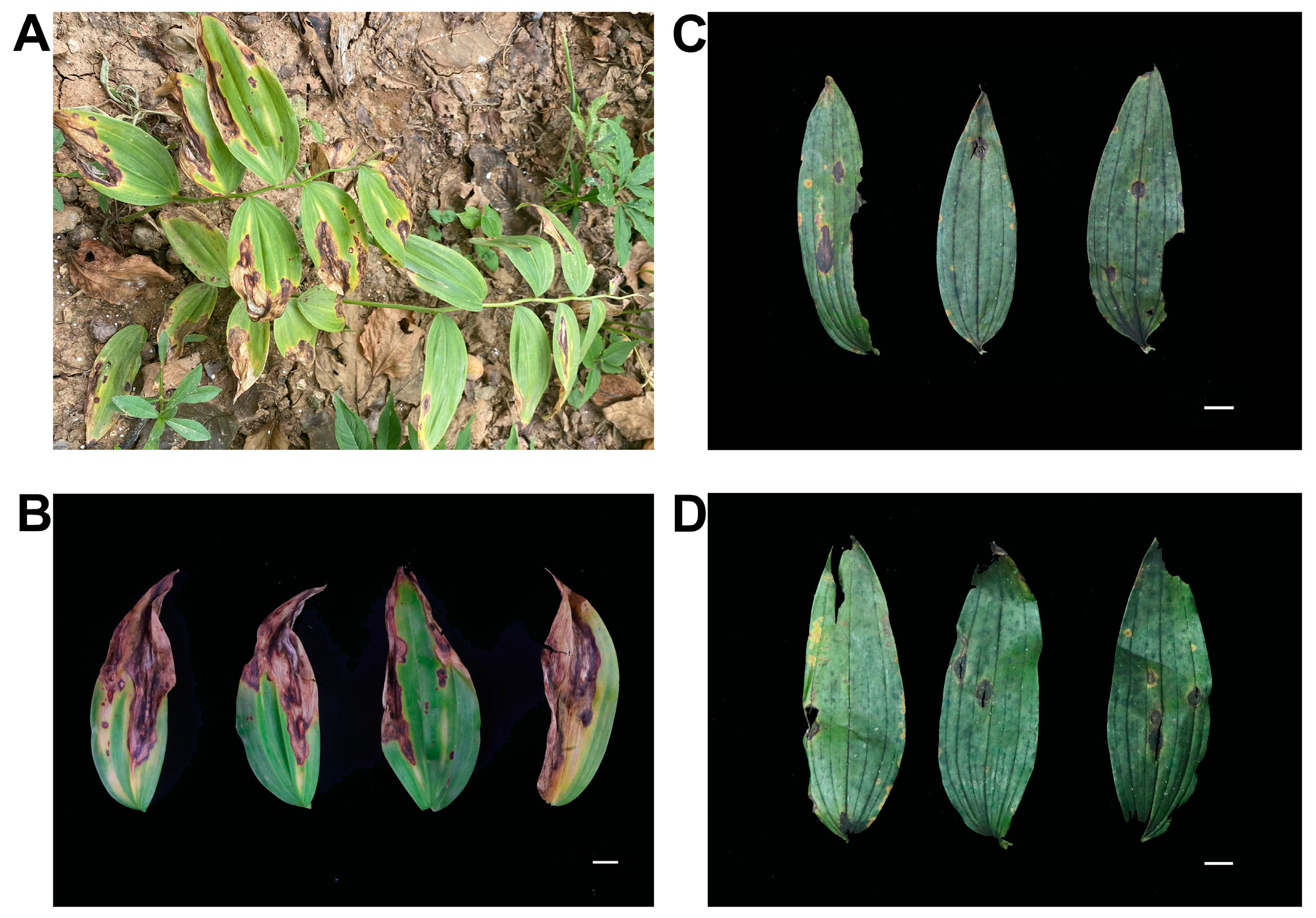
3.1. Disease Symptoms of Anthracnose of P. cyrtonema in Fields and Pathogen Isolation

Disease incidence was notably high during the hot and rainy season, affecting 50% to 70% of the plant population in the fields. Initially, the symptomatic leaves exhibited circular to oval-shaped yellowish-brown or dark-brown depressed spots, each encircled by a distinct yellow halo. As the disease advanced, these lesions expanded and merged into extensive black-brown areas, eventually resulting in plant mortality in severe instances (Figure 1A). Additionally, some affected plants initially displayed irregular dark spots with slight central indentations, which subsequently expanded into larger black lesions (Figure 1B–D). According to the results of field investigation and literature comparison [9,12,13,15,28], we preliminarily judged that this serious disease was anthracnose, which is potentially caused by Colletotrichum species. Pathogen isolation was conducted using the diseased leaves (Figure 1B–D), resulting in the recovery of 120 isolates from 30 infected P. cyrtonema leaves.

3.2. Molecular Identification and Phylogenetic Analysis

To confirm the identity of species, the ITS region was first amplified and sequenced for each of these isolates. A total of 15 genera were identified from the 120 isolates. Among them, Colletotrichum spp. represented 41.67% of the total, while Trichoderma sp. and Alternaria sp. each constituted 11.67%, Botryosphaeria sp. accounted for 7.5%, Fusarium sp. for 5%, and the remaining seven genera collectively comprised 12.5% (Figure 2A). Given that Colletotrichum spp. constituted the largest proportion, and isolates from this genus were selected for further investigation. To achieve species-level identification, the ACT, GAPDH, and CHS genes were amplified and sequenced. The BLAST analysis divided these strains into six categories: one isolate of Colletotrichum aenigma, three isolates of Colletotrichum siamense, four isolates of Colletotrichum gloeosporioides, seventeen isolates of Colletotrichum spaethianum, twenty-four isolates of Colletotrichum fructicola, and one isolate of Colletotrichum karsti. In summary, C. fructicola and C. spaethianum were predominant, comprising 82% of the population, followed by C. gloeosporioides and C. siamense at 8% and 6%, respectively. C. aenigma and C. karsti were the least represented, each accounting for only 2% (Figure 2B). Consequently, six isolates designated as HJ-11, HJ-18, HJ-34, HJ-41, HJ-43, and HJ-Y2 were selected as representatives of six Colletotrichum pathogens for further study.
The HJ-11 strain exhibited the highest sequence similarity to Colletotrichum aenigma, with average identity values of 99.44%, 99.59%, 99.19%, and 99.23% for the ITS, ACT, GAPDH, and CHS sequences, respectively, compared to reference sequences. The HJ-18 strain demonstrated sequence homology of 100%, 99.17%, 99.67%, and 100% with the reference strains of Colletotrichum siamense for the same genetic markers. Similarly, HJ-34 exhibited homology rates of 99.44%, 99.60%, 99.68%, and 100% with Colletotrichum gloeosporioides. The HJ-41 strain shared sequence identities of 100%, 99.17%, 99.66%, and 100% with Colletotrichum spaethianum. Additionally, HJ-43 displayed sequence homology of 99.63%, 99.61%, 99.84%, and 100% with Colletotrichum fructicola. Lastly, HJ-Y2 exhibited homology levels of 100%, 99.61%, 99.02%, and 99.22% with Colletotrichum karsti.
The gene sequences of the six pathogenic isolates were submitted to the GenBank database to obtain the corresponding accession numbers (Table 1). A phylogenetic tree was constructed utilizing the maximum likelihood method in MEGA 7 software, based on the concatenated sequences of the ACT, GADPH, and CHS genes from six representative isolates and forty-four Colletotrichum isolates. As illustrated in Figure 3, the isolates HJ-11, HJ-18, HJ-34, HJ-41, HJ-43, and HJ-Y2 were individually clustered with C. aenigma, C. siamense, C. gloeosporioides, C. spaethianum, C. fructicola, and C. karsti, respectively. In summary, we identified six distinct Colletotrichum species as pathogens responsible for P. cyrtonema anthracnose.

3.3. Morphological Characteristics of Colletotrichum Pathogens of P. cyrtonema Anthracnose

The morphological characteristics of the six pathogens were assessed on the seventh day of growth on PDA. Notable differences in colony morphology and conidia dimensions were observed among the species (Figure 4 and Table 2). Six isolates formed dense aerial mycelium. Among them, C. aenigma and C. spaethianum produced greyish-white colonies, C. siamense, C. gloeosporioides, and C. karsti produced milky-white colonies, and C. fructicola produced whitish-green colonies. On the back side of medium, in addition to the milky white of C. karsti in the whole medium, the middle of C. aenigma, C. spaethianum, and C. fructicola were black, and the middle of C. siamense and C. gloeosporioides were orange, but the color of C. siamense was lighter than that of C. gloeosporioides. The conidia of these isolates were hyaline and smooth. C. aenigma, C. siamense, C. gloeosporioides, and C. karsti were cylindrical, C. spaethianum was acinacifoliate, and C. fructicola was oval or spindle-shaped, slightly curved. The length and width of conidia of the six isolates were similar. Whereas the conidial lengths of C. karsti were the shortest among them, its width was the widest. Overall, the morphological characteristics of these isolates determined in this study were consistent with the previous descriptions and were preliminarily identified as Colletotrichum spp. [13,29,30,31].

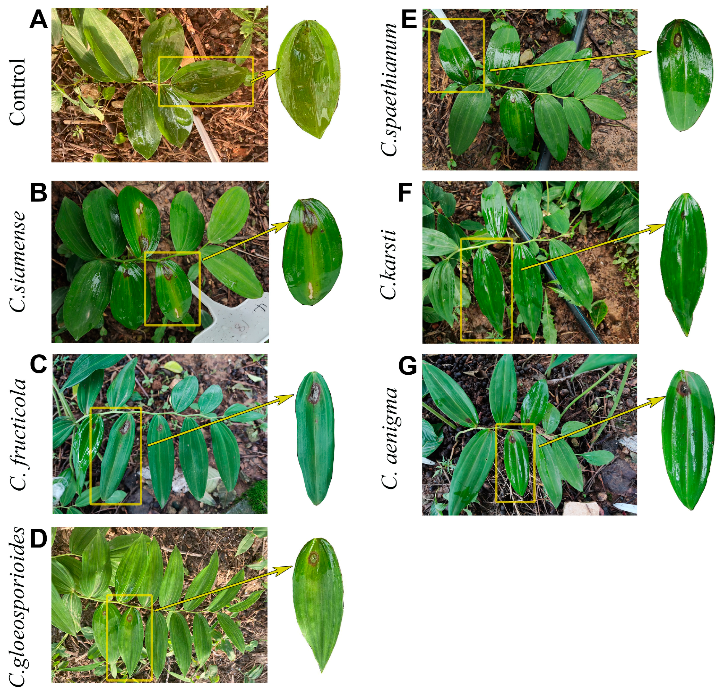
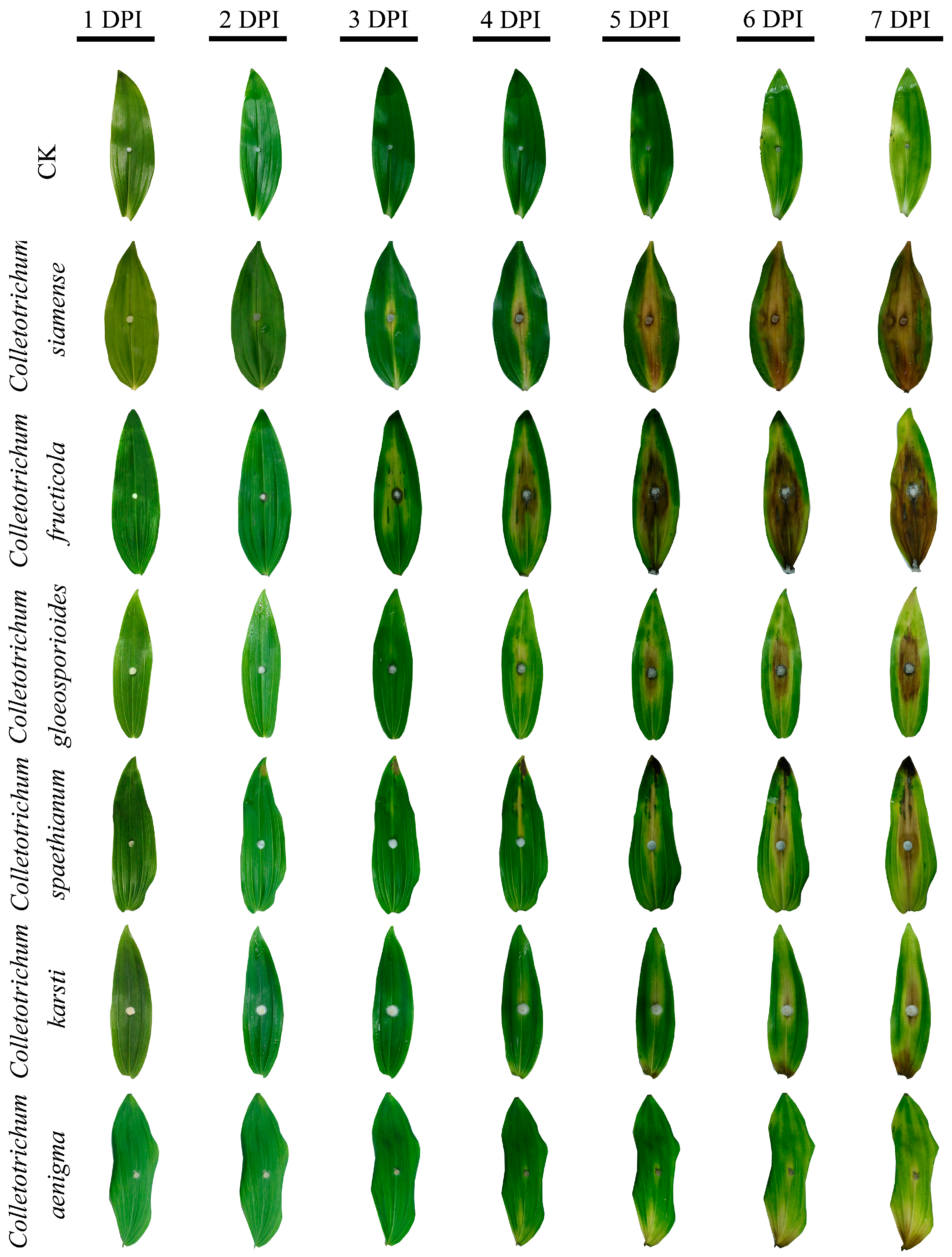
3.4. Pathogenicity Test and Comparison of Six Colletotrichum Species

Representative isolates of all Colletotrichum spp. were pathogenic on plant leaf surfaces (Figure 5). Inoculation with C. siamense (Figure 5B), C. fructicola (Figure 5C), C. gloeosporioides (Figure 5D), C. spaethianum (Figure 5E), C. karsti (Figure 5F), and C. aenigma (Figure 5G) resulted in the manifestation of characteristic anthracnose symptoms. Initially, lesions appeared as water-soaked spots on the leaves, which progressively expanded into larger, circular necrotic lesions. These symptoms were consistent with those observed in field conditions. Leaves inoculated with sterile PDA plugs remained asymptomatic (Figure 5A). Furthermore, the morphological and molecular characteristics of fungal pathogens re-isolated from infected leaves were identical to those of the originally inoculated strains. Consequently, it has been confirmed that these six species are pathogens of P. cyrtonema anthracnose.
To compare the pathogenicity of these six pathogens, an in vitro leaf inoculation experiment was conducted, and the disease index was calculated. The results demonstrated that C. siamense and C. fructicola exhibited high pathogenicity in inducing anthracnose lesions. By three days post-inoculation (DPI), both C. siamense and C. fructicola had successfully infected the leaves and initiated visible lesion formation, whereas leaves inoculated with other strains did not exhibit disease phenotype (Figure 6). By seven days post-inoculation, all inoculated leaves had developed lesions. The disease indices for leaves inoculated with C. siamense, C. fructicola, C. gloeosporioides, C. spaethium, C. karsti, and C. aenigma were 66, 62, 44, 40, 32, and 28, respectively (Figure S1).

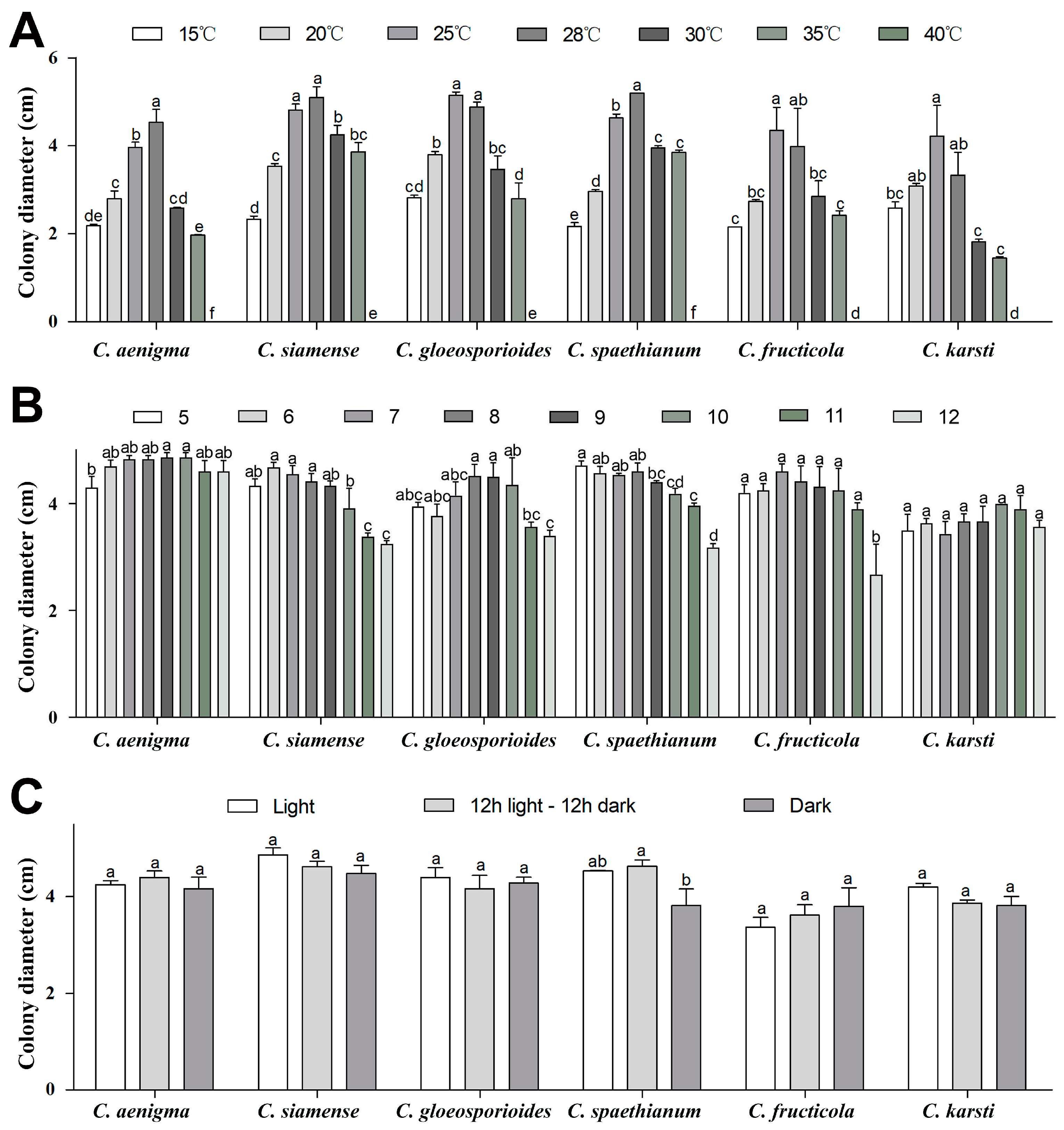
3.5. Cultural Characteristics of the Six Colletotrichum Species

The results in Figure 7A and Figure S2 showed that the six strains were able to grow at temperatures between 15 and 35 °C, whereas their mycelium did not survive at 40 °C. All strains demonstrated a consistent response to temperature variations, with a significant increase in colony growth rate observed as the temperature rose from 15 to 28 °C. However, growth rates declined markedly when temperatures exceeded 28 °C. Specifically, the optimal growth temperature for strains C. karsti and C. fructicola was identified as 25 °C, while strains C. siamense and C. gloeosporioides exhibited optimal growth between 25 and 28 °C, and for strains C. aenigma and C. spaethianum it was 28 °C.
The six strains demonstrated growth in a pH range of 5 to 12 (Figure 7B and Figure S3), although their sensitivity to pH variations differed among strains. Notably, C. aenigma exhibited a relatively rapid growth rate, with optimal growth occurring at pH value levels between 9 and 10, and its growth rate diminished when the pH fell below 5. C. siamense thrived within a pH range of 6 to 8, but its growth rate significantly declined at pH values exceeding 11. C. gloeosporioides was found to be optimal for growth at pH levels of 8 to 9, with significant growth inhibition observed at pH values above 12. In contrast, C. spaethianum achieved optimal mycelial growth at pH 5, while C. fructicola was suitable for growth across a pH range of 5 to 11. C. karsti has the strongest tolerance to both acidic and alkaline conditions, showing no significant difference in mycelium growth across varying pH levels.
Furthermore, the photoperiod did not significantly impact the growth of all isolates except C. spaethianum, which displayed accelerated growth under a 12-h light/12-h darkness cycle compared to continuous light or darkness conditions (Figure 7C and Figure S4).

3.6. Fungicide Sensitivity Testing of Colletotrichum Species

To identify highly effective fungicides against P. cyrtonema anthracnose, fungicide sensitivity assays were performed on four of the most aggressive Colletotrichum species. Various fungicide types and concentrations resulted in different inhibition rates among the four strains after a 4-day incubation on fungicide-containing PDA (Table 3). Among the three fungicides tested, prochloraz demonstrated the highest efficacy in suppressing the mycelial growth of C. siamense and C. gloeosporioides, with EC50 values of 0.239 and 0.265 μg/mL, respectively. This was followed by 50% carbendazim, with EC50 values of 0.753 and 0.688 μg/mL, and 30% pyraclostrobin, with EC50 values of 0.825 and 1.861 μg/mL, respectively. However, pyraclostrobin exhibited the most potent inhibitory effect on the growth of C. spaethianum and C. fructicola with EC50 values of 0.017 and 0.087 μg/mL, respectively. This was followed by 450 g/l prochloraz, with EC50 values of 0.363 and 0.213 μg/mL, and 50% carbendazim, with EC50 values of 0.852 and 0.540 μg/mL. These findings indicate that the three fungicides could be effectively utilized for the management of Colletotrichum spp. that caused P. cyrtonema anthracnose.

4. Discussion

The objective of this study is to identify and compare the Colletotrichum spp. associated with anthracnose of P. cyrtonema, combining morphological characteristics with phylogenetic analysis. The complex infection by multiple Colletotrichum species has led to the occurrence of anthracnose. Our analysis of 50 isolates revealed six distinct taxa: C. aenigma, C. siamense, C. gloeosporioides, C. spaethianum, C. fructicola, and C. karsti. Previous studies have identified C. gloeosporioides and C. fructicola as causal agents of anthracnose in P. cyrtonema in Chongqing, China, while C. spaethianum has been reported in Anhui and Guizhou Province, China [13,14,16]. Our findings corroborate previous research, indicating that the above-mentioned species are the predominant pathogen responsible for P. cyrtonema anthracnose in China. Moreover, we isolated C. aenigma, C. siamense, and C. karsti for the first time from the diseased samples, and verified their pathogenicity through in vitro and in vivo inoculation, enriching the pathogen library of P. cyrtonema anthracnose. While this study primarily investigated Colletotrichum populations in Hubei and Hunan provinces, Colletotrichum is distributed worldwide [32], suggesting potential pathogen adaptability to diverse environments. Future studies should expand sampling to other major P. cyrtonema cultivation zones and compare pathogen composition under varying climatic and soil conditions. This would clarify whether the observed species dominance reflects regional ecological selection or universal pathogenicity patterns.
The co-occurrence of six Colletotrichum species raises questions about potential interspecific interactions. Notably, C. siamense and C. fructicola exhibited the highest pathogenicity individually, but field observations of complex infections suggest possible synergistic effects. Such as Fusarium oxysporum and Alternaria tenuissima complex infection causes stem blight in sweetpotato [33]. In the future, double inoculation experiments can be used to explore the synergistic effects of these pathogens and then study the mode of interspecies interactions and synergistic pathogenic mechanisms between these pathogens.
In our systematic investigation of the predominant populations and pathogenicity of Colletotrichum species in the Hubei and Hunan provinces, we identified C. fructicola and C. spaethianum as the principal pathogens, comprising over 80% of the total isolates. Currently, C. fructicola has been documented in a variety of hosts, including Ixora chinensis [34], pecan [35], peach [36], Camellia chrysantha [37], grape [38], apple [39], Ixora chinensis [40], Paeonia lactiflora [41], Rosa chinensis [42], and Epimedium sagittatum [43]. This species exhibits a broad geographical distribution and has been reported across all five continents, including the Americas, Western and Eastern Asia, Western Europe, Western Africa, and Australia [40]. C. spaethianum is a very common causal agent of anthracnose worldwide [44], it was originally discovered on Funkia univittata in Germany and subsequently classified within the Colletotrichum genus as a separate species [45]. Furthermore, C. siamense and C. fructicola demonstrate the highest pathogenicity on P. cyrtonema compared to other Colletotrichum species. In summary, C. fructicola and C. spaethianum are characterized by their extensive distribution, diverse host range, and strong infectivity, thereby playing a critical role in the global threat of plant anthracnose.
Temperature has been considered the primary factor influencing the prevalence of plant diseases [46]. There was no significant difference in the minimum and optimal growth temperatures of the six Colletotrichum spp., while C. siamense has an extremely high tolerance to high temperatures and can grow normally even at 35 °C. Anthracnose always occurs in hot and rainy seasons. Studies have shown that increasing temperatures, changing precipitation patterns, and extreme weather events can directly affect the life cycle, population dynamics, and geographic distribution of plant pathogens [47]. Understanding the complex relationship between climate change and Polygonatum cyrtonema diseases is essential for developing adaptive strategies. This requires establishing integrated monitoring networks that correlate meteorological patterns with pathogen dynamics, enabling targeted interventions to reduce disease incidence while ensuring sustainable cultivation of medicinal crops. Furthermore, the effects of pH and photoperiod on the growth rate of Colletotrichum spp. were investigated. Highly pathogenic pathogens, including C. siamense, C. fructicola, C. gloeosporioides, and C. spaethianum, exhibited vigorous growth under acidic to mildly alkaline conditions, whereas their growth was markedly inhibited in strong alkaline environments. In contrast, weakly pathogenic pathogens (C. aenigma and C. karsti) were capable of normal growth across a broader pH spectrum. This provides valuable insights for the ecological prevention of anthracnose through soil pH adjustment. Regarding photoperiod, the growth rate of various Colletotrichum spp. was minimally impacted.
Currently, synthetic fungicide application is the most efficacious strategy for anthracnose management [48]. Notably, pyraclostrobin, carbendazim, and prochloraz have demonstrated significant protective and therapeutic effects and are recommended for the early intervention of anthracnose [49,50]. Among these, research indicates that prochloraz exhibits a substantial inhibitory effect on numerous C. gloeosporioides species by disrupting ergosterol biosynthesis within the cell membrane of the pathogenic fungus [51,52,53]. Carbendazim, classified as a benzimidazole fungicide, exerts its antifungal activity by affecting fungal cell division and deforming the germ tubes emerging from spores [54]. Furthermore, pyraclostrobin belongs to the strobilurin and strobilurin-related fungicides, which function by inhibiting mitochondrial respiration [55]. Despite these advancements, there remains a lack of comprehensive screening and research concerning effective fungicides for the prevention and treatment of P. cyrtonema anthracnose. This study evaluates the sensitivity levels of three fungicides (pyraclostrobin, prochloraz, and carbendazim) against C. siamense, C. gloeosporioides, C. fructicola, and C. spaethianum. C. siamense and C. gloeosporioides isolates were more sensitive to prochloraz, while C. spaethianum and C. fructicola were most sensitive to pyraclostrobin. These species-specific sensitivity patterns highlight the need for precision fungicide selection based on regional pathogen composition. However, the current evaluation focused on three widely used fungicides, leaving other commercially available compounds untested. Moreover, long-term monitoring is essential given that resistance to pyraclostrobin in C. siamense has been documented to be associated with the G143A point mutation [56]. Integrating molecular diagnostics with fungicide rotation schemes could mitigate resistance development while maintaining control efficacy.
Notably, the present study found significant differences in the pathogenicity of different anthracnose pathogens on P. cyrtonema, suggesting that harnessing host resistance resources may serve as a core strategy for sustainable disease management. In the future, we can analyze the molecular mechanism differences between high and low aggressive Colletotrichum species to identify key virulence factors. Concurrently, the resistance or tolerance of P. cyrtonema resources to major pathogens should be systematically evaluated, with particular emphasis on their performance under high temperatures, high humidity, and other stress-prone environmental conditions. By resolving the genetic and biochemical mechanisms related to resistance, the selection and breeding of resistant varieties can be accelerated. Combining resistant varieties, precise fungicide application, and ecological regulation, it is expected to build an integrated management system for P. cyrtonema anthracnose and reduce the dependence on chemical agents.

5. Conclusions

In this study, the pathogens responsible for P. cyrtonema anthracnose were identified as Colletotrichum aenigma, Colletotrichum siamense, Colletotrichum gloeosporioides, Colletotrichum spaethianum, Colletotrichum fructicola, and Colletotrichum karsti through the integration of multilocus phylogenetic analyses, morphological characterization, and pathogenicity validation. This finding highlights the capability of multiple Colletotrichum species to infect a single host (P. cyrtonema). Notably, this study provides the first report identifying C. aenigma, C. siamense, and C. karsti as causal agents of P. cyrtonema anthracnose in China. Biological characterization indicated that different factors, such as temperature, pH, and light, could affect the growth of Colletotrichum species. In addition, the sensitivity test suggests that carbendazim, pyraclostrobin, and prochloraz are effective in controlling the Colletotrichum spp. responsible for P. cyrtonema anthracnose. These discoveries elucidate the epidemiological factors, primary pathogens, and control agents associated with P. cyrtonema anthracnose, thereby providing a theoretical foundation for the development of prevention and management strategies for this disease.

Supplementary Materials

The following supporting information can be downloaded at: https://www.mdpi.com/article/10.3390/agronomy15051215/s1, Figure S1: Disease index of six Colletotrichum spp. infecting P. cyrtonema leaves. Figure S2: Mycelial growth of six isolates under different temperatures. Figure S3: Mycelial growth of six isolates under different pH values. Figure S4: Mycelial growth of six isolates under different photoperiods.

Author Contributions

Conceptualization, H.C. (Huixia Cai) and J.L.; methodology, H.C. (Huixia Cai); software, H.C. (Huixia Cai) and K.Q.; validation, H.C. (Huixia Cai) and J.L.; formal analysis, H.C. (Hong Chen); investigation, J.L. and Y.D.; resources, D.L.; data curation, H.C. (Huixia Cai), D.W. and J.C.; writing—original draft preparation, H.C. (Huixia Cai); writing—review and editing, J.L. and Y.M.; visualization, K.Q.; supervision, J.L. and D.L.; project administration, Y.M.; funding acquisition, D.L. All authors have read and agreed to the published version of the manuscript.

Funding

This research was funded by the project for National key research and development plan (2023YFC3503804); Science and technology innovation program of Hubei Province (2024BBB088); The fourth batch of modern agricultural industrial technology system projects in Hubei Province (2023HBSTX4-07); special fund for the construction of modern agricultural industrial technology system (CARS-21) and Traditional Chinese medicine innovation team project of hubei provincial administration of TCM (ZY2025J002).

Data Availability Statement

The data presented in this study area available within the paper.

Conflicts of Interest

Author Jinxin Li was employed by the company Hubei Monyan Pharmaceutical Co., Ltd. The remaining authors declare that the research was conducted in the absence of any commercial or financial relationships that could be construed as a potential conflict of interest.

References

  1. Fan, Q.J.; Zhou, Q.Y.; Zhang, S.; Li, Y.; Li, J.; Chen, X.; Sun, L. First report of leaf spot disease caused by Alternaria alternata on Polygonatum cyrtonema Hua in Hunan province of China. Plant Dis. 2024, 108, 530. [Google Scholar] [CrossRef]
  2. Li, M.; Chen, X.; Li, Y.; Yuan, Y. Research progress on pharmacological effects of Polygonatum polysaccharides. Acta Chin. Med. Pharmacol. 2024, 52, 95–101. [Google Scholar] [CrossRef]
  3. Wang, Y.; Liu, W.; Shen, B.; Zhao, R.; Xu, L.; Xie, M.; Kang, T. Textual research of Polygonatum sibiricum. Acta Chin. Med. Pharmacol. 2019, 47, 81–86. [Google Scholar] [CrossRef]
  4. Chen, H.; Feng, S.; Sun, Y.; Hao, Z.; Feng, W.; Zheng, X. Advances in studies on chemical constituents of three medicinal plants from Polygonatum Mill. and their pharmacological activities. Chin. Tradit. Herb. Drugs 2015, 46, 2329–2338. [Google Scholar] [CrossRef]
  5. Su, W.; Liu, Y.; Jiang, Y.; Xie, J.; Pan, X.; Liu, J.; Si, J. Status of Polygonati rhizome industry and suggestion for its sustainable development. China J. Chin. Mater. Medica 2018, 43, 2831–2835. [Google Scholar] [CrossRef]
  6. Chen, X.; Ye, W.; Zhong, J. Research on the application and industrial development of the medicinal plant Polygonatum sibiricum Red. Hortic. Seed 2024, 44, 62–63+66. [Google Scholar] [CrossRef]
  7. Luo, M.; Zhang, W.; Deng, C.; Tan, Q.; Luo, C.; Luo, S. Advances in studies of medicinal crop Polygonatum cyrtonema Hua. Lishizhen Med. Mater. Medica Res. 2016, 27, 1467–1469. [Google Scholar] [CrossRef]
  8. Li, J.; Cai, H.; Du, Y.; Chen, H.; Qu, K.; Wei, L.; Miao, Y.; Liu, D. Morphological, molecular and pathogenic characterization of Botryosphaeria dothidea causing leaf blight disease of Polygonatum cyrtonema in China. Physiol. Mol. Plant Pathol. 2024, 134, 102424. [Google Scholar] [CrossRef]
  9. Dan, Y.; Ma, W.; Tang, Z.; Xu, Q.; Song, Z.; Liu, M.; Yin, F. Biological characterization and fungicide screening of Colletotrichum fioriniae causing Polygonatum cyrtonema Hua anthracnose. J. Chin. Med. Mater. 2023, 46, 1876–1881. [Google Scholar] [CrossRef]
  10. Jeffries, P.; Dodd, J.C.; Jeger, M.J.; Plumbley, R.A. The biology and control of Colletotrichum species on tropical fruit crops. Plant Pathol. 1999, 39, 343–366. [Google Scholar] [CrossRef]
  11. Wang, Y.; Hao, X.; Wang, L.; Bin, X.; Wang, X.; Yang, Y. Diverse Colletotrichum species cause anthracnose of tea plants (Camellia sinensis (L.) O. Kuntze) in China. Sci. Rep. 2016, 6, 35287. [Google Scholar] [CrossRef] [PubMed]
  12. Dan, Y.; Liu, M.; Cui, X.; Ma, W.; Tang, Z.; Yin, F. Identification of pathogen causing anthracnose on Polygonatum cyrtonema Hua. Plant Prot. 2022, 49, 288–293. [Google Scholar] [CrossRef]
  13. Dan, Y.; Tang, Z.; Ma, W.; Cui, X.; Xu, Q.; Song, Z.; Yin, F.; Liu, M. Identification, biological characterization, and fungicide screening of the pathogen causing Polygonatum cyrtonema Hua anthracnose. Acta Phytopathol. Sin. 2023, 53, 796–809. [Google Scholar] [CrossRef]
  14. Ma, J.; Xiao, X.; Wang, X.; Guo, M. Colletotrichum spaethianum causing anthracnose on Polygonatum cyrtonema Hua in Anhui province, China. Plant Dis. 2020, 105, 509. [Google Scholar] [CrossRef] [PubMed]
  15. Qiu, Z.; Chen, J.; Zhang, Z.; Zhong, J.; Zhu, J. Identification and fungicide screening of pathogen causing anthracnose on Polygonatum cyrtonema (Huangjing) in Hunan province of China. Southwest China J. Agric. Sci. 2023, 36, 91–97. [Google Scholar] [CrossRef]
  16. Tan, Q.; Li, J.; Xue, Y.; Zhao, D.; Yang, C.; He, H. Identification of the pathogen of anthracnose of Polygonatum cyrtonema from Guizhou. J. Chin. Med. Mater. 2023, 46, 12–16. [Google Scholar] [CrossRef]
  17. Kim, C.H.; Hassan, O.; Chang, T. Diversity, pathogenicity, and fungicide sensitivity of Colletotrichum species associated withapple anthracnose in South Korea. Plant Dis. 2020, 104, 2866–2874. [Google Scholar] [CrossRef]
  18. Prasad, M.S.L.; Sujatha, K.; Naresh, N.; Rao, S.C. Variability in Sclerotium rolfsii associated with collar rot of sunflower. Indian Phytopathol. 2012, 65, 161–165. [Google Scholar] [CrossRef]
  19. Talbot, N.J. Nucleic Acid Isolation and Analysis; Oxford University Press: Oxford, UK, 2001. [Google Scholar]
  20. White, T.J.; Bruns, T.; Lee, S.; Taylor, J. Amplification and direct sequencing of fungal ribosomal RNA genes for phylogenetics. PCR protocols: A guide to methods and applications. Mycologia 1990, 18, 315–322. [Google Scholar] [CrossRef]
  21. Carbone, I.; Kohn, L.M. A method for designing primer sets for speciation studies in filamentous ascomycetes. Mycologia 1999, 91, 553–556. [Google Scholar] [CrossRef]
  22. O’Donnell, K.; Cigelnik, E. Two divergent Intragenomic rDNA ITS2 types within a monophyletic lineage of the fungus Fusarium are nonorthologous. Mol. Phylogenetics Evol. 1997, 7, 103–116. [Google Scholar] [CrossRef] [PubMed]
  23. Templeton, M.D.; Rikkerink, E.H.A.; Solon, S.L.; Crowhurst, R.N. Cloning and molecular characterization of the glyceraldehyde-3-phosphate dehydrogenase-encoding gene and cDNA from the plant pathogenic fungus Glomerella cingulata. Gene 1992, 122, 225–230. [Google Scholar] [CrossRef]
  24. Zhang, L.; Li, X.; Zhou, Y.; Tan, G.; Zhang, L. Identification and characterization of Colletotrichum species associated with Camellia sinensis anthracnose in Anhui province, China. Plant Dis. 2021, 105, 2649–2657. [Google Scholar] [CrossRef]
  25. Hou, Y.; Xin, H.; Zhang, X.; Fan, X.; Liu, S.; Xu, J. Sensitivity of Fusarium pseudograminis to epoxiconazole in Henan province. Acta Phytopathol. Sin. 2023, 53, 307–316. [Google Scholar] [CrossRef]
  26. Machuca, A.; Durán, N. Optimization of some parameters influencing Thermoascus aurantiacus growth: Effects of lignin-related compounds. J. Ind. Microbiol. 1996, 16, 224–229. [Google Scholar] [CrossRef]
  27. Wang, K.; Jiang, S.; Pu, T.; Fan, L.; Su, F.; Ye, M. Antifungal activity of phenolic monoterpenes and structure-related compounds against plant pathogenic fungi. Nat. Prod. Res. 2019, 33, 1423–1430. [Google Scholar] [CrossRef]
  28. Deng, H.; Wang, Y.; Chen, S.; Jin, C.; Chen, Y.; Guo, K. Identification and laboratory toxicity test of the pathogen of leaf blight of Polygonatum cyrtonema in Xinhua of Hunan province. Hunan Agric. Sci. 2023, 4, 61–65. [Google Scholar] [CrossRef]
  29. Feng, W.; Wang, C.; Ju, Y.; Chen, Z.; Wu, X.; Fang, D. Identifying the biological characteristics of anthracnose pathogens of Blueberries (Vaccinium corymbosum L.) in China. Forests 2024, 15, 117. [Google Scholar] [CrossRef]
  30. Kaur, D.; Kaur, A.; Singh, H.; Arora, A. First report of Colletotrichum siamense causing fruit drop in kinnow mandarin (Citrus nobilis x Citrus deliciosa) in Punjab, India. Crop Prot. 2024, 175, 106464. [Google Scholar] [CrossRef]
  31. Zhang, A.; Li, L.; Xie, X.; Chai, A.; Shi, Y.; Xing, D.; Yu, Z.; Li, B. Identification and genetic diversity analysis of the pathogen of anthracnose of pepper in Guizhou. Plants 2024, 13, 728. [Google Scholar] [CrossRef]
  32. Talhinhas, P.; Baroncelli, R. Colletotrichum species and complexes: Geographic distribution, host range and conservation status. Fungal Divers. 2021, 110, 109–198. [Google Scholar] [CrossRef]
  33. Qian, H.; Xu, P.; Chi, M.; Huang, J. Mixed infection by Fusarium oxysporum and Alternaria tenuissima on sweet potato Fusarium wilt. J. Plant Prot. 2017, 44, 867–868. [Google Scholar] [CrossRef]
  34. Qin, R.; Zhang, Y.; Li, Q.; Huang, S.; Chen, X.; Guo, T.; Tang, L. Leaf spot caused by Colletotrichum siamense, C. fructicola, and C. aeschynomenes on Ixora chinensis in Guangxi, China. Plant Dis. 2024, 108, 225. [Google Scholar] [CrossRef]
  35. Chang, J.; Zhai, F.; Zhang, Y.; Wang, D.; Shu, J.; Yao, X. Identification and characterization of Colletotrichum fioriniae and C. fructicola that cause anthracnose in pecan. Front. Plant Sci. 2022, 13, 1043750. [Google Scholar] [CrossRef]
  36. Muhammad, U.H.; Tan, Q.; Mazharul, K.M.; Muhammad, A.; Yin, W.; Zhu, F.; Luo, C. Sensitivity of C. fructicola and C. siamense of peach in China to multiple classes of fungicides and characterization of pyraclostrobin-resistant isolates. Plant Dis. 2021, 105, 3459–3465. [Google Scholar] [CrossRef]
  37. Zhao, J.; Liu, T.; Zhang, D.; Wu, H.; Pan, L.; Liao, N.; Liu, W. First report of anthracnose caused by Colletotrichum siamense and C. fructicola of Camellia chrysantha in China. Plant Dis. 2021, 105, 2020. [Google Scholar] [CrossRef] [PubMed]
  38. Guginski-Piva, C.A.; Bogo, A.; Gomes, B.R.; Menon, J.K.; Nodari, R.O.; Welter, L.J. Morphological and molecular characterization of Colletotrichum nymphaeae and C. fructicola associated with anthracnose symptoms of grape in Santa Catarina State, southern Brazil. J. Plant Dis. Prot. 2018, 125, 405–413. [Google Scholar] [CrossRef]
  39. Park, M.S.; Kim, B.R.; Park, I.H.; Hahm, S.S. First report of two Colletotrichum Species associated with bitter rot on apple fruit in Korea—C. fructicola and C. siamense. Mycobiology 2018, 46, 154–158. [Google Scholar] [CrossRef]
  40. Li, H.; Zhou, G.; Liu, J.; Xu, J. Population Genetic Analyses of the Fungal Pathogen Colletotrichum fructicola on Tea-Oil Trees in China. PLoS ONE 2017, 11, e0156841. [Google Scholar] [CrossRef]
  41. Kang, N.; Xiang, M.; Xiao, L.; Zheng, Z.; Yang, H.; Zeng, J.; Jiang, H.; Chen, J.; Chen, M. First report of Colletotrichum fructicola causing anthracnose on Paeonia lactiflora in China. Plant Dis. 2023, 107, 4022. [Google Scholar] [CrossRef]
  42. Du, L.; Du, C.; Ding, C. First report of Colletotrichum fructicola causing anthracnose on Rosa chinensis in China. Plant Dis. 2023, 107, 3316. [Google Scholar] [CrossRef] [PubMed]
  43. Hou, W.; Chu, L.; Yang, L.; Dong, N.; Dan, Z.; Zhong, H.; Dong, C. First report of Colletotrichum fructicola causing anthracnose on Epimedium sagittatum in China. Plant Dis. 2024, 108, 813. [Google Scholar] [CrossRef]
  44. Huang, D.; Li, M.; Qin, Q.P. Identification, pathogenicity, and fungicide sensitivity of Colletotrichum Spaethianum isolates, the causal agents of anthracnose of daylily in Shanghai, China. Res. Sq. 2022. [Google Scholar] [CrossRef]
  45. Hyde, K.D.; Cai, L.; Cannon, P.F.; Crouch, J.A.; Zhang, J.Z. Colletotrichum—Names in current use. Fungal Divers. 2009, 39, 147–182. [Google Scholar] [CrossRef]
  46. Bonde, M.R.; Nester, S.E.; Berner, D.K. Effects of daily temperature highs on development of Phakopsora pachyrhizi on soybean. Phytopathology 2012, 102, 761–768. [Google Scholar] [CrossRef] [PubMed]
  47. Nega, A. Climate change impacts on agriculture: A review of plant diseases and insect pests in Ethiopia and East Africa, with adaptation and mitigation strategies. Adv. Agric. 2025, 2025, 5606701. [Google Scholar] [CrossRef]
  48. Ciofini, A.; Negrini, F.; Baroncelli, R.; Baraldi, E. Management of Post-Harvest Anthracnose: Current Approaches and Future Perspectives. Plants 2022, 11, 1856. [Google Scholar] [CrossRef]
  49. Ma, J.; Yuan, Q.; Wang, Y.; Miao, L.; Xin, B. Screening of chemical Fungicides for controlling Alfalfa anthracnose caused by Colletotrichum linicola. Chin. J. Grassl. 2016, 38, 84–90+115. [Google Scholar] [CrossRef]
  50. Fan, K.; Qi, Y.; Fu, L.; Li, L.; Liu, X.; Qu, J.; Li, D.; Dong, A.; Peng, Y.; Wang, Q. Identification and Fungicide Screening of Fungal Species Associated with Walnut Anthracnose in Shaanxi and Liaoning Provinces, China. Plant Dis. 2024, 108, 599–607. [Google Scholar] [CrossRef]
  51. Mora-Aguilera, J.A.; Ríos-López, E.G.; Yáñez-Zúñiga, M.; Rebollar-Alviter, A.; Nava-Díaz, C.; Leyva-Mir, S.G.; Sandoval-Islas, J.S.; Tovar-Pedraza, M.J. Sensitivity to MBC fungicides and prochloraz of Colletotrichum gloeosporioides species complex isolates from mango orchards in Mexico. J. Plant Dis. Prot. 2021, 128, 481–491. [Google Scholar] [CrossRef]
  52. Soe, Z.Z.; Shin, Y.H.; Kang, H.S.; Jeun, Y.C. Chemical Resistance of Diaporthe citri against Systemic Fungicides on Citrus. Plant Pathol. J. 2023, 39, 351–360. [Google Scholar] [CrossRef] [PubMed]
  53. Vinggaard, A.M.; Hass, U.; Dalgaard, M.; Andersen, H.R.; BonefeldJørgensen, E.; Christiansen, S.; Laier, P.; Poulsen, M.E. Prochloraz: An imidazole fungicide with multiple mechanisms of action. Int. J. Androl. 2006, 29, 186–192. [Google Scholar] [CrossRef] [PubMed]
  54. Bai, S.; Zhang, M.; Tang, S.; Li, M.; Wu, R.; Wan, S.; Chen, L.; Wei, X.; Li, F. Research Progress on Benzimidazole Fungicides: A Review. Molecules 2024, 29, 1218. [Google Scholar] [CrossRef] [PubMed]
  55. Karadimos, D.A.; Karaoglanidis, G.S.; Tzavella–Klonari, K. Biological activity and physical modes of action of the Qo inhibitor fungicides trifloxystrobin and pyraclostrobin against Cercospora Beticola. Crop Prot. 2005, 24, 23–29. [Google Scholar] [CrossRef]
  56. Hu, S.; Zhang, S.; Xiao, W.; Liu, Y.; Yu, H.; Zhang, C. Diversity and characterization of resistance to pyraclostrobin in Colletotrichum spp. from Strawberry. Agronomy 2023, 13, 2824. [Google Scholar] [CrossRef]
Figure 1. Typical symptoms of Polygonatum cyrtonema anthracnose under field conditions. (A) Field symptoms. (BD) The diseased leaves used for pathogen isolation. (Scale bars: 1 cm).
Agronomy 15 01215 g001
Figure 2. The proportion of different genera of fungi isolated from disease leaves of P. cyrtonema (A) and the proportion of different species cause P. cyrtonema anthracnose (B).
Agronomy 15 01215 g002
Figure 3. Maximum likelihood phylogenetic tree based on the concatenated dataset (ACT, GAPDH, and CHS) from HJ-11, HJ-18, HJ-34, HJ-41, HJ-43, and HJ-Y2 with 500 bootstrap replicates. Bar = 0.05 substitutions per nucleotide position. Red font is the sequence of this study.
Agronomy 15 01215 g003
Figure 4. Morphological characterization of six pathogenic isolates. (A) top and (B) reverse sides on PDA medium after 7-day culture at 28 °C. (C) Conidia. Bar = 10 μm.
Agronomy 15 01215 g004
Figure 5. Pathogenicity assays of P. cyrtonema anthracnose pathogens in vivo. (A) Control. (B) C. siamense. (C) C. fructicola. (D) C. gloeosporioides. (E) C. spaethianum. (F) C. karsti. (G) C. aenigma.
Agronomy 15 01215 g005
Figure 6. Comparison of pathogenic intensity of six Colletotrichum species on P. cyrtonema.
Agronomy 15 01215 g006
Figure 7. Mycelial growth of six pathogenic isolates under different conditions. (A) temperatures, (B) pH, (C) Photoperiod. Different lower-case letters represent a significant difference amongst colony diameter data within each isolate according to One-way ANOVA (p < 0.05).
Agronomy 15 01215 g007
Table 1. Information of isolates identified in this study for phylogenetic analyses.
TaxonStrainsGenBank Accession Numbers
ITSACTGAPDHCHS
Colletotrichum aenigmaHJ-11PP551230PP227177PP227179PP5558343
Colletotrichum siamenseHJ-18PP551582PP227180PP227181PP227182
Colletotrichum gloeosporioidesHJ-34PP551515PP227183PP227167PP227184
Colletotrichum spaethianumHJ-41PP551521PP227168PP227170PP227169
Colletotrichum fructicolaHJ-43PP551516PP227171PP227173PP227172
Colletotrichum karstiHJ-Y2PP551573PP227174PP227176PP227175
Table 2. Morphological characteristics of conidia in each of the six isolates.
IsolatesAppearanceLength
Mean (Range, SD) (µm) (n = 50)
Width
Mean (Range, SD) (µm) (n = 50)
C. aenigmacylindrical, aseptate, transparent14.9 (10.71~22.16, 2.24)6.26 (4.23~9.60, 1.16)
C. siamensecylindrical, aseptate, transparent15.01 (10.26~17.71, 1.59)5.31 (3.36~6.45, 0.6)
C. gloeosporioidescylindrical, aseptate, transparent13.74 (10.77~17.44, 1.55)4.52 (3.04~6.34, 0.7)
C. spaethianumacinacifoliate, aseptate, transparent15.40 (9.24~19.87, 2.08)4.50 (3.37~8.95, 0.81)
C. fructicolamonocytes, transparent, oval, or spindle-shaped, slightly curved16.93 (9.62~21.07, 2.32)5.85 (4.16~7.37, 0.81)
C. karsticylindrical with both ends rounded, transparent, smooth, aseptate12.33 (9.40~15.59, 1.52)7.09 (5.87~7.93, 0.57)
Table 3. Toxicity of three fungicides.
IsolatesFungicide NameToxic Regression EquationREC50 (μg/mL)
C. siamense30% Pyraclostrobiny = 1.0522x + 5.09040.94170.825
450g/L Prochlorazy = 0.9473x + 5.58690.99650.239
50% Carbendazimy = 2.1008x + 5.27530.94010.753
C. gloeosporioides30% Pyraclostrobiny = 0.693x + 4.80460.88631.861
450 g/L Prochlorazy = 1.2494x + 5.7160.99160.265
50% Carbendazimy = 3.5325x + 5.62010.94740.688
C. spaethianum30% Pyraclostrobiny = 0.3603x + 5.63880.85030.017
450 g/L Prochlorazy = 1.4154x + 5.63370.98690.363
50% Carbendazimy = 2.0481x + 5.1440.97790.852
C. fructicola30% Pyraclostrobiny = 0.2613x + 5.27710.96880.087
450 g/L Prochlorazy = 0.9313x + 5.62210.97320.213
50% Carbendazimy = 2.3866x + 5.73480.93420.540
Disclaimer/Publisher’s Note: The statements, opinions and data contained in all publications are solely those of the individual author(s) and contributor(s) and not of MDPI and/or the editor(s). MDPI and/or the editor(s) disclaim responsibility for any injury to people or property resulting from any ideas, methods, instructions or products referred to in the content.

