


Polymers, Volume 9, Issue 9 (September 2017) – 80 articles
Cover Story (view full-size image):
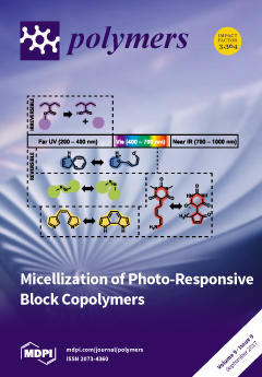
A large variety of photo-responsive molecules have been incorporated into block copolymers, in order to form micelles for potential applications ranging from drug delivery, sensing or the preparation of photochromic hybrid materials. Thereby, the photo-responses were mainly distinguished according to whether the process is: (a) irreversible such as for photo-cleavage reactions of nitrobenzyl or pyrenyl esters; or (b) reversible such as well-known examples for photoisomerizations of spiropyrans and azobenzenes. Moreover, we subdivide the chromophores, according to the wavelength at which the irradiation has to be carried out to achieve photo-response. The cover image schematically illustrates a combination of both approaches. We provide an overview on both irreversible and reversible processes and at which wavelength light-stimulation can be triggered. View the paper
- Issues are regarded as officially published after their release is announced to the table of contents alert mailing list.
- You may sign up for e-mail alerts to receive table of contents of newly released issues.
- PDF is the official format for papers published in both, html and pdf forms. To view the papers in pdf format, click on the "PDF Full-text" link, and use the free Adobe Reader to open them.
Previous Issue
Next Issue