
Sarcoid-like Granulomatosis Associated with Immune Checkpoint Inhibitors in Melanoma

 , , , ,
Simple Summary
Abstract
1. Introduction
2. Materials and Methods
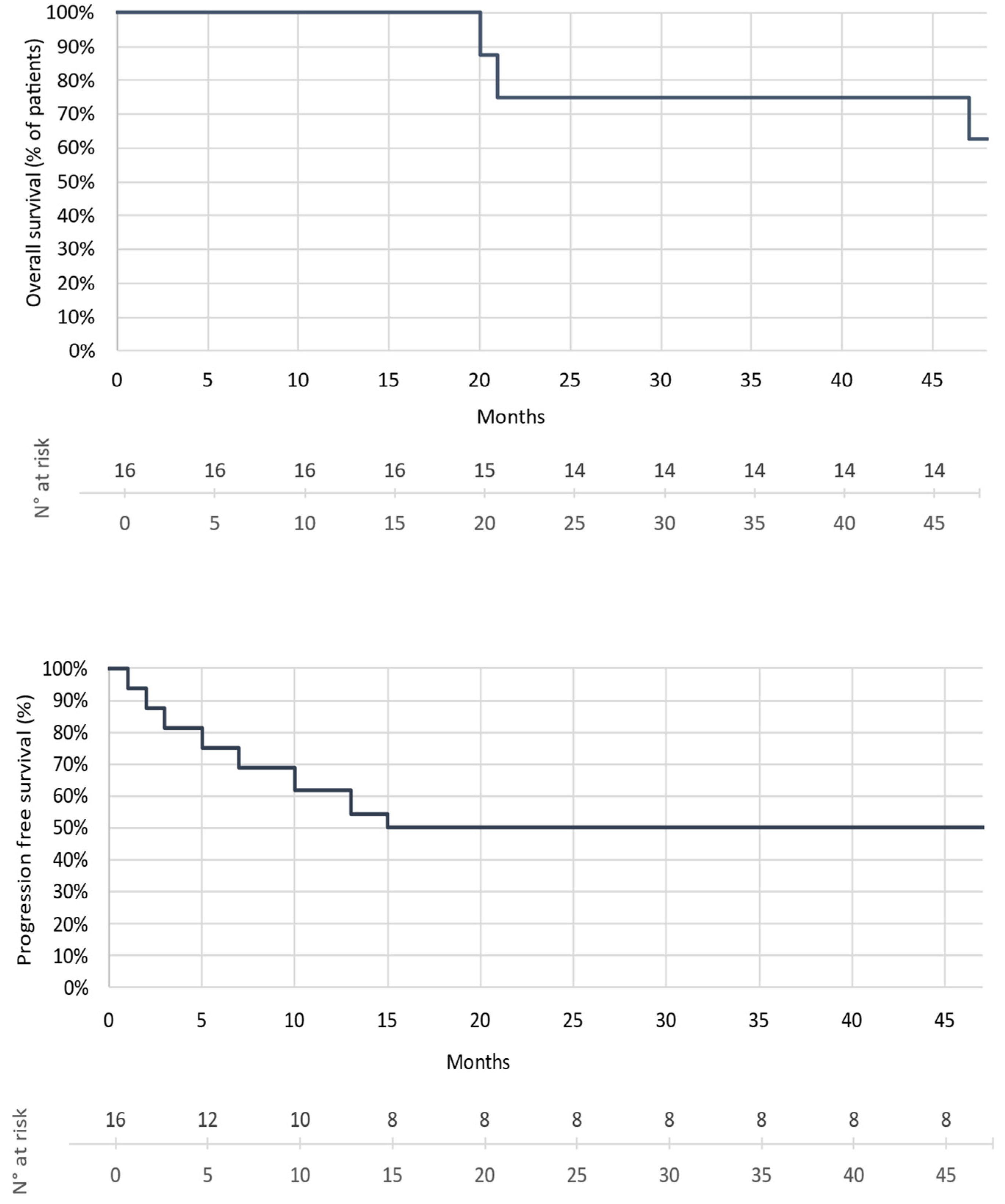
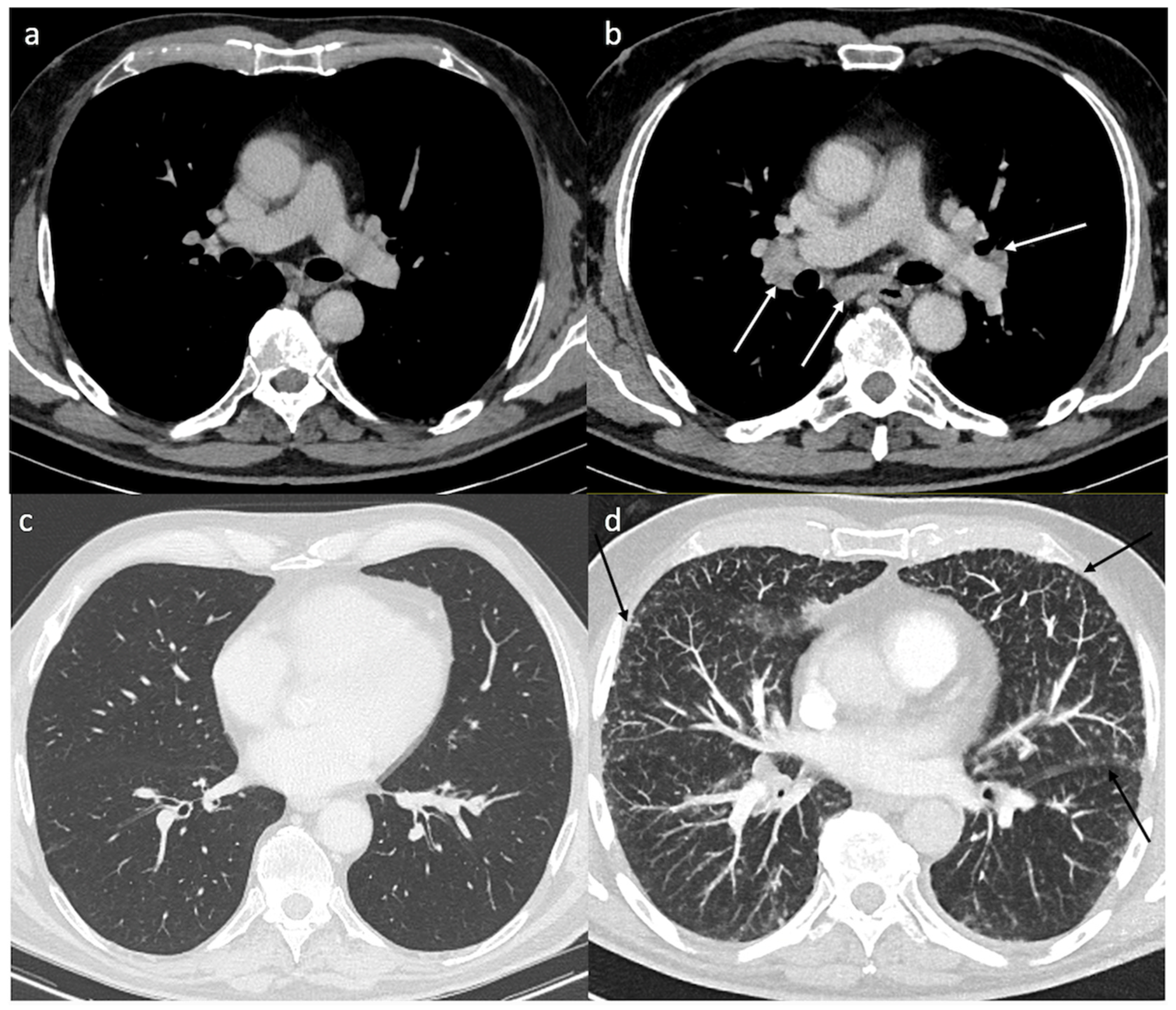
3. Results
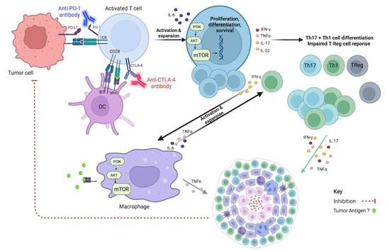
4. Discussion
5. Conclusions
Author Contributions
Funding
Institutional Review Board Statement
Informed Consent Statement
Data Availability Statement
Acknowledgments
Conflicts of Interest
References
- Schadendorf, D.; Hodi, F.S.; Robert, C.; Weber, J.S.; Margolin, K.; Hamid, O.; Patt, D.; Chen, T.-T.; Berman, D.M.; Wolchok, J.D. Pooled Analysis of Long-Term Survival Data from Phase II and Phase III Trials of Ipilimumab in Unresectable or Metastatic Melanoma. J. Clin. Oncol. 2015, 33, 1889–1894. [Google Scholar] [CrossRef]
- Robert, C.; Long, G.V.; Brady, B.; Dutriaux, C.; Maio, M.; Mortier, L.; Hassel, J.C.; Rutkowski, P.; McNeil, C.; Kalinka-Warzocha, E.; et al. Nivolumab in Previously Untreated Melanoma without BRAF Mutation. N. Engl. J. Med. 2015, 372, 320–330. [Google Scholar] [CrossRef]
- Larkin, J.; Chiarion-Sileni, V.; Gonzalez, R.; Grob, J.-J.; Rutkowski, P.; Lao, C.D.; Cowey, C.L.; Schadendorf, D.; Wagstaff, J.; Dummer, R.; et al. Five-Year Survival with Combined Nivolumab and Ipilimumab in Advanced Melanoma. N. Engl. J. Med. 2019, 381, 1535–1546. [Google Scholar] [CrossRef]
- National Cancer Institute. Common Terminology Criteria for Adverse Events v3.0 (CTCAE); National Cancer Institute: Bethesda, MD, USA, 2006.
- Cohen, P.R.; Kurzrock, R. Sarcoidosis and Malignancy. Clin. Dermatol. 2007, 25, 326–333. [Google Scholar] [CrossRef]
- Bonifazi, M.; Bravi, F.; Gasparini, S.; La Vecchia, C.; Gabrielli, A.; Wells, A.U.; Renzoni, E.A. Sarcoidosis and Cancer Risk: Systematic Review and Meta-Analysis of Observational Studies. Chest 2015, 147, 778–791. [Google Scholar] [CrossRef]
- Askling, J.; Grunewald, J.; Eklund, A.; Hillerdal, G.; Ekbom, A. Increased Risk for Cancer Following Sarcoidosis. Am. J. Respir. Crit. Care Med. 1999, 160, 1668–1672. [Google Scholar] [CrossRef]
- Seve, P.; Schott, A.M.; Pavic, M.; Broussolle, C.; Gilis, L.; Thomas, L. Sarcoidosis and Melanoma: A Referral Center Study of 1,199 Cases. Dermatology 2009, 219, 25–31. [Google Scholar] [CrossRef]
- Beutler, B.D.; Cohen, P.R. Sarcoidosis in Melanoma Patients: Case Report and Literature Review. Cancers 2015, 7, 1005–1021. [Google Scholar] [CrossRef]
- Robert, C.; Schoenlaub, P.; Avril, M.F.; Lok, C.; Grosshans, E.; Valeyre, D.; Bourgeois, C.; Pinquier, L.; Dubertret, L.; Guillaume, J.C. Malignant Melanoma and Granulomatosis. Br. J. Dermatol. 1997, 137, 787–792. [Google Scholar] [CrossRef]
- Echigo, T.; Saito, A.; Takehara, K.; Takata, M.; Hatta, N. Coexistence of Micrometastatic Melanoma Cells and Sarcoid Granulomas in All Regional Lymph Nodes in a Patient with Acral Melanoma. Clin. Exp. Dermatol. 2003, 28, 375–376. [Google Scholar] [CrossRef]
- Rubinstein, I.; Baum, G.L.; Yellin, A.; Herczeg, E. Sarcoidosis: A Cause of Bilateral Hilar Lymphadenopathy after Excision of Malignant Melanoma of the Arm. South. Med. J. 1985, 78, 1139–1140. [Google Scholar] [CrossRef]
- Sacks, E.L.; Donaldson, S.S.; Gordon, J.; Dorfman, R.F. Epithelioid Granulomas Associated with Hodgkin’s Disease: Clinical Correlations in 55 Previously Untreated Patients. Cancer 1978, 41, 562–567. [Google Scholar] [CrossRef]
- Miedema, J.; Nunes, H. Drug-Induced Sarcoidosis-like Reactions. Curr. Opin. Pulm. Med. 2021, 27, 439–447. [Google Scholar] [CrossRef]
- Cleven, K.L.; Ye, K.; Zeig-Owens, R.; Hena, K.M.; Montagna, C.; Shan, J.; Hosgood, H.D.; Jaber, N.; Weiden, M.D.; Colbeth, H.L.; et al. Genetic Variants Associated with FDNY WTC-Related Sarcoidosis. Int. J. Environ. Res. Public Health 2019, 16, 1830. [Google Scholar] [CrossRef]
- Kishore, A.; Petrek, M. Next-Generation Sequencing Based HLA Typing: Deciphering Immunogenetic Aspects of Sarcoidosis. Front. Genet. 2018, 9, 503. [Google Scholar] [CrossRef]
- Grunewald, J.; Spagnolo, P.; Wahlström, J.; Eklund, A. Immunogenetics of Disease-Causing Inflammation in Sarcoidosis. Clin. Rev. Allergy Immunol. 2015, 49, 19–35. [Google Scholar] [CrossRef]
- Koth, L.L.; Solberg, O.D.; Peng, J.C.; Bhakta, N.R.; Nguyen, C.P.; Woodruff, P.G. Sarcoidosis Blood Transcriptome Reflects Lung Inflammation and Overlaps with Tuberculosis. Am. J. Respir. Crit. Care Med. 2011, 184, 1153–1163. [Google Scholar] [CrossRef]
- Gupta, D.; Agarwal, R.; Aggarwal, A.N.; Jindal, S.K. Molecular Evidence for the Role of Mycobacteria in Sarcoidosis: A Meta-Analysis. Eur. Respir. J. 2007, 30, 508–516. [Google Scholar] [CrossRef]
- Song, Z.; Marzilli, L.; Greenlee, B.M.; Chen, E.S.; Silver, R.F.; Askin, F.B.; Teirstein, A.S.; Zhang, Y.; Cotter, R.J.; Moller, D.R. Mycobacterial Catalase-Peroxidase Is a Tissue Antigen and Target of the Adaptive Immune Response in Systemic Sarcoidosis. J. Exp. Med. 2005, 201, 755–767. [Google Scholar] [CrossRef]
- Milman, N.; Lisby, G.; Friis, S.; Kemp, L. Prolonged Culture for Mycobacteria in Mediastinal Lymph Nodes from Patients with Pulmonary Sarcoidosis. A Negative Study. Sarcoidosis Vasc. Diffus. Lung Dis. 2004, 21, 25–28. [Google Scholar]
- Eishi, Y.; Suga, M.; Ishige, I.; Kobayashi, D.; Yamada, T.; Takemura, T.; Takizawa, T.; Koike, M.; Kudoh, S.; Costabel, U.; et al. Quantitative Analysis of Mycobacterial and Propionibacterial DNA in Lymph Nodes of Japanese and European Patients with Sarcoidosis. J. Clin. Microbiol. 2002, 40, 198–204. [Google Scholar] [CrossRef]
- Ebe, Y.; Ikushima, S.; Yamaguchi, T.; Kohno, K.; Azuma, A.; Sato, K.; Ishige, I.; Usui, Y.; Takemura, T.; Eishi, Y. Proliferative Response of Peripheral Blood Mononuclear Cells and Levels of Antibody to Recombinant Protein from Propionibacterium Acnes DNA Expression Library in Japanese Patients with Sarcoidosis. Sarcoidosis Vasc. Diffus. Lung Dis. 2000, 17, 256–265. [Google Scholar]
- Song, J.; Zhao, M.; Li, Q.; Lu, L.; Zhou, Y.; Zhang, Y.; Chen, T.; Tang, D.; Zhou, N.; Yin, C.; et al. IL-17A Can Promote Propionibacterium acnes-Induced Sarcoidosis-Like Granulomatosis in Mice. Front. Immunol. 2019, 10, 1923. [Google Scholar] [CrossRef]
- Ishige, I.; Eishi, Y.; Takemura, T.; Kobayashi, I.; Nakata, K.; Tanaka, I.; Nagaoka, S.; Iwai, K.; Watanabe, K.; Takizawa, T.; et al. Propionibacterium Acnes Is the Most Common Bacterium Commensal in Peripheral Lung Tissue and Mediastinal Lymph Nodes from Subjects without Sarcoidosis. Sarcoidosis Vasc. Diffus. Lung Dis. 2005, 22, 33–42. [Google Scholar]
- Kreider, M.E.; Christie, J.D.; Thompson, B.; Newman, L.; Rose, C.; Barnard, J.; Bresnitz, E.; Judson, M.A.; Lackland, D.T.; Rossman, M.D. Relationship of Environmental Exposures to the Clinical Phenotype of Sarcoidosis. Chest 2005, 128, 207–215. [Google Scholar] [CrossRef]
- Newman, L.S.; Rose, C.S.; Bresnitz, E.A.; Rossman, M.D.; Barnard, J.; Frederick, M.; Terrin, M.L.; Weinberger, S.E.; Moller, D.R.; McLennan, G.; et al. A Case Control Etiologic Study of Sarcoidosis: Environmental and Occupational Risk Factors. Am. J. Respir. Crit. Care Med. 2004, 170, 1324–1330. [Google Scholar] [CrossRef]
- Eckert, A.; Schoeffler, A.; Dalle, S.; Phan, A.; Kiakouama, L.; Thomas, L. Anti-CTLA4 Monoclonal Antibody Induced Sarcoidosis in a Metastatic Melanoma Patient. Dermatology 2009, 218, 69–70. [Google Scholar] [CrossRef]
- Berthod, G.; Lazor, R.; Letovanec, I.; Romano, E.; Noirez, L.; Mazza Stalder, J.; Speiser, D.E.; Peters, S.; Michielin, O. Pulmonary Sarcoid-Like Granulomatosis Induced by Ipilimumab. J. Clin. Oncol. 2012, 30, e156–e159. [Google Scholar] [CrossRef]
- Vogel, W.V.; Guislain, A.; Kvistborg, P.; Schumacher, T.N.M.; Haanen, J.B.A.G.; Blank, C.U. Ipilimumab-Induced Sarcoidosis in a Patient with Metastatic Melanoma Undergoing Complete Remission. J. Clin. Oncol. 2012, 30, e7–e10. [Google Scholar] [CrossRef]
- Wilgenhof, S.; Morlion, V.; Seghers, A.C.; Du Four, S.; Vanderlinden, E.; Hanon, S.; Vandenbroucke, F.; Everaert, H.; Neyns, B. Sarcoidosis in a Patient with Metastatic Melanoma Sequentially Treated with Anti-CTLA-4 Monoclonal Antibody and Selective BRAF Inhibitor. Anticancer Res. 2012, 32, 1355–1359. [Google Scholar]
- Reule, R.B.; North, J.P. Cutaneous and Pulmonary Sarcoidosis-like Reaction Associated with Ipilimumab. J. Am. Acad. Dermatol. 2013, 69, e272–e273. [Google Scholar] [CrossRef] [PubMed]
- Andersen, R.; Nørgaard, P.; Al-Jailawi, M.K.M.; Svane, I.M. Late Development of Splenic Sarcoidosis-like Lesions in a Patient with Metastatic Melanoma and Long-Lasting Clinical Response to Ipilimumab. Oncoimmunology 2014, 3, e954506. [Google Scholar] [CrossRef] [PubMed]
- Murphy, K.P.; Kennedy, M.P.; Barry, J.E.; O’Regan, K.N.; Power, D.G. New-Onset Mediastinal and Central Nervous System Sarcoidosis in a Patient with Metastatic Melanoma Undergoing CTLA4 Monoclonal Antibody Treatment. Oncol. Res. Treat. 2014, 37, 351–353. [Google Scholar] [CrossRef] [PubMed]
- Danlos, F.-X.; Pagès, C.; Baroudjian, B.; Vercellino, L.; Battistella, M.; Mimoun, M.; Jebali, M.; Bagot, M.; Tazi, A.; Lebbé, C. Nivolumab-Induced Sarcoid-Like Granulomatous Reaction in a Patient with Advanced Melanoma. Chest 2016, 149, e133–e136. [Google Scholar] [CrossRef] [PubMed]
- Martínez Leboráns, L.; Esteve Martínez, A.; Victoria Martínez, A.M.; Alegre de Miquel, V.; Berrocal Jaime, A. Cutaneous Sarcoidosis in a Melanoma Patient under Ipilimumab Therapy. Dermatol. Ther. 2016, 29, 306–308. [Google Scholar] [CrossRef] [PubMed]
- Koelzer, V.H.; Rothschild, S.I.; Zihler, D.; Wicki, A.; Willi, B.; Willi, N.; Voegeli, M.; Cathomas, G.; Zippelius, A.; Mertz, K.D. Systemic Inflammation in a Melanoma Patient Treated with Immune Checkpoint Inhibitors-an Autopsy Study. J. Immunother. Cancer 2016, 4, 13. [Google Scholar] [CrossRef]
- Reuss, J.E.; Kunk, P.R.; Stowman, A.M.; Gru, A.A.; Slingluff, C.L.; Gaughan, E.M. Sarcoidosis in the Setting of Combination Ipilimumab and Nivolumab Immunotherapy: A Case Report & Review of the Literature. J. Immunother. Cancer 2016, 4, 94. [Google Scholar] [CrossRef]
- Montaudié, H.; Pradelli, J.; Passeron, T.; Lacour, J.-P.; Leroy, S. Pulmonary Sarcoid-like Granulomatosis Induced by Nivolumab. Br. J. Dermatol. 2017, 176, 1060–1063. [Google Scholar] [CrossRef]
- Reddy, S.B.; Possick, J.D.; Kluger, H.M.; Galan, A.; Han, D. Sarcoidosis Following Anti-PD-1 and Anti-CTLA-4 Therapy for Metastatic Melanoma. J. Immunother. 2017, 40, 307–311. [Google Scholar] [CrossRef]
- Dunn-Pirio, A.M.; Shah, S.; Eckstein, C. Neurosarcoidosis Following Immune Checkpoint Inhibition. Case Rep. Oncol. 2018, 11, 521–526. [Google Scholar] [CrossRef]
- Nishino, M.; Sholl, L.M.; Awad, M.M.; Hatabu, H.; Armand, P.; Hodi, F.S. Sarcoid-Like Granulomatosis of the Lung Related to Immune-Checkpoint Inhibitors: Distinct Clinical and Imaging Features of a Unique Immune-Related Adverse Event. Cancer Immunol. Res. 2018, 6, 630–635. [Google Scholar] [CrossRef] [PubMed]
- Faviez, G.; Bousquet, E.; Rabeau, A.; Rouquette, I.; Collot, S.; Goumarre, C.; Meyer, N.; Prevot, G.; Mazieres, J. Sarcoid-like granulomatosis in cancer patients treated with immune checkpoints inhibitors. Rev. Mal. Respir. 2018, 35, 963–967. [Google Scholar] [CrossRef] [PubMed]
- Laroche, A.; Alarcon Chinchilla, E.; Bourgeault, E.; Doré, M.-A. Erythema Nodosum as the Initial Presentation of Nivolumab-Induced Sarcoidosis-Like Reaction. J. Cutan. Med. Surg. 2018, 22, 627–629. [Google Scholar] [CrossRef]
- Yatim, N.; Mateus, C.; Charles, P. Sarcoidosis Post-Anti-PD-1 Therapy, Mimicking Relapse of Metastatic Melanoma in a Patient Undergoing Complete Remission. Rev. Med. Interne 2018, 39, 130–133. [Google Scholar] [CrossRef] [PubMed]
- Jespersen, H.; Bjursten, S.; Ny, L.; Levin, M. Checkpoint Inhibitor-Induced Sarcoid Reaction Mimicking Bone Metastases. Lancet Oncol. 2018, 19, e327. [Google Scholar] [CrossRef]
- Dimitriou, F.; Frauchiger, A.L.; Urosevic-Maiwald, M.; Naegeli, M.C.; Goldinger, S.M.; Barysch, M.; Franzen, D.; Kamarachev, J.; Braun, R.; Dummer, R.; et al. Sarcoid-like Reactions in Patients Receiving Modern Melanoma Treatment. Melanoma Res. 2018, 28, 230–236. [Google Scholar] [CrossRef] [PubMed]
- Lu, Y. FDG PET/CT Course of Pembrolizumab-Associated Multiorgan Sarcoidosis. Clin. Nucl. Med. 2019, 44, 167–168. [Google Scholar] [CrossRef]
- Tetzlaff, M.T.; Nelson, K.C.; Diab, A.; Staerkel, G.A.; Nagarajan, P.; Torres-Cabala, C.A.; Chasen, B.A.; Wargo, J.A.; Prieto, V.G.; Amaria, R.N.; et al. Granulomatous/Sarcoid-like Lesions Associated with Checkpoint Inhibitors: A Marker of Therapy Response in a Subset of Melanoma Patients. J. Immunother. Cancer 2018, 6, 14. [Google Scholar] [CrossRef]
- Fukuchi, K.; Hikawa, M.; Sano, Y.; Kasuya, A.; Aoshima, M.; Tatsuno, K.; Nakamura, Y.; Kosugi, I.; Tokura, Y. Sarcoid-like Reaction and Vitiligo Occurring after Nivolumab Therapy in a Patient with Metastatic Melanoma. J. Dermatol. 2019, 46, e359–e360. [Google Scholar] [CrossRef]
- Cervantes, J.; Rosen, A.; Dehesa, L.; Dickinson, G.; Alonso-Llamazares, J. Granulomatous Reaction in a Patient with Metastatic Melanoma Treated with Ipilimumab: First Case Reported with Isolated Cutaneous Findings. Actas Dermo-Sifiliográficas 2019, 110, 43–49. [Google Scholar] [CrossRef]
- Toumeh, A.; Sakhi, R.; Shah, S.; Arudra, S.K.C.; De Las Casas, L.E.; Skeel, R.T. Ipilimumab-Induced Granulomatous Disease Occurring Simultaneously with Disease Progression in a Patient with Metastatic Melanoma. Am. J. Ther. 2016, 23, e1068–e1071. [Google Scholar] [CrossRef] [PubMed]
- Lidar, M.; Giat, E.; Garelick, D.; Horowitz, Y.; Amital, H.; Steinberg-Silman, Y.; Schachter, J.; Shapira-Frommer, R.; Markel, G. Rheumatic Manifestations among Cancer Patients Treated with Immune Checkpoint Inhibitors. Autoimmun. Rev. 2018, 17, 284–289. [Google Scholar] [CrossRef] [PubMed]
- Burillo-Martinez, S.; Morales-Raya, C.; Prieto-Barrios, M.; Rodriguez-Peralto, J.-L.; Ortiz-Romero, P.-L. Pembrolizumab-Induced Extensive Panniculitis and Nevus Regression: Two Novel Cutaneous Manifestations of the Post-Immunotherapy Granulomatous Reactions Spectrum. JAMA Dermatol. 2017, 153, 721–722. [Google Scholar] [CrossRef] [PubMed]
- Nandavaram, S.; Nadkarni, A. Ipilimumab-Induced Sarcoidosis and Thyroiditis. Am. J. Ther. 2018, 25, e379–e380. [Google Scholar] [CrossRef]
- Tan, I.; Malinzak, M.; Salama, A.K.S. Delayed Onset of Neurosarcoidosis after Concurrent Ipilimumab/Nivolumab Therapy. J. Immunother. Cancer 2018, 6, 77. [Google Scholar] [CrossRef]
- Van Willigen, W.W.; Gerritsen, W.R.; Aarntzen, E.H.J.G. 18F-FDG PET/CT of Multiorgan Sarcoid-Like Reaction During Anti-PD-1 Treatment for Melanoma. Clin. Nucl. Med. 2019, 44, 905–906. [Google Scholar] [CrossRef]
- Wang, L.L.; Patel, G.; Chiesa-Fuxench, Z.C.; McGettigan, S.; Schuchter, L.; Mitchell, T.C.; Ming, M.E.; Chu, E.Y. Timing of Onset of Adverse Cutaneous Reactions Associated with Programmed Cell Death Protein 1 Inhibitor Therapy. JAMA Dermatol. 2018, 154, 1057–1061. [Google Scholar] [CrossRef]
- Woodbeck, R.; Metelitsa, A.I.; Naert, K.A. Granulomatous Tumoral Melanosis Associated with Pembrolizumab Therapy: A Mimicker of Disease Progression in Metastatic Melanoma. Am. J. Dermatopathol. 2018, 40, 523–526. [Google Scholar] [CrossRef]
- Tissot, C.; Carsin, A.; Freymond, N.; Pacheco, Y.; Devouassoux, G. Sarcoidosis Complicating Anti-Cytotoxic T-Lymphocyte-Associated Antigen-4 Monoclonal Antibody Biotherapy. Eur. Respir. J. 2013, 41, 246–247. [Google Scholar] [CrossRef]
- Firwana, B.; Ravilla, R.; Raval, M.; Hutchins, L.; Mahmoud, F. Sarcoidosis-like Syndrome and Lymphadenopathy Due to Checkpoint Inhibitors. J. Oncol. Pharm. Pract. 2017, 23, 620–624. [Google Scholar] [CrossRef]
- Rodriguez, E.F.; Lipson, E.; Suresh, K.; Cappelli, L.C.; Monaco, S.E.; Maleki, Z. Immune Checkpoint Blocker-Related Sarcoid-like Granulomatous Inflammation: A Rare Adverse Event Detected in Lymph Node Aspiration Cytology of Patients Treated for Advanced Malignant Melanoma. Hum. Pathol. 2019, 91, 69–76. [Google Scholar] [CrossRef] [PubMed]
- Chorti, E.; Kanaki, T.; Zimmer, L.; Hadaschik, E.; Ugurel, S.; Gratsias, E.; Roesch, A.; Bonella, F.; Wessendorf, T.E.; Wälscher, J.; et al. Drug-Induced Sarcoidosis-like Reaction in Adjuvant Immunotherapy: Increased Rate and Mimicker of Metastasis. Eur. J. Cancer 2020, 131, 18–26. [Google Scholar] [CrossRef] [PubMed]
- Frohlich, M.; Wang, H.; Sakr, L. Sarcoid-like Reaction Discovered on EBUS-TBNA of Intrathoracic Lymph Nodes During Immunotherapy for Metastatic Melanoma. J. Immunother. 2020, 43, 75–78. [Google Scholar] [CrossRef] [PubMed]
- Tulbah, R.I.; Rowe, S.P.; Solnes, L.B.; Javadi, M.S. Nivolumab-Associated Pulmonary and Bone Sarcoidosis in a Patient With Melanoma of Unknown Primary. Clin. Nucl. Med. 2019, 44, e519–e521. [Google Scholar] [CrossRef]
- Urrego-Callejas, T.; Sandoval-Álvarez, S.; Gómez-Wolff, R.; Vásquez, G. Cutaneous and Pulmonary Sarcoid-Like Reaction Induced by Nivolumab: Case Report and Brief Literature Review. J. Clin. Rheumatol. 2019, 27, S460–S464. [Google Scholar] [CrossRef]
- Marcoval, J.; Bauer-Alonso, A.; Fornons-Servent, R.; Jiménez-Colomo, L.; Sabaté-Llobera, A.; Penín, R.M. Subcutaneous Sarcoidosis Induced by Pembrolizumab in a Melanoma Patient Mimicking Subcutaneous Metastasis at 18F-FDG PET/CT. Rev. Esp. Med. Nucl. Imagen. Mol. 2021, 40, 255–256. [Google Scholar] [CrossRef]
- Apalla, Z.; Kemanetzi, C.; Papageorgiou, C.; Bobos, M.; Manoli, M.; Fotiadou, C.; Hatzibougias, D.; Boukovinas, I.; Stergiou, E.; Levva, S.; et al. Challenges in Sarcoidosis and Sarcoid-like Reactions Associated to Immune Checkpoint Inhibitors: A Narrative Review apropos of a Case. Dermatol. Ther. 2021, 34, e14618. [Google Scholar] [CrossRef]
- Keukeleire, S.D.; Schwarze, J.; Awada, G.; Everaert, H.; Van Binst, A.M.; Cras, L.; Neyns, B.; Aspeslagh, S. An Atypical Sarcoid-like Reaction during Anti-Protein Death 1 Treatment in a Patient with Metastatic Melanoma. Melanoma Res. 2020, 30, 524–527. [Google Scholar] [CrossRef]
- Garanzini, E.M.; Scaramuzza, D.; Spadarella, G.; Di Guardo, L.; Marchianò, A. Sarcoidosis-like Disease Mimicking Metastases during Adjuvant Ipilimumab Therapy in Advanced Melanoma Patient: CT Scan and MRI Help in Managing Difficult Clinical Decision. BJR Case Rep. 2020, 6, 20190065. [Google Scholar] [CrossRef]
- Mobini, N.; Dhillon, R.; Dickey, J.; Spoon, J.; Sadrolashrafi, K. Exclusive Cutaneous and Subcutaneous Sarcoidal Granulomatous Inflammation Due to Immune Checkpoint Inhibitors: Report of Two Cases with Unusual Manifestations and Review of the Literature. Case Rep. Dermatol. Med. 2019, 2019, 6702870. [Google Scholar] [CrossRef]
- Chahin, M.; Stack, A.; Siddiqi, A.; Shaikh, M. Is It Metastatic Melanoma or Is It Sarcoidosis? Non-Caseating Granulomas Due to Pembrolizumab. BMJ Case Rep. 2020, 13, e240701. [Google Scholar] [CrossRef] [PubMed]
- Abdel-Wahab, N.; Shah, M.; Suarez-Almazor, M.E. Adverse Events Associated with Immune Checkpoint Blockade in Patients with Cancer: A Systematic Review of Case Reports. PLoS ONE 2016, 11, e0160221. [Google Scholar] [CrossRef] [PubMed]
- Bronstein, Y.; Ng, C.S.; Hwu, P.; Hwu, W.-J. Radiologic Manifestations of Immune-Related Adverse Events in Patients with Metastatic Melanoma Undergoing Anti-CTLA-4 Antibody Therapy. Am. J. Roentgenol. 2011, 197, W992–W1000. [Google Scholar] [CrossRef] [PubMed]
- Tirumani, S.H.; Ramaiya, N.H.; Keraliya, A.; Bailey, N.D.; Ott, P.A.; Hodi, F.S.; Nishino, M. Radiographic Profiling of Immune-Related Adverse Events in Advanced Melanoma Patients Treated with Ipilimumab. Cancer Immunol. Res. 2015, 3, 1185–1192. [Google Scholar] [CrossRef] [PubMed]
- Hodi, F.S.; Ribas, A.; Daud, A.; Hamid, O.; Robert, C.; Kefford, R.; Hwu, W.-J.; Gangadhar, T.C.; Joshua, A.M.; Hersey, P.; et al. Patterns of Response in Patients with Advanced Melanoma Treated with Pembrolizumab (MK-3475) and Evaluation of Immune-Related Response Criteria (IrRC). J. ImmunoTherapy Cancer 2014, 2, P103. [Google Scholar] [CrossRef]
- Markovic, S.N.; Galli, F.; Suman, V.J.; Nevala, W.K.; Paulsen, A.M.; Hung, J.C.; Gansen, D.N.; Erickson, L.A.; Marchetti, P.; Wiseman, G.A.; et al. Non-Invasive Visualization of Tumor Infiltrating Lymphocytes in Patients with Metastatic Melanoma Undergoing Immune Checkpoint Inhibitor Therapy: A Pilot Study. Oncotarget 2018, 9, 30268–30278. [Google Scholar] [CrossRef]
- Statement on Sarcoidosis. Joint Statement of the American Thoracic Society (ATS), the European Respiratory Society (ERS) and the World Association of Sarcoidosis and Other Granulomatous Disorders (WASOG) Adopted by the ATS Board of Directors and by the ERS Executive Committee, February 1999. Am. J. Respir. Crit. Care Med. 1999, 160, 736–755. [Google Scholar] [CrossRef]
- Judson, M.A. Advances in the Diagnosis and Treatment of Sarcoidosis. F1000Prime Rep 2014, 6, 89. [Google Scholar] [CrossRef]
- Hao, W. Mathematical Model of Sarcoidosis. Proc. Natl. Acad. Sci. USA 2014, 111, 16065–16070. [Google Scholar] [CrossRef]
- Fischer, A.; Ellinghaus, D.; Nutsua, M.; Hofmann, S.; Montgomery, C.G.; Iannuzzi, M.C.; Rybicki, B.A.; Petrek, M.; Mrazek, F.; Pabst, S.; et al. Identification of Immune-Relevant Factors Conferring Sarcoidosis Genetic Risk. Am. J. Respir. Crit. Care Med. 2015, 192, 727–736. [Google Scholar] [CrossRef]
- Cinetto, F.; Agostini, C. Advances in Understanding the Immunopathology of Sarcoidosis and Implications on Therapy. Expert Rev. Clin. Immunol. 2016, 12, 973–988. [Google Scholar] [CrossRef] [PubMed]
- Facco, M.; Cabrelle, A.; Teramo, A.; Olivieri, V.; Gnoato, M.; Teolato, S.; Ave, E.; Gattazzo, C.; Fadini, G.P.; Calabrese, F.; et al. Sarcoidosis Is a Th1/Th17 Multisystem Disorder. Thorax 2011, 66, 144–150. [Google Scholar] [CrossRef] [PubMed]
- Facco, M.; Baesso, I.; Miorin, M.; Bortoli, M.; Cabrelle, A.; Boscaro, E.; Gurrieri, C.; Trentin, L.; Zambello, R.; Calabrese, F.; et al. Expression and Role of CCR6/CCL20 Chemokine Axis in Pulmonary Sarcoidosis. J. Leukoc. Biol. 2007, 82, 946–955. [Google Scholar] [CrossRef] [PubMed]
- Li, Q.; Laumonnier, Y.; Syrovets, T.; Simmet, T. Recruitment of CCR6-Expressing Th17 Cells by CCL20 Secreted from Plasmin-Stimulated Macrophages. Acta Biochim. Biophys. Sin. 2013, 45, 593–600. [Google Scholar] [CrossRef]
- Ramstein, J.; Broos, C.E.; Simpson, L.J.; Ansel, K.M.; Sun, S.A.; Ho, M.E.; Woodruff, P.G.; Bhakta, N.R.; Christian, L.; Nguyen, C.P.; et al. IFN-γ-Producing T-Helper 17.1 Cells Are Increased in Sarcoidosis and Are More Prevalent than T-Helper Type 1 Cells. Am. J. Respir. Crit. Care Med. 2016, 193, 1281–1291. [Google Scholar] [CrossRef]
- Huang, H.; Lu, Z.; Jiang, C.; Liu, J.; Wang, Y.; Xu, Z. Imbalance between Th17 and Regulatory T-Cells in Sarcoidosis. Int. J. Mol. Sci. 2013, 14, 21463–21473. [Google Scholar] [CrossRef]
- Mortaz, E.; Rezayat, F.; Amani, D.; Kiani, A.; Garssen, J.; Adcock, I.M.; Velayati, A. The Roles of T Helper 1, T Helper 17 and Regulatory T Cells in the Pathogenesis of Sarcoidosis. Iran. J. Allergy Asthma Immunol. 2016, 15, 334–339. [Google Scholar]
- Moller, D.R.; Rybicki, B.A.; Hamzeh, N.Y.; Montgomery, C.G.; Chen, E.S.; Drake, W.; Fontenot, A.P. Genetic, Immunologic, and Environmental Basis of Sarcoidosis. Ann. Am. Thorac. Soc. 2017, 14, S429–S436. [Google Scholar] [CrossRef]
- Gkiozos, I.; Kopitopoulou, A.; Kalkanis, A.; Vamvakaris, I.N.; Judson, M.A.; Syrigos, K.N. Sarcoidosis-Like Reactions Induced by Checkpoint Inhibitors. J. Thorac. Oncol. 2018, 13, 1076–1082. [Google Scholar] [CrossRef]
- Ying, H.; Yang, L.; Qiao, G.; Li, Z.; Zhang, L.; Yin, F.; Xie, D.; Zhang, J. Cutting Edge: CTLA-4–B7 Interaction Suppresses Th17 Cell Differentiation. J. Immunol. 2010, 185, 1375–1378. [Google Scholar] [CrossRef]
- Von Euw, E.; Chodon, T.; Attar, N.; Jalil, J.; Koya, R.C.; Comin-Anduix, B.; Ribas, A. CTLA4 Blockade Increases Th17 Cells in Patients with Metastatic Melanoma. J. Transl. Med. 2009, 7, 35. [Google Scholar] [CrossRef] [PubMed]
- Broos, C.E.; van Nimwegen, M.; In ’t Veen, J.C.C.M.; Hoogsteden, H.C.; Hendriks, R.W.; van den Blink, B.; Kool, M. Decreased Cytotoxic T-Lymphocyte Antigen 4 Expression on Regulatory T Cells and Th17 Cells in Sarcoidosis: Double Trouble? Am. J. Respir. Crit. Care Med. 2015, 192, 763–765. [Google Scholar] [CrossRef] [PubMed]
- Parry, R.V.; Chemnitz, J.M.; Frauwirth, K.A.; Lanfranco, A.R.; Braunstein, I.; Kobayashi, S.V.; Linsley, P.S.; Thompson, C.B.; Riley, J.L. CTLA-4 and PD-1 Receptors Inhibit T-Cell Activation by Distinct Mechanisms. Mol. Cell. Biol. 2005, 25, 9543–9553. [Google Scholar] [CrossRef]
- D’Addio, F.; Riella, L.V.; Mfarrej, B.G.; Chabtini, L.; Adams, L.T.; Yeung, M.; Yagita, H.; Azuma, M.; Sayegh, M.H.; Guleria, I. The Link between the PDL1 Costimulatory Pathway and Th17 in Fetomaternal Tolerance. J. Immunol. 2011, 187, 4530–4541. [Google Scholar] [CrossRef] [PubMed]
- Zhang, Y.; Liu, Z.; Tian, M.; Hu, X.; Wang, L.; Ji, J.; Liao, A. The Altered PD-1/PD-L1 Pathway Delivers the ‘One-Two Punch’ Effects to Promote the Treg/Th17 Imbalance in Pre-Eclampsia. Cell. Mol. Immunol. 2018, 15, 710–723. [Google Scholar] [CrossRef] [PubMed]
- Georas, S.N.; Chapman, T.J.; Crouser, E.D. Sarcoidosis and T-Helper Cells. Th1, Th17, or Th17.1? Am. J. Respir. Crit. Care Med. 2016, 193, 1198–1200. [Google Scholar] [CrossRef] [PubMed]
- Ten Berge, B.; Paats, M.S.; Bergen, I.M.; van den Blink, B.; Hoogsteden, H.C.; Lambrecht, B.N.; Hendriks, R.W.; Kleinjan, A. Increased IL-17A Expression in Granulomas and in Circulating Memory T Cells in Sarcoidosis. Rheumatology 2012, 51, 37–46. [Google Scholar] [CrossRef] [PubMed]
- Lomax, A.J.; McGuire, H.M.; McNeil, C.; Choi, C.J.; Hersey, P.; Karikios, D.; Shannon, K.; van Hal, S.; Carr, U.; Crotty, A.; et al. Immunotherapy-Induced Sarcoidosis in Patients with Melanoma Treated with PD-1 Checkpoint Inhibitors: Case Series and Immunophenotypic Analysis. Int. J. Rheum. Dis. 2017, 20, 1277–1285. [Google Scholar] [CrossRef]
- Linke, M.; Pham, H.T.T.; Katholnig, K.; Schnöller, T.; Miller, A.; Demel, F.; Schütz, B.; Rosner, M.; Kovacic, B.; Sukhbaatar, N.; et al. Chronic Signaling via the Metabolic Checkpoint Kinase MTORC1 Induces Macrophage Granuloma Formation and Marks Sarcoidosis Progression. Nat. Immunol. 2017, 18, 293–302. [Google Scholar] [CrossRef]


| Patient Characteristics (N = 18) | |
|---|---|
| Gender | N = 18 |
| Female | 11 (61%) |
| Age | |
| Mean age at melanoma diagnosis (min–max) (years) | 47 (22–73) |
| Melanoma medical history | |
| Melanoma type | |
| Cutaneous | 14 (78%) |
| Mucosal | 2 (11%) |
| Unknown primitive | 2 (11%) |
| Oncological approach | |
| Adjuvant | 2 (11%) |
| Metastatic | 16 (89%) |
| 1st line of treatment | 12 (75%) |
| 2nd or 3rd line of treatment | 4 (25%) |
| Type of immunotherapy | |
| Anti-CTLA-4 monotherapy | 2 (11%) |
| Anti-PD-1 monotherapy | 7 (39%) |
| Anti-CTLA-4 + Anti-PD-1 combined | 9 (50%) |
| Immune-induced granulomatosis | |
| Clinical features | 10 (56%) |
| Median time since ICI initiation (min–max) (months) | 2 (1–11) |
| Thoracic | 6 (33%) |
| Dermatological | 2 (11%) |
| Ophthalmologic | 2 (11%) |
| Hepatic | 2 (11%) |
| Renal | 1 (6%) |
| Radiological features | |
| Consistent radiological signs | 17 (94%) |
| Median time since ICI initiation (min–max) (months) | 2 (1–10) |
| 18-FDG PET/CT; n = 14 | |
| Mediastino-hilar nodes | 14 (100%) |
| Interstitial involvement | 6 (43%) |
| Subcutaneous nodules | 4 (29%) |
| CT scanner; n = 15 | |
| Mediastino-hilar nodes | 11 (73%) |
| Interstitial involvement | 1 (7%) |
| Subcutaneous nodules | 1 (7%) |
| Biological features | |
| Lymphopenia | 5 (28%) |
| Anicteric cholestasis | 4 (22%) |
| Hypergammaglobulinemia; n = 7 | 1 (14%) |
| Elevated angiotensin converting enzyme; n = 8 | 2 (25%) |
| Hypercalcemia | 0 (0%) |
| Histological confirmation | 18 (100%) |
| Median time since ICI initiation (min–max) (months) | 4 (1–11) |
| Therapeutic management | |
| Systemic corticosteroids | 7 (39%) |
| Discontinuation of immunotherapy | 7 (39%) |
| Granulomatosis outcome | |
| Regression of clinical/biological features | 18 (100%) |
| Radiological outcome of granulomatosis; n = 16 | |
| Stability | 8 (50%) |
| Partial or complete regression | 8 (50%) |
| Oncological outcome at the time of granulomatosis | |
| Patient in adjuvant condition | N = 2 |
| Relapse | 0 (0%) |
| Patients in metastatic stage | N = 16 |
| Objective response | 12 (75%) |
| Complete response | 5 (42%) |
| Complete or partial response | 2 (17%) |
| Partial response | 5 (42%) |
| Stability | 2 (13%) |
| Progression | 2 (13%) |
| Oncological outcome at data collection | |
| Mean follow-up time for melanoma (min–max) (months) | 22 (6–50) |
| Patient in adjuvant condition | N = 2 |
| Relapse | 0 (0%) |
| Patients in metastatic stage | N = 16 |
| Objective response | 8 (50%) |
| Complete response | 5 (63%) |
| Partial response | 3 (38%) |
| Stability | 0 (0%) |
| Progression | 8 (50%) |
| Death | 3 (38%) |
| LITERATURE REVIEW: PATIENT CHARACTERISTICS (N = 67) | |
|---|---|
| Gender | N = 67 |
| Male | 37 (55%) |
| Age | |
| Mean age at melanoma diagnosis (min–max) (years) | 58 (26–83) |
| Melanoma medical history | |
| Oncological approach | |
| Adjuvant | 25 (37%) |
| Metastatic | 42 (63%) |
| Type of immunotherapy | |
| Anti-CTLA-4 monotherapy | 19 (28%) |
| Anti-PD-1 monotherapy | 27 (40%) |
| Anti-CTLA-4 + Anti-PD-1 combined | 10 (15%) |
| Anti-PD-1 +/− Anti-CTLA-4 | 11 (16%) |
| Immune-induced granulomatosis | |
| Median time since initiation of ICI (min–max) (months) | 3 (1–43) |
| Thoracic involvement | 61 (91%) |
| Grade 1 impairment | 38 (63%) |
| Grade 2 impairment | 17 (28%) |
| Grade 3 or 4 impairment | 6 (10%) |
| Dermatological involvement | 32 (48%) |
| Lymph node invasion | 8 (12%) |
| Hepatic involvement | 2 (3%) |
| Bone involvement | 5 (7%) |
| Histological confirmation | 62 (93%) |
| Therapeutic management | |
| Systemic corticosteroids | 25 (37%) |
| Discontinuation of immunotherapy | 33 (49%) |
| Granulomatosis outcome | |
| Radiological outcome of granulomatosis | N = 62 |
| Stability | 8 (13%) |
| Partial or complete regression | 54 (87%) |
| Oncological outcome at the time of granulomatosis | |
| Patient in adjuvant condition | N = 25 |
| Relapse | 3 (12%) |
| Patients in metastatic stage | N = 42 |
| Objective response | 29 (69%) |
| Complete response | 16 (55%) |
| Partial response | 13 (45%) |
| Stability | 5 (12%) |
| Progression | 8 (19%) |
| Oncological outcome at last reported evaluation | |
| Mean follow-up time for melanoma (min–max) (months) | 8 (1–34) |
| Patient in adjuvant condition | N = 25 |
| Relapse | 3 (12%) |
| Patients in metastatic stage | N = 42 |
| Objective response | 24 (57%) |
| Complete response | 16 (67%) |
| Partial response | 8 (33%) |
| Stability | 4 (10%) |
| Progression | 14 (33%) |
Publisher’s Note: MDPI stays neutral with regard to jurisdictional claims in published maps and institutional affiliations. |
© 2022 by the authors. Licensee MDPI, Basel, Switzerland. This article is an open access article distributed under the terms and conditions of the Creative Commons Attribution (CC BY) license (https://creativecommons.org/licenses/by/4.0/).
Share and Cite
Melin, A.; Routier, É.; Roy, S.; Pradere, P.; Le Pavec, J.; Pierre, T.; Chanson, N.; Scoazec, J.-Y.; Lambotte, O.; Robert, C. Sarcoid-like Granulomatosis Associated with Immune Checkpoint Inhibitors in Melanoma. Cancers 2022, 14, 2937. https://doi.org/10.3390/cancers14122937
Melin A, Routier É, Roy S, Pradere P, Le Pavec J, Pierre T, Chanson N, Scoazec J-Y, Lambotte O, Robert C. Sarcoid-like Granulomatosis Associated with Immune Checkpoint Inhibitors in Melanoma. Cancers. 2022; 14(12):2937. https://doi.org/10.3390/cancers14122937
Chicago/Turabian StyleMelin, Audrey, Émilie Routier, Séverine Roy, Pauline Pradere, Jerome Le Pavec, Thibaut Pierre, Noémie Chanson, Jean-Yves Scoazec, Olivier Lambotte, and Caroline Robert. 2022. "Sarcoid-like Granulomatosis Associated with Immune Checkpoint Inhibitors in Melanoma" Cancers 14, no. 12: 2937. https://doi.org/10.3390/cancers14122937
APA StyleMelin, A., Routier, É., Roy, S., Pradere, P., Le Pavec, J., Pierre, T., Chanson, N., Scoazec, J.-Y., Lambotte, O., & Robert, C. (2022). Sarcoid-like Granulomatosis Associated with Immune Checkpoint Inhibitors in Melanoma. Cancers, 14(12), 2937. https://doi.org/10.3390/cancers14122937