


Due to scheduled maintenance work on our servers, there may be short service disruptions on this website between 11:00 and 12:00 CEST on March 28th.
Audiol. Res., Volume 15, Issue 2 (April 2025) – 27 articles
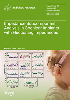
Cover Story (view full-size image):
Some cochlear implant users show unexplained fluctuating/rising electrode impedances, which counterintuitively decrease during rest and increase with stimulation. Stable impedances in inactive electrodes suggest that the cochlear environment is not solely responsible. The impedance decrease during rest offers a chance to study impedance subcomponents, revealing that spikes correlate with increased near-field and Faradaic resistance, and decreased Warburg capacitance. View this paper
- Issues are regarded as officially published after their release is announced to the table of contents alert mailing list.
- You may sign up for e-mail alerts to receive table of contents of newly released issues.
- PDF is the official format for papers published in both, html and pdf forms. To view the papers in pdf format, click on the "PDF Full-text" link, and use the free Adobe Reader to open them.
Previous Issue
Next Issue