You are currently viewing a new version of our website. To view the old version click .
Pharmaceutics
  • Review
  • Open Access

19 November 2025

Controlled Release Technologies for Diltiazem Hydrochloride: A Comprehensive Review of Solid Dosage Innovations

,
and
1
Grupo de Investigación en Química y Biotecnología (QUIBIO), Facultad de Ciencias Básicas, Universidad Santiago de Cali, Cali 760035, Colombia
2
Departamento de Farmacia, Facultad de Ciencias Farmacéuticas y Alimentarias, Universidad de Antioquia, Calle 70 No. 52-21, Medellín 050010, Colombia
3
Grupo de Investigación Ciencia de Materiales Avanzados, Departamento de Química, Facultad de Ciencias, Universidad Nacional de Colombia Sede Medellín, Cra. 65 #59a-110, Medellín 050034, Colombia
4
Grupo de Investigación Cecoltec, Cecoltec Services SAS, Medellín 050034, Colombia
This article belongs to the Special Issue Optimizing Pharmacokinetics Through Formulation Science and Technology

Abstract

Introduction: Diltiazem hydrochloride (DH) is a calcium channel blocker used in the treatment of hypertension, angina pectoris, and arrhythmias. Its short half-life and frequent dosing requirements limit patient adherence and cause plasma concentration fluctuations. Objective: This review critically examines recent pharmaceutical technologies and formulation strategies for modified-release dosage forms (MRDFs) of diltiazem hydrochloride, emphasizing their impact on pharmacokinetics, clinical performance, and regulatory aspects. Methodology: A structured literature review (2010–2025) was conducted using databases such as PubMed, ScienceDirect, MDPI, and ACS Publications. Studies were selected based on relevance to solid oral MRDFs of DH and their associated manufacturing techniques. Results: Techniques including direct compression, granulation, extrusion–spheronization, spray drying, solvent evaporation, and ionotropic gelation have enabled the development of hydrophilic matrices, coated pellets, microspheres, and osmotic systems. Functional polymers such as HPMC, Eudragit®, and ethylcellulose play a central role in modulating release kinetics and improving bioavailability. Conclusions: This review not only synthesizes current formulation strategies but also explores reverse engineering of ideal release profiles and the integration of advanced modeling tools such as physiologically based pharmacokinetic (PBPK) modeling and in vitro–in vivo correlation (IVIVC). These approaches support the rational design of personalized, regulatory-compliant DH therapies.

1. Introduction

Diltiazem hydrochloride (DH) is a cornerstone in the pharmacological management of cardiovascular diseases, particularly hypertension, angina pectoris, and certain supraventricular arrhythmias. As a non-dihydropyridine calcium channel blocker, its primary mechanism involves the inhibition of calcium influx into cardiac and vascular smooth muscle cells during depolarization, resulting in vasodilation and reduced peripheral vascular resistance. DH also reduces heart rate and prolongs diastole, improving myocardial oxygen balance []. According to the Biopharmaceutical Classification System (BCS), DH is categorized as a Class I drug, characterized by high solubility and high permeability []. Despite its favorable solubility and permeability, immediate-release DH formulations have notable clinical limitations. These include dose-dependent adverse effects such as bradycardia and hypotension, which may compromise safety in sensitive populations []. Moreover, DH has a relatively short elimination half-life (approximately 3–4.5 h), requiring multiple daily doses to maintain therapeutic plasma concentrations—an issue that can negatively impact patient adherence and long-term treatment outcomes []. To address these challenges, the development of modified-release (MR) formulations has gained prominence. These systems aim to sustain plasma drug levels, reduce dosing frequency, and enhance both safety and therapeutic compliance []. Recent advances in pharmaceutical technology have introduced platforms such as pellets, microspheres, and osmotic tablets, which demonstrate superior pharmacokinetic profiles and improved tolerability compared to immediate-release counterparts [,]. The design and optimization of these MR formulations rely on diverse manufacturing techniques, including direct compression, wet granulation, microencapsulation, emulsification, gelification, lyophilization, and fusion granulation. Each method contributes uniquely to the modulation of drug release kinetics and the enhancement of the pharmacotechnical profile of DH [,,,].
This work synthesizes the current literature on DH-based modified-release dosage forms and their associated manufacturing strategies. By integrating biopharmaceutical, technological, and clinical perspectives, it establishes a comprehensive framework for the development of safer and more effective formulations—ultimately contributing to improved therapeutic management and patient quality of life. Beyond the descriptive analysis of formulation strategies, the scope of the review has been expanded to include the reverse engineering of ideal release profiles, aiming to align drug delivery kinetics with therapeutic needs and circadian rhythms. In addition, advanced modeling tools—such as physiologically based pharmacokinetic (PBPK) simulations and in vitro–in vivo correlation (IVIVC) frameworks—are considered for their potential to optimize formulation design, predict clinical performance, and support regulatory decision-making. This broader perspective bridges experimental development with predictive modeling, offering translational insights for the rational design of modified-release systems.

2. Methodology

A literature review was conducted covering the period from 2010 to 2025, using major scientific databases and repositories such as Multidisciplinary Digital Publishing Institute (MDPI), American Chemical Society (ACS) Publications, Biomed Central (BMC), PubMed, Scientific Electronic Library Online (SciELO), Sage Journals, ScienceDirect, Taylor & Francis, and Google Patents. The objective was to identify peer-reviewed studies on manufacturing methods and formulation strategies for solid modified-release forms of DH. The search algorithm and selection process are illustrated in Figure 1.
Figure 1. Information search algorithm.

3. Modified Release Dosage Forms (MRDFs)

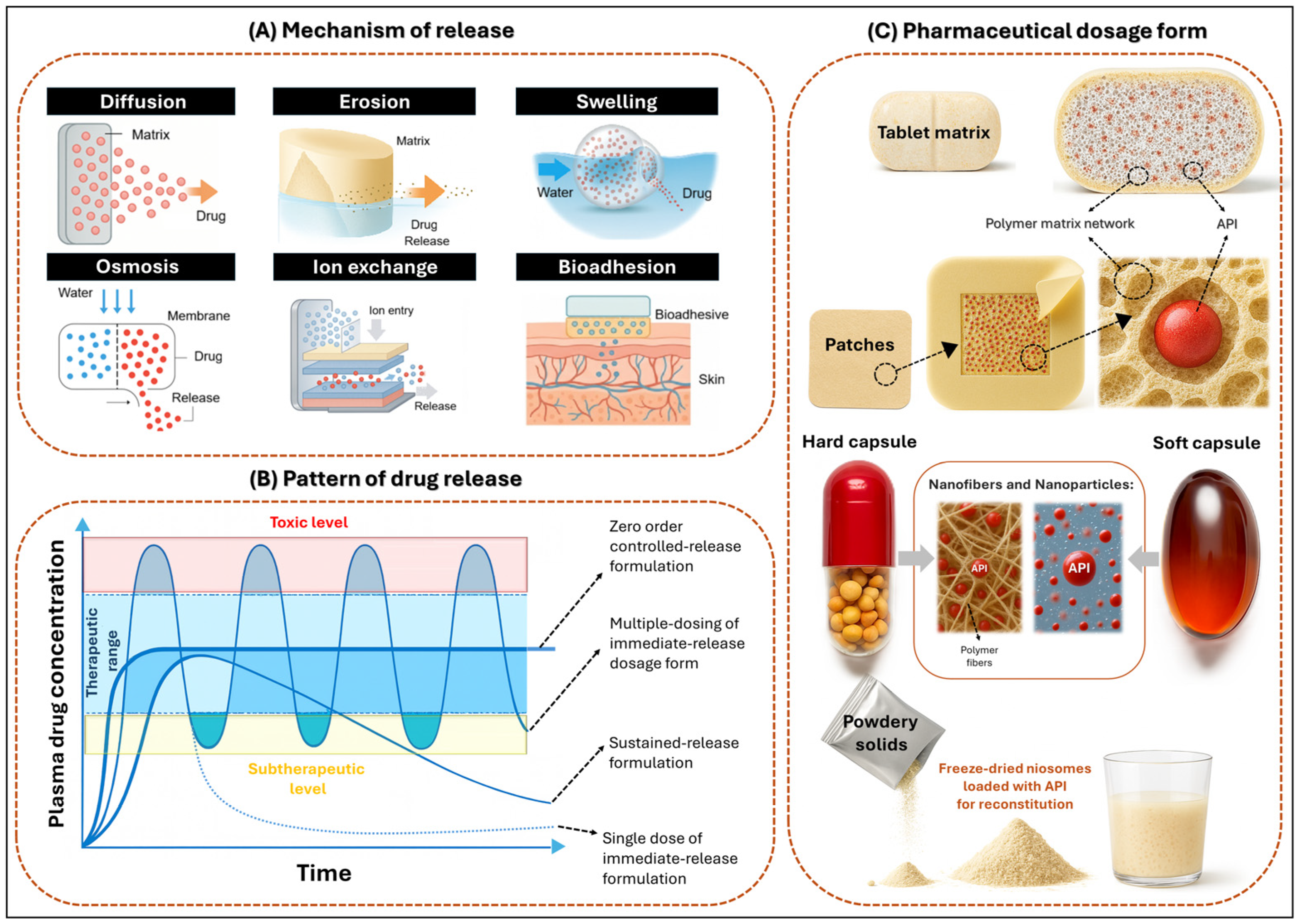
Modified release dosage forms (MRDFs) are pharmaceutical systems designed to alter the rate and/or site of release of the active substance compared to conventional formulations. Their primary objective is to maintain therapeutic plasma concentrations of diltiazem hydrochloride (DH) over extended periods, thereby reducing the frequency of administration and minimizing fluctuations in plasma levels [,]. MRDFs can be classified according to (i) release mechanism, (ii) release pattern, and (iii) dosage form as depicted in Figure 2.
Figure 2. Conceptual framework for MRDFs. (A) Principal mechanisms of drug release, including diffusion, erosion, swelling, osmosis, ion exchange, and bioadhesion. (B) Representative plasma concentration–time profiles illustrating immediate, sustained, delayed, and pulsatile release patterns in relation to therapeutic windows. (C) Pharmaceutical dosage forms commonly employed in MRDFs, such as matrix tablets, transdermal patches, capsules, powders, nanofibers, and nanoparticles.

3.1. Mechanism of Release

Modified-release systems can be classified according to their underlying mechanism of drug release, which includes diffusion, erosion, dissolution, swelling, osmosis, ion exchange, and bioadhesion []. In diffusion-controlled systems, the drug migrates through a polymeric matrix or semi-permeable membrane without inducing significant structural changes in the carrier. This migration may occur through hydrated polymer networks, insoluble frameworks, or the liquid boundary layer surrounding the device [,]. The process typically involves water ingress, drug dissolution, and molecular diffusion []. In contrast, erosion- or dissolution-based systems rely on the gradual degradation or solubilization of the matrix, whereby the drug is released as the carrier disintegrates [,]. Furthermore, swelling-dependent systems initiate drug release upon water absorption, which causes matrix expansion and the formation of gel layers that regulate diffusion. Polymers such as polyethylene oxide (PEO) exhibit rapid swelling behavior, significantly influencing gel formation and the duration of release [,,,,]. Additionally, osmotic systems function through water influx across a semi-permeable membrane, generating internal pressure that forces the drug out through a calibrated orifice. These systems often employ cross-linked gelatin capsules or osmotic pumps [,]. On the other hand, ion exchange systems operate via reversible interactions between drug ions and resin matrices, with release rates modulated by the ionic composition of the surrounding medium [,]. Finally, bioadhesive systems are designed to adhere to biological surfaces, such as mucosal tissues, thereby prolonging residence time and enhancing localized drug delivery [,]. These mechanisms are schematically illustrated in Figure 2A, which outlines the structural and physicochemical strategies employed to modulate drug release in modified systems.

3.2. Pattern of Drug Release

Based on their release patterns, MRDFs can be engineered to achieve specific pharmacokinetic profiles. To begin with, sustained or prolonged release systems are designed to maintain drug concentrations within the therapeutic window over extended periods, thereby minimizing plasma fluctuations and reducing dosing frequency [,,,]. In contrast, delayed release systems postpone drug liberation until reaching a specific physiological site or time, often triggered by factors such as pH, enzymatic activity, or gastrointestinal transit. This approach enables targeted delivery and improves therapeutic efficacy [,]. Moreover, pulsatile release systems deliver the drug in programmed bursts, mimicking circadian rhythms or adapting to variable therapeutic demands. These systems are particularly valuable in chronotherapy or in conditions requiring intermittent dosing []. In addition, bimodal release platforms integrate an initial immediate-release phase for rapid onset of action with a subsequent sustained-release phase, ensuring prolonged therapeutic coverage and enhanced patient adherence [,]. Collectively, these release patterns—schematically illustrated in Figure 2B—reflect the strategic modulation of drug delivery kinetics aimed at optimizing pharmacological outcomes while minimizing adverse effects.

3.3. Pharmaceutical Dosage Form

Structurally, MRDFs fall into three primary categories—monolithic or matrix-type, reservoir-type, and multiparticulate platforms—each offering distinct strategies for controlling drug release kinetics and optimizing therapeutic outcomes. One widely employed approach is the use of monolithic or matrix-type systems, in which the drug is uniformly dispersed within a continuous polymeric material. This configuration enables release through mechanisms such as diffusion, swelling, or erosion [,,]. Alternatively, reservoir-type systems encapsulate the drug within a central core surrounded by a semi-permeable or selective membrane, allowing controlled release via diffusion or osmotic pressure []. In addition, multiparticulate systems—including pellets [], microspheres [,], nanospheres [,,] and microsponges [,]—are composed of discrete units that can be engineered to exhibit tailored release profiles.
These units are commonly incorporated into tablets or capsules and offer enhanced flexibility in formulation design, improved gastrointestinal distribution, and reduced inter-subject variability. As illustrated in Figure 2C, the diversity of pharmaceutical dosage forms—ranging from tablets and capsules to transdermal patches—demonstrates the adaptability of these structural systems to various routes of administration and therapeutic requirements.

3.3.1. Monolithic Matrix Systems

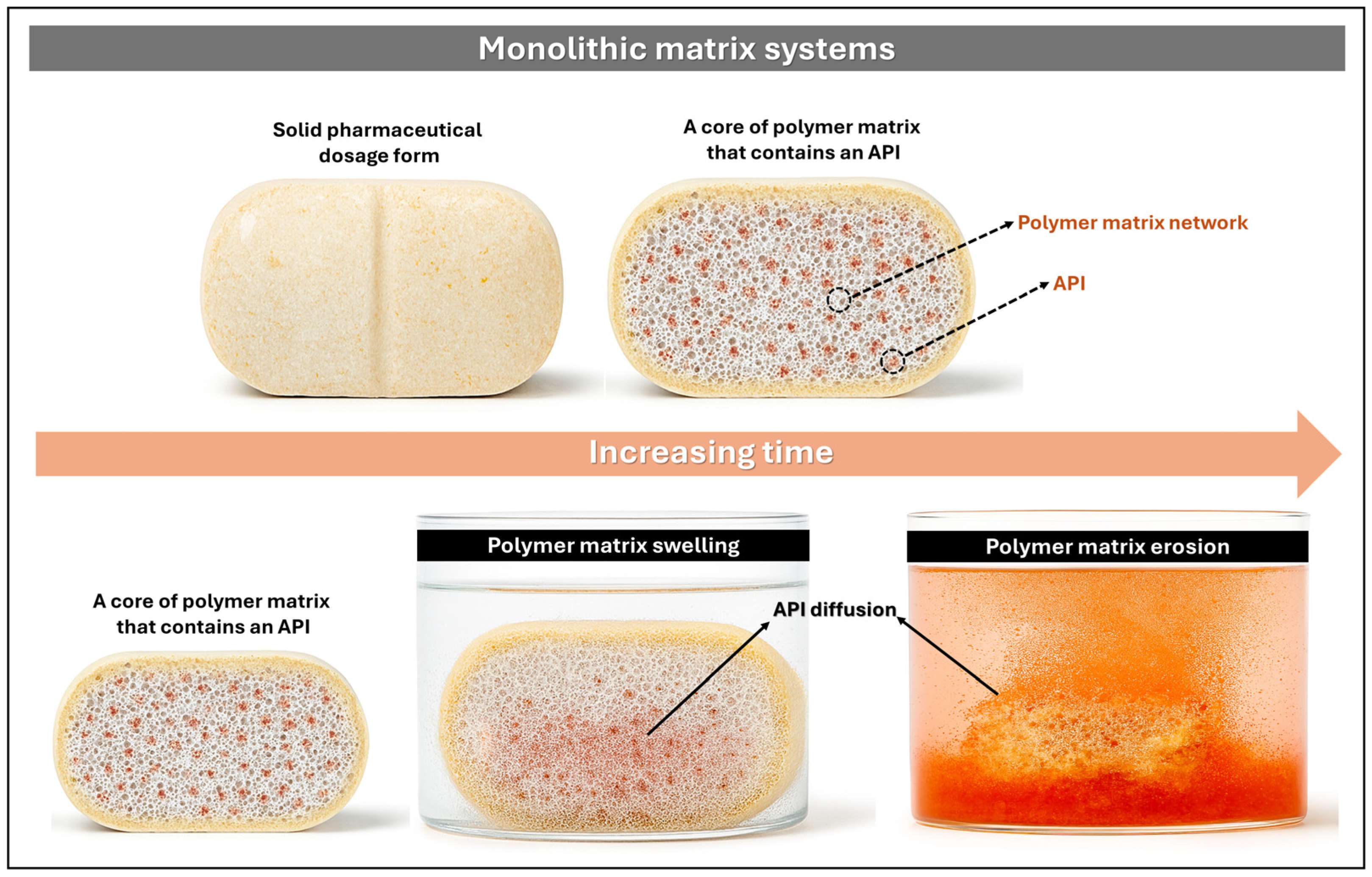
Monolithic matrix systems are pharmaceutical platforms in which the active pharmaceutical ingredient (API) is either uniformly or heterogeneously dispersed within a polymeric matrix or biocompatible excipient. Drug release from these systems is governed by the matrix structure through mechanisms such as diffusion, swelling, or erosion, with the objective of sustaining therapeutic plasma concentrations over a defined period [,]. The release kinetics may be modulated by diffusion through a hydrated gel layer in hydrophilic matrices, by diffusion across an insoluble framework in hydrophobic matrices, or by a combination of both in mixed systems. These platforms contribute to stabilizing plasma levels, minimizing adverse effects, and improving patient adherence (Figure 3) [].
Figure 3. Schematic representation of drug release from monolithic matrix systems, illustrating the sequential processes of swelling, erosion, and diffusion of the API into the surrounding medium.
Recent investigations on hydrophilic matrices have compared the performance of polymers such as hydroxypropyl methylcellulose (HPMC) and polyethylene oxide (PEO) in oral delivery systems. For instance, Tsuji et al. [] evaluated matrices composed of PEO and polyethylene glycol (PEG), reporting that PEO-based tablets released 85.7% of DH within 6 h, whereas HPMC matrices released only 63.1% over the same period. These findings underscore notable differences in hydration kinetics and gel formation between the two polymers. Furthermore, matrices formulated with HPMC K15M exhibited robust gel-forming capacity, effectively delaying DH release for up to 12 h via an anomalous diffusion mechanism involving both swelling and polymer relaxation [].
In contrast, hydrophobic matrices offer the advantage of reducing the influence of gastrointestinal physiological variables. These systems incorporate water-insoluble polymers such as ethylcellulose and Eudragit®, along with lipids and waxes, to form inert structures through which the drug diffuses via channels generated during partial dissolution. The hydrophobic nature of these materials enhances resistance to pH and enzymatic fluctuations, resulting in more predictable and stable release profiles compared to hydrophilic systems [].
Complementarily, transdermal matrix films containing DH have been developed using Eudragit RS100 in combination with HPMC K4M []. Eudragit RS100, being water-insoluble, enables modulation of drug release [] and is also employed as a coating for microspheres to attenuate the initial burst release of DH. In a related strategy, Alkhamis et al. [] formulated an insoluble matrix using polystyrene cation-exchange resins for modified DH release, eliminating the need for additional polymeric coatings. This approach exemplifies how low-permeability or insoluble matrices are essential for designing sustained-release formulations of DH.

3.3.2. Multiparticulate Systems

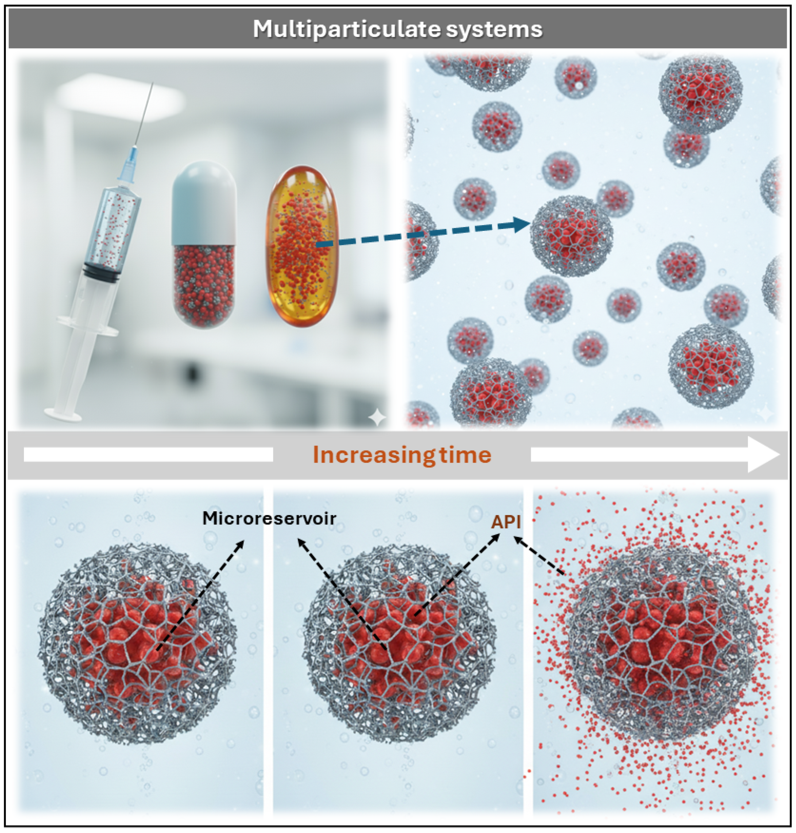
Multiparticulate systems consist of numerous discrete units—typically microparticles ranging from 1 to 1000 μm—designed to encapsulate the API within polymeric matrices or structured cores. These units function as autonomous microreservoirs, capable of modulating drug release both temporally and spatially within the organism [,]. Their architecture enables customized pharmacokinetic profiles, enhanced bioavailability, and reduced local irritation, particularly in gastrointestinal and transdermal applications. Moreover, for parenteral administration, microparticles must meet strict size specifications—typically below 5 μm for intravenous use—to prevent phlebitis and vascular complications. As illustrated in Figure 4, each dosage form (e.g., capsule, gel, or injectable) contains multiple microparticles with internal reservoirs from which the API diffuses progressively over time.
Figure 4. Schematic representation of multiparticulate systems: dosage forms containing microparticles with polymeric microreservoirs that release the API progressively over time.
This schematic representation reinforces the concept of temporal modulation of drug release and highlights the structural integrity of reservoir-based platforms. These systems are fabricated using a wide range of materials, including inorganic substrates, synthetic and natural polymers, and mineral-based excipients. Depending on the formulation strategy, they may adopt diverse structural configurations such as microspheres, micropellets, microcapsules, micro- and nanosponges, microemulsions, magnetic particles, and lipid-based vesicular systems (e.g., liposomes and niosomes) []. Each configuration offers distinct advantages in terms of drug loading capacity, release kinetics, and targeting potential.
Microsponges and Nanosponges
Microsponges and nanosponges are porous polymeric systems designed for controlled drug release. Their primary distinction lies in particle size and biological distribution: microsponges, typically ranging from 5 to 300 μm, are polymeric spheres that remain localized on tissue surfaces—such as skin, mucosa, or the gastrointestinal tract—providing extended drug release without systemic absorption [,]. In contrast, nanosponges, sized between 50 and 300 nm, consist of cross-linked polymeric networks (often cyclodextrin-based) capable of systemic circulation and targeted, sustained drug delivery.
In a comprehensive review, Utzeri et al. described the synthesis, structural features, and applications of cyclodextrin-based nanosponges in drug delivery and environmental systems, emphasizing how cross-linking strategies influence porosity and release behavior []. Complementarily, Rao et al. developed a nanosponge formulation loaded with DH for oral administration via the solvent emulsion-diffusion technique, employing ethylcellulose and β-cyclodextrin as polymeric carriers []. The process involved dissolving the drug and polymers in dichloromethane, followed by emulsification with an aqueous polyvinyl alcohol (PVA) phase, homogenization, filtration, and drying. SEM analysis revealed spherical, porous nanosponges with particle sizes ranging from 186 to 476 nm. In vitro release studies demonstrated that ethylcellulose enabled sustained DH release following zero-order kinetics, governed by a non-Fickian diffusion mechanism.
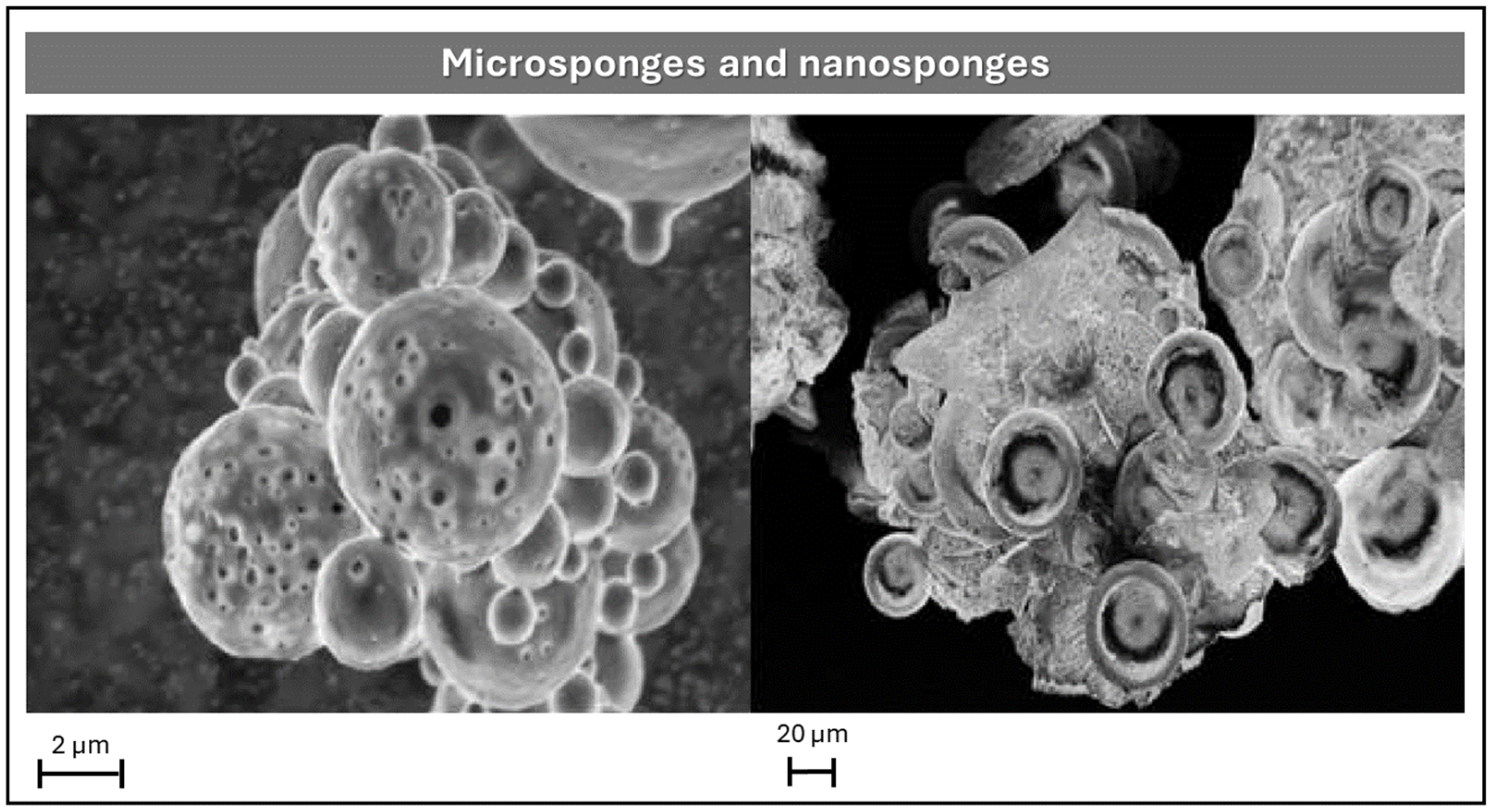
On the other hand, Ivanova et al. formulated topical gels containing DH-loaded microsponges for the treatment of chronic anal fissures (Figure 5) []. These microsponges were synthesized using Eudragit RS 100 via quasi-emulsion solvent diffusion, employing diltiazem base (poorly water-soluble) to enhance encapsulation efficiency. After encapsulation, the base was converted back to its hydrochloride salt using an HCl-generating chamber. The resulting microsponges were dispersed in two hydrogel matrices: 2% methylcellulose and 20% poloxamer 407. These formulations exhibited prolonged DH release for up to 24 h, significantly exceeding the 6–12 h release profile of conventional gels. Additionally, microsponge-based gels showed reduced permeation rates and approximately double the drug deposition in rectal mucosa compared to standard formulations.
Figure 5. SEM micrographs comparing microsponges and nanosponges: porous polymeric spheres (left, 2 μm scale) designed for localized drug delivery, and cross-linked nanoscale structures (right, 20 μm scale) intended for systemic administration. Figures Reprinted from Nadezhda Antonova Ivanova, Adriana Trapani, Cinzia Di Franco, Delia Mandracchia, Giuseppe Trapani, Carlo Franchini, Filomena Corbo, Giuseppe Tripodo, Iliyan Nikolov Kolev, Georgi Stoyanov Stoyanov, Kameliya Zhechkova Bratoeva, In vitro and ex vivo studies on diltiazem hydrochloride-loaded microsponges in rectal gels for chronic anal fissures treatment, Pages No. 62, Copyright (2025), with permission from Elsevier [].
Further insights were provided by Kolev et al., who investigated the physicochemical behavior of DH-loaded microsponges prepared with Eudragit RS 100 via quasi-emulsion solvent diffusion []. To elucidate drug distribution and compatibility, two reference models were employed: (i) physical drug–polymer mixtures, representing heterogeneous dispersions with surface-bound drug, and (ii) homogeneous solid dispersions obtained by molding, where the drug is molecularly dispersed within the polymer matrix. Characterization techniques included ATR-FTIR spectroscopy, differential scanning calorimetry (DSC), SEM coupled with energy-dispersive X-ray analysis (SEM-EDX), and dissolution studies. ATR-FTIR spectra indicated molecular dispersion of DH within the microsponge matrix, while DSC confirmed amorphization and compatibility. SEM-EDX revealed porous morphology and elemental distribution, suggesting partial surface localization of the drug and effective removal of residual solvents. Dissolution profiles showed that the porous architecture of microsponges enabled faster and more complete drug release compared to solid dispersions. Although this study did not culminate in a final dosage form, it established a robust analytical framework for understanding drug–matrix interactions and their impact on release kinetics.
Microspheres
Microspheres are solid spherical particles composed of polymeric matrices typically ranging from 1 to 1000 μm in diameter, composed of a homogeneous polymeric matrix in which the API is uniformly dispersed [,]. Owing to their structural simplicity and formulation versatility, they are suitable for multiple administration routes, including nasal, oral, and parenteral delivery.
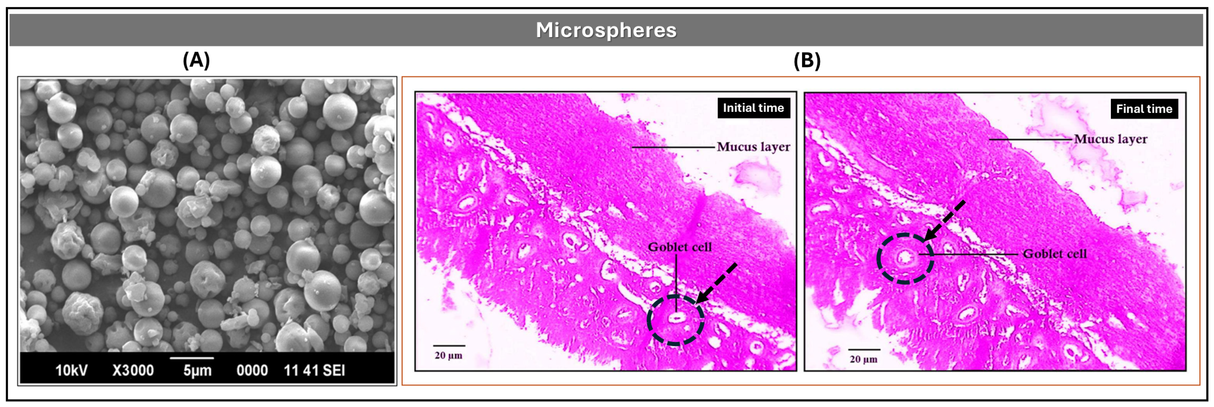
In one approach, Kulkarni et al. developed nasal mucoadhesive microspheres using chitosan (CS) and DH, prepared via spray drying from aqueous solution []. The formulation was optimized using a central composite experimental design, adjusting polymer concentration, atomization rate, and drying temperature to maximize encapsulation efficiency (EE > 90%) and mucoadhesive performance. The resulting solid microspheres had an average diameter of ~11 μm (Figure 6A) and exhibited strong adhesion to nasal mucosa. Ex vivo permeation studies in sheep nasal tissue revealed 95.6% DH permeation after 240 min, attributed to the high degree of deacetylation and molecular weight of chitosan. Histopathological analysis confirmed preservation of epithelial architecture, with no signs of irritation or damage (Figure 6B). Accelerated stability studies over three months (ICH guidelines) showed minimal variation in particle size (10.8–12.4 μm) and EE (90.66–89.40%), with consistent mucoadhesive strength.
Figure 6. (A) Optimized DH-loaded microsphere formulation (D7); (B) Optical photomicrographs of untreated sheep nasal mucosa and mucosa treated with 100 mL of 1% v/v DH microsphere suspension for 7 h. Kulkarni, Deepak B. Bari, Sanjay J. Surana, Chandrakantsing V. Pardeshi, In vitro, ex vivo and in vivo performance of chitosan-based spray-dried nasal mucoadhesive microspheres of diltiazem hydrochloride, Pages No. 113 and 115, Copyright (2016), with permission from Elsevier [].
From an oral delivery perspective, Bolourchian et al. formulated DH-loaded microspheres using Eudragit RL and RS polymers via emulsion solvent evaporation []. Formulations with varying drug and polymer concentrations were characterized for yield, encapsulation efficiency, particle size, and surface morphology. Uncoated microspheres achieved EE values between 56% and 93%, with particle sizes ranging from 470 μm to over 1000 μm. Coating with Eudragit RS improved EE (82–92%) and mitigated the initial burst release. Sustained release behavior was influenced by drug-to-polymer ratio and particle size; formulations with DH–Eudragit RL ratios of 1:3 and 1:4 met USP criteria for extended-release DH products, supporting 12 and 24 h dosing intervals.
Additionally, Tareq et al. and Sahoo et al. investigated gelatin-based microsphere systems for the controlled release of DH, employing distinct formulation strategies [,]. Tareq et al. developed semi-interpenetrated microspheres using a natural polymer blend of sodium alginate and gelatin, stabilized with glutaraldehyde []. These systems demonstrated sustained DH release over 24 h, with efficiencies of 82.1% in acidic medium and 63.1% in neutral medium, following first order and Higuchi kinetics. Microspheres with an alginate–gelatin ratio of 80:20 showed optimal swelling and diffusion-controlled behavior, supporting their suitability for nasal or oral prolonged-release applications.
In contrast, Sahoo et al. formulated gelatinous microspheres via emulsification–polymerization, incorporating ethylcellulose, Eudragit RSPO, and Span 80 to enhance encapsulation and modulate release []. The resulting systems achieved production efficiencies of 73.33–82.53% and encapsulation efficiencies of 77.25–89.01%. In vitro studies at pH 7.4 revealed a biphasic release profile: an initial burst (~30% DH in the first hour) followed by sustained release with t50 values between 2.5 and 3 h. The hydrophobic polymers and glutaraldehyde crosslinking contributed to a rigid hydrogel network, reducing drug leaching, and promoting controlled release kinetics.
Pellets
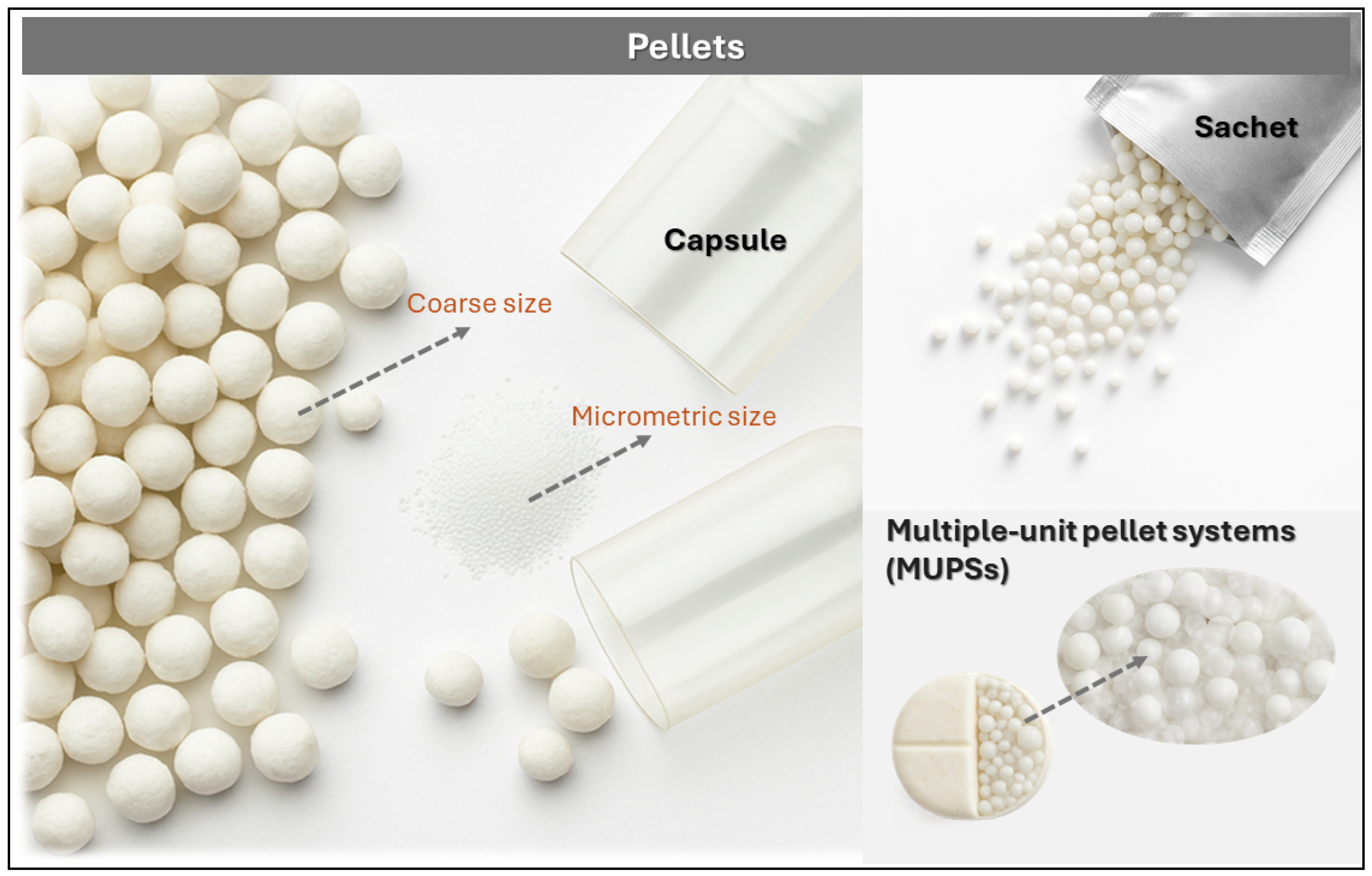
Pellets are small, free-flowing spherical particles typically ranging from 500 to 1500 μm in diameter, produced through the controlled agglomeration of fine drug powders and excipients [,]. These multiparticulate units are manufactured using techniques such as extrusion–spheronization, fluid bed granulation, freeze-drying, or spray drying, yielding mechanically robust granules with uniform size and excellent flow properties. As illustrated in Figure 7, pellet-based systems exhibit notable versatility in terms of size, formulation, and packaging formats, including encapsulation in gelatin capsules, dispensing from sachets, and integration into multiple-unit pellet systems (MUPSs), which support oral sustained-release applications.
Figure 7. Visual representation of pellet-based drug delivery systems: coarse and micrometric pellets encapsulated in gelatin capsules, dispensed from sachets, and integrated into multiple-unit pellet systems (MUPSs). These formats illustrate the structural diversity and packaging flexibility of sustained-release pellet formulations.
In one formulation strategy, Rashmitha et al. developed DH-loaded pellets via extrusion–spheronization, employing microcrystalline cellulose (MCC) as the structuring agent and carboxymethylcellulose (CMC) as the binder []. DH HCl, MCC, and CMC powders (sieved through mesh #40) were mixed for 10 min in a planetary mixer to ensure homogeneity. The dry blend was moistened with demineralized water to achieve suitable plasticity and extruded through a cylindrical roller extruder with a 1 mm nozzle, producing uniform filaments. These were processed in a spheronizer equipped with a ribbed rotor plate, where centrifugal force and controlled friction shaped them into spheres. Pellets were dried at 40 °C for 8 h, yielding particles with an average size of 1026–1061 μm, high sphericity, and favorable flow characteristics, confirmed by SEM and micromorphological analysis. The EE ranged from 89.17% to 95.27%, and in vitro release studies at pH 6.8 showed 88.1% DH release over 12 h. Stability was maintained for 90 days at 40 °C/75% RH, with no significant changes in drug content or release kinetics.
In a complementary study, Kanteti et al. formulated nine DH pellet variants via extrusion–spheronization, incorporating a hydrophobic phase of molten Gelucire 43/01 (processed at 50 °C, ground and sieved to 250 μm) combined with almond gum in varying proportions (20%, 30%, 40%), Gelucire (10%, 20%, 30%), and lactose as filler, following a 32 factorial design []. The resulting spherical pellets ranged from 1128 to 1458 μm in size, with high EE (89–95%) and adequate mechanical strength, confirmed by SEM, DSC, and FTIR. In vitro release profiles revealed that pellets with lower Gelucire content (10%) released DH within 6 h, while intermediate formulations (20%) extended release to 8 h. The combination of 30% Gelucire and 40% almond gum achieved 88.1% release over 12 h. Statistical analysis confirmed that Gelucire primarily governed drug retention, while almond gum enhanced cohesion and reduced friability, enabling precise modulation of sustained-release profiles.
Nanofibers and Nanoparticles
Nanofibers and nanoparticles represent advanced platforms for controlled release, offering high surface area and tunable physicochemical properties. Nanofibers, primarily obtained via electrospinning, exhibit diameters typically ranging from 90 to 120 nm [] or 100 to 300 nm [], and possess a high surface-to-volume ratio that favors sustained drug release. Their structural resemblance to the extracellular matrix (ECM) enhances cellular behavior and fibroblast proliferation, making them suitable for applications such as wound dressings and tissue engineering scaffolds. In contrast, nanoparticles are colloidal solid carriers whose small size enables tissue penetration and targeted release, contributing to improved bioavailability [].
In one formulation strategy, Etemadi et al. developed nanofibrous membranes composed of PVA, CS, and polycaprolactone (PCL), incorporating diltiazem hydrochloride (DH) at 10% w/w []. Electrospinning of individual polymer–drug solutions yielded flexible, porous membranes with diameters between 150 and 300 nm, reduced by the increased polarity and conductivity of the DH-containing solution. PCL, a hydrophobic polymer, enhanced mechanical strength, degradation control, and drug release modulation, while PVA and CS, both hydrophilic, facilitated faster release. FTIR confirmed chemical compatibility, and sustained DH release extended to 48 h. In vitro biocompatibility assays with L929 cells showed viability rates > 85%, reaching 99.89% in DH-loaded formulations. Fibroblasts covered nearly the entire membrane surface, suggesting favorable drug–cell interactions and enhanced proliferation. Additionally, DH incorporation improved wettability and mechanical properties, supporting the use of these nanofibers as advanced wound dressings.
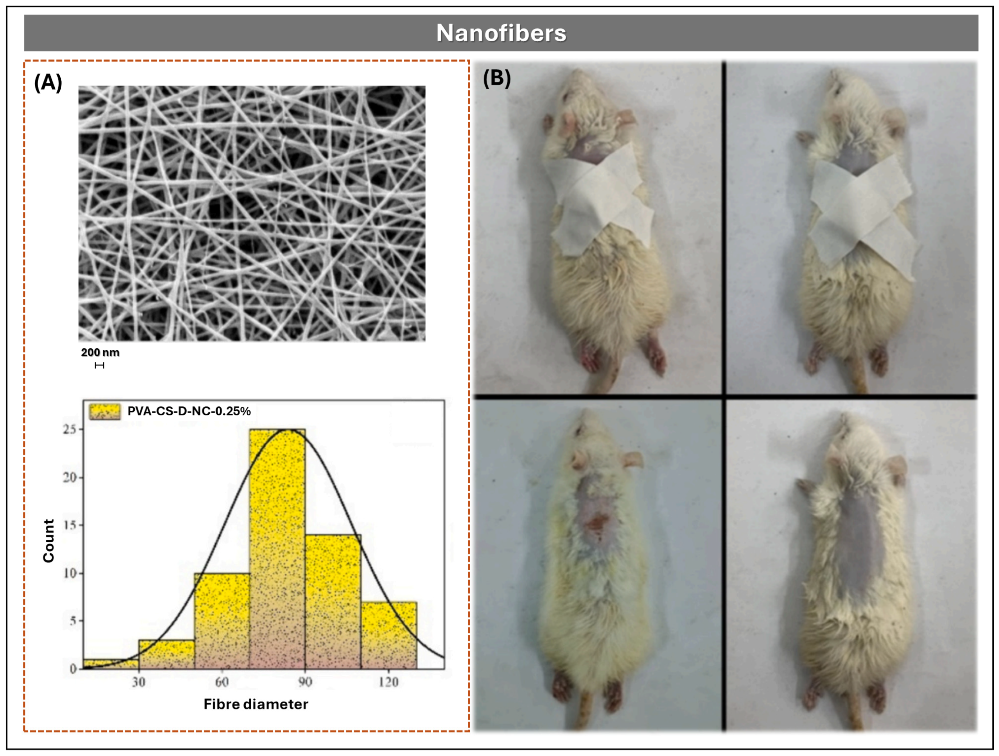
In a complementary approach, Samanta et al. designed a nanocellulose-reinforced nanofiber scaffold using PVA/CS (80:20) matrices for transdermal DH delivery (Figure 8) []. Nanocellulose, extracted from jute waste and functionalized with ethylenediamine, was incorporated at 0.25–0.5% w/w. Electrospinning was performed at 25 kV, 0.8 mL/h, and 14 cm distance, producing smooth fibers of 150–300 nm. FTIR confirmed chemical modification, and SEM revealed uniform morphology (Figure 8A). In vitro release studies showed 67.9 ± 3.4% DH release over 72 h, compared to 92.6 ± 4.6% in scaffolds without nanocellulose, following the Korsmeyer–Peppas model. MTT assays with RAW 264.7 cells and rat skin irritation tests demonstrated >85% viability and good biocompatibility (Figure 8B). Antibacterial activity (>97% reduction in E. coli and S. aureus) was attributed to the amine groups of the nanocellulose.
Figure 8. (A) SEM micrograph and diameter distribution histogram of electrospun nanofibers composed of PVA/CS reinforced with amino-nanocellulose and loaded with DH, showing uniform morphology and nanoscale dimensions. (B) In vivo wound healing progression in rats treated with DH-loaded nanofiber membranes, demonstrating enhanced epithelial regeneration and reduced lesion size. Figures Reprinted from Arpita Priyadarshini Samanta, Adrija Ghosh, Koushik Dutta, Debashmita Mandal, Surajit Tudu, Kunal Sarkar, Beauty Das, Swapan Kumar Ghosh, Dipankar Chattopadhyay, Biofabrication of aminated nanocellulose reinforced polyvinyl alcohol/chitosan nanofibrous scaffold for sustained release of diltiazem hydrochloride, Pages No. 6, Copyright (2024), with permission from Elsevier [].
Further insights were provided by Seyedian et al., who fabricated PVA nanofibrous mats (6% w/v) containing 0%, 2%, and 4% DH, electrospun at 15–17 kV and 12 cm distance and cross-linked with 10% glutaraldehyde vapor for 12 h []. SEM revealed fibers of ~152.7 nm and porosity of ~88.4% in the 4% DH formulation. The mats exhibited a contact angle of 29.1°, swelling of 110.4%, and in vitro release of ~94% in 120 min, fitting the Korsmeyer–Peppas model (R2 = 0.96, n = 0.52). In human fibroblast cultures, 4% DH mats were non-cytotoxic and increased proliferation by 263% after 5 days. In a murine wound model, lesion size was reduced to 14.7% after 14 days, with improved histopathological parameters and reductions in malondialdehyde (−63%) and nitrite (−59%) levels.
Representative examples of nanoparticles in DH formulations include CS conjugated with L-leucine for pulmonary inhalation, synthesized via water-in-oil emulsification and glutaraldehyde crosslinking []. These nanoparticles exhibited a leucine substitution degree of 0.89, creating an amphiphilic environment that enhanced swelling and prolonged DH release at pH 7.3. Similarly, Khairnar et al. developed CS nanoparticles loaded with repaglinide and DH via ion gelation with tripolyphosphate, producing particles of 200–300 nm with sustained release of ~19 h for repaglinide and ~20 h for DH, without burst effect []. Lastly, modified silica nanoporters (Silica-03) functionalized with silane KH-570 demonstrated superior DH adsorption and extended release compared to unmodified silica (Silica-01), maintaining crystal stability per DSC and FTIR, due to enhanced hydrophobic affinity from the organofunctional coating.
Niosomes
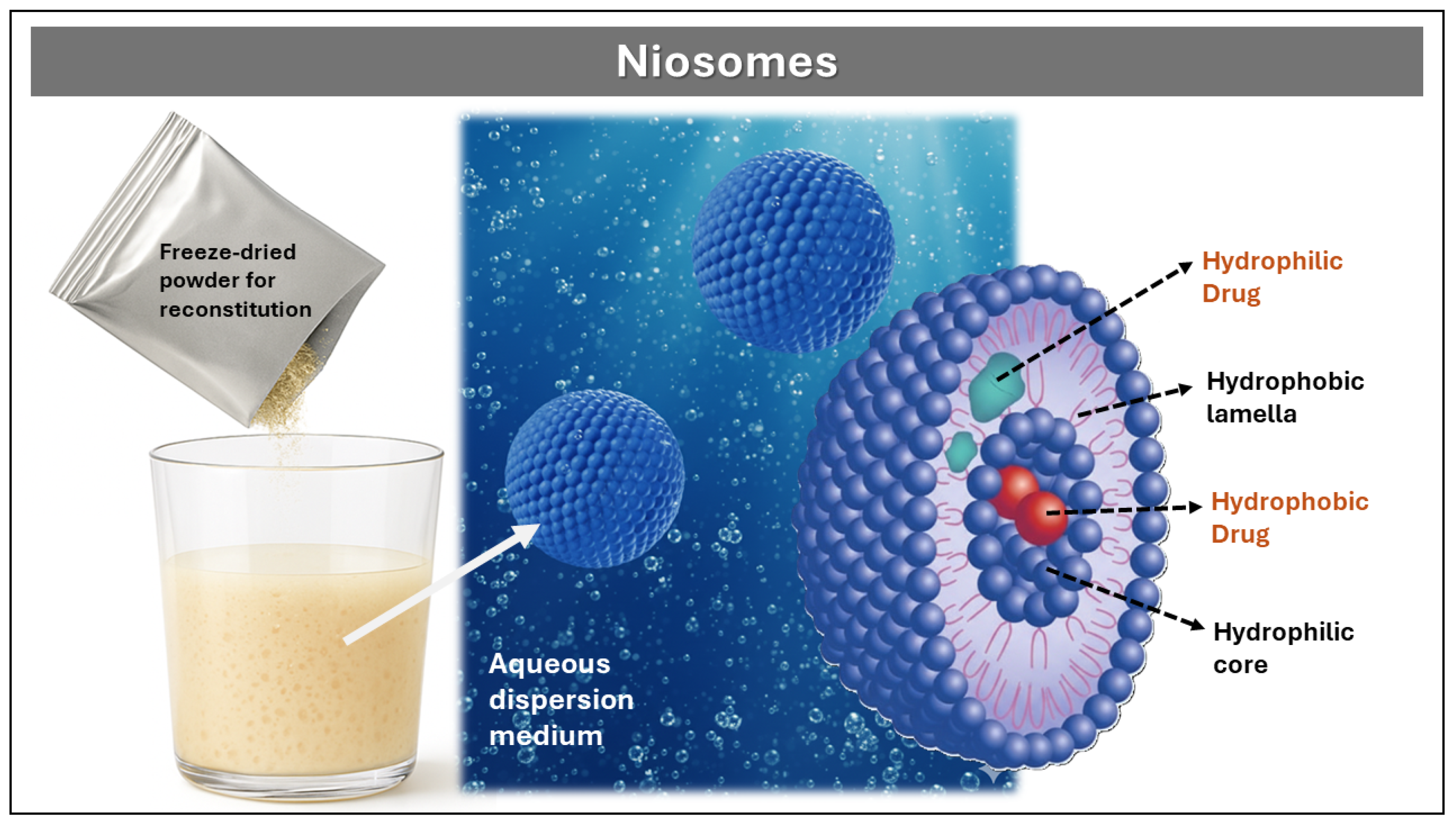
Niosomes are multiparticulate vesicular systems formed by the self-assembly of non-ionic surfactants—such as Spans, Brijs, and polysorbates—in combination with cholesterol as a stabilizing excipient, which can encapsulate hydrophilic drugs within their internal aqueous core and lipophilic drugs within the lipid bilayer, offering high physical and chemical stability during preparation and storage, as well as versatility for the controlled release of molecules with diverse polarity profiles []. As illustrated in Figure 9, freeze-dried niosomal powder can be reconstituted in aqueous dispersion media to form bilayer vesicles capable of dual drug loading and targeted release. Importantly, such reconstitution requires that the vesicles have been previously formed and stabilized—commonly via spray drying or lyophilization—since non-ionic surfactants alone do not spontaneously self-assemble into uniform bilayer structures upon simple aqueous dispersion.
Figure 9. Schematic representation of niosome formation and structure. Freeze-dried powder is reconstituted in aqueous dispersion medium to form bilayer vesicles. Hydrophilic drugs are encapsulated in the aqueous core and surface, while hydrophobic drugs are embedded within the lipid bilayer.
Niosomes are typically prepared by methods such as thin film hydration, ether injection, reverse-phase evaporation, and microfluidic techniques, each offering distinct control over vesicle size, lamellarity, and encapsulation efficiency. Thin film hydration is widely employed for its simplicity and scalability, while ether injection and reverse-phase evaporation allow finer control of bilayer formation and drug entrapment. Microfluidic approaches, although more recent, enable precise modulation of particle size and polydispersity through controlled flow dynamics. Regardless of the method, the resulting niosomes generally exhibit diameters between 10 nm and 1 μm, low polydispersity, and high biocompatibility. Their release kinetics can be modulated by adjusting the surfactant-to-cholesterol ratio [].
In a complementary design, Ammar et al. developed DH-loaded niosomes for nasal administration using the thin film hydration technique []. Non-ionic surfactants (Span 60 or Brij-52) and cholesterol were dissolved in a chloroform/methanol mixture (2:1 v/v) to form a thin lipid film. Following solvent evaporation via rotary evaporation, the film was rehydrated with phosphate buffer (pH 7.4) at 60 °C for 30 min and matured overnight at 4 °C. The resulting suspension contained spherical unilamellar vesicles with mean sizes ranging from 0.82 to 1.59 μm. In vivo pharmacokinetic studies in male Wistar rats revealed a significant increase in mean residence time, elimination half-life, and area under the curve (AUC), along with a decrease in the elimination constant, indicating prolonged therapeutic action and enhanced bioavailability of DH compared to control solutions.
In a complementary approach, Akbari et al. formulated a topical DH-loaded niosomal gel using the ultrasonic method, optimizing the cholesterol-to-surfactant ratio to achieve vesicles with controlled particle size, low polydispersity index, and high encapsulation efficiency (~94%) []. In vitro release studies demonstrated sustained DH release over 24 h. In vivo pharmacological evaluation in an animal wound model showed that the niosomal gel achieved wound closure efficacy comparable or superior to a commercial reference, with enhanced tissue regeneration and no signs of skin irritation. Histopathological analysis and skin sensitivity tests confirmed the therapeutic potential of this nanoparticulate platform for dermal applications.

3.3.3. Reservoir-Type

Reservoir-type formulations are strategic platforms for modulating the rate and timing of drug release, thereby improving bioavailability and patient adherence. These systems are defined by the presence of a membrane or barrier that regulates drug diffusion into the surrounding medium.
A representative example for DH was reported by Ishrath et al., who developed a PulsinCap®-type system consisting of hard gelatin capsules cross-linked with formaldehyde (12 h vapor exposure followed by drying) []. The capsules contained DH granules obtained via wet granulation and a hydropolymer plug (HPMC and ethylcellulose) that controlled the lag time. Optimization of plug composition revealed that 90 mg produced a 3–4 h delay, while 100 mg extended it to 7–11 h, depending on polymer ratios. The PF5 formulation (HPMC 50 mg/ethylcellulose 50 mg) was selected for its minimal release during latency and rapid post-delay release. Accelerated stability studies confirmed consistent performance, demonstrating that capsule cross-linking and plug engineering enable precise temporal control—ideal for chronotherapy and targeted administration. Additionally, another reservoir strategy is the “tablets-in-a-capsule” technique, where the capsule shell acts as a barrier layer that dissolves or degrades, releasing the drug from internal tablets arranged as a multiparticulate core [].
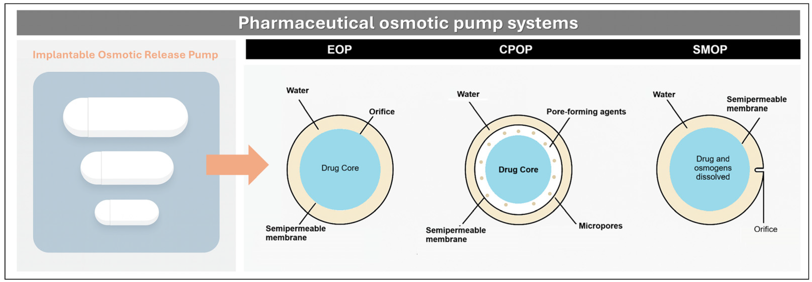
A specialized subcategory of reservoir systems is the osmotic pump, illustrated in Figure 10. These systems rely on water permeation through a semipermeable membrane, which hydrates the drug-loaded core and generates internal pressure that expels the drug through a precisely sized orifice, achieving zero-order kinetics and minimizing variability due to pH or intestinal motility. In the elementary osmotic pump (EOP), drug granules are compressed with hydrophilic excipients and coated with cellulose acetate, followed by orifice drilling. Upon hydration, the core expands and steadily releases the drug. In controlled porosity osmotic pumps (CPOP), the coating includes soluble porogens (e.g., PEGs) that dissolve in situ to form microchannels, eliminating the need for mechanical drilling. In modulated solubility osmotic pumps (SMOP), salts such as NaCl are added to reduce drug solubility, maintaining a constant osmotic gradient, and extending release up to 16–18 h [].
Figure 10. Schematic representation of pharmaceutical osmotic systems. (Left): implantable osmotic release pump. (Right): cross-sectional diagrams of EOP (semipermeable membrane with drilled orifice), CPOP (porogen-induced micropores), and SMOP (salt-modulated solubility gradient).
In one formulation strategy, Joshi et al. applied a Quality by Design (QbD) approach to develop DH EOP tablets via wet granulation, using HPMC and cellulose acetate coatings []. Optimization of polymer ratios and coating weight yielded zero-order release for 14–16 h, with robust mechanical properties and strong in vivo correlation. In contrast, Soliman and Ibrahim formulated CPOP tablets by mixing DH with mannitol (1:1.5) as an osmotic agent, followed by coating with Eudragit RS-100 and PEG-400 []. Upon PEG dissolution, pores formed that enabled ~90% release in 8 h, independent of pH or agitation. In a complementary study, they investigated two key variables: (i) coating thickness, which modulates water ingress and osmotic pressure, and (ii) PEG molecular weight, which affects porosity and release rate. Thicker coatings delayed release, while low molecular weight PEGs produced more porous membranes and faster release. The optimal balance achieved sustained zero-order kinetics, demonstrating the importance of membrane engineering in reservoir systems.
Further refinement was achieved by Monton and Kulvanich, who designed a multi-chamber push-pull osmotic pump (PPOP) for oral DH delivery []. The system used formaldehyde-crosslinked gelatin capsules containing a drag layer (10 mg DH, 130 mg PEO Mw 200K, 40 mg lactose) and a push layer (55 mg PEO Mw 5000K, 34.5 mg NaCl, 0.5 mg pigment), coated with HPMC/PEG 4000 and eight layers of cellulose acetate (6% w/w), ending with a 0.6 mm outlet orifice. Upon hydration, water penetrates both chambers, dissolving the drug and expanding the polymer, generating pressure that ejects the DH suspension with zero-order kinetics, independent of pH, stirring, or solubility. The formulation remained stable for 12 months, with consistent Higuchi-model release and latency times, supporting its industrial scalability.
Table 1 presents a comparison of the advantages and disadvantages of MDRFs compared to conventional dosage forms.
Table 1. Comparison between MDRFs and conventional dosage forms.
In summary, the formulation of DH MDRFs has enabled precise modulation of pharmacokinetic behavior, improved therapeutic adherence, and broadened the range of administration routes. Through platforms such as matrix tablets, pellets, microspheres, nanofibers, and osmotic systems, researchers have addressed the limitations associated with DH’s short half-life, variable bioavailability, and chronopharmacological requirements. These advances reflect the strategic integration of polymer selection, structural design, and release mechanisms to optimize clinical performance. Building on these formulation principles, the following section outlines the general manufacturing methods employed in MRDFs development, emphasizing key unit operations, process variables, and scale-up considerations.

4. General Methods of Manufacture of MRDFs

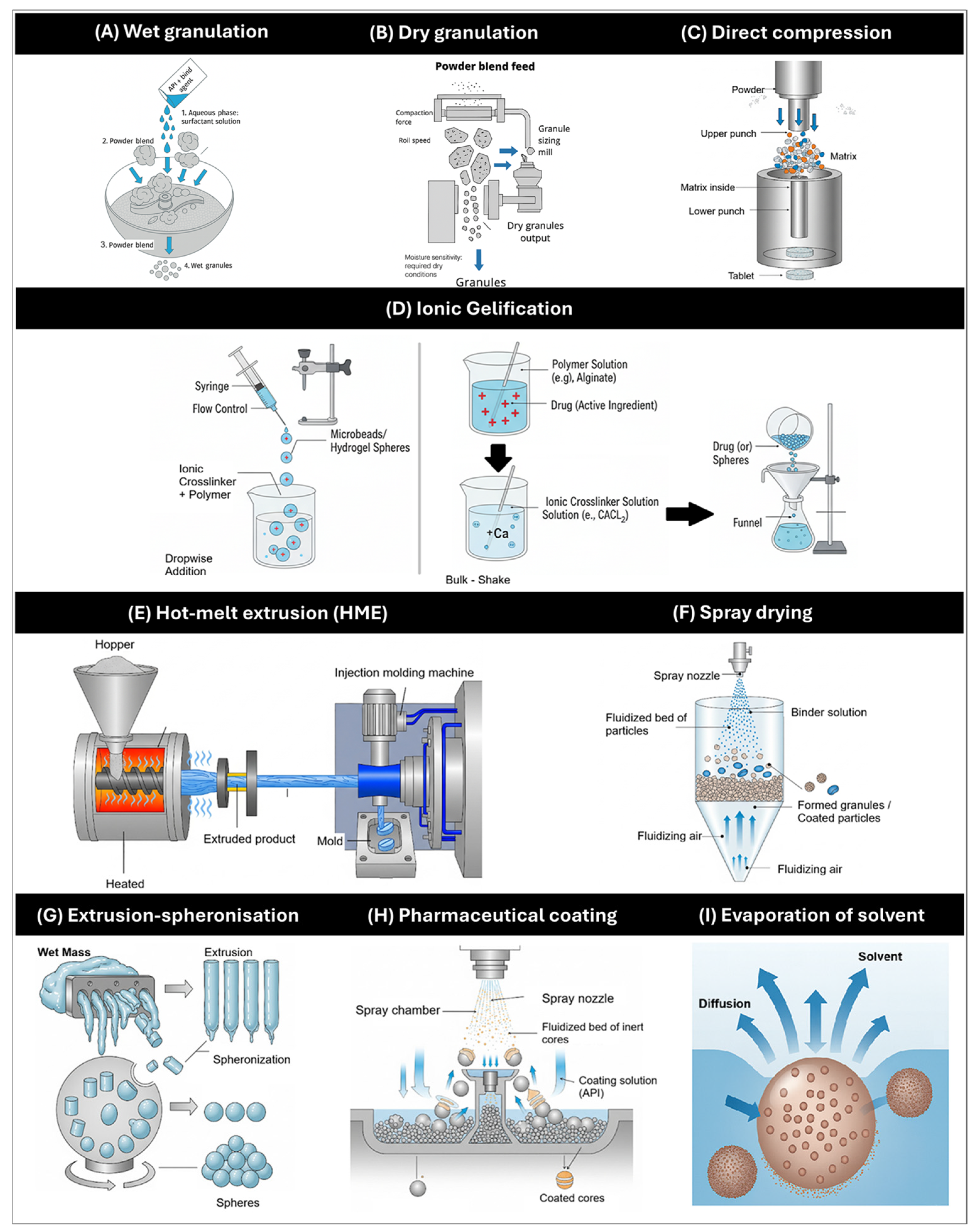
The manufacture of MRDFs requires specialized technologies and process controls to ensure the quality, reproducibility, and functional performance of the final product. Figure 11 illustrates the principal manufacturing methods employed in the pharmaceutical industry for solid MRDFs platforms, highlighting their relevance across formulation types and dosage formats.
Figure 11. Schematic overview of advanced pharmaceutical technologies used in the design and optimization of modified-release and targeted drug delivery systems. The illustration includes: (A) Wet granulation; (B) Dry granulation; (C) direct compression; (D) ionic gelation; (E) Hot melt extrusion; (F) spray drying; (G) Extrusion-spheronization; (H) Pharmaceutical coating; (I) Evaporation solvent.

4.1. Optimal Pharmacokinetics and Integration with Drug Delivery Technology

An optimal controlled release (CR) formulation of DH should maintain plasma concentrations within the therapeutic window—typically 50 to 200 ng/mL—over an extended period, ideally 24 h. This target aims to minimize peak–trough fluctuations, reduce dose-dependent adverse effects such as bradycardia and hypotension, and improve adherence in chronic cardiovascular therapy. Achieving zero-order release kinetics is considered ideal, as it ensures a constant drug input rate independent of physiological variables. Recent studies have demonstrated the feasibility of approximating this ideal profile using scalable technologies and validated modeling approaches. Joshi et al. (2022) [] developed an osmotic DH formulation using a Quality by Design (QbD) strategy, achieving near zero-order kinetics and robust performance across physiological conditions. Arafat et al. (2021) [] evaluated a polymer matrix-based DH formulation against a commercial reference (Tildiem®), showing sustained release up to 10 h and comparable pharmacokinetic parameters, reinforcing the clinical viability of matrix systems. Complementarily, Taha and Emara (2022) [] applied convolution and deconvolution modeling to predict plasma profiles of extended-release DH tablets, achieving strong in vitro–in vivo correlation (IVIVC) and minimal prediction error for Cmax and AUC.
These findings support the rational design of CR formulations that approximate ideal pharmacokinetic behavior, combining therapeutic consistency with industrial scalability. To translate these pharmacokinetic objectives into functional dosage forms, the selection and optimization of manufacturing technologies becomes critical. The ability to approximate zero-order kinetics or sustain therapeutic plasma levels over extended periods depends not only on the choice of excipients and release mechanisms, but also on the precision and reproducibility of the underlying processes. The following subsections outline the principal manufacturing methods employed in the development of MRDFs, highlighting their relevance in aligning formulation performance with pharmacokinetic targets.

4.2. Wet and Dry Granulation

Wet granulation (Figure 11A) remains widely used in DH MRDF development for enhancing compressibility and flow. This method involves the agglomeration of powder particles using a liquid binder to form granules with enhanced compressibility, flowability, and uniformity. Its versatility has enabled the incorporation of natural polymers—such as karaya gum and carob gum—as matrix formers, with polyvinylpyrrolidone K-30 (PVP K-30) serving as a binder to modulate drug release kinetics []. In parallel, pectin (PEC) has been employed as a matrix material in wet-granulated tablets to achieve controlled release profiles, demonstrating the adaptability of this technique to diverse excipient functionalities [].
By contrast, dry granulation (Figure 11B) is preferred when the API or excipients exhibit sensitivity to moisture, as it eliminates the need for liquid binders. Techniques such as roller compaction have been successfully applied to produce sustained-release tablets containing hydrophilic and hydrophobic polymers—including HPMC, hydroxypropylcellulose (HPC), and methacrylic acid copolymers—ensuring mechanical integrity and controlled release without compromising chemical stability []. Moreover, dry processing has proven particularly advantageous in MRDFs systems based on cation exchange resins, where moisture-free conditions help preserve drug stability and optimize powder flow and compressibility [].

4.3. Direct Compression

Direct compression (Figure 11C) is one of the most efficient and scalable techniques for tablet production, as it eliminates intermediate steps such as granulation and drying, thereby reducing processing time and operational complexity []. This approach requires excipients with excellent flowability and compressibility, including MCC, dicalcium phosphate dihydrate, and spray-dried lactose, which facilitate uniform die filling and robust tablet formation [].
In the context of MRDFs, direct compression has been successfully employed using HPMC matrices of varying viscosities. These polymers serve dual functions: they act as compression aids during tableting and as release modulators within the gastrointestinal tract, enabling sustained drug delivery profiles []. Furthermore, the absence of moisture and heat exposure during processing helps preserve drug integrity, particularly for thermolabile or hydrolytically sensitive compounds. By minimizing process variables, direct compression also enhances batch-to-batch reproducibility and formulation robustness, making it a valuable strategy for MRDFs development under both pilot and industrial conditions.

4.4. Ionic Gelification

Ionic gelification (Figure 11D) refers to the capacity of specific polymers to form a semi-solid, three-dimensional network that entraps the drug, thereby creating a matrix system capable of modulating its release profile []. Depending on the triggering mechanism, gelification processes can be classified as ionotropic, thermal, covalent, or pH-sensitive, each offering distinct structural and kinetic characteristics.
For diltiazem hydrochloride formulations, MRDFs, ionotropic gelification has been extensively investigated. This technique employs anionic polymers such as sodium alginate, which undergo gelation upon contact with multivalent cations (e.g., Ca2+, Zn2+, Fe3+). Typically, DH is dispersed in an alginate solution and dropped into a calcium chloride bath, where ionic crosslinking transforms the droplets into spherical microspheres. The resulting gelled particles exhibit mechanical integrity and sustained release behavior, making them suitable for MRDFs applications under gastrointestinal or mucosal conditions [,]

4.5. Hot-Melt Extrusion (HME)

HME (Figure 11E) enables continuous mixing and shaping of drug–polymer blends, allowing controlled release and integration with 3D printing for personalized dosage design.
A notable advancement in this domain is the integration of HME with 3D Fused Filament Deposition (FDM) printing, where HME enables the fabrication of drug-loaded filaments suitable for additive manufacturing. These filaments, incorporating DH and polymers such as HPMC, have demonstrated thermal stability and programmable release profiles governed by print geometry, infill density, and surface area-to-volume ratios []. As such, HME serves not only as a conventional MRDFs production method but also as a foundational technology for personalized pharmaceutical manufacturing via 3D printing, offering precise control overdose, release kinetics, and spatial distribution of the API [,].

4.6. Spray Drying

Spray drying (Figure 11F) is a widely employed technique for converting liquid formulations—such as solutions, suspensions, or emulsions—into dry solid particles via atomization in a heated air stream. This method is particularly valuable for microencapsulation and the production of spherical microparticles with uniform size distribution, enhanced flow properties, and compatibility with thermolabile drugs, owing to rapid solvent evaporation and minimal thermal exposure []. Key process parameters—including inlet and outlet temperatures, feed rate, atomization pressure, and solvent composition—enable precise control over particle morphology, encapsulation efficiency, and release behavior.
For DH-containing MRDFs systems, spray drying has been applied using polymers such as Eudragit L100, HPMC K4M, guar gum, and xanthan gum. A recent study optimized a formulation based on Eudragit L100 (drug-to-polymer ratio 1:2 in isopropanol), achieving controlled release over 12 h, content uniformity of approximately 70%, and zero-order kinetics []. These findings underscore the versatility of spray drying in tailoring release profiles and enhancing the physicochemical stability of diltiazem hydrochloride formulations.

4.7. Extrusion-Spheronization

Extrusion–spheronization (Figure 11G) is a two-step manufacturing process designed to produce spherical pellets with consistent size, shape, and mechanical integrity. Initially, a wet mass containing DH and selected excipients—such as polymers, waxes, or natural gums—is extruded through a perforated die to generate cylindrical extrudates. These are subsequently processed in a spheronizer, where rotational motion and frictional forces reshape them into uniform spheres []. Key process variables—including rotation speed, frictional energy, and moisture content—directly influence pellet morphology, surface characteristics, and drug release kinetics.
This technique has been successfully applied to develop sustained-release DH pellets using excipients such as gelucire 43/01 and almond gum. Resulting formulations demonstrated high production yields (86–92%) and prolonged drug release, confirming the suitability of extrusion–spheronization for MRDFs systems requiring precise control over particle architecture and release dynamics [].

4.8. Pharmaceutical Coating

Pharmaceutical coating (Figure 11H) is a versatile and widely adopted technique for modifying both the site of drug release and its kinetic profile, particularly in solid dosage forms such as tablets, pellets, and multiparticulate systems. Among the most established approaches is fluid-bed coating, wherein particles are suspended in an upward air stream while a polymeric solution is sprayed. This method commonly employs polymers such as ethylcellulose for sustained release, hydroxypropyl methylcellulose phthalate (HPMCP) for enteric protection, and methacrylic acid copolymers like Eudragit L100/S100 for pH-dependent release.
An alternative strategy is perforated drum coating, in which the cores rotate while the coating solution or suspension is applied. This configuration allows precise control of temperature and humidity, making it particularly suitable for formulations involving organic solvents. Additionally, the layering technique on inert nuclei—such as cellulose microspheres or sugar spheres—enables the sequential deposition of drug and excipients, allowing fine-tuned control over content uniformity and release modulation [,].
For diltiazem hydrochloride formulations, various coating strategies have been explored. One example involves aqueous HPMC coatings derived from raw cotton, successfully applied to compressed tablets. In this study, different viscosity grades of HPMC (18–52 cps) and coating levels (2–8%) were evaluated, demonstrating that higher polymer viscosity and thicker films markedly delayed drug release, in contrast to uncoated tablets which released ~96% of the drug within 1 h []. Alternatively, fluid-bed coating of DH pellets with Eudragit FS 30D at 35% w/w has been implemented to achieve a 12 h pulsatile release profile under variable pH conditions []. These examples underscore the potential of coating technologies to design programmable and environmentally responsive MRDFs systems.

4.9. Evaporation of Solvent

Solvent evaporation (Figure 11I) is a widely employed technique in pharmaceutical microencapsulation, particularly for the development of MRDFs [,]. Grounded in mass transfer principles, this method utilizes water-insoluble polymers as encapsulating matrices to form microparticles or microspheres with controlled release properties. The process typically involves the creation of a two-phase system, wherein the drug and polymer are dissolved in a volatile organic solvent to form a homogeneous solution. This solution is then emulsified into an aqueous phase containing surfactants under high-shear homogenization.
The core mechanism relies on the diffusion of the organic solvent from the emulsion droplets into the continuous aqueous phase. As the solvent migrates, the polymer solubility decreases, triggering its controlled precipitation around the drug. This results in the formation of encapsulated particles with tailored morphology and release kinetics [].
For DH MRDFs systems, solvent evaporation has been successfully applied using polymers such as poly(lactic-co-glycolic acid) (PLGA), ethylcellulose, and Eudragit derivatives. These encapsulating agents enable the design of sustained and programmable release profiles, while preserving drug stability and enhancing formulation versatility [].

4.10. Combination of Manufacturing Methods

In contemporary pharmaceutical development, most diltiazem hydrochloride formulations are produced through combinations of manufacturing techniques, allowing for the optimization of drug performance and formulation robustness. This integrated approach leverages the strengths of individual methods while mitigating their limitations, enabling the design of complex systems with tailored release profiles.
Representative examples include matrix systems incorporating polymers such as poloxamer-188 in combination with HPMC, which modulate hydration dynamics and diffusion rates []. Other strategies involve multi-layer coated pellets produced via extrusion–spheronization, using MCC as a spheroizing agent, followed by sequential coatings—HPMC for core protection and ethylcellulose for sustained release control. Additionally, PEO of varying molecular weights has been employed to fine-tune matrix behavior and drug release kinetics [].
Among the most impactful innovations are osmotic technologies, which have significantly advanced the design of DH MRDF. These include osmotic tablets and pumps capable of delivering drugs with zero-order kinetics, independent of pH and gastrointestinal motility []. Recent developments have integrated 3D printing—particularly semi-solid extrusion—to fabricate tablets with programmable osmotic cores and semipermeable shells []. Furthermore, push–pull systems combining enteric coatings and ion-exchange resin complexes offer dual-release mechanisms, enhancing bioavailability and therapeutic precision. These hybrid platforms complement conventional osmotic systems and represent a cornerstone in the evolution of modified-release technologies for DH [].

4.11. Emerging Technologies: 3D Printing

Three-dimensional (3D) printing, also referred to as additive manufacturing, has emerged as a disruptive technology in pharmaceutical development, enabling the fabrication of personalized solid dosage forms through computer-aided design and layer-by-layer deposition. Among the pioneering platforms, ZipDose® technology stands out as the first formulation system to employ 3D printing for drug manufacturing, utilizing aqueous fluid binding of powdered layers without reliance on compression forces, punches, or dies [,]. This approach enables the production of highly porous structures with rapid disintegration and precise dose control.
Recent studies demonstrate the potential of 3D printing to fabricate multi-drug dosage forms with programmable release []. In diltiazem hydrochloride formulations, one study reported the integration of 3D printing with hot-melt extrusion (HME) to fabricate dual filaments: one composed of PVA, DH, and osmogens (NaCl and/or mannitol) for the osmotic core; and another of cellulose acetate for the semipermeable shell []. Using a dual-extruder fused deposition modeling (FDM) printer, the authors constructed core–shell units with engineered geometries, including micro-orifices to facilitate water ingress and lateral weakening cavities designed to rupture under osmotic pressure.
The release onset was programmable at 0, 120, or 360 min, depending on the structural configuration of the shell, followed by sustained drug release. Physicochemical characterization of the filaments and printed constructs included TGA, DSC, mechanical testing, and X-ray diffraction (XRD), confirming DH amorphization post-HME/FDM and retention of NaCl crystallinity. In situ monitoring via micro-computed tomography (micro-CT) revealed volumetric expansion of the osmotic core during hydration, leading to targeted rupture of the shell and transition from latency to controlled release.
This highlights the potential of HME-FDM integration for personalized MRDFs fabrication, where geometric modulation of the dosage form is as critical as its composition in determining the release profile. The integration of HME and FDM printing thus represents a promising avenue for the development of programmable, patient-specific therapies.

4.12. Current Commercial Formulations of DH

Several extended-release formulations of DH are currently available on the pharmaceutical market, including Cardizem CD®, Cardizem LA®, Cartia XT®, Tiazac®, and Dilacor XR® in the United States, as well as Tiazac XC® in Canada [,]. Although all products are presented as modified-release capsules or tablets, they differ in the technological platforms employed to regulate drug release. For instance, Cardizem CD® utilizes a multiparticulate system composed of microgranules coated with semipermeable polymers, whereas Cartia XT® and Tiazac® rely on hydrophilic matrix systems that swell upon hydration to form a gel barrier, thereby modulating drug diffusion [].
These technological distinctions result in varied pharmacokinetic profiles. Formulations such as Cardizem LA® and Tiazac® provide sustained release over 24 h, enabling once-daily administration, while others—including Cartia XT®—exhibit 12 h release, requiring twice-daily dosing. Despite these differences, all formulations demonstrate clinical bioequivalence in the management of hypertension and angina pectoris, with 24 h systems offering improved therapeutic adherence due to simplified dosing regimens [,].
From a pharmacotechnical perspective, MRDFs of DH offer multiple advantages that contribute to therapeutic optimization. The reduction in dosing frequency—from three or four times daily to one or two—simplifies administration and enhances patient compliance. These systems also minimize fluctuations in plasma concentrations, thereby reducing the incidence of adverse effects associated with peak levels. The incorporation of protective matrices or coatings confers stability against acid-mediated degradation, improving drug performance in gastric environments. Furthermore, MRDFs platforms enable chronomodulated release, which is particularly relevant in the treatment of hypertension—a condition characterized by circadian variation and early morning blood pressure surges. Finally, gradual or site-specific release along the gastrointestinal tract contributes to the attenuation of first-pass metabolism, enhancing systemic bioavailability and ensuring more consistent therapeutic outcomes.
To consolidate the diverse formulation strategies and technological platforms described throughout this section, Table 1 presents a structured overview of the principal investigations focused on MRDFs of DH. The table highlights key aspects of each study, including the manufacturing method employed, the therapeutic objective pursued—such as sustained, pulsatile, or chronomodulated release—and the compositional features that define each system. This synthesis facilitates comparative analysis across approaches, offering insight into how polymer selection, process variables, and structural design converge to optimize drug performance and clinical applicability.

4.13. Industrial Viability and Comparative Implementation Analysis

Although MRDFs offer significant therapeutic advantages, their industrial viability depends on factors beyond pharmacokinetic performance, such as scalability, cost-efficiency, compatibility with existing manufacturing infrastructure, and regulatory compliance [,]. Osmotic pump technologies are widely recognized for delivering drugs at a near zero-order rate, independent of physiological variables like pH, motility, and food intake. Their modular design and membrane-controlled release make them highly reproducible and scalable []. More recent innovations, such as magnetic nanoparticles, have enhanced their performance, supporting long-term use and reducing dosing frequency, which could be a promising strategy for this active pharmaceutical ingredient.
Polymer matrix systems—particularly hydrophilic and hydrophobic matrices—represent an attractive alternative due to their simplicity, cost-effectiveness, and compatibility with conventional tableting equipment. These systems allow flexible modulation of drug release and are suitable for hot-melt extrusion, solvent evaporation, and compression molding. Advances in polymer science and 3D printing further reinforce their scalability and personalization potential []. Regulatory acceptance of matrix systems is well established, especially when supported by dissolution testing and bioavailability studies.
Additionally, Level A IVIVC modeling has enabled the linkage of dissolution profiles with clinical pharmacokinetics, making virtual bioequivalence studies and regulatory approvals possible without extensive in vivo trials. Although it requires robust data, its integration can accelerate development and facilitate post-approval adjustments. Mechanistic models such as PBPK are also gaining acceptance by agencies like the FDA and EMA for complex formulations [].
To provide a comprehensive perspective, this analysis also considers the full range of manufacturing methods described in Section 4, organized from conventional to emerging technologies. These include wet and dry granulation [], direct compression [], ionic gelification [], hot-melt extrusion [], spray drying [], extrusion-spheronization [], pharmaceutical coating, solvent evaporation, and hybrid or additive manufacturing approaches such as 3D printing. Each method presents distinct advantages and limitations in terms of industrial scalability, equipment compatibility, cost, and regulatory alignment. Table 2 summarizes the industrial possibilities of each platform to support formulation selection and technology transfer decisions.
Table 2. Comparative Analysis of Industrial Viability for MRDF Technologies.

4.14. Market Traction and Bibliometric Visualization

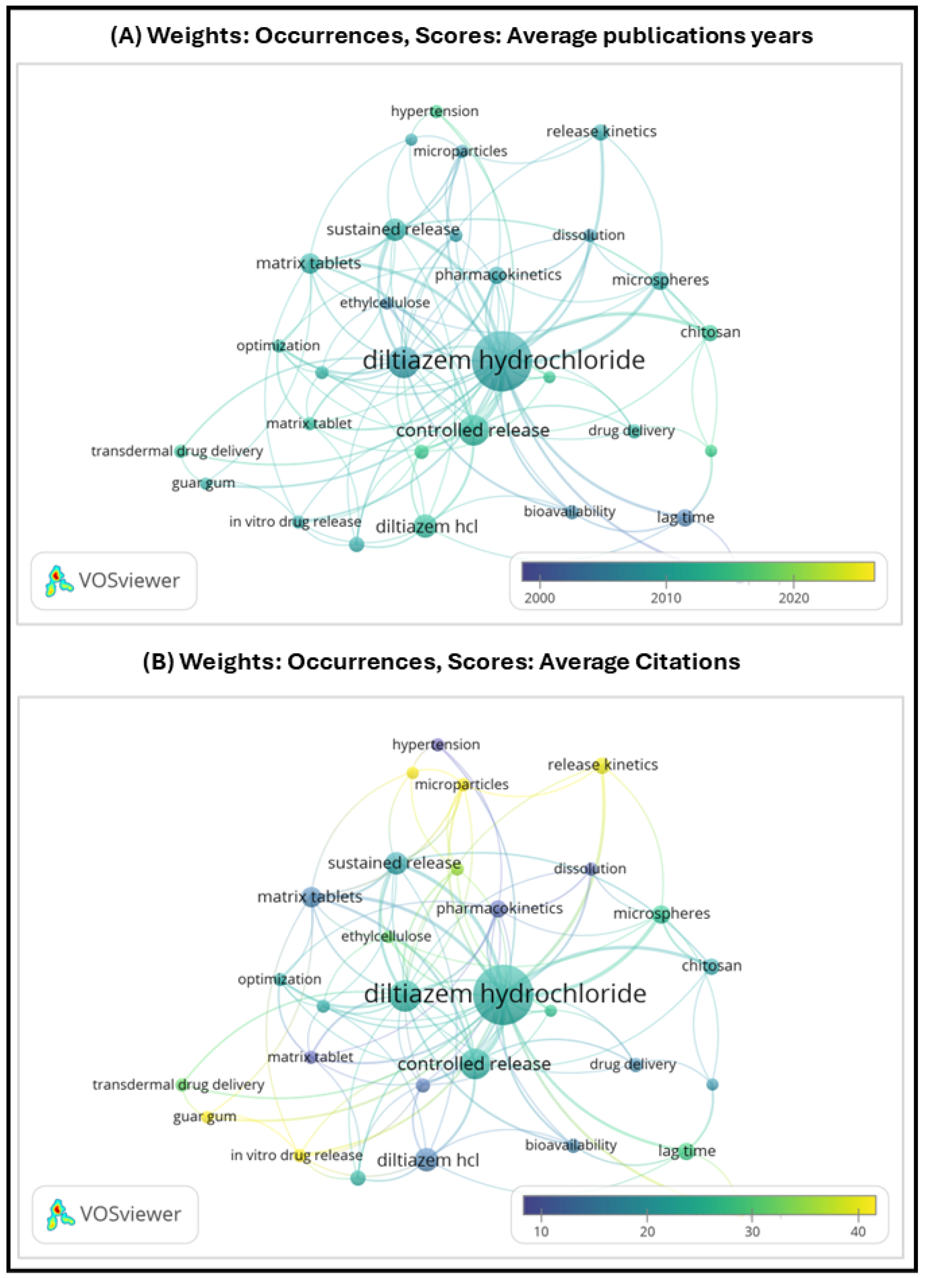
To complement the technological and pharmacokinetic analysis of controlled release strategies for DH, this section addresses the financial and scientific traction of the field. From a market perspective, DH remains a relevant molecule in the cardiovascular segment, with sustained-release formulations representing a niche but stable share of the total addressable market (TAM). While generic immediate-release products dominate in terms of volume, controlled-release formats—particularly once-daily tablets—offer differentiated value in terms of adherence and therapeutic consistency, justifying their continued development in select patient populations and regulatory environments [,]. To visualize the scientific traction of controlled release technologies applied to DH, a bibliometric analysis was conducted using VOSviewer software version 1.6.20. The co-occurrence map (Figure 12) was generated from Scopus-indexed publications between 2000 and 2025, filtered by keywords such as Diltiazem hydrochloride, Emerging technologies, Methods of manufacture, and Modified release dosage forms.
Figure 12. VOSviewer co-occurrence map generated from Scopus-indexed publications (2010–2025) using the keywords: Diltiazem hydrochloride, Emerging technologies, Methods of manufacture, and Modified release dosage forms. The clusters illustrate thematic concentrations in formulation strategies, manufacturing approaches, and innovation trends related to controlled release systems.
From this bibliometric visualization, it becomes evident that while Diltiazem hydrochloride remains a central node in the co-occurrence network, the color gradient in visualization (A)—coded by average publication year—shows that most relevant studies were published prior to 2020. Dominant purple tones around keywords such as controlled release, microspheres, pharmacokinetics, and drug delivery suggest that research activity on MRDFs of DH has declined in recent years. This trend may reflect technological maturity, a shift toward newer therapeutic agents, or reduced editorial prioritization in high-impact journals.
In contrast, visualization (B)—coded by average citation count—indicates that the most cited studies are also associated with established technologies, reinforcing their historical relevance but also highlighting a lack of thematic renewal. This temporal and thematic gap underscores the need for a critical review like the present one, which consolidates accumulated knowledge and connects it with emerging platforms and industrial feasibility criteria.
Based on this analysis, Table 3 provides a structured synthesis of key investigations on modified-release dosage forms of DH, organized by system type, manufacturing method, therapeutic application, and core composition.
Table 3. Investigations from MRDF of DH.

5. Conclusions

MRDFs of DH offer a practical solution to overcome the drug’s short half-life and the need for frequent dosing. The reviewed studies show that various manufacturing techniques—such as direct compression, granulation, extrusion–spheronization, spray drying, solvent evaporation, and gelation—have enabled the development of platforms like matrices, pellets, microspheres, osmotic systems, and transdermal devices. These approaches have been effective in controlling drug release, improving bioavailability, and enhancing patient adherence.
From a formulation point of view, the use of functional polymers—including HPMC, Eudragit®, ethylcellulose, and chitosan—has played a key role in adjusting release profiles under different physiological conditions. In addition, combining manufacturing methods has proven useful for creating multiparticulate systems and matrix-based formulations with better control over release kinetics and mechanical properties.
On the clinical side, MRDFs of DH help maintain stable plasma levels, reduce side effects related to peak concentrations, and support chronotherapy in conditions like hypertension. These benefits contribute to better treatment outcomes and improved patient compliance.
Looking ahead, technologies such as hot-melt extrusion combined with 3D printing, and systems based on nanomaterials, show promise for developing personalized and programmable MRDFs. These innovations allow precise control over dosage form geometry and release behavior, aligning with current trends in precision medicine. Moreover, the rational design of these systems increasingly involves the reverse engineering of ideal release profiles and the integration of advanced modeling tools—such as physiologically based pharmacokinetic (PBPK) simulations and in vitro–in vivo correlation (IVIVC)—to predict clinical performance and support regulatory decision-making. Further research and industrial application will be important to bring these advanced systems into routine clinical use.

Author Contributions

Conceptualization, E.T.-M. and Y.C.; methodology, E.T.-M. and Y.C.; formal analysis E.T.-M., Y.C. and C.H.S.; investigation, E.T.-M.; resources, E.T.-M.; data curation, E.T.-M. and Y.C.; writing—original draft preparation, E.T.-M.; writing—review and editing, Y.C. and C.H.S.; visualization, Y.C. and C.H.S.; supervision, Y.C. and C.H.S.; project administration, Y.C.; funding acquisition, Y.C. All authors have read and agreed to the published version of the manuscript.

Funding

This research has been funded by Dirección General de Investigaciones of Universidad Santiago de Cali under call No. DGI-01-2025, by Proyecto de fortalecimeinto de grupos number 939-621124-749 and Grant number 939-621122-071.

Data Availability Statement

No new data were created or analyzed in this study. Data sharing is not applicable.

Acknowledgments

Figures Reprinted from Abhijeet D. Kulkarni, Deepak B. Bari, Sanjay J. Surana, Chandrakantsing V. Pardeshi—in vitro, ex vivo and in vivo performance of chitosan-based spray-dried nasal mucoadhesive microspheres of diltiazem hydrochloride, Pages No. 113 and 115, Copyright (2016), with permission from Elsevier []. Figures Reprinted from Arpita Priyadarshini Samanta, Adrija Ghosh, Koushik Dutta, Debashmita Mandal, Surajit Tudu, Kunal Sarkar, Beauty Das, Swapan Kumar Ghosh, Dipankar Chattopadhyay—biofabrication of aminated nanocellulose reinforced polyvinyl alcohol/chitosan nanofibrous scaffold for sustained release of diltiazem hydrochloride, Pages No. 6, Copyright (2024), with permission from Elsevier []. Figures Reprinted from Nadezhda Antonova Ivanova, Adriana Trapani, Cinzia Di Franco, Delia Mandracchia, Giuseppe Trapani, Carlo Franchini, Filomena Corbo, Giuseppe Tripodo, Iliyan Nikolov Kolev, Georgi Stoyanov Stoyanov, Kameliya Zhechkova Bratoeva—in vitro and ex vivo studies on diltiazem hydrochloride-loaded microsponges in rectal gels for chronic anal fissures treatment, Pages No. 62, Copyright (2025), with permission from Elsevier []. During the preparation of this manuscript, the author used Gemini GenAI Version 2.5 Pro and Copilot IA Version 2025 for the purposes of obtaining Figure 1, Figure 2, Figure 3, Figure 4, Figure 7, Figure 9, Figure 10 and Figure 11.

Conflicts of Interest

Author Constain H. Salamanca was employed by the company Cecoltec Services SAS. The remaining authors declare that the research was conducted in the absence of any commercial or financial relationships that could be construed as a potential conflict of interest.

Abbreviations

APIActive Pharmaceutical Ingredient
BCSBiopharmaceutical Classification System
CAPCellulose Acetate Phthalate
CSChitosan
CPOPControlled Porosity Osmotic Pump
DHDiltiazem Hydrochloride
DSCDifferential Scanning Calorimetry
ECMExtracellular Matrix
EEEncapsulation Efficiency
EOPElementary Osmotic Pump
FDMFused Deposition Modeling
FTIRFourier Transform Infrared Spectroscopy
HMEHot-Melt Extrusion
HPMCHydroxypropyl Methylcellulose
HPCHydroxypropylcellulose
ICHInternational Council for Harmonisation
IVIVCIn Vitro–In Vivo Correlation
MCCMicrocrystalline Cellulose
MRModified Release
MRDFsModified Release Dosage Forms
MUPSMultiple-Unit Pellet Systems
NaCMCSodium Carboxymethylcellulose
PCLPolycaprolactone
PECPectin
PEGPolyethylene Glycol
PEOPolyethylene Oxide
PVAPolyvinyl Alcohol
QbDQuality by Design
SEMScanning Electron Microscopy
SEDDSSelf-Emulsifying Drug Delivery System
SMOPSolubility-Modulated Osmotic Pump
TGAThermogravimetric Analysis
TPPTripolyphosphate
USPUnited States Pharmacopeia
XRDX-ray Diffraction

References

  1. Escobar, C.; Ariza, A.; Barrios, V.; Campuzano, R.; Freixa-Pamias, R.; Gámez, J.M.; Fernández Olmo, M.R.; Jorge-Pérez, P.; Tamargo, J. Actualización Del Uso de Los Fármacos Antianginosos En El Tratamiento Del Síndrome Coronario Crónico: Enfoque Práctico. Rev. Esp. Cardiol. 2022, 22, 1–10. [Google Scholar] [CrossRef]
  2. Ivanova, N.A.; Trapani, A.; Di Franco, C.; Mandracchia, D.; Trapani, G.; Franchini, C.; Corbo, F.; Tripodo, G.; Kolev, I.N.; Stoyanov, G.S.; et al. In Vitro and Ex Vivo Studies on Diltiazem Hydrochloride-Loaded Microsponges in Rectal Gels for Chronic Anal Fissures Treatment. Int. J. Pharm. 2019, 557, 53–65. [Google Scholar] [CrossRef] [PubMed]
  3. Ahmed, V.; Sharma, S.; Bhatt, P. Formulation and Evaluation of Sustained Release Tablet of Diltiazem Hydrochloride. Int. J. Pharm. Sci. Res. 2020, 11, 2193. [Google Scholar] [CrossRef]
  4. Jones, K.E.; Hayden, S.L.; Meyer, H.R.; Sandoz, J.L.; Arata, W.H.; Dufrene, K.; Ballaera, C.; Lopez Torres, Y.; Griffin, P.; Kaye, A.M.; et al. The Evolving Role of Calcium Channel Blockers in Hypertension Management: Pharmacological and Clinical Considerations. Curr. Issues Mol. Biol. 2024, 46, 6315–6327. [Google Scholar] [CrossRef] [PubMed]
  5. Kavishetti, R.; Halkeri, M.; Nargund, S.L.; Shahapur, A. Formulation and Evaluation of Mucoadhesive Tablets of Diltiazem Hydrochloride. World J. Pharm. Pharm. Sci. 2024, 13, 1022–1037. [Google Scholar]
  6. Rashmitha, N.; Somashekar, C.; Shravya, S.; Mani, T. Formulation and Evaluation of Pellets Loaded with Diltiazem Hydrochloride for Sustained Release. World J. Pharm. Res. 2019, 8, 1607. Available online: www.wjpr.Net (accessed on 24 May 2025).
  7. Aulton, M.; Taylor, K. Aulton’s Pharmaceutics: The Design and Manufacture of Medicines, 5th ed.; Elsevier: Amsterdam, The Netherlands, 2018; ISBN 978-0-7020-7005-1. [Google Scholar]
  8. Saxena, A.; Kitawat, S.; Gaur, K.; Singh, V. Formulation, Development and Characterization of Floating Beads of Diltiazem Hydrochloride. Int. J. Drug Deliv. Technol. 2016, 6, 1–6. [Google Scholar] [CrossRef]
  9. Höskuldsdóttir, A.H.; Freydís, E.; Ína, T.; Gísladóttir, L.; Káradóttir, K. Diltiazem Hydrochloride with First Order Modified Release. Bachelor’s Thesis, University of Iceland, Reykjavík, Iceland, 2022. [Google Scholar]
  10. Kolev, I.; Ivanova, N.; Topouzova-Hristova, T.; Dimova, T.; Koseva, P.; Vasileva, I.; Ivanova, S.; Apostolov, A.; Alexieva, G.; Tzonev, A.; et al. Ammonio Methacrylate Copolymer (Type B)-Diltiazem Interactions in Solid Dispersions and Microsponge Drug-Delivery Systems. Polymers 2022, 14, 2125. [Google Scholar] [CrossRef]
  11. Gupta, M.K.; Khunteta, A. Optimization of the Release Kinetics of Diltiazem Hydrochloride from Tableted Microspheres. J. Drug Deliv. Ther. 2018, 8, 57–63. [Google Scholar] [CrossRef]
  12. Viramgama, P.H.; Modi, C.D.; Patel, D.J.; Chaudhary, A.B. Amalgamation of QbD and Alcohol Induced Dose Dumping Studies on Diltiazem Hydrochloride Modified Release Tablets. Int. J. Pharm. Investig. 2022, 12, 462–469. [Google Scholar] [CrossRef]
  13. Adepu, S.; Ramakrishna, S. Controlled Drug Delivery Systems: Current Status and Future Directions. Molecules 2021, 26, 5905. [Google Scholar] [CrossRef]
  14. Elgaied-Lamouchi, D.; Descamps, N.; Lefèvre, P.; Mackin-Mohamour, A.R.; Neut, C.; Siepmann, F.; Siepmann, J.; Muschert, S. Robustness of Controlled Release Tablets Based on a Cross-Linked Pregelatinized Potato Starch Matrix. AAPS PharmSciTech 2020, 21, 1–14. [Google Scholar] [CrossRef]
  15. Zhang, P.; Jiang, Q.; Li, J. Comparative Study of Silica Nanoparticles and Surface Modified Silica Nanoparticles: Drug Adsorption Process and Controlled Release Behavior. Mater. Sci. Eng. B 2020, 259, 114609. [Google Scholar] [CrossRef]
  16. Emara, L.H.; El-Ashmawy, A.A.; Taha, N.F. Stability and Bioavailability of Diltiazem/Polyethylene Oxide Matrix Tablets. Pharm. Dev. Technol. 2018, 23, 1057–1066. [Google Scholar] [CrossRef] [PubMed]
  17. Li, J.; Luo, C.; Zhang, D.; Li, M.; Fu, Q.; He, Z. Formulation and Development of Ternary Hybrid Matrix Tablets of Diltiazem Hydrochloride. Powder Technol. 2016, 294, 66–70. [Google Scholar] [CrossRef]
  18. Samanta, A.P.; Ghosh, A.; Dutta, K.; Mandal, D.; Tudu, S.; Sarkar, K.; Das, B.; Ghosh, S.K.; Chattopadhyay, D. Biofabrication of Aminated Nanocellulose Reinforced Polyvinyl Alcohol/Chitosan Nanofibrous Scaffold for Sustained Release of Diltiazem Hydrochloride. Int. J. Biol. Macromol. 2024, 277, 134395. [Google Scholar] [CrossRef]
  19. Seyedian, R.; Shabankareh Fard, E.; Hashemi, S.S.; Hasanzadeh, H.; Assadi, M.; Zaeri, S. Diltiazem-Loaded Electrospun Nanofibers as a New Wound Dressing: Fabrication, Characterization, and Experimental Wound Healing. Pharm. Dev. Technol. 2021, 26, 167–180. [Google Scholar] [CrossRef]
  20. Kim, K.-J.; Kang, J.H.; Se-Woon, C.; Yun, Y.H.; Yoon, S. Do Synthesis, Recognition Properties and Drug Release Behavior of Diltiazem-Imprinted Chitosan-Based Biomaterials. J. Appl. Polym. Sci. 2025, 142, e56307. [Google Scholar] [CrossRef]
  21. Almoshari, Y. Osmotic Pump Drug Delivery Systems—A Comprehensive Review. Pharmaceuticals 2022, 15, 1430. [Google Scholar] [CrossRef]
  22. Monton, C.; Kulvanich, P. Push-Pull Osmotic Pumps Using Crosslinked Hard Gelatin Capsule as a Structural Assembly for Delivery of Drugs with Different Water Solubilities. J. Pharm. Innov. 2022, 17, 791–805. [Google Scholar] [CrossRef]
  23. Alkhamis, K.A.; Al-Nimry, S.S. Preparation of Diltiazem HCl-Modified Release Formulation Using Cation-Exchange Resin as a Single Excipient. Pharm. Dev. Technol. 2025, 30, 306–313. [Google Scholar] [CrossRef] [PubMed]
  24. Allaboun, H.; Alkhamis, K.A.; Al-Nimry, S.S. Preparation of Sustained Release Formulation of Verapamil Hydrochloride Using Ion Exchange Resins. AAPS PharmSciTech 2023, 24, 1–10. [Google Scholar] [CrossRef] [PubMed]
  25. Kulkarni, A.D.; Bari, D.B.; Surana, S.J.; Pardeshi, C.V. In Vitro, Ex Vivo and in Vivo Performance of Chitosan-Based Spray-Dried Nasal Mucoadhesive Microspheres of Diltiazem Hydrochloride. J. Drug Deliv. Sci. Technol. 2016, 31, 108–117. [Google Scholar] [CrossRef]
  26. Derle, D.V.; Barhate, R.S.; Umberkar, P.A.; Derle, N.D. Formulation Development and Evaluation of Diltiazem Hydrochloride Sustained Release Matrix Tablet. Indo Am. J. Pharm. Res. 2017, 7, 3. [Google Scholar]
  27. Ishrath, A.; Devadas, V.; Ahmed, M.M. Formulation Development and Characterization of Diltiazem Pulsin Cap for Pulsatile Drug Delivery. Am. J. Pharmtech Res. 2019, 9, 97–113. [Google Scholar]
  28. Varasteghan, H.; Shokri, J.; Asnaashari, S.; Javadzadeh, Y. Formulation and Evaluation of Novel Bilayer Floating and Sustained Release Drug Delivery System of Diltiazem HCl. Int. J. Drug Dev. Res. 2019, 11, 1–3. [Google Scholar] [CrossRef]
  29. Shaikh, N.; Zuber, P.; Khan, G.; Patel, S.; Zakir, S. Design and Evaluation of Bilayer Floating Tablets of Diltiazem HCl. Indo Am. J. Pharm. Sci. 2017, 4, 2612–2621. [Google Scholar] [CrossRef]
  30. Baviskar, D.; Sharma, R.; Jain, D. Formulation and in Vitro/in Vivo Evaluation of Sustained Release Diltiazem Matrix Tablets. Trop. J. Pharm. Res. 2013, 12, 311–316. [Google Scholar] [CrossRef]
  31. Magnúsdóttir, I.L.; Halldórsdóttir, L.; Davíðsdóttir, N. Development of Zero-Order Release Diltiazem Tablets. Bachelor’s Thesis, University of Iceland, Reykjavík, Iceland, 2024. [Google Scholar]
  32. Kanteti, R.V.; Sarheed, O.; Yadav, H.; Islam, Q.; Boateng, J. Studies on Almond Gum and Gelucire-Based Pellets Prepared by Extrusion and Spheronization for Sustained Release. Turk. J. Pharm. Sci. 2022, 19, 521–529. [Google Scholar] [CrossRef]
  33. Bolourchian, N.; Bahjat, M. Design and in Vitro Evaluation of Eudragit-Based Extended Release Diltiazem Microspheres for Once- and Twice-Daily Administration: The Effect of Coating on Drug Release Behavior. Turk. J. Pharm. Sci. 2019, 16, 340–347. [Google Scholar] [CrossRef]
  34. Muhsin, M.D.A.; George, G.; Beagley, K.; Ferro, V.; Wang, H.; Islam, N. Effects of Chemical Conjugation of l -Leucine to Chitosan on Dispersibility and Controlled Release of Drug from a Nanoparticulate Dry Powder Inhaler Formulation. Mol. Pharm. 2016, 13, 1455–1466. [Google Scholar] [CrossRef]
  35. Akbari, J.; Saeedi, M.; Morteza-Semnani, K.; Ghasemi, M.; Eshaghi, M.; Eghbali, M.; Jafarkhani, B.; Rahimnia, S.M.; Negarandeh, R.; Babaei, A.; et al. An Eco-Friendly and Hopeful Promise Platform for Delivering Hydrophilic Wound Healing Agents in Topical Administration for Wound Disorder: Diltiazem-Loaded Niosomes. J. Pharm. Innov. 2023, 18, 1111–1127. [Google Scholar] [CrossRef]
  36. Subedi, G.; Shrestha, A.K.; Shakya, S. Study of Effect of Different Factors in Formulation of Micro and Nanospheres with Solvent Evaporation Technique. Open Pharm. Sci. J. 2016, 3, 182–195. [Google Scholar] [CrossRef]
  37. Mondal, N. The Role of Matrix Tablet in Drug Delivery System: Review Article. Int. J. Appl. Pharm. 2018, 10, 1–6. [Google Scholar] [CrossRef]
  38. Tsuji, T.; Ono, T.; Taguchi, H.; Leong, K.; Hayashi, Y.; Kumada, S.; Okada, K.; Onuki, Y. Continuous Monitoring of the Hydration Behavior of Hydrophilic Matrix Using Time-Domain NMR. Chem. Pharm. Bull. 2023, 71, 576–583. [Google Scholar] [CrossRef]
  39. Hirun, N.; Kraisit, P. Drug-Polymers Composite Matrix Tablets: Effect of Hydroxypropyl Methylcellulose (HPMC) K-Series on Porosity, Compatibility, and Release Behavior of the Tablet Containing a BCS Class I Drug. Polymers 2022, 14, 3406. [Google Scholar] [CrossRef]
  40. Vasvári, G.; Kalmár, J.; Veres, P.; Vecsernyés, M.; Bácskay, I.; Fehér, P.; Ujhelyi, Z.; Haimhoffer, Á.; Rusznyák, Á.; Fenyvesi, F.; et al. Matrix Systems for Oral Drug Delivery: Formulations and Drug Release. Drug Discov. Today Technol. 2018, 27, 71–80. [Google Scholar] [CrossRef]
  41. Dos Santos, J.; da Silva, G.S.; Velho, M.C.; Beck, R.C.R. Eudragit®: A Versatile Family of Polymers for Hot Melt Extrusion and 3D Printing Processes in Pharmaceutics. Pharmaceutics 2021, 13, 1424. [Google Scholar] [CrossRef]
  42. Rafiee, M.H.; Rasool, B.K.A. An Overview of Microparticulate Drug Delivery System and Its Extensive Therapeutic Applications in Diabetes. Adv. Pharm. Bull. 2022, 12, 730–746. [Google Scholar] [CrossRef]
  43. Kállai-Szabó, N.; Farkas, D.; Lengyel, M.; Basa, B.; Fleck, C.; Antal, I. Microparticles and Multi-Unit Systems for Advanced Drug Delivery. Eur. J. Pharm. Sci. 2024, 194, 106704. [Google Scholar] [CrossRef]
  44. Nidhi, K.; Verma, S.; Kumar, S. Microsponge. J. Clin. Sci. Res. 2021, 10, 108–111. [Google Scholar] [CrossRef]
  45. Biharee, A.; Bhartiya, S.; Yadav, A.; Thareja, S.; Jain, A.K. Microsponges as Drug Delivery System: Past, Present, and Future Perspectives. Curr. Pharm. Des. 2023, 29, 1026–1045. [Google Scholar] [CrossRef] [PubMed]
  46. Utzeri, G.; Matias, P.M.C.; Murtinho, D.; Valente, A.J.M. Cyclodextrin-Based Nanosponges: Overview and Opportunities. Front. Chem. 2022, 10, 859406. [Google Scholar] [CrossRef] [PubMed]
  47. Rao, N.; Reddy, R.; Fathima, R.; Preethi, P. Design, Development and Evaluation of Diltiazem Hydrochloride Loaded Nanosponges for Oral Delivery. Int. J. Curr. Pharm. Res. 2020, 12, 116–122. [Google Scholar] [CrossRef]
  48. Zárate-Hernández, E.; Hernández-Esquivel, R.A.; Pérez-Urizar, J.T. Microcápsulas y Microesferas: Una Visión a La Caracterización Integral y Aplicación Para La Liberación de Medicamentos Biotecnológicos. CienciaUAT 2021, 15, 21–36. [Google Scholar] [CrossRef]
  49. Tareq, A.Z.; Hussein, M.S. Sodium Alginate-Gelatin Cross-Linked Microspheres for Releasing Diltiazem Hcl. Sci. J. Univ. Zakho 2016, 4, 226–235. [Google Scholar] [CrossRef]
  50. Sahoo, J.; Mishra, B.; Kumar Biswal, P.; Dixit, P.K. Formulation, Evaluation and in-Vitro Release Study of Diltiazem Hcl Loaded Gelatinous Microsphere. Biopharm. J. 2016, 2016, 27–33. [Google Scholar]
  51. Tawde, V.V. Multiparticulate Drug Delivery Systems-a Brief Review on Pharmaceutical Pellets and Pelletization Techniques. J. Emerg. Technol. Innov. Res. 2024, 11, b386. [Google Scholar]
  52. Priese, F.; Wiegel, D.; Funaro, C.; Mondelli, G.; Wolf, B. Comparison of Mini-Tablets and Pellets as Multiparticulate Drug Delivery Systems for Controlled Drug Release. Coatings 2023, 13, 1891. [Google Scholar] [CrossRef]
  53. Etemadi, N.; Mehdikhani, M.; Huri, P.Y.; Poursamar, S.A.; Rafienia, M. Novel Electrospun Polyvinyl Alcohol/Chitosan/Polycaprolactone-Diltiazem Hydrochloride Nanocomposite Membranes for Wound Dressing Applications. Emergent Mater. 2024, 7, 1103–1113. [Google Scholar] [CrossRef]
  54. Khairnar, G.; Mokale, V.; Mujumdar, A.; Naik, J. Development of Nanoparticulate Sustained Release Oral Drug Delivery System for the Antihyperglycemic with Antihypertensive Drug. Mater. Technol. 2019, 34, 880–888. [Google Scholar] [CrossRef]
  55. Ge, X.; Wei, M.; He, S.; Yuan, W.E. Advances of Non-Ionic Surfactant Vesicles (Niosomes) and Their Application in Drug Delivery. Pharmaceutics 2019, 11, 55. [Google Scholar] [CrossRef]
  56. Pandey, P.; Pal, R.; Khadam, V.K.R.; Chawra, H.S.; Singh, R.P. Advancement and Characteristics of Non-Ionic Surfactant Vesicles (Niosome) and Their Application for Analgesics. Int. J. Pharm. Investig. 2024, 14, 616–632. [Google Scholar] [CrossRef]
  57. Ammar, H.O.; Haider, M.; Ibrahim, M.; El Hoffy, N.M. In Vitro and in Vivo Investigation for Optimization of Niosomal Ability for Sustainment and Bioavailability Enhancement of Diltiazem after Nasal Administration. Drug Deliv. 2017, 24, 414–421. [Google Scholar] [CrossRef]
  58. Joshi, M.; Gokhale, C.; Kenjale, P.; Pokharkar, V. Optimization of Diltiazem Hydrochloride Osmotic Formulation Using QBD Approach. Braz. J. Pharm. Sci. 2022, 58, e19779. [Google Scholar] [CrossRef]
  59. Soliman, L.; Ibrahim, W. Effect of Coating Thickness and Polyethylene Glycol’s Molecular Weight on Diltiazem Hydrochloride Release from Controlled Porosity Osmotic Pump Tablets. Res. J. Pharm. Technol. 2022, 15, 4043–4047. [Google Scholar] [CrossRef]
  60. Kardile, R.D.; Bhagat, V.C.; Birajdar, R.S.; Shete, R.V.; Lathi, S.S.; Mathdevru, B.R.; Pawar, O.P.; Mhetre, R. A Comprehensive Review of Recent Studies on Matrix Tablets for Drug Delivery with Oral Controlled Release. Biol. Forum 2023, 15, 789–798. [Google Scholar]
  61. Yadav, R.P.; Sheeba, F.R.; Sharma, M.; Bhargav, A.; Kumar, Y.; Patel, A.K. The Role of Matrix Tablet of Drug De-Livery. J. Pharm. Res. Dev. 2021, 9, 80–86. [Google Scholar] [CrossRef]
  62. Tiwari, K.; Bhattacharya, S. The Ascension of Nanosponges as a Drug Delivery Carrier: Preparation, Characterization, and Applications. J. Mater. Sci. Mater. Med. 2022, 33, 28. [Google Scholar] [CrossRef] [PubMed]
  63. Garg, A.; Lai, W.C.; Chopra, H.; Agrawal, R.; Singh, T.; Chaudhary, R.; Dubey, B.N. Nanosponge: A Promising and Intriguing Strategy in Medical and Pharmaceutical Science. Heliyon 2024, 10, e23303. [Google Scholar] [CrossRef]
  64. Srinatha, N.; Battu, S.; Vishwanath, B.A. Microsponges: A Promising Frontier for Prolonged Release-Current Perspectives and Patents. Beni-Suef Univ. J. Basic. Appl. Sci. 2024, 13, 60. [Google Scholar] [CrossRef]
  65. Padhi, R.K.; Ahmed, F. Advances in Microsponge Technology: A Comprehensive Review. Res. J. Pharm. Life Sci. 2024, 5, 23–44. [Google Scholar]
  66. Yadav, N.; Verma, A. Pharmaceutical Pellets: A Versatile Carrier for Oral Controlled Delivery of Drugs. Indian. J. Pharm. Educ. Res. 2016, 50, 9–24. [Google Scholar] [CrossRef]
  67. Hiwrale, A.; Pingale, P.; Rajput, A. Nanofibers: A Current Era in Drug Delivery System. Heliyon 2023, 9, e18917. [Google Scholar] [CrossRef] [PubMed]
  68. Bathia, T.; Singh, P. Comparative versus Nanotechnology for Drug Delivery System—A Review. Int. J. Recent. Sci. Res. 2025, 16, 189–205. [Google Scholar]
  69. Petrovic, S.; Bita, B.; Barbinta-Patrascu, M.E. Nanoformulations in Pharmaceutical and Biomedical Applications: Green Perspectives. Int. J. Mol. Sci. 2024, 25, 5842. [Google Scholar] [CrossRef]
  70. Liga, S.; Paul, C.; Moacă, E.A.; Péter, F. Niosomes: Composition, Formulation Techniques, and Recent Progress as Delivery Systems in Cancer Therapy. Pharmaceutics 2024, 16, 223. [Google Scholar] [CrossRef]
  71. Deulkar, D.A.; Kubde, J.A.; Hatwar, P.R.; Bakal, R.L.; Motwani, A.N. Niosomes: A Promising Approach for Targeted Drug Delivery. GSC Biol. Pharm. Sci. 2024, 29, 179–195. [Google Scholar] [CrossRef]
  72. Arafat, M.; Sarfraz, M.; Bostanudin, M.F.; Esmaeil, A.; Salam, A.; AbuRuz, S. In Vitro and in Vivo Evaluation of Oral Controlled Release Formulation of a BCS Class I Drug Using Polymer Matrix Sys-Tem. Pharmaceuticals 2021, 14, 929. [Google Scholar] [CrossRef]
  73. Taha, N.F.; Emara, L.H. Convolution- and Deconvolution-Based Approaches for Prediction of Pharmacokinetic Parameters of Diltiazem Extend-Ed-Release Products in Flow-through Cell Dissolution Tester. AAPS PharmSciTech 2022, 23, 1–13. [Google Scholar] [CrossRef]
  74. Moin, A.; Gangadharappa, H.V.; Adnan, M.; Rizvi, S.M.; Ashraf, S.A.; Patel, M.; Abu Lila, A.S.; Allam, A.N. Modulation of Drug Release from Natural Polymer Matrices by Response Surface Methodology: In Vitro and in Vivo Evaluation. Drug Des. Devel Ther. 2020, 14, 5325–5336. [Google Scholar] [CrossRef]
  75. Deshmukh, M.; Shete, R.V.; Deshmukh, V.; Deshmukh, T. Preparation and Evaluation of Sustained Release Drug Delivery of Diltiazem Hydrochloride. J. Curr. Pharma Res. 2016, 6, 1882–1889. [Google Scholar] [CrossRef]
  76. George, M.; Joseph, L.; Shivade, S.; Anjana, M.N. Design and in Vitro Evaluation of Sustained Release Tablets of Diltiazem Hydrochloride by Dry Granulation Method. Int. J. Pharm. Ther. 2017, 8, 1–10. [Google Scholar]
  77. Onofre, F.; O’Donnell, K. Leveraging Direct Compression Technology to Improve Tableting Efficiency|American Pharmaceutical Review—The Review of American Pharmaceutical Business & Technology. Available online: https://www.americanpharmaceuticalreview.com/Featured-Articles/581695-Leveraging-Direct-Compression-Technology-to-Improve-Tableting-Efficiency/ (accessed on 19 September 2025).
  78. Al-Zoubi, N.; Gharaibeh, S.; Aljaberi, A.; Nikolakakis, I. Spray Drying for Direct Compression of Pharmaceuticals. Processes 2021, 9, 267. [Google Scholar] [CrossRef]
  79. Qazi, F.; Shoaib, M.H.; Yousuf, R.I.; Qazi, T.M.; Mehmood, Z.A.; Muhammad, S.; Hasan, F. Formulation Development and Evaluation of Diltiazem HCl Sustained Release Matrix Tablets Using HPMC K4M and K100M. Pak. J. Pharm. Sci. 2013, 26, 653–663. [Google Scholar] [PubMed]
  80. Gadziński, P.; Froelich, A.; Jadach, B.; Wojtyłko, M.; Tatarek, A.; Białek, A.; Krysztofiak, J.; Gackowski, M.; Otto, F.; Osmałek, T. Ionotropic Gelation and Chemical Crosslinking as Methods for Fabrication of Modified-Release Gellan Gum-Based Drug Delivery Systems. Pharmaceutics 2023, 15, 108. [Google Scholar] [CrossRef]
  81. Maheswaran, A.; Padmavathy, J.; Nandhini, V.; Saravanan, D.; Angel, P. Formulation and Evaluation of Floating Oral in Situ Gel of Diltiazem Hydrochloride. Int. J. Appl. Pharm. 2017, 9, 50–53. [Google Scholar] [CrossRef]
  82. Kadry, H.; Al-Hilal, T.A.; Keshavarz, A.; Alam, F.; Xu, C.; Joy, A.; Ahsan, F. Multi-Purposable Filaments of HPMC for 3D Printing of Medications with Tailored Drug Release and Timed-Absorption. Int. J. Pharm. 2018, 544, 285–296. [Google Scholar] [CrossRef]
  83. Bandari, S.; Nyavanandi, D.; Dumpa, N.; Repka, M.A. Coupling Hot Melt Extrusion and Fused Deposition Modeling: Critical Properties for Successful Performance. Adv. Drug Deliv. Rev. 2021, 172, 52–63. [Google Scholar] [CrossRef]
  84. Dumpa, N.; Butreddy, A.; Wang, H.; Komanduri, N.; Bandari, S.; Repka, M.A. 3D Printing in Personalized Drug Delivery: An Overview of Hot-Melt Extrusion-Based Fused Deposition Modeling. Int. J. Pharm. 2021, 600, 120501. [Google Scholar] [CrossRef]
  85. Kamble, S.R.; Kothari, P.P.; Kajale, A.D.; Channawar, M.A.; Gawande, S.R. Formulation and Evaluation of Microspheres of Diltiazem Hydrochloride by Spray Drying Technique. Int. J. Pharm. Sci. Rev. Res. 2022, 77, 212–219. [Google Scholar] [CrossRef]
  86. Garekani, H.A.; Dolatabadi, R.; Akhgari, A.; Abbaspour, M.R.; Sadeghi, F. Evaluation of Ethylcellulose and Its Pseudolatex (Surelease) in Preparation of Matrix Pellets of Theophylline Using Extrusion-Spheronization. Iran. J. Basic. Med. Sci. 2017, 20, 9–16. [Google Scholar] [CrossRef]
  87. Salawi, A. Pharmaceutical Coating and Its Different Approaches, a Review. Polymers 2022, 14, 3318. [Google Scholar] [CrossRef]
  88. Kállai-szabó, N.; Lengyel, M.; Farkas, D.; Barna, Á.T.; Fleck, C.; Basa, B.; Antal, I. Review on Starter Pellets: Inert and Functional Cores. Pharmaceutics 2022, 14, 1299. [Google Scholar] [CrossRef] [PubMed]
  89. Salpe, H.G.; Ghugre, A.; Devhare, L.D.; Singh, N. Formulation and Evaluation of HPMC Coated Diltiazem HCl Tablet and Its Comparison with Other Marketed Preparation. Res. Chron. Health Sci. 2017, 3, 8–14. [Google Scholar]
  90. Krishan, B.V.; Rao, C.B.; Kishore, V.S. Design and Development of Pulsatile Drug Delivery of Diltiazem Hydrochloride. Res. J. Pharm. Tech. 2020, 13, 2315. [Google Scholar] [CrossRef]
  91. Ahangaran, F. Microencapsulation: Solvent Evaporation. In Principles of Biomaterials Encapsulation: Volume 1; Woodhead Publishing: Cambridge, UK, 2023; Volume 1, pp. 377–392. ISBN 9780323859479. [Google Scholar]
  92. Hossain, K.M.Z.; Patel, U.; Ahmed, I. Development of Microspheres for Biomedical Applications: A Review. Prog. Biomater. 2015, 4, 1–19. [Google Scholar] [CrossRef]
  93. Chaturvedi, P.; Sharma, P. A Review on Microencapsulation as Method of Drug Delivery. BIO Web Conf. 2024, 86, 01033. [Google Scholar] [CrossRef]
  94. Loureiro, M.V.; Aguiar, A.; dos Santos, R.G.; Bordado, J.C.; Pinho, I.; Marques, A.C. Design of Experiment for Optimizing Microencapsulation by the Solvent Evaporation Technique. Polymers 2023, 16, 111. [Google Scholar] [CrossRef]
  95. Chen, H.; Fang, D.; Wang, X.; Gong, Y.; Ji, Y.; Pan, H. Fabrication of Osmotic Pump Tablets Utilizing Semisolid Extrusion Three-Dimensional Printing Technology. Int. J. Pharm. 2024, 665, 124668. [Google Scholar] [CrossRef]
  96. Naikodi, S. An Osmotic Drug Delivery System as a Component of a Modified Release Dosage Form-A Comprehensive Review. Int. J. Pharm. Res. Appl. 2023, 8, 1190–1201. [Google Scholar]
  97. Khaled, S.A.; Burley, J.C.; Alexander, M.R.; Yang, J.; Roberts, C.J. 3D Printing of Tablets Containing Multiple Drugs with Defined Release Profiles. Int. J. Pharm. 2015, 494, 643–650. [Google Scholar] [CrossRef]
  98. Serrano, D.R.; Kara, A.; Yuste, I.; Luciano, F.C.; Ongoren, B.; Anaya, B.J.; Molina, G.; Diez, L.; Ramirez, B.I.; Ramirez, I.O.; et al. 3D Printing Technologies in Personalized Medicine, Nanomedicines, and Biopharmaceuticals. Pharmaceutics 2023, 15, 313. [Google Scholar] [CrossRef] [PubMed]
  99. Anaya, B.J.; Cerda, J.R.; D’Atri, R.M.; Yuste, I.; Luciano, F.C.; Kara, A.; Ruiz, H.K.; Ballesteros, M.P.; Serrano, D.R. Engineering of 3D Printed Personalized Polypills for the Treatment of the Metabolic Syndrome. Int. J. Pharm. 2023, 642, 123194. [Google Scholar] [CrossRef] [PubMed]
  100. Gioumouxouzis, C.I.; Tzimtzimis, E.; Katsamenis, O.L.; Dourou, A.; Markopoulou, C.; Bouropoulos, N.; Tzetzis, D.; Fatouros, D.G. Fabrication of an Osmotic 3D Printed Solid Dosage Form for Controlled Release of Active Pharmaceutical Ingredients. Eur. J. Pharm. Sci. 2020, 143, 105176. [Google Scholar] [CrossRef] [PubMed]
  101. Diltiazem (Oral Route)—Side Effects & Dosage—Mayo Clinic. Available online: https://www.mayoclinic.org/drugs-supplements/diltiazem-oral-route/description/drg-20071775 (accessed on 19 September 2025).
  102. Diltiazem: MedlinePlus Medicinas. Available online: https://medlineplus.gov/spanish/druginfo/meds/a684027-es.html (accessed on 19 September 2025).
  103. DailyMed—DH Capsule, Extended Release. Available online: https://dailymed.nlm.nih.gov/dailymed/lookup.cfm?setid=1e2f2025-0350-40c4-ab42-3b57f3d105a6 (accessed on 19 September 2025).
  104. Patil, C.; Patil, R.; Patil, A. A Comprehensive Review of Matrix Tablets and Assessment Techniques. Int. J. Pharm. Sci. 2025, 3, 1047–1057. [Google Scholar] [CrossRef]
  105. FDA. Mechanistic Modeling of Complex Injectables: Recommendations to Navigate Regulatory Challenges; Office of Generic Drugs, CDER: Silver Spring, MD, USA, 2022.
  106. Alzhrani, R.F.; Fitaihi, R.A.; Majrashi, M.A.; Zhang, Y.; Maniruzzaman, M. Toward a Harmonized Regulatory Framework for 3D-Printed Pharmaceutical Products: The Role of Critical Feedstock Materials and Process Parameters. Drug Deliv. Transl. Res. 2025, 15, 4501–4518. [Google Scholar] [CrossRef]
  107. Forster, S.P.; Dippold, E.; Haser, A.; Emanuele, D.; Meier, R. Integrated Continuous Wet Granulation and Drying: Process Evaluation and Comparison with Batch Processing. Pharmaceutics 2023, 15, 2317. [Google Scholar] [CrossRef]
  108. Na, M.S.; Lee, Y.S.; Ha, E.S.; Kim, M.-S.; Park, H. Comprehensive Review in Continuous Direct Compression Process. J. Pharm. Investig. 2025, 55, 415–462. [Google Scholar] [CrossRef]
  109. Ortiz-Romero, N.; Ochoa-Martínez, L.A.; González-Herrera, S.M.; Rutiaga-Quiñones, O.M.; Gallegos-Infante, J.A. Advances in Researchinto Encapsulation through Ionic Gelation: A Systematic Review. TecnoLógicas 2021, 24, e1962. [Google Scholar] [CrossRef]
  110. Patil, H.; Vemula, S.K.; Narala, S.; Lakkala, P.; Munnangi, S.R.; Narala, N.; Jara, M.O.; Williams, R.O.; Terefe, H.; Repka, M.A. Hot-Melt Extrusion: From Theory to Application in Pharmaceutical Formulation—Where Are We Now? AAPS PharmSciTech 2024, 25, 1–25. [Google Scholar] [CrossRef]
  111. Gurrea, J. Formulation and Process Considerations for Optimising Spray-Dried Solid Dispersions. Int. Pharm. Ind. 2021, 13, 56–59. [Google Scholar]
  112. Thommes, M.; Kleinebudde, P. The Science and Practice of Extrusion-Spheronization. In Multiparticulate Drug Delivery. Advances in Delivery Science and Technology; Springer: Berlin/Heidelberg, Germany, 2017; pp. 37–63. [Google Scholar]
  113. Dataintelo Consulting Pvt Ltd. Diltiazem Hydrochloride Market Report|Global Forecast From 2025 To 2033; Dataintelo: Ontario, CA, USA, 2024. [Google Scholar]
  114. Verified Market Reports. Diltiazem Hydrochloride Market 2026–2033; Verified Market Reports: Washington, DC, USA, 2024. [Google Scholar]
  115. Winarti, L.; Sari, L.O.R.K.; Nurahmanto, E.D.I.D.; Rosyidi, V.A.; Ameliana, L.; Barikah, K.Z.A.; Anjarani, R.A. Optimization of Diltiazem Hydrochloride Nanoparticles Formula and Its Release Kinetics Evaluation. Int. J. Appl. Pharm. 2021, 13, 194–199. [Google Scholar] [CrossRef]
  116. Suttee, A.; Pavani, S. Formulation and Evaluation of Buccoadhesive Tablets of Diltiazem Hydrochloride. Int. J. Drug Deliv. Technol. 2022, 12, 981–984. [Google Scholar] [CrossRef]
  117. Monti, D.; Egiziano, E.; Burgalassi, S.; Chetoni, P.; Chiappe, C.; Sanzone, A.; Tampucci, S. Ionic Liquids as Potential Enhancers for Transdermal Drug Delivery. Int. J. Pharm. 2017, 516, 45–51. [Google Scholar] [CrossRef] [PubMed]
  118. Parhi, R.; Suresh, P. Transdermal Delivery of Diltiazem HCl from Matrix Film: Effect of Penetration Enhancers and Study of Antihypertensive Activity in Rabbit Model. J. Adv. Res. 2016, 7, 539–550. [Google Scholar] [CrossRef] [PubMed]
  119. Wen, H.; Li, X.; Li, Y.; Wang, H.; Wang, Y.; Wang, T.; Pan, W.; Yang, X. In Vitro and in Vivo Evaluation of Controlled-Release Matrix Tablets of Highly Water-Soluble Drug Applying Different Mw Polyethylene Oxides (PEO) as Retardants. Drug Dev. Ind. Pharm. 2018, 44, 544–552. [Google Scholar] [CrossRef]
  120. Chandira, R.M.; Pethappachetty, P.; Murugan, T.; Antony Samy, D. Designing a Nanofiber Mats as a Targeted Approach of Diltiazem Hydrochloride for Management of Hypertensive Conditions. Asian J. Biol. Life Sci. 2022, 11, 570–577. [Google Scholar] [CrossRef]
  121. Shravani, S.; Battu, S.; Abbulu, K. Formulation and In-Vitro Evaluation of Diltiazem Hydrochloride Non Effervescent Floating Tablets. Int. J. Sci. Res. 2018, 9, 387–392. [Google Scholar]
  122. Surini, S.; Gotalia, F.; Putri, K.S.S. Formulation of Mucoadhesive Buccal Films Using Pregelatinized Cassava Starch Phthalate as a Film-Forming Polymer. Int. J. Appl. Pharm. 2018, 10, 225–229. [Google Scholar] [CrossRef]
  123. Garudaiahgari, S.; Shaik, N.; Kovvasu, S.P.; Gudhanti, S.; Kolapalli, M. Response Surface Optimization of Diltiazem HCl Gastric Floating Matrix Tablets. Asian J. Pharm. 2020, 14, 422. [Google Scholar]
  124. Sharma, G.; Verma, V.S.; Sharma, M.; Chandrakar, S.; Gupta, S.; Solanki, H.; Dewangan, K.; Sahu, V.D.; Majumdar, M.; Tripathi, D.K.; et al. Formulation and Evaluation of Extended Release Tablets of Diltiazem Hydrochloride. Res. J. Pharm. Technol. 2016, 9, 782–788. [Google Scholar] [CrossRef]
  125. Koradia, K.D.; Jotaniya, B.K.; Koradia, H.D. Diltiazem Hydrochloride Floating Matrix Tablet: Formulation and in Vitro-in Vivo Evaluation. Cardiovasc. Hematol. Disord. Drug Targets 2024, 24, 110–124. [Google Scholar] [CrossRef] [PubMed]
  126. Bhukyanagaraju; Raj, M.A.; Nagaraju, B. Design & Evaluation of Pulsatile Drug Delivery of Diltiazem HCL. Int. J. Multidiscip. Res. 2023, 5, 1–29. [Google Scholar]
  127. Al-Zoubi, N.; Al-obaidi, G.; Tashtoush, B.; Malamataris, S. Sustained Release of Diltiazem HCl Tableted after Co-Spray Drying and Physical Mixing with PVAc and PVP. Drug Dev. Ind. Pharm. 2016, 42, 270–279. [Google Scholar] [CrossRef]
  128. Reddy, P.D.; Bharathi, D.V.; Swarnalatha, D.; Gopinath, C. Formulation and Characterization of Diltiazem Hydrochloride Matrix Tablets by Using Natural Polymers. Int. J. Pharm. Sci. Res. 2016, 7, 3764. [Google Scholar] [CrossRef]
  129. Alhawmdeh, E.H.; Barqawi, A.; Alkhatib, H.S. Effect of Casein Incorporation on the Release of Diltiazem HCl from Hypromellose-Based Matrix Tablets. Jordan J. Pharm. Sci. 2018, 11, 93–103. [Google Scholar]
  130. Badwaik, H.R.; Sakure, K.; Nakhate, K.T.; Kashayap, P.; Dhongade, H.; Alexander, A.; Ajazuddin; Tripathi, D.K. Effect of Ca+2 Ion on the Release of Diltiazem Hydrochloride from Matrix Tablets of Carboxymethyl Xanthan Gum Graft Polyacrylamide. Int. J. Biol. Macromol. 2017, 94, 691–697. [Google Scholar] [CrossRef]
  131. Yong-Bin, W.; Lian, Z.X.; Chen, M.N.; Zhang, L.; Zhou, C.Y.; Wei, W. Bioadhesive Drug Delivery System of Diltiazem Hydrochloride for Improved Bioavailability in Cardiac Therapy. Trop. J. Pharm. Res. 2016, 15, 1375–1380. [Google Scholar] [CrossRef]
Disclaimer/Publisher’s Note: The statements, opinions and data contained in all publications are solely those of the individual author(s) and contributor(s) and not of MDPI and/or the editor(s). MDPI and/or the editor(s) disclaim responsibility for any injury to people or property resulting from any ideas, methods, instructions or products referred to in the content.

Article Metrics

Citations

Article Access Statistics

Article metric data becomes available approximately 24 hours after publication online.