Alternatives to Biological Skin in Permeation Studies: Current Trends and Possibilities

 ,
, 
 and
and
Abstract
1. Introduction
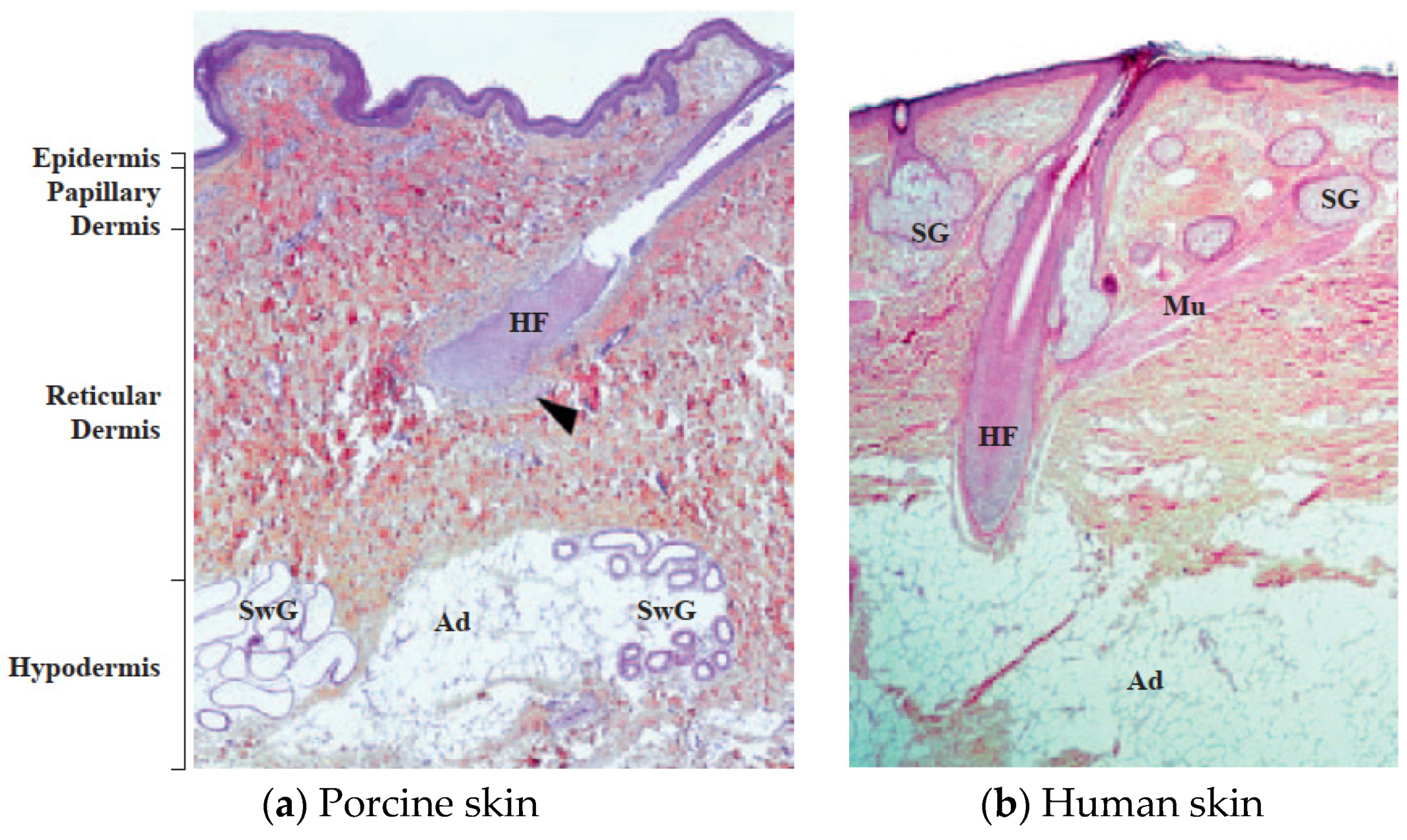
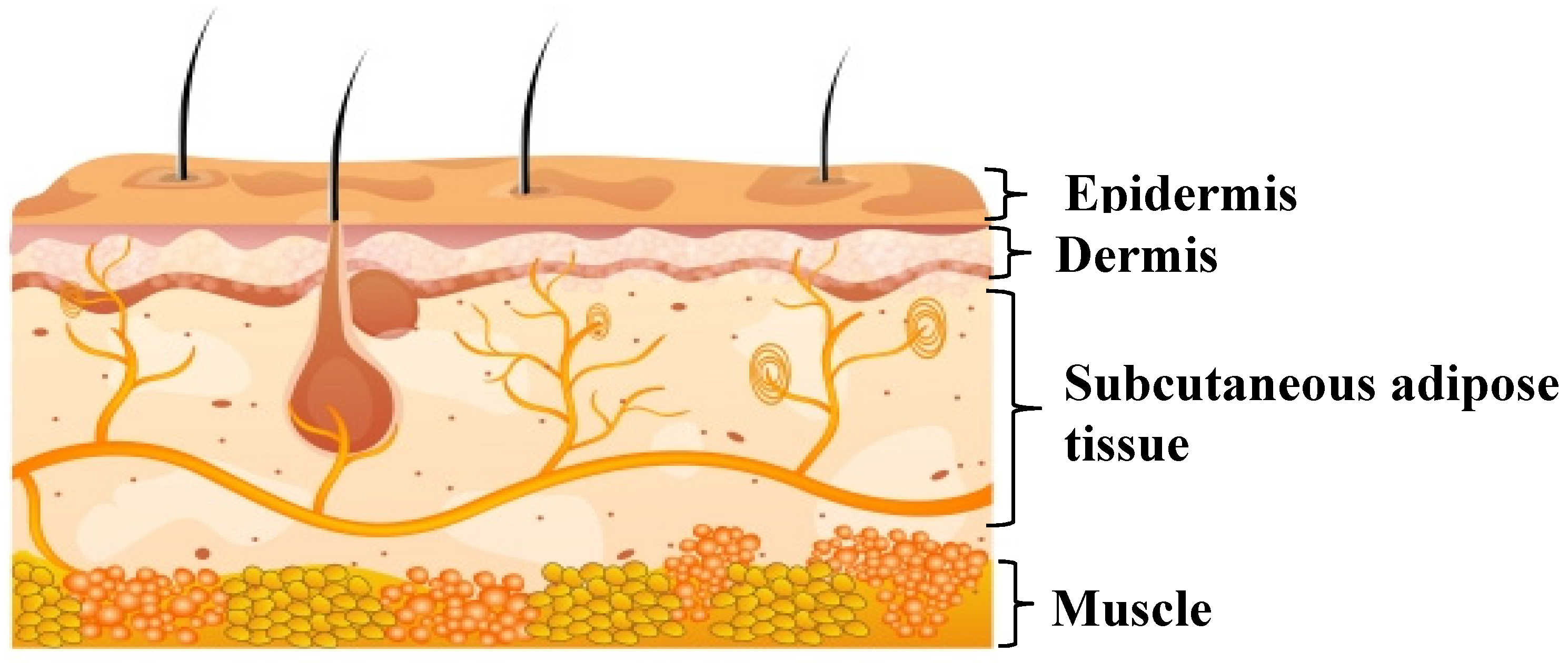
2. Skin
3. Permeation Study
3.1. Summary of European Medicines Agency Guideline (EMA) for Permeation Study
3.2. Experimental Parameters Affecting the Permeation Study
3.3. Significance of In Vitro Permeation Study Using Excised Human Skin
3.4. Effect of Drug Properties on the Permeation Mechanism Through the Skin
3.5. Need of Human Skin Alternatives
4. Skin Surrogates in Permeation Studies
4.1. Artificial Cultured Human Skin Model
4.2. Skin Parallel Artificial Membrane Permeability Assay (Skin-PAMPA)
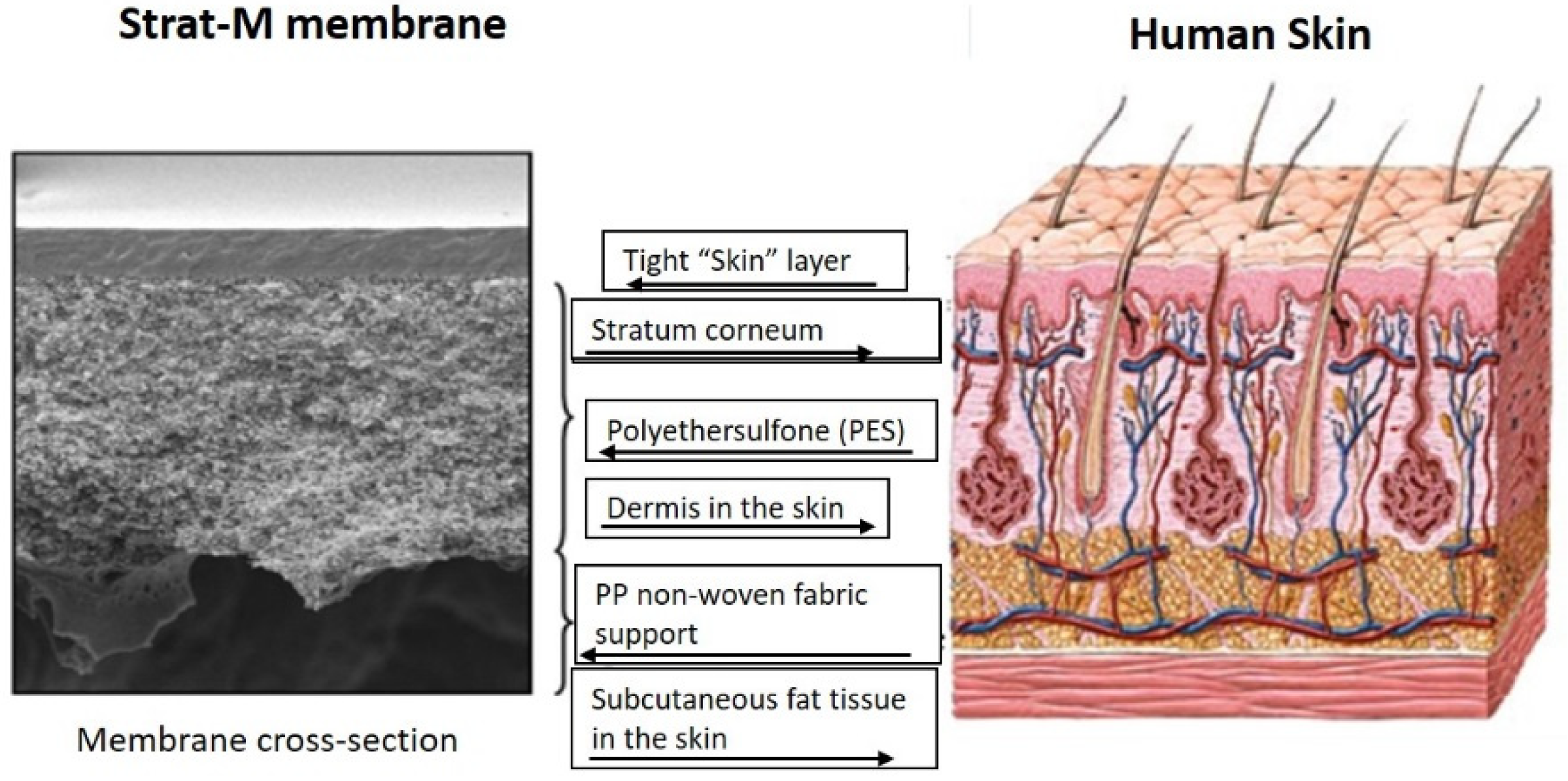
4.3. Strat-M™ and Other Artificial Membranes
5. Conclusions and Future Directions
Author Contributions
Funding
Conflicts of Interest
References
- Mali, A.D. An updated review on transdermal drug delivery systems. Int. J. Adv. Sci. Res. 2015, 8, 9. [Google Scholar] [CrossRef]
- Dedakia, A.; Matholiya, C.; Koyani, V.; Bhimani, D. Three generations: Pimary, secondary and tertiary generations of transdermal drug delivery systems: A review. Int. J. Pharm. Sci. Res. 2013, 4, 2159–2173. [Google Scholar]
- Wilson, E.J. Three generations: The past, present, and future of transdermal drug delivery systems. SC USA Pharmcon. 2011. Available online: https://pdfs.semanticscholar.org/e283/1944c712a8fa5db387969e422cdd4b75926f.pdf (accessed on 15 February 2019).
- Wiedersberg, S.; Guy, R.H. Transdermal drug delivery: 30+ years of war and still fighting! J. Control. Release Off. J. Control. Release Soc. 2014, 190, 150–156. [Google Scholar] [CrossRef] [PubMed]
- Alsaab, H.; Alzhrani, R.M.; Boddu, S.H. Evaluation of the percutaneous absorption of chlorpromazine from PLO gels across porcine ear and human abdominal skin. Drug Dev. Ind. Pharm. 2016, 42, 1258–1266. [Google Scholar] [CrossRef]
- Alsaab, H.; Bonam, S.P.; Bahl, D.; Chowdhury, P.; Alexander, K.; Boddu, S.H. Organogels in drug delivery: A special emphasis on pluronic lecithin organogels. J. Pharm. Pharm. Sci. 2016, 19, 252–273. [Google Scholar] [CrossRef]
- Gaucher, S.; Elie, C.; Verola, O.; Jarraya, M. Viability of cryopreserved human skin allografts: Effects of transport media and cryoprotectant. Cell Tissue Bank. 2012, 13, 147–155. [Google Scholar] [CrossRef]
- Kagan, R.J.; Robb, E.C.; Plessinger, R.T. Human skin banking. Clin. Lab. Med. 2005, 25, 587–605. [Google Scholar] [CrossRef]
- Aggarwal, S.J.; Baxter, C.R.; Diller, K.R. Cryopreservation of skin: An assessment of current clinical applicability. J. Burn Care Rehabil. 1985, 6, 469–476. [Google Scholar] [CrossRef]
- Bätz, F.M.; Klipper, W.; Korting, H.C.; Henkler, F.; Landsiedel, R.; Luch, A.; von Fritschen, U.; Weindl, G.; Schäfer-Korting, M. Esterase activity in excised and reconstructed human skin—Biotransformation of prednicarbate and the model dye fluorescein diacetate. Eur. J. Pharm. Biopharm. 2013, 84, 374–385. [Google Scholar] [CrossRef]
- Bravo, D.; Rigley, T.H.; Gibran, N.; Strong, D.M.; Newman-Gage, H. Effect of storage and preservation methods on viability in transplantable human skin allografts. Burns J. Int. Soc. Burn Inj. 2000, 26, 367–378. [Google Scholar] [CrossRef]
- Barbero, A.M.; Frasch, H.F. Effect of frozen human epidermis storage duration and cryoprotectant on barrier function using two model compounds. Skin Pharmacol. Physiol. 2016, 29, 31–40. [Google Scholar] [CrossRef] [PubMed]
- Kerimoğlu, O.; Şahbaz, S. Animal Skin Models for Percutaneous Absorption Studies. J. Biopharm. Ther. Chal. 2018, 2, 1–2. [Google Scholar]
- Todo, H. Transdermal permeation of drugs in various animal species. Pharmaceutics 2017, 9, 33. [Google Scholar] [CrossRef] [PubMed]
- Shim, J.; Seok Kang, H.; Park, W.S.; Han, S.H.; Kim, J.; Chang, I.S. Transdermal delivery of mixnoxidil with block copolymer nanoparticles. J. Control. Release Off. J. Control. Release Soc. 2004, 97, 477–484. [Google Scholar] [CrossRef]
- Carrer, D.C.; Vermehren, C.; Bagatolli, L.A. Pig skin structure and transdermal delivery of liposomes: A two photon microscopy study. J. Control. Release Off. J. Control. Release Soc. 2008, 132, 12–20. [Google Scholar] [CrossRef] [PubMed]
- Sintov, A.C. Cumulative evidence of the low reliability of frozen/thawed pig skin as a model for in vitro percutaneous permeation testing. Eur. J. Pharm. Sci. 2017, 102, 261–263. [Google Scholar] [CrossRef]
- Kikwai, L.; Kanikkannan, N.; Babu, R.J.; Singh, M. Effect of vehicles on the transdermal delivery of melatonin across porcine skin in vitro. J. Control. Release Off. J. Control. Release Soc. 2002, 83, 307–311. [Google Scholar] [CrossRef]
- Debeer, S.; Le Luduec, J.-B.; Kaiserlian, D.; Laurent, P.; Nicolas, J.-F.; Dubois, B.; Kanitakis, J. Comparative histology and immunohistochemistry of porcine versus human skin. Eur. J. Dermatol. 2013, 23, 456–466. [Google Scholar] [CrossRef]
- Haq, A.; Goodyear, B.; Ameen, D.; Joshi, V.; Michniak-Kohn, B. Strat-M® synthetic membrane: Permeability comparison to human cadaver skin. Int. J. Pharm. 2018, 547, 432–437. [Google Scholar] [CrossRef]
- Waters, L.J. Recent developments in skin mimic systems to predict transdermal permeation. Curr. Pharm. Des. 2015, 21, 2725–2732. [Google Scholar] [CrossRef] [PubMed]
- Abd, E.; Yousef, S.A.; Pastore, M.N.; Telaprolu, K.; Mohammed, Y.H.; Namjoshi, S.; Grice, J.E.; Roberts, M.S. Skin models for the testing of transdermal drugs. Clin. Pharmacol. Adv. Appl. 2016, 8, 163–176. [Google Scholar] [CrossRef] [PubMed]
- Menon, G.K. New insights into skin structure: Scratching the surface. Adv. Drug Deliv. Rev. 2002, 54, S3–S17. [Google Scholar] [CrossRef]
- Benson, H.A. Transdermal drug delivery: Penetration enhancement techniques. Curr. Drug Deliv. 2005, 2, 23–33. [Google Scholar] [CrossRef]
- Barry, B.W. Drug delivery routes in skin: A novel approach. Adv. Drug Deliv. Rev. 2002, 54 (Suppl. 1), S31–S40. [Google Scholar] [CrossRef]
- Wester, R.C.; Christoffel, J.; Hartway, T.; Poblete, N.; Maibach, H.I.; Forsell, J. Human cadaver skin viability for in vitro percutaneous absorption: Storage and detrimental effects of heat-separation and freezing. Pharm. Res. 1998, 15, 82–84. [Google Scholar] [CrossRef]
- Bouwstra, J.A.; Honeywell-Nguyen, P.L.; Gooris, G.S.; Ponec, M. Structure of the skin barrier and its modulation by vesicular formulations. Prog. Lipid Res. 2003, 42, 1–36. [Google Scholar] [CrossRef]
- Prausnitz, M.R.; Langer, R. Transdermal drug delivery. Nat. Biotechnol. 2008, 26, 1261–1268. [Google Scholar] [CrossRef]
- Fang, C.-L.; Aljuffali, I.A.; Li, Y.-C.; Fang, J.-Y. Delivery and targeting of nanoparticles into hair follicles. Ther. Deliv. 2014, 5, 991–1006. [Google Scholar] [CrossRef]
- Agency, E.M. Available online: https://www.ema.europa.eu/documents/scientific-guideline/guideline-quality-transdermal-patches_en.pdf (accessed on 15 February 2019).
- EMA. The European Agency for the Evaluation of Medicinal Products. Available online: https://www.ema.europa.eu/documents/scientific-guideline/note-guidance-quality-modified-release-products-oral-dosage-forms-b-transdermal-dosage-forms-section_en.pdf (accessed on 15 February 2019).
- Ruela, A.L.M.; Perissinato, A.G.; Lino, M.E.d.S.; Mudrik, P.S.; Pereira, G.R. Evaluation of skin absorption of drugs from topical and transdermal formulations. Braz. J. Pharm. Sci. 2016, 52, 527–544. [Google Scholar] [CrossRef]
- Ng, S.-F.; Rouse, J.J.; Sanderson, F.D.; Meidan, V.; Eccleston, G.M. Validation of a static Franz diffusion cell system for in vitro permeation studies. AAPS PharmSciTech 2010, 11, 1432–1441. [Google Scholar] [CrossRef] [PubMed]
- Fritsch, W.C.; Stoughton, R.B. The effect of temperature and humidity on the penetration of C14 acetylsalicylic acid in excised human skin. J. Investig. Dermatol. 1963, 41, 307–311. [Google Scholar] [CrossRef] [PubMed]
- Yin, Q.; Wang, R.; Yang, S.; Wu, Z.; Guo, S.; Dai, X.; Qiao, Y.; Shi, X. Influence of temperature on transdermal penetration enhancing mechanism of borneol: A multi-scale study. Int. J. Mol. Sci. 2017, 18, 195. [Google Scholar] [CrossRef]
- Golden, G.M.; Guzek, D.B.; Harris, R.R.; McKie, J.E.; Potts, R.O. Lipid thermotropic transitions in human stratum corneum. J. Investig. Dermatol. 1986, 86, 255–259. [Google Scholar] [CrossRef] [PubMed]
- Akomeah, F.; Nazir, T.; Martin, G.P.; Brown, M.B. Effect of heat on the percutaneous absorption and skin retention of three model penetrants. Eur. J. Pharm. Sci. Off. J. Eur. Fed. Pharm. Sci. 2004, 21, 337–345. [Google Scholar] [CrossRef] [PubMed]
- Kulkarni, U.D.; Mahalingam, R.; Li, X.; Pather, I.; Jasti, B. Effect of experimental temperature on the permeation of model diffusants across porcine buccal mucosa. AAPS PharmSciTech 2011, 12, 579–586. [Google Scholar] [CrossRef]
- Guideline, O.T. Test no. 428. Skin absorption: In vitro method. In OECD Guidelines for the Testing of Chemicals; Section 4; OECD: Paris, France, 2004. [Google Scholar]
- White, E.A.; Horne, A.; Runciman, J.; Orazem, M.E.; Navidi, W.C.; Roper, C.S.; Bunge, A.L. On the correlation between single-frequency impedance measurements and human skin permeability to water. Toxicol. In Vitro 2011, 25, 2095–2104. [Google Scholar] [CrossRef]
- Davies, D.J.; Ward, R.J.; Heylings, J.R. Multi-species assessment of electrical resistance as a skin integrity marker for in vitro percutaneous absorption studies. Toxicol. In Vitro. 2004, 18, 351–358. [Google Scholar] [CrossRef]
- Imhof, R.; De Jesus, M.; Xiao, P.; Ciortea, L.; Berg, E. Closed-chamber transepidermal water loss measurement: Microclimate, calibration and performance. Int. J. Cosmet. Sci. 2009, 31, 97–118. [Google Scholar] [CrossRef]
- Akdeniz, M.; Gabriel, S.; Lichterfeld-Kottner, A.; Blume-Peytavi, U.; Kottner, J. TEWL reference values in healthy adults. Br. J. Dermatol. 2018, 179, e204. [Google Scholar] [CrossRef]
- Momota, Y.; Shimada, K.; Gin, A.; Matsubara, T.; Azakami, D.; Ishioka, K.; Nakamura, Y.; Sako, T. Measurement of transepidermal water loss (TEWL) in cats with experimental skin barrier dysfunction using a closed chamber system. J. Vet. Dermatol. 2016, 27, 428-e110. [Google Scholar] [CrossRef] [PubMed]
- Boer, M.; Duchnik, E.; Maleszka, R.; Marchlewicz, M. Structural and biophysical characteristics of human skin in maintaining proper epidermal barrier function. Postep. Dermatol. Alergol. 2016, 33, 1–5. [Google Scholar] [CrossRef] [PubMed]
- Davies, D.J.; Heylings, J.R.; McCarthy, T.J.; Correa, C.M. Development of an in vitro model for studying the penetration of chemicals through compromised skin. J. Toxicol. Vitro 2015, 29, 176–181. [Google Scholar] [CrossRef] [PubMed]
- Davies, D.J.; Heylings, J.R.; Gayes, H.; McCarthy, T.J.; Mack, M.C. Further development of an in vitro model for studying the penetration of chemicals through compromised skin. J. Toxicol. Vitro 2017, 38, 101–107. [Google Scholar] [CrossRef] [PubMed][Green Version]
- Lau, W.M.; Ng, K.W. Finite and infinite dosing. In Percutaneous Penetration Enhancers Drug Penetration Into/Through the Skin; Springer: Berlin/Heidelberg, Germany, 2017; pp. 35–44. [Google Scholar]
- Bahl, D.; Daftardar, S.; Devi Bachu, R.; Boddu, S.H.; Altorok, N.; Kahaleh, B. Evaluation of topical econazole nitrate formulations with potential for treating Raynaud’s phenomenon. Pharm. Dev. Technol. 2019, 24, 689–699. [Google Scholar] [CrossRef] [PubMed]
- Diez-Sales, O.; Watkinson, A.; Herráez-Domínguez, M.; Javaloyes, C.; Hadgraft, J. A mechanistic investigation of the in vitro human skin permeation enhancing effect of Azone®. Int. J. Pharm. 1996, 129, 33–40. [Google Scholar] [CrossRef]
- Gallagher, S.J.; Trottet, L.; Carter, T.P.; Heard, C.M. Effects of membrane type and liquid/liquid phase boundary on in vitro release of ketoprofen from gel formulations. J. Drug Targets 2003, 11, 373–379. [Google Scholar] [CrossRef]
- Akomeah, F.K.; Martin, G.P.; Brown, M.B. Variability in human skin permeability in vitro: Comparing penetrants with different physicochemical properties. J. Pharm. Sci. 2007, 96, 824–834. [Google Scholar] [CrossRef]
- Heuber, F.; Ouvrard-Baraton, F.; Biesse, J.; Courtellemont, P.; Vincent, C.; Marty, J. Pig ear skin as a model for in vitro percutaneous absorption studies: Preliminary results of an inter-laboratory validation. Perspect. Percutan. Penetr. 1998, 6, 81. [Google Scholar]
- Franz, T.; Lehman, P.; Raney, S. Use of excised human skin to assess the bioequivalence of topical products. Skin Pharmacol. Physiol. 2009, 22, 276–286. [Google Scholar] [CrossRef]
- FDA. Guidance for Industry: Topical Dermatologic Corticosteroids: In vivo Bioequivalence. In Rockville, Center for Drug Evaluation and Research; Food and Drug Administration: Silver Spring, MD, USA, 1995. [Google Scholar]
- Finnin, B.; Walters, K.A.; Franz, T.J. In vitro skin permeation methodology. In Transdermal and Topical Drug Delivery: Principles and Practice; John Wiley & Sons: Hoboken, NJ, USA, 2012; pp. 85–108. [Google Scholar]
- Mitragotri, S. Modeling skin permeability to hydrophilic and hydrophobic solutes based on four permeation pathways. J. Control. Release Off. J. Control. Release Soc. 2003, 86, 69–92. [Google Scholar] [CrossRef]
- Marqusee, J.; Dill, K.A. Solute partitioning into chain molecule interphases: Monolayers, bilayer membranes, and micelles. J. Chem. Phys. 1986, 85, 434–444. [Google Scholar] [CrossRef]
- Marrink, S.J.; Berendsen, H.J. Permeation process of small molecules across lipid membranes studied by molecular dynamics simulations. J. Phys. Chem. 1996, 100, 16729–16738. [Google Scholar] [CrossRef]
- Xiang, T.X.; Anderson, B.D. Molecular distributions in interphases: Statistical mechanical theory combined with molecular dynamics simulation of a model lipid bilayer. Biophys. J. 1994, 66, 561–572. [Google Scholar] [CrossRef]
- Potts, R.O.; Guy, R.H. Predicting skin permeability. Pharm. Res. 1992, 9, 663–669. [Google Scholar] [CrossRef]
- Elias, P.M.; Cooper, E.R.; Korc, A.; Brown, B.E. Percutaneous transport in relation to stratum corneum structure and lipid composition. J. Investig. Dermatol. 1981, 76, 297–301. [Google Scholar] [CrossRef]
- Rougier, A.; Lotte, C.; Maibach, H.I. In vivo percutaneous penetration of some organic compounds related to anatomic site in humans: Predictive assessment by the stripping method. J. Pharm. Sci. 1987, 76, 451–454. [Google Scholar] [CrossRef]
- Roberts, M.S. Targeted drug delivery to the skin and deeper tissues: Role of physiology, solute structure and disease. Clin. Exp. Pharmacol. Physiol. 1997, 24, 874–879. [Google Scholar] [CrossRef]
- Banning, T.P.; Heard, C.M. Binding of doxycycline to keratin, melanin and human epidermal tissue. Int. J. Pharm. 2002, 235, 219–227. [Google Scholar] [CrossRef]
- Hayashi, T.; Sugibayashi, K.; Morimoto, Y. Calculation of skin permeability coefficient for ionized and unionized species of indomethacin. Chem. Pharm. Bull. 1992, 40, 3090–3093. [Google Scholar] [CrossRef]
- Kielhorn, J.; Melching-Kollmuss, D.; Mangelsdorf, I. Dermal Absorption—Environmental Health Criteria 235; World Health Organization: Geneva, Switzerland, 2006. [Google Scholar]
- Michaels, A.; Chandrasekaran, S.; Shaw, J. Drug permeation through human skin: Theory and in vitro experimental measurement. AIChE J. 1975, 21, 985–996. [Google Scholar] [CrossRef]
- Toyoda, H. Regulation of the animal experiments and testing in EU. Environ. Mutagen. Res. 2005, 27, 125–128. [Google Scholar] [CrossRef]
- FDA. Guidance for Industry. Nonsterile semisolid dosage forms, scale-up and postapproval changes: Chemistry, manufacturing, and controls; in vitro release testing and in vivo bioequivalence documentation. Center Drug Eval. Res. 1997, CMC 7, 20. [Google Scholar]
- EMA. Guideline on Quality of Transdermal Patches; EMA: London, UK, 2012. [Google Scholar]
- de Almeida Borges, V.R.; Simon, A.; Sena, A.R.; Cabral, L.M.; de Sousa, V.P. Nanoemulsion containing dapsone for topical administration: A study of in vitro release and epidermal permeation. Int. J. Nanomed. 2013, 8, 535. [Google Scholar]
- Henning, A.; Schaefer, U.F.; Neumann, D. Potential pitfalls in skin permeation experiments: Influence of experimental factors and subsequent data evaluation. Eur. J. Pharm. Biopharm. 2009, 72, 324–331. [Google Scholar] [CrossRef] [PubMed]
- Ponec, M. In vitro cultured human skin cells as alternatives to animals for skin irritancy screening. Int. J. Cosmet. Sci. 1992, 14, 245–264. [Google Scholar] [CrossRef]
- Van Gele, M.; Geusens, B.; Brochez, L.; Speeckaert, R.; Lambert, J. Three-dimensional skin models as tools for transdermal drug delivery: Challenges and limitations. Exp. Opin. Drug Deliv. 2011, 8, 705–720. [Google Scholar] [CrossRef]
- De Wecer, B.; Petersohn, D.; Mewes, K.R. Overview of human three-dimensional (3D) skin models used for dermal toxicity assessment. HPC Today 2013, 8, 18–22. [Google Scholar]
- Schmook, F.P.; Meingassner, J.G.; Billich, A. Comparison of human skin or epidermis models with human and animal skin in in-vitro percutaneous absorption. Int. J. Pharm. 2001, 215, 51–56. [Google Scholar] [CrossRef]
- Dreher, F.; Fouchard, F.; Patouillet, C.; Andrian, M.; Simonnet, J.-T.; Benech-Kieffer, F. Comparison of cutaneous bioavailability of cosmetic preparations containing caffeine or α-tocopherol applied on human skin models or human skin ex vivo at finite doses. Skin Pharmacol. Physiol. 2002, 15, 40–58. [Google Scholar] [CrossRef]
- Schreiber, S.; Mahmoud, A.; Vuia, A.; Rubbelke, M.K.; Schmidt, E.; Schaller, M.; Kandarova, H.; Haberland, A.; Schafer, U.F.; Bock, U.; et al. Reconstructed epidermis versus human and animal skin in skin absorption studies. Toxicol. Vitro Int. J. Publ. Assoc. BIBRA 2005, 19, 813–822. [Google Scholar] [CrossRef] [PubMed]
- Schafer-Korting, M.; Bock, U.; Gamer, A.; Haberland, A.; Haltner-Ukomadu, E.; Kaca, M.; Kamp, H.; Kietzmann, M.; Korting, H.C.; Krachter, H.U.; et al. Reconstructed human epidermis for skin absorption testing: Results of the German prevalidation study. Altern. Lab. Anim. ATLA 2006, 34, 283–294. [Google Scholar] [CrossRef] [PubMed]
- Schäfer-Korting, M.; Bock, U.; Diembeck, W.; Düsing, H.-J.; Gamer, A.; Haltner-Ukomadu, E.; Hoffmann, C.; Kaca, M.; Kamp, H.; Kersen, S. The use of reconstructed human epidermis for skin absorption testing: Results of the validation study. Altern. Lab. Anim. ATLA 2008, 36, 161–187. [Google Scholar] [CrossRef] [PubMed]
- Ackermann, K.; Borgia, S.L.; Korting, H.C.; Mewes, K.; Schäfer-Korting, M. The Phenion® full-thickness skin model for percutaneous absorption testing. Skin Pharmacol. Physiol. 2010, 23, 105–112. [Google Scholar] [CrossRef]
- Tfayli, A.; Bonnier, F.; Farhane, Z.; Libong, D.; Byrne, H.J.; Baillet-Guffroy, A. Comparison of structure and organization of cutaneous lipids in a reconstructed skin model and human skin: Spectroscopic imaging and chromatographic profiling. Exp. Dermatol. 2014, 23, 441–443. [Google Scholar] [CrossRef]
- Thakoersing, V.S.; Gooris, G.S.; Mulder, A.; Rietveld, M.; El Ghalbzouri, A.; Bouwstra, J.A. Unraveling barrier properties of three different in-house human skin equivalents. Tissue Eng. Part C Methods 2012, 18, 1–11. [Google Scholar] [CrossRef]
- Klicks, J.; von Molitor, E.; Ertongur-Fauth, T.; Rudolf, R.; Hafner, M. In vitro skin three-dimensional models and their applications. J. Cell. Biotechnol. 2017, 3, 21–39. [Google Scholar] [CrossRef]
- Teimouri, A.; Yeung, P.; Agu, R. 2D vs. 3D cell culture models for in vitro topical (dermatological) medication testing. In Cell Culture; IntechOpen: London, UK, 2018. [Google Scholar]
- Niehues, H.; Bouwstra, J.A.; El Ghalbzouri, A.; Brandner, J.M.; Zeeuwen, P.L.; van den Bogaard, E.H. 3D skin models for 3R research: The potential of 3D reconstructed skin models to study skin barrier function. Exp. Dermatol. 2018, 27, 501–511. [Google Scholar] [CrossRef]
- Chi, C.-T.; Lee, M.-H.; Weng, C.-F.; Leong, M.K. In Silico Prediction of PAMPA Effective Permeability Using a Two-QSAR Approach. Int. J. Mol. Sci. 2019, 20, 3170. [Google Scholar] [CrossRef]
- Geinoz, S.; Guy, R.H.; Testa, B.; Carrupt, P.A. Quantitative structure-permeation relationships (QSPeRs) to predict skin permeation: A critical evaluation. Pharm. Res. 2004, 21, 83–92. [Google Scholar] [CrossRef]
- Ottaviani, G.; Martel, S.; Carrupt, P.A. Parallel artificial membrane permeability assay: A new membrane for the fast prediction of passive human skin permeability. J. Med. Chem. 2006, 49, 3948–3954. [Google Scholar] [CrossRef] [PubMed]
- Sinkó, B.; Garrigues, T.M.; Balogh, G.T.; Nagy, Z.K.; Tsinman, O.; Avdeef, A.; Takács-Novák, K. Skin—PAMPA: A new method for fast prediction of skin penetration. Eur. J. Pharm. Sci. 2012, 45, 698–707. [Google Scholar] [CrossRef] [PubMed]
- Luo, L.; Patel, A.; Sinko, B.; Bell, M.; Wibawa, J.; Hadgraft, J.; Lane, M.E. A comparative study of the in vitro permeation of ibuprofen in mammalian skin, the PAMPA model and silicone membrane. Int. J. Pharm. 2016, 505, 14–19. [Google Scholar] [CrossRef] [PubMed]
- Vizserálek, G.; Berkó, S.; Tóth, G.; Balogh, R.; Budai-Szűcs, M.; Csányi, E.; Sinkó, B.; Takács-Novák, K. Permeability test for transdermal and local therapeutic patches using Skin PAMPA method. Eur. J. Pharm. Sci. 2015, 76, 165–172. [Google Scholar] [CrossRef]
- Balázs, B.; Vizserálek, G.; Berkó, S.; Budai-Szűcs, M.; Kelemen, A.; Sinkó, B.; Takács-Novák, K.; Szabó-Révész, P.; Csányi, E. Investigation of the efficacy of transdermal penetration enhancers through the use of human skin and a skin mimic artificial membrane. J. Pharm. Sci. 2016, 105, 1134–1140. [Google Scholar] [CrossRef]
- Csizmazia, E.; Erős, G.; Berkesi, O.; Berkó, S.; Szabó-Révész, P.; Csányi, E. Penetration enhancer effect of sucrose laurate and Transcutol on ibuprofen. J. Drug Deliv. Sci. 2011, 21, 411–415. [Google Scholar] [CrossRef]
- Zhang, Y.; Lane, M.E.; Hadgraft, J.; Heinrich, M.; Chen, T.; Lian, G.; Sinko, B. A comparison of the in vitro permeation of niacinamide in mammalian skin and in the Parallel Artificial Membrane Permeation Assay (PAMPA) model. Int. J. Pharm. 2019, 556, 142–149. [Google Scholar] [CrossRef]
- Köllmer, M.; Mossahebi, P.; Sacharow, E.; Gorissen, S.; Gräfe, N.; Evers, D.-H.; Herbig, M.E. Investigation of the Compatibility of the Skin PAMPA Model with Topical Formulation and Acceptor Media Additives Using Different Assay Setups. AAPS PharmSciTech 2019, 20, 89. [Google Scholar] [CrossRef]
- Engesland, A.; Škalko-Basnet, N.; Flaten, G.E. In vitro models to estimate drug penetration through the compromised stratum corneum barrier. Drug Dev. Ind. Pharm. 2016, 42, 1742–1751. [Google Scholar] [CrossRef]
- Palac, Z.; Engesland, A.; Flaten, G.E.; Škalko-Basnet, N.; Filipović-Grčić, J.; Vanić, Ž. Liposomes for (trans) dermal drug delivery: The skin-PVPA as a novel in vitro stratum corneum model in formulation development. J. Liposome Res. 2014, 24, 313–322. [Google Scholar] [CrossRef]
- Zhang, H.; Zhu, X.; Shen, J.; Xu, H.; Ma, M.; Gu, W.; Jiang, Q.; Chen, J.; Duan, J. Characterization of a liposome-based artificial skin membrane for in vitro permeation studies using Franz diffusion cell device. J. Liposome Res. 2017, 27, 302–311. [Google Scholar] [CrossRef] [PubMed]
- Ameri, M.; Lewis, H.; Lehman, P. Effect of Skin Model on In Vitro Performance of an Adhesive Dermally Applied Microarray Coated with Zolmitriptan. J. Pharm. 2018, 2018, 7459124. [Google Scholar] [CrossRef] [PubMed]
- Haq, A.; Dorrani, M.; Goodyear, B.; Joshi, V.; Michniak-Kohn, B. Membrane properties for permeability testing: Skin versus synthetic membranes. Int. J. Pharm. 2018, 539, 58–64. [Google Scholar] [CrossRef] [PubMed]
- Uchida, T.; Kadhum, W.R.; Kanai, S.; Todo, H.; Oshizaka, T.; Sugibayashi, K. Prediction of skin permeation by chemical compounds using the artificial membrane, Strat-M™. Eur. J. Pharm. Sci. 2015, 67, 113–118. [Google Scholar] [CrossRef]
- Uchida, T.; Nishioka, K.; Motoki, A.; Yakumaru, M.; Sano, T.; Todo, H.; Sugibayashi, K. Effect of Esters on the Permeation of Chemicals with Different Polarities through Synthetic Artificial Membranes Using a High-Throughput Diffusion Cell Array. Chem. Pharm. Bull. 2016, 64, 1597–1606. [Google Scholar] [CrossRef] [PubMed]
- Uchida, T.; Yakumaru, M.; Nishioka, K.; Higashi, Y.; Sano, T.; Todo, H.; Sugibayashi, K. Evaluation of a Silicone Membrane as an Alternative to Human Skin for Determining Skin Permeation Parameters of Chemical Compounds. Chem. Pharm. Bull. 2016, 64, 1338–1346. [Google Scholar] [CrossRef]
- Simon, A.; Amaro, M.I.; Healy, A.M.; Cabral, L.M.; de Sousa, V.P. Comparative evaluation of rivastigmine permeation from a transdermal system in the Franz cell using synthetic membranes and pig ear skin with in vivo-in vitro correlation. Int. J. Pharm. 2016, 512, 234–241. [Google Scholar] [CrossRef]
- Kaur, L.; Singh, K.; Paul, S.; Singh, S.; Singh, S.; Jain, S.K. A Mechanistic Study to Determine the Structural Similarities Between Artificial Membrane Strat-M™ and Biological Membranes and Its Application to Carry Out Skin Permeation Study of Amphotericin B Nanoformulations. AAPS PharmSciTech 2018, 19, 1606–1624. [Google Scholar] [CrossRef]
- Zsikó, S.; Cutcher, K.; Kovács, A.; Budai-Szűcs, M.; Gácsi, A.; Baki, G.; Csányi, E.; Berkó, S. Nanostructured Lipid Carrier Gel for the Dermal Application of Lidocaine: Comparison of Skin Penetration Testing Methods. Pharmaceutics 2019, 11, 310. [Google Scholar] [CrossRef]
- Osmałek, T.; Milanowski, B.; Froelich, A.; Górska, S.; Białas, W.; Szybowicz, M.; Kapela, M. Technology. Novel organogels for topical delivery of naproxen: Design, physicochemical characteristics and in vitro drug permeation. Pharm. Dev. 2017, 22, 521–536. [Google Scholar] [CrossRef]
- Chellappan, D.K.; Yee, N.J.; Kaur Ambar Jeet Singh, B.J.; Panneerselvam, J.; Madheswaran, T.; Chellian, J.; Satija, S.; Mehta, M.; Gulati, M.; Gupta, G.; et al. Formulation and characterization of glibenclamide and quercetin-loaded chitosan nanogels targeting skin permeation. Ther. Deliv. 2019, 10, 281–293. [Google Scholar] [CrossRef] [PubMed]
- Gonçalves, L.; Seabra, A.; Pelegrino, M.; De Araujo, D.; Bernardes, J.; Haddad, P. Superparamagnetic iron oxide nanoparticles dispersed in Pluronic F127 hydrogel: Potential uses in topical applications. RSC Adv. 2017, 7, 14496–14503. [Google Scholar] [CrossRef]
- Xie, Y.; Yao, Y. Octenylsuccinate hydroxypropyl phytoglycogen enhances the solubility and in-vitro antitumor efficacy of niclosamide. Int. J. Pharm. 2018, 535, 157–163. [Google Scholar] [CrossRef] [PubMed]
- Lee, J.-J.; Han, Y.-M.; Kwon, T.-W.; Kim, D.H.; Lee, H.S.; Jung, W.J.; Kim, J.; Kang, S.; Kim, S.K.; Cho, C.-W. Functional Fragments of AIMP1-Derived Peptide (AdP) and Optimized Hydrosol for Their Topical Deposition by Box-Behnken Design. Molecules 2019, 24, 1967. [Google Scholar] [CrossRef] [PubMed]
- Mennini, N.; Cirri, M.; Maestrelli, F.; Mura, P. Comparison of liposomal and NLC (nanostructured lipid carrier) formulations for improving the transdermal delivery of oxaprozin: Effect of cyclodextrin complexation. Int. J. Pharm. 2016, 515, 684–691. [Google Scholar] [CrossRef] [PubMed]
- Grillo, R.; Dias, F.V.; Querobino, S.M.; Alberto-Silva, C.; Fraceto, L.F.; de Paula, E.; de Araujo, D.R. Influence of hybrid polymeric nanoparticle/thermosensitive hydrogels systems on formulation tracking and in vitro artificial membrane permeation: A promising system for skin drug-delivery. Colloids Surf. B Biointerfaces 2019, 174, 56–62. [Google Scholar] [CrossRef] [PubMed]
- Carrer, V.; Guzmán, B.; Martí, M.; Alonso, C.; Coderch, L. Lanolin-based synthetic membranes as percutaneous absorption models for transdermal drug delivery. Pharmaceutics 2018, 10, 73. [Google Scholar] [CrossRef]
- Karadzovska, D.; Riviere, J.E. Assessing vehicle effects on skin absorption using artificial membrane assays. Eur. J. Pharm. Sci. 2013, 50, 569–576. [Google Scholar] [CrossRef]
- Kim, J.H.; Ko, J.A.; Kim, J.T.; Cha, D.S.; Cho, J.H.; Park, H.J.; Shin, G.H. Preparation of a capsaicin-loaded nanoemulsion for improving skin penetration. J. Agric. Food Chem. 2014, 62, 725–732. [Google Scholar] [CrossRef]
- Nallagundla, S.; Patnala, S.; Kanfer, I. Comparison of in vitro release rates of acyclovir from cream formulations using vertical diffusion cells. AAPS PharmSciTech 2014, 15, 994–999. [Google Scholar] [CrossRef]
- Nayak, A.; Das, D.B.; Chao, T.C.; Starov, V.M. Spreading of a lidocaine formulation on microneedle-treated skin. J. Pharm. Sci. 2015, 104, 4109–4116. [Google Scholar] [CrossRef] [PubMed][Green Version]
- Carmona-Moran, C.A.; Zavgorodnya, O.; Penman, A.D.; Kharlampieva, E.; Bridges, S.L., Jr.; Hergenrother, R.W.; Singh, J.A.; Wick, T.M. Development of gellan gum containing formulations for transdermal drug delivery: Component evaluation and controlled drug release using temperature responsive nanogels. Int. J. Pharm. 2016, 509, 465–476. [Google Scholar] [CrossRef] [PubMed]
- Kang, M.K.; Kim, Y.; Gil, S.; Lee, S.; Jang, J.; Kim, S.J.; Yoon, M.S.; Yoo, K.J.; Lee, J.B.; Yoo, H.S. Effects of liquid crystal-based formulation on transdermal delivery of retinyl palmitate and proliferation of epidermal cells. Macromol. Res. 2016, 24, 44–50. [Google Scholar] [CrossRef]
- Mura, P.; Bragagni, M.; Mennini, N.; Cirri, M.; Maestrelli, F. Development of liposomal and microemulsion formulations for transdermal delivery of clonazepam: Effect of randomly methylated β-cyclodextrin. Int. J. Pharm. 2014, 475, 306–314. [Google Scholar] [CrossRef]


| Compounds | P (cm/s) × 10−6 Strat-M™ | P (cm/s) × 10−6 Hairless Rat Skin | P (cm/s) × 10−6 Human Skin |
|---|---|---|---|
| MP | 4.02 ± 0.6 | 2.39 ± 0.31 | 2.49 ± 0.03 |
| EP | 4.21 ± 0.2 | 2.96 ± 0.06 | 3.86 ± 0.49 |
| PP | 4.01 ± 0.16 | 3.63 ± 0.15 | 3.63 ± 0.15 |
| BP | 4.24 ± 0.34 | 2.31 ± 0.26 | 3.54 ± 0.18 |
| ISMN | 0.15 ± 0.02 | 0.07 ± 0.01 | 0.11 ± 0.01 |
| AMP | 0.87 ± 0.25 | 0.29 ± 0.11 | 0.33 ± 0.02 |
| ISDN | 3.14 ± 0.01 | 2.57 ± 0.15 | 3.14 ± 0.15 |
| LCH (pH5.0) | 0.14 ± 0.02 | 0.04 ± 0.01 | 0.05 ± 0.02 |
| LCH (pH10) | 5.55 ± 0.53 | 9.21 ± 3.68 | 1.97 ± 0.15 |
| LCH (pH7.9) | 1.2 ± 0.12 | 1.3 ± 0.11 | 0.14 ± 0.01 |
| CAF | 0.47 ± 0.07 | 0.26 ± 0.02 | 0.18 ± 0.02 |
| M-PABA | 8.95 ± 0.26 | 7.22 ± 0.17 | 18.4 ± 0.83 |
| E-PABA | 11.25 ± 0.41 | 10.6 ± 0.25 | 20.1 ± 2.24 |
| P-PABA | 13.55 ± 0.58 | 19.6 ± 1.61 | 22.3 ± 3.03 |
| B-PABA | 10.14 ± 0.53 | 9.49 ± 0.79 | 20.1 ± 3.02 |
| Study | Important Findings | References |
|---|---|---|
| Topical delivery of a soluble form of naproxen from organogel. | Organogels containing a soluble form of naproxen could be a possible substitute to marketed topical products with the crystalline form of naproxen, as shown using a Strat-M™ membrane in the permeation study. | [109] |
| Topical delivery of a combination of glibenclamide and quercetin using chitosan nanogel. | Nanogel could be potential formulation for the delivery of an antidiabetic drug through the skin was demonstrated using a Strat-M™ membrane in the permeation study. Although such formulation enables controlled release of the drug, further improvement of the release mechanism is required for commercialization. | [110] |
| Super magnetic iron oxide nanoparticles dispersed in Pluronic F127 gel by topical delivery of nitric oxide (NO). | Permeation study carried using a Strat-M™ membrane demonstrated that the iron will not permeate from the nanoparticle dispersed in Pluronic. | [111] |
| Topical delivery of niclosamide (niclo) using octenylsuccinate hydroxypropyl phytoglycogen (OHPP) as a solubilizing agent. | Permeation study across the Strat-M™ membrane of niclo-OHPP solid dispersion was 5.3 times higher than niclosamide alone. This study can be used further in the development of the formulation of niclosamide. | [112] |
| Topical delivery of functional fragments of an AIMP1-derived peptide (AdP) in a hydrosol system. | A Strat-M™ membrane was used in the determination of in vitro deposition from FA-AdP hydrogel compared with FA-AdP alone. The in vitro deposition from optimized FA-AdP hydrosole in the Strat-M™ membrane was 127-fold higher than FA-Adp alone. | [113] |
| To develop a topical formulation of oxaprozin in liposomal or nanostructured lipid carriers (NLCs) formulation following a drug complexation with randomly-methylated-ß-CD and arginine. | The permeation of oxaprozin was screened first using the nitrocellulose membrane and later using the excised human skin. Both the formulations (deformable liposomes and NLCs) showed increased permeations compared to the plain drug. The deformable liposomes had significantly greater drug permeations than the NLCs across the human skin. | [114] |
| Topical delivery of benzocaine loaded with a polymeric nanoparticle/thermosensitive hydrogel system. | Benzocaine-loaded nanoparticles in hydrogel permeated through the artificial membrane Strat-M™, acting as a depot system for long duration action when applied on skin. | [115] |
| To check whether the use of lanolin in a synthetic membrane (Strat-M™ and nucleophore) would mimic the skin-barrier function using three different model drugs (lidocaine, diclofenac, and betamethasone). | The barrier function of the artificial membrane with lanolin was higher than without lanolin. The absorption of model drug substances on the lanolin-containing artificial membranes was found to be similar to that of the skin, which indicates the lanolin-containing membranes mimic topical active absorption. | [116] |
| To compare penetration profiles of lidocaine incorporated in the nanostructured lipid-carrier gel using different membranes (Strat-M™, skin-PAMPA, cellulose, and heat-separated human epidermis). | Drug permeability profile of lidocaine across the Strat-M™ membrane and skin-PAMPA correlated favorably with heat-separated human skin epidermis. | [108] |
| To evaluate the ability of the Strat-M™, isopropyl myristate, and certramides to predict the percutaneous absorption of six different compounds in saturated and unsaturated concentration in three different vehicles (water, ethanol, and propylene glycol). | The correlation of the absorption through membranes was drawn in relation to the porcine skin. The correlation was better with saturated concentrations than the unsaturated concentration of the compounds. Similarly, the Strat-M™ membrane had the best correlation, followed by certramides and IPM, for the amount remaining in the membrane and retained in the porcine skin. | [117] |
| To investigate the optimum condition of a capsaicin-loaded nano-emulsion formulation for topical application using the Strat-M™ membrane. | The nano-emulsion of sizes between 20 to 62 nm loaded with capsaicin successfully penetrated the layers in the Strat-M™ membrane. Thus, it was presumed to be suitable for evaluation in transdermal delivery. | [118] |
| To check whether the in vitro release profile of generic acyclovir creams is equivalent to the innovator product using different types of artificial membranes (Magna Nylon, Tuffryn membrane, Durapore, Nitrocellulose, Fluoropore, and Strat-M™). | The generic acyclovir cream formulations demonstrated similar release profiles, which were comparable to the reference product, Zovirax®. This study indicated the possibility of biowaivers for generic topical products based on in vitro studies using artificial membranes. | [119] |
| To examine the spreadability of the transdermal formulation in microneedle-treated and untreated skin by the study of parameters like the spreading radius, droplet height, and dynamic contact angle. | A Strat-M™ membrane was used as a control for the skin. The spreading parameters were lower for the lidocaine hydrogel compared to the lidocaine solution. The lidocaine hydrogel on the microneedle-treated skin resulted in slower dynamic reduction spreadability parameters because of microneedle cavities. | [120] |
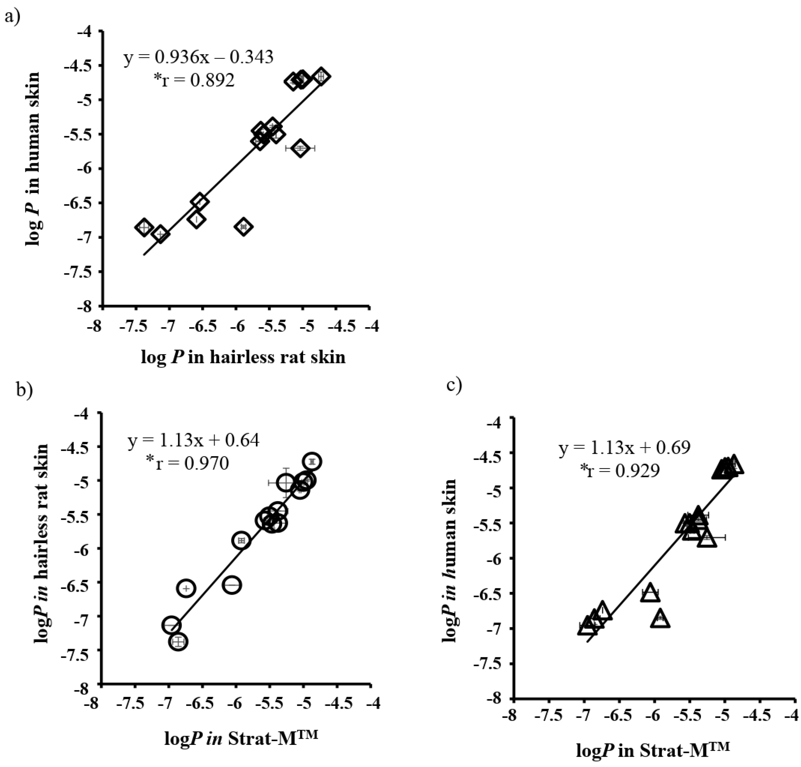
| To determine the usefulness of synthetic membranes as an alternative of human and animal skins to evaluate the skin permeability. | Thirteen different compounds with molecular weights in the range of 152–289 and lipophilicity (log Ko/w) in 0.9–3.5 were used in the permeation study through the Strat-M™, excised human skin, and hairless rat skin. The Strat-M™ had similar characteristics to that of the skin membrane in terms of the log P values, diffusion, and partition coefficient. | [103] |
| To develop an in vitro drug permeation test utilizing a synthetic membrane that can be a substitute for the permeation study where the animal or human skin is used. | Out of six different synthetic membranes used in the study, the Strat-M® membrane adequately mimicked the barrier property of the skin, where the rivastigmine permeation profile through the Strat-M™ was similar to that of pig skin (R2 − 0.920). Additionally, the in vitro-in vivo correlation was linear (R2 − 0.991) for the Strat-M™ membrane. | [106] |
| To demonstrate the feasibility of incorporating the thermo-responsive nanogels in the semisolid gel or hydrogel film formulation to enable the adjustment in drug transport kinetics using diclofenac in a formulation containing gellan gum. | A Strat-M™ membrane was used as the substitute to the skin barrier in this study, which yielded high reproducible results with the ease of use. It was demonstrated that the combination of thermo-responsive nanogel with a semisolid gel or a hydrogel of diclofenac would result in a formulation that can provide fast drug penetration for rapid pain relief, as well as sustained drug delivery over a period. | [121] |
| To investigate the effect of 25 different esters in the permeation of four model compounds (caffeine, aminopyrine, benzoic acid, and flurbiprofen) with different polarity through the synthetic membranes (silicone and Strat-M™). | The amount of the model compounds that permeated through the silicon and the Strat-M™ membrane had significant correlation with the wettability, surface tension, and uptake of esters into the membrane. Therefore, the type of ester used as a vehicle is pivotal to control the skin permeability of topical formulations. | [104] |
| To formulate and evaluate the percutaneous absorption of a liquid crystal emulsion of retinyl palmitate through the skin barrier using a Strat-M™ membrane. | The liquid crystal emulsion showed increased retention at the membrane and high permeation to the acceptor chamber in a permeation study compared to the plain oil-water emulsion. | [122] |
| The aim of the study was to develop an ultra-deformable liposomal and microemulsion formulation for the transdermal delivery of clonazepam using cyclodextrin as a penetration enhancer. | A nitrocellulose membrane was used for preliminary screening of the formulation. Later, the best formulations were screened using animal skins. The permeability of microemulsion with cyclodextrin had a 4-fold higher permeability than of clonazepam. Similarly, the liposomes without cyclodextrin had a 2-fold higher permeability. | [123] |
| To develop a topical formulation of oxaprozin in liposomal or nanostructured lipid carriers (NLCs) formulation following a drug complexation with randomly-methylated-ß-CD and arginine. | The permeation of oxaprozin was screened first using the nitrocellulose membrane and later using the excised human skin. Both the formulations (deformable liposomes and NLCs) showed increased permeations compared to the plain drug. The deformable liposomes had significantly greater drug permeations than the NLCs across the human skin. |
© 2020 by the authors. Licensee MDPI, Basel, Switzerland. This article is an open access article distributed under the terms and conditions of the Creative Commons Attribution (CC BY) license (http://creativecommons.org/licenses/by/4.0/).
Share and Cite
Neupane, R.; Boddu, S.H.S.; Renukuntla, J.; Babu, R.J.; Tiwari, A.K. Alternatives to Biological Skin in Permeation Studies: Current Trends and Possibilities. Pharmaceutics 2020, 12, 152. https://doi.org/10.3390/pharmaceutics12020152
Neupane R, Boddu SHS, Renukuntla J, Babu RJ, Tiwari AK. Alternatives to Biological Skin in Permeation Studies: Current Trends and Possibilities. Pharmaceutics. 2020; 12(2):152. https://doi.org/10.3390/pharmaceutics12020152
Chicago/Turabian StyleNeupane, Rabin, Sai H.S. Boddu, Jwala Renukuntla, R. Jayachandra Babu, and Amit K. Tiwari. 2020. "Alternatives to Biological Skin in Permeation Studies: Current Trends and Possibilities" Pharmaceutics 12, no. 2: 152. https://doi.org/10.3390/pharmaceutics12020152
APA StyleNeupane, R., Boddu, S. H. S., Renukuntla, J., Babu, R. J., & Tiwari, A. K. (2020). Alternatives to Biological Skin in Permeation Studies: Current Trends and Possibilities. Pharmaceutics, 12(2), 152. https://doi.org/10.3390/pharmaceutics12020152