Abstract
Populus species are highly susceptible to wetwood formation, which adversely affects tree growth, timber quality, and wood processing. In this study, 28 aerobic and 7 anaerobic bacterial strains were isolated and purified from I-69 poplar trees infected with wetwood using tissue-based pathogen isolation techniques. Preliminary screening identified three highly pathogenic isolates, including two aerobic strains (AB4 and AB14) and one anaerobic strain (ANAB1), all of which induced wetwood symptoms in 100% of inoculated seedlings with pronounced severity. Through comprehensive characterization, including morphological analysis, physiological–biochemical profiling, and 16S rRNA gene sequencing, these strains were taxonomically classified as Pantoea agglomerans (AB4), Escherichia fergusonii (AB14), and Enterobacter hormaechei (ANAB1). These 35 strains were subsequently inoculated into one-year-old healthy poplar seedlings through three distinct methods, including stem injection, root infection, and leaf infection. Experimental results demonstrated that only stem injection successfully induced wetwood symptoms, while root and leaf infection failed to produce pathological manifestations. For stem-inoculated specimens, pathogenicity was evaluated based on three diagnostic parameters, including heartwood discoloration length, pigmentation intensity, and affected tissue area ratio. Significant variability in symptom severity was observed among different bacterial strains. These findings expand the known diversity of bacterial species associated with wetwood development and provide valuable insights for understanding its etiology and for guiding future disease management strategies.
1. Introduction
Wetwood, also known as slime flux or water-soaked heartwood, is a physiological disorder in trees characterized by water-soaked, discolored heartwood infiltrated by bacteria—often causing elevated internal moisture and pressure, leading to structural weakening and external exudation, which occurs in both broad-leaved and coniferous trees [,]. The phenomenon is particularly prevalent in broad-leaved trees such as poplars (Populus spp.) and elms (Ulmus pumila), with an incidence of 100% in some areas of China [,]. The wetwood mainly occurs in living trees’ pith and central xylem []. Visually, the affected areas exhibit as water-stained patterns, with colors significantly darker than the surrounding healthy wood, ranging from brown and red to tan and even black []. When using an increment borer to extract a core sample from a diseased tree, the exposed wetwood will gradually darken in color, showing a clear boundary between it and the sapwood. In severe cases, the wetwood may generate significant internal pressure, leading to the outflow or even forceful ejection of liquid, accompanied by the release of foul-smelling gas [,,]. Wetwood has also been reported to affect the mechanical properties of wood, often showing reduced bonding strength in the compound middle lamella compared to normal wood, which can weaken structural integrity []. In addition, previous work [] provided a schematic illustration of microbial distribution between wetwood and sapwood tissues, which can serve as a useful reference for infection patterns across the tree crown. Microscopic observations of wetwood-affected poplar growth rings demonstrated a markedly higher bacterial concentration in tissues near the pith, whereas the sapwood region exhibited minimal bacterial presence [,].
The secretions produced by bacteria decompose and degrade the vessel ray pits and the pit membranes between the vessels, leading to distinct structural differences between the wetwood and sapwood. Analysis of the gas and liquid extracted from the wetwood section revealed that the primary components of the gas are methane, nitrogen, and carbon dioxide, with trace amounts of hydrogen and a complete absence of oxygen. The liquid was found to contain elevated concentrations of acetates (esters), fatty acids, and fatty alcohols, including propionate (ester), butyrate (ester), and ethanol, all of which are characteristic byproducts of microbial fermentation metabolism [,]. The formation of poplar wetwood involves multiple interacting factors, including microbial communities, environmental conditions, and host physiological status, yet its exact mechanism remains uncertain []. It is widely acknowledged that wetwood arises from the invasion and subsequent proliferation of various microorganisms within the tree. This phenomenon is also associated with the tree’s genetic regulation, physiological metabolism, site conditions, climatic variations, mechanical damage (such as pruning or animal browsing), and biological damage (e.g., insect infestations, particularly by longhorn beetles) [,,]. Scholars have extracted a significant number of microbial metabolites from wetwood and isolated a variety of bacteria and fungi, demonstrating a strong correlation between microorganisms and the formation of wetwood. Due to differences in the composition of pathogenic bacteria across various tree varieties, as well as variations in laboratory isolation and culturing techniques, the identified pathogenic bacteria often differ, resulting in frequent inconsistencies in experimental results []. Consequently, no definitive conclusion has been reached regarding the microbial composition of wetwood. The bacterial community within wetwood is highly diverse, exhibiting substantial variations across different regions, tree species, and even within different sections of wetwood in the same tree []. Microscopic analyses have examined each growth ring of wetwood-afflicted trees and revealed a higher density of bacteria closer to the pith, whereas almost no bacteria were detected in the healthy wood sections [,]. To date, numerous strains have been isolated from poplar wetwood through pathogen isolation, including Xanthomonas, Agrobacterium, Acinetobacter, Corynebacterium, Erwinia, Clostridium, Erwinia spp., Edwardia spp., Klebsiella spp., and Lactobacillus spp. In recent years, molecular biological techniques, such as 16S rRNA and ITS sequencing [], have been applied to investigate poplar wetwood, enabling the identification of differences in microbial community composition between wetwood and normal sapwood. These studies revealed the presence of 43 bacterial phyla (comprising 395 genera) and 11 fungal phyla (comprising 309 genera) within poplar trunks. However, the most significant differences between wetwood and sapwood were observed in bacterial communities rather than fungal communities []. This evidence suggests that the majority of microorganisms colonizing the plant are of external origin rather than endophytic. Therefore, exploring the species of pathogenic bacteria that may contribute to wetwood formation and their modes of invasion is crucial for understanding the underlying mechanisms of wetwood occurrence. In this context, the objective of this study was to investigate the diversity and composition of bacterial communities in poplar wetwood and to identify the predominant bacterial species isolated from the affected tissue. Specifically, the study had the following aims: (1) survey the bacterial taxa present in wetwood; and (2) isolate and characterize the dominant bacterial strains.
2. Materials and Methods
2.1. Materials
Nine-year-old I-69 poplar trees (Populus deltoides ‘Lux’) were selected as experimental materials from the plantations at Huazhong Agricultural University. The sampled trees had an average diameter at breast height (DBH) of 24.6 cm and an average height of 23.8 m. Wetwood samples were collected for pathogen isolation.
For bacterial isolation and culture, standard media were used, including Nutrient Agar (NA; Hopebio, Qingdao, China), Nutrient Broth (NB; Hopebio, Qingdao, China), Lysogeny Broth (LB; Sangon Biotech, Shanghai, China), and Super Optimal Broth (SOB; Sangon Biotech, Shanghai, China). The compositions of these media follow the manufacturer’s formulations, with liquid media prepared identically to their solid counterparts except without agar.
In May, naturally pollinated seeds of I-69 poplar were collected and sown in pots (21 cm × 15 cm × 18 cm; Taoyang Horticulture, Wuhan, China) filled with a mixed soil composition (natural light loam:sand:peat = 6:1:1, pH = 6.2). The soil consisted of 2%–5% N, P2O5, and K2O, with more than 20% organic matter (dry weight). Following seed germination, the seedlings were cultivated in a greenhouse. Tap water and 1/2 Hoagland nutrient solution (prepared in our laboratory according to standard formulation) were irrigated once a week, respectively. One-year-old healthy poplar seedlings with a height of 1.5–2 m were used for pathogen inoculation and subsequent pathogenicity assessment.
2.2. Methods
Bacterial Isolation
Healthy poplar trees exhibiting no visible symptoms were selected for the study. The bark was disinfected with 75% ethanol at a height of 1.3 m, after which a sterile increment borer was used to extract a wood core. The infected wetwood section was carefully excised and divided into two portions for the isolation of aerobic and anaerobic bacteria, respectively.
Pathogen isolation was conducted within an ultra-clean workbench. The surface of the wood core was washed with sterile water, air-dried, and cut into 5 mm cubes. Then, the surfaces of the wood samples were disinfected with 75% ethanol and 0.1% HgCl2 completely [,]. For microbial isolation, the wood sample was finely chopped, and 1.0 g was accurately weighed and transferred into a 15 mL centrifuge tube containing 9 mL of sterile water. The tube was shaken at 180 rpm for 20 min to disperse microbial cells, followed by a settling period of 20–30 s to prepare a 10−1× dilution. Serial dilutions were then performed using sterile water to achieve concentrations ranging from 10−2× to 10−7× [].
For the isolation of aerobic bacteria, 100 μL of the bacterial suspension from each dilution was spread onto Nutrient Agar (NA) plates (Sangon Biotech, Shanghai, China). Three replicates were prepared for each concentration. The plates were incubated at 30 °C for 3 days and observed every 12 h until the number and size of the colonies remained constant and no further growth or morphological changes were observed. Based on the morphological characteristics of the colonies, distinct individual colonies were selected and transferred onto fresh NA medium. The streak plate method was then used to purify the colonies, ensuring the isolation of pure cultures [].
For the isolation of anaerobic bacteria, 100 μL of the bacterial suspension from each dilution was spread onto six plates. Vesicular beef broth medium (NB medium) was then poured into each plate and mixed thoroughly with the bacterial suspension to ensure even distribution. The plates were placed in an anaerobic bag (Qingdao High-Tech Industrial Park Haibo Biotechnology Co., Ltd., Qingdao, China) and incubated at 30 °C until bacterial colonies became visible for enumeration. Based on the morphological characteristics of the colonies, distinct individual colonies were selected and transferred onto fresh NB medium. The streak plate method was employed for purification to obtain pure colonies, and the process was repeated until colony growth was observed for counting [].
While enumerating the single colonies, those exhibiting unique morphological characteristics were marked and assigned specific numbers. Detailed descriptions of the morphological attributes of each single colony were documented, including moisture level, color, shape, size, elevation, edge characteristics, surface texture, and transparency [,]. Individual colonies were inoculated into Luria–Bertani (LB) medium (Wuhan Fengsheng Beier Biotechnology Co., Ltd., Wuhan, China) and cultured at 180 rpm and 30 °C until the optical density at 600 nm (OD600) reached 0.8. Subsequently, 200 μL of the bacterial culture was preserved using the glycerol suspension method [].
2.3. Pathogen Inoculation and Pathogenicity Identification
2.3.1. Growth Curve
A single colony was inoculated into 3 mL of liquid culture medium and cultivated at 30 °C, 180 rpm, for 12 h to prepare the seed culture. Subsequently, 20 μL of the seed culture was transferred into 3 mL of Super Optimal Broth (SOB) liquid medium. OD600 was measured as the baseline (zero-time point) for growth curve construction [].
Quantitative analysis of aerobic bacterial growth kinetics: 20 µL of the seed culture (prepared as described above) was inoculated into 3 mL of SOB liquid medium and incubated at 30 °C, 200 rpm. OD600 was measured every 3 h over 24 h (8 time points). Following each measurement, 10 μL of the culture was serially diluted and spread onto LB agar in triplicate. Plates were incubated at 30 °C for 3–5 days until no new colonies were observed for 24 h. Colony-forming units (CFUs) were enumerated, and the mean values were calculated exclusively from plates with 20–300 colonies [].
Quantification of anaerobic bacterial growth: 20 µL of the seed culture (prepared as described above) was inoculated into 3 mL of NB liquid medium, and an overlay with 2 mL of liquid paraffin to establish anaerobic conditions. The culture was incubated at 30 °C. OD600 was measured every 12 h (6 time points). Concurrently, 10 μL aliquots were plated on LB agar plates in triplicate and incubated in anaerobic bags at 30 °C for 5–7 days, with incubation continuing until no new colonies were observed. The CFUs were enumerated, and only plates containing 20–300 colonies were included for statistical analysis [,].
2.3.2. Pathogen Culture
Aerobic bacteria cultivation: A single colony was inoculated into 3 mL of LB liquid medium at 30 °C, 200 rpm until mid-log phase, after which the suspension was diluted to a final concentration of CFU = 106 CFU/mL for subsequent experiments [].
Anaerobic bacteria cultivation: A single colony of anaerobic bacteria was transferred into a sterile, pre-reduced, nitrogen-purged 10 mL glass ampoule containing NB liquid medium. The ampoule was sealed and incubated at 30 °C until mid-log phase, after which the culture was diluted to a final concentration of CFU = 105 CFU/mL for downstream applications [].
2.3.3. Bacterial Inoculation and Pathogenicity Verification
- (1)
- Inoculation of pathogenic bacteria
To verify the pathogenicity of the bacterial strains, three distinct inoculation methods (stem injection, root infection, and leaf infection) were employed. For each inoculation method, two types of control groups were established to ensure the reliability of the experimental results.
Stem injection: The inoculation sites were selected at a height of 1.0–1.2 m above the ground on the stems of one-year-old healthy poplar seedlings, where the stem diameter ranged from 5 to 10 mm. Prior to inoculation, the selected sites were disinfected with 75% ethanol. Using a sterile syringe, 100 μL of the bacterial suspension was slowly injected into the pith tissue. The inoculation sites were then covered with sterile cotton balls and sealed with cling film to prevent contamination and maintain moisture. Three plants were inoculated for each pathogen strain following the method described by Sherald [].
Root infection: The soil surrounding the root system was carefully excavated to expose the roots. Using a sterile surgical blade, longitudinal incisions (approximately 2–3 cm in length) were made on the secondary roots. A total of 5 mL of the bacterial suspension was evenly applied to the wounded root surfaces using a sterile sprayer. After inoculation, the excavated soil was gently backfilled to restore the original root environment. Three plants were inoculated for each pathogen strain [].
Leaf infection: Three consecutive leaves with intact morphology were selected for inoculation. The leaf surfaces were first disinfected by wiping with 75% ethanol. Using a sterile scalpel, four parallel incisions (each 2 cm in length) were made along the main leaf vein. Subsequently, 1 mL of the bacterial suspension was evenly sprayed onto the wounded areas using a sterile sprayer. Three plants were inoculated for each pathogen strain [].
Control treatments: Two control groups (CK1 and CK2) were established for each inoculated treatment (stem, root, and leaf). CK1 consisted of inoculation with sterile culture medium (devoid of bacteria) to evaluate the potential effects of the medium itself. CK2 involved creating wounds without bacterial inoculation, while ensuring all other procedures remained identical to those in the inoculated treatments [].
- (2)
- Determination of plant growth parameters
The stem injection method involved measurements of seedling height, ground diameter, and diameter at the inoculation site for each plant both before inoculation (0 days) and 80 days post-inoculation. For both root infection and leaf scarification methods, the height and ground diameter of each seedling were measured before inoculation (0 days) and again 80 days after inoculation.
For the stem injection method, plant growth was assessed by measuring seedling height, ground diameter, and diameter at the inoculation site for each plant. Measurements were taken at two time points: before inoculation (0 d) and 80 d post-inoculation. For the root infection and leaf infection methods, seedling height and ground diameter were measured at the same time points (0 d and 80 d).
- (3)
- Symptom observation and pathogen isolation of wetwood
For the stem injection method, treated poplar seedlings were harvested 80 d post-inoculation. In an ultra-clean workbench, the stems were longitudinally dissected to assess wetwood symptoms. Several parameters were recorded, including the longitudinal length of the wetwood, the diameter growth at the inoculation site, and the proportional area of wetwood 1.5 cm above the inoculation point. Using these measurements, the incidence rate of wetwood was calculated. Additionally, pathogenic bacteria were isolated and purified from the wetwood tissues of each plant, following the previously described methodology. For plants inoculated via the root infection and leaf infection, wetwood symptoms were observed 80 days post-inoculation using the same procedures as described above. The disease area ratio was calculated using the formula (d/2)2/(D/2)2, where d represents the average diameter of the wetwood measured both parallel and perpendicular to the injection direction, and D denotes the diameter of the stem segment measured in the same orientations [].
2.3.4. Pathogenic Bacteria Identification
Strains that elicited significant wetwood symptoms in poplar following inoculation were selected for further analysis. These strains, which were isolated and purified both prior to and after inoculation, underwent comprehensive identification. This included morphological, physiological, and biochemical characterization, as well as molecular identification, to determine their specific traits and their role in the development of wetwood symptoms.
- (1)
- Morphological identification
All strains were inoculated onto NA medium using the plate streaking method. They were then incubated at 30 °C until colony formation was observed. Subsequently, the morphological characteristics of the colonies were carefully examined and recorded.
- (2)
- Molecular identification
Genomic DNA of the pathogenic bacteria was extracted using the cetyltrimethylammonium bromide (CTAB) method []. The 16S rDNA amplification was performed using bacterial universal primers 27F (5′-AGAGTTTGATCCTGGCTCAG-3′) and 1492R (5′-TACCTTGTTACGACTT-3). The PCR reaction system (50 μL total volume) was prepared as follows: 20 μL of 2 × T5 Super PCR Mix (Colony), 2 μL of each primer (10 μmol/L), 1 μL of DNA (100–120 ng/μL), and 25 μL of dd H2O. The PCR amplification protocol was as follows: 95 °C for 5 min (1 cycle); 35 cycles of 94 °C for 30 s, 55 °C for 30 s, and 72 °C for 60 s; followed by a final extension at 72 °C for 8 min.
Following the completion of the PCR reaction, the products were analyzed using electrophoresis. Gel fragments containing the target bands (approximately 1500 bp) were excised and purified using a gel recovery kit (Axygen, Union City, CA, USA). The purified gene fragments were subsequently sequenced by Wuhan Qingke Innovative Biotechnology Co., Ltd., Wuhan, China. Then, sequence alignment was performed between the strains isolated before and after inoculation. A sequence homology greater than 97% indicated that the pathogens before and after inoculation belonged to the same species. Subsequently, closely related strains for each isolate were identified using the NCBI database (https://www.ncbi.nlm.nih.gov, accessed on 1 January 2019), and their corresponding 16S rRNA gene sequences were downloaded. The phylogenetic tree of the 16S rRNA gene sequence was constructed by MEGA6.0, employing the Neighbor-Joining (NJ) method, with 1000 bootstrap replications, and bootstrap values above 50% being displayed [].
- (3)
- Physiological and biochemical identification
Single colonies were selected from NA plates and inoculated into a series of media, including semi-solid medium, ornithine decarboxylase medium, lysine decarboxylase medium, amino acid decarboxylase medium, Simmons’ citrate medium, hydrogen sulfide medium, urease medium, peptone water, MR (Methyl Red) medium, VP (Voges-Proskauer) medium, phenylalanine medium, mannitol medium, inositol medium, sorbitol medium, melibiose medium, ribitol medium, and cotton sugar medium. The inoculated cultures were incubated at 37 °C. After incubation, the reactions of each strain were recorded. Based on the observed characteristics of the strains, the corresponding bacterial biochemical identification kit (HBIG08) (Qingdao Haibo Biotechnology Co., Ltd., Qingdao, China) was used for further identification to determine the species names of each strain [].
2.4. Data Processing
All experimental data were statistically analyzed using SPSS Statistics software (v26). Graphs were generated using OriginPro software (v2024). Analysis of variance (ANOVA) followed by multiple comparisons using Duncan’ s test was performed.
3. Results
3.1. Bacterial Isolation and Purification
The number of viable bacteria (CFU/g) was calculated using the following formula: CFU/g = (average number of colonies on the plate × dilution factor)/sample weight (only plates with 20–300 colonies were counted.) The viable bacterial counts (CFU) on the NA medium are presented in Table 1. For aerobic bacteria, colony counts were obtained from serial dilutions of 10−2× to 10−7×, corresponding to CFUAN-1 to CFUAN-6, respectively. For anaerobic bacteria, colony counts were obtained from the same dilution series, corresponding to CFUANAB-1 to CFUANAB-6, respectively. The average CFU for aerobic bacteria was calculated as: CFU = (CFUAN-2 + CFUAN-3 + CFUAN-4)/3 = 2.33 × 10⁶/g. The average CFU for anaerobic bacteria was calculated as: CFU = (CFUANAB-1 + CFUANAB-2)/2 = 3.09 × 10⁵/g.

Table 1.
Number of viable bacteria on the medium (CFU).
A total of 35 bacterial strains were successfully isolated and purified, comprising 28 aerobic bacterial strains (designated AB1-AB28;) and 7 anaerobic bacterial strains (designated ANAB1-ANAB7), which are summarized together in Table 2.

Table 2.
Morphological characteristics of aerobic and anaerobic bacterial strains.
3.2. Logistic Growth Curve of Pathogenic Bacteria
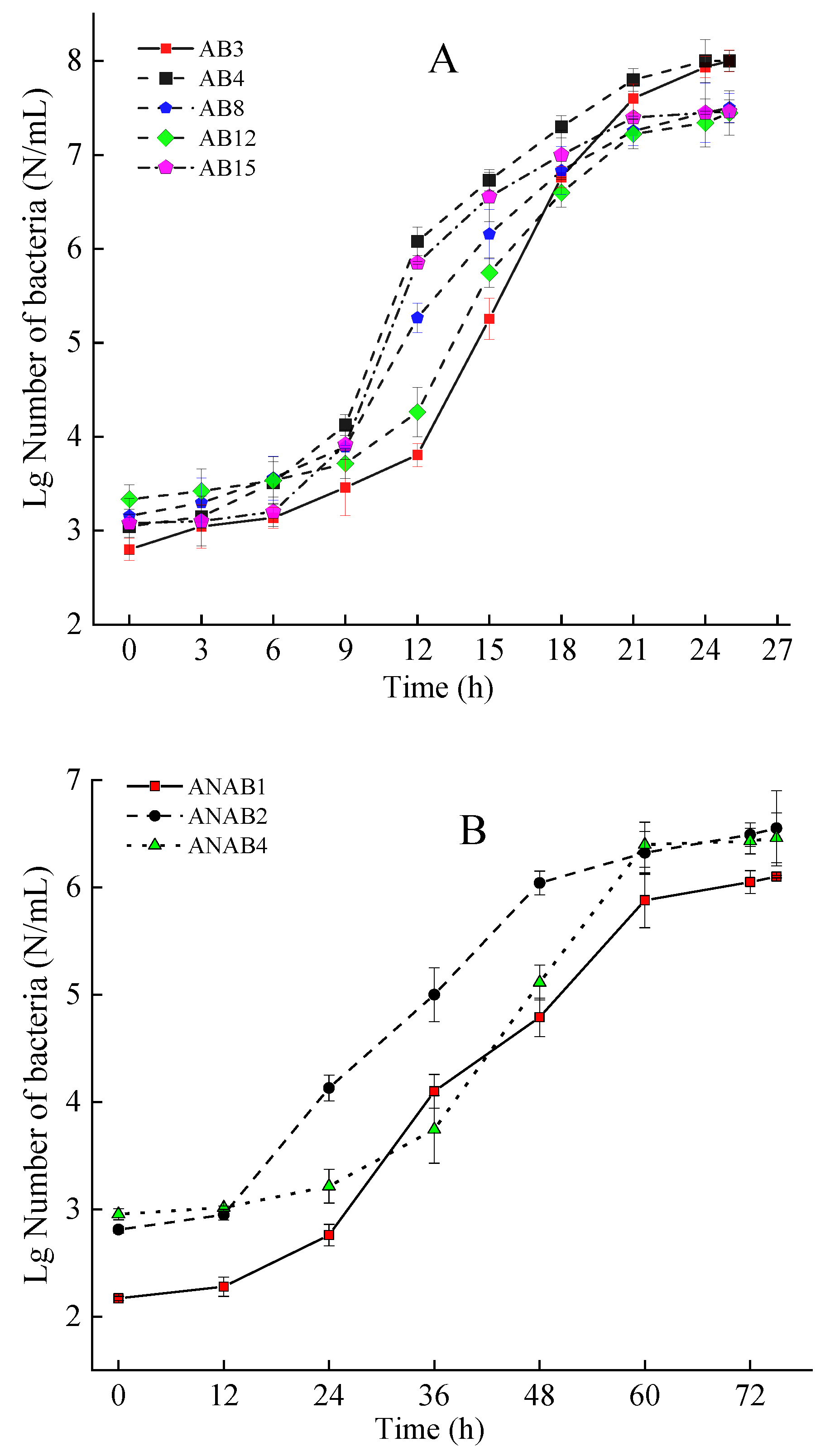
Under specific environmental conditions, bacterial populations typically progress through four distinct phases: the lag phase, the exponential (log) phase, the stationary phase, and the decline phase. During the exponential phase, the bacterial population increases at a constant geometric rate, demonstrating robust metabolic activity and vitality. Utilizing bacteria in this phase enhances experimental efficiency. Figure 1 illustrates the logistic growth curves for a subset of the bacterial strains analyzed in this study.

Figure 1.
Growth curve of aerobic bacteria (A) and anaerobic bacteria (B) strains.
Based on the logistic growth curves, it was determined that all aerobic bacteria in this experiment reached the exponential phase at a concentration of CFU = 106/mL, while all anaerobic bacteria entered the exponential phase at a concentration of CFU = 105/mL. Prior to inoculation, the concentrations of the aerobic and anaerobic bacterial solutions were adjusted using normal saline to achieve CFU = 106/mL for aerobic bacteria and CFU = 105/mL for anaerobic bacteria.
3.3. Pathogenicity Testing of Bacteria
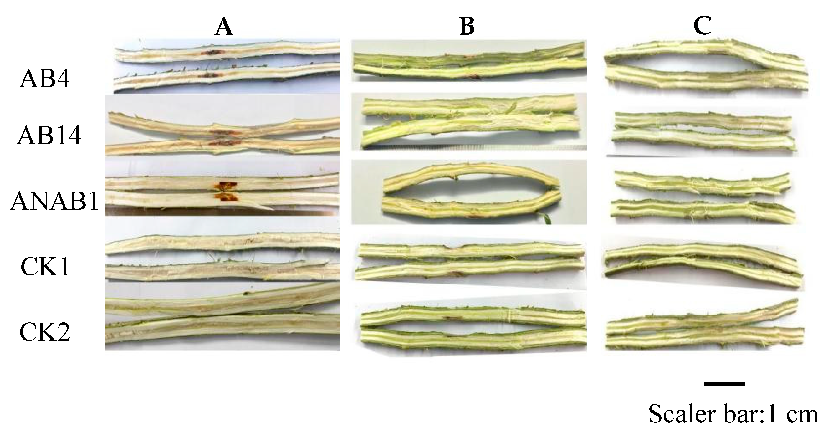
No significant effect on seedling height or ground diameter growth was observed across the various inoculation methods (p > 0.05, Table 3). However, significant differences in the incidence of wetwood disease were detected among the inoculation methods (multiple comparisons with CK2, p < 0.05). Notably, all poplar seedlings inoculated using the stem injection method developed wetwood symptoms, whereas no such symptoms were observed in CK1, CK2, and the seedlings inoculated by the root or leaf infection methods (Figure 2).

Table 3.
Effects of inoculation methods on poplar growth and occurrence of wetwood.

Figure 2.
Discoloration of stems induced by the bacterial strains under different inoculation methods. Note: (A) stem injection method; (B) root infection method; (C) leaf infection method.
Inoculation with pathogenic bacteria induced noticeable swelling at the inoculation site, leading to a significant increase in the diameter of the stem at the inoculation point (t-test, p < 0.05). Furthermore, significant variations were observed among different bacterial strains (t-test, p < 0.05; Figure 3). Among the aerobic bacteria, strains AB3 caused the most substantial increase in diameter at the inoculation site, with growth increments of 2.57 cm. Its values correspond to 3.36-fold CK1 and 3.76-fold increases compared to CK2. Among the anaerobic bacteria, strain ANAB1 showed the highest growth increment, measuring 1.91 cm, which represents 3.38-fold and 4.59-fold increase relative to CK1 and CK2, respectively.

Figure 3.
Stem diameter growth at the inoculation site. Note: * indicates significant difference with CK2 (t-test, p < 0.05).
Eighty days after pathogen inoculation via the stem injection method, dissection of the stem segments revealed clear symptoms of wetwood near the inoculation site. Water-soaked areas were observed in the heartwood, accompanied by a color change to brown-yellow or brown, which contrasted sharply with the normal white coloration of healthy tissue. The severity and manifestation of wetwood symptoms varied significantly among the different inoculated strains (Figure 2A).
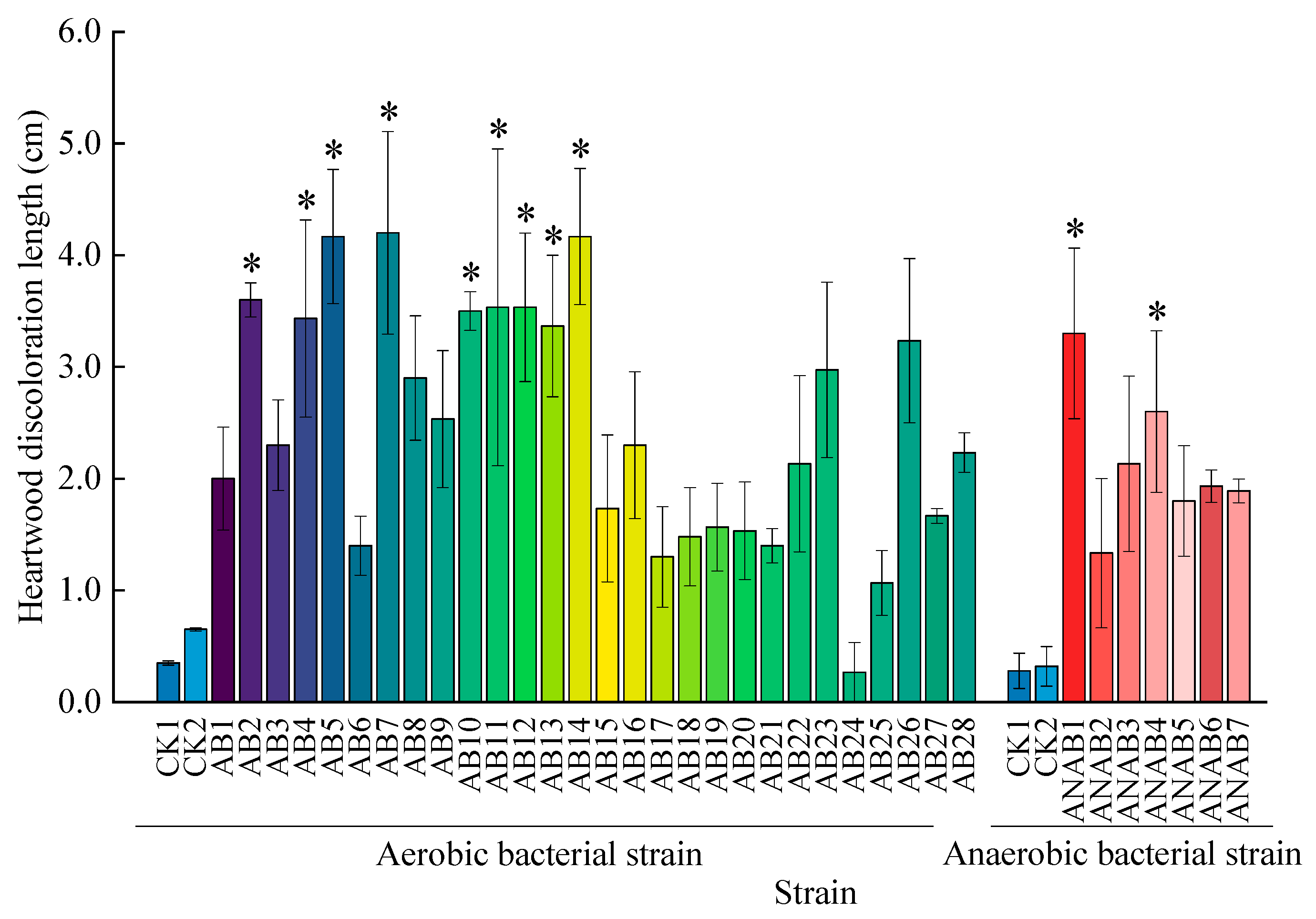
When different strains were inoculated using the stem injection method, significant variations in the length of heartwood discoloration were observed among the strains (t-test, p < 0.05; Figure 4). Among the 28 aerobic bacterial strains, AB2, AB4, AB5, AB8, AB10, AB11, AB12, AB13, and AB14 caused significantly greater heartwood discoloration lengths compared to the control treatment (multiple comparisons with CK2, t-test, p < 0.05). Strain AB14 induced the longest heartwood discoloration, with an average length of 4.17 cm, which represents 11.91-fold and 6.42-fold increase relative to CK1 and CK2, respectively. Among the seven anaerobic bacterial strains, ANAB1 and ANAB4 resulted in significantly longer heartwood discoloration than the control, with ANAB1 producing the longest average discoloration length of 3.3 cm, which represents 11.79-fold and 10.32-fold increase relative to CK1 and CK2, respectively.

Figure 4.
Discoloration length of heartwood inoculated with different strains. Note: * indicates significant difference with CK2 (t-test, p < 0.05).
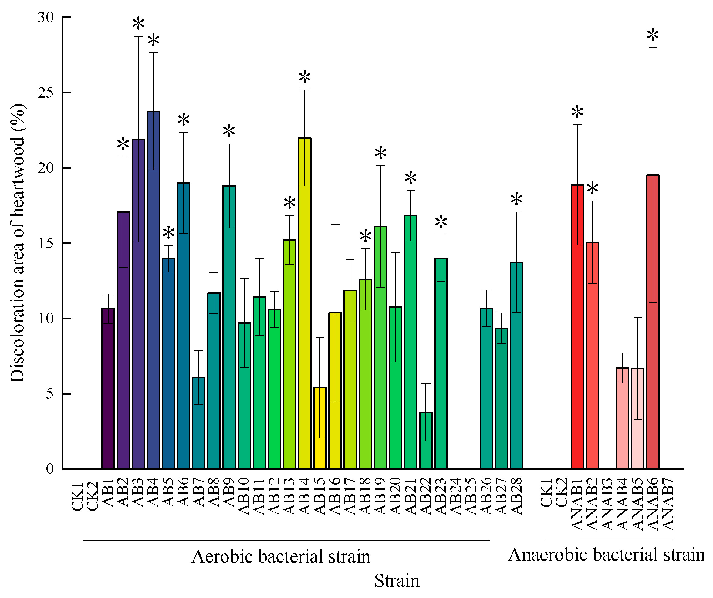
Statistical analysis revealed significant differences (t-test, p < 0.05) in discoloration area proportions among different strains (Figure 5). Among the 28 aerobic bacterial strains tested, AN2, AB3, AB4, AB5, AB6, AB9, AB13, AB14, AB18, AB19, AB21, AB23, and AB28 showed significant differences compared to the control group (multiple comparisons with CK2, t-test, p < 0.05). Notably, AB4 demonstrated the most pronounced effect, with discolored tissue accounting for an average of 23.76% of the cross-sectional wood area. Within the anaerobic bacterial group (7 strains), ANAB1, ANAB2, and ANAB6 showed significant differences compared to the control group (multiple comparisons with CK2, t-test, p < 0.05). ANAB6 demonstrated the most pronounced effect, with discolored tissue accounting for an average of 19.51% of the cross-sectional wood area.

Figure 5.
Discolored area ratios of heartwood inoculated with different strains. Note: * indicates significant difference with CK2 (t-test, p < 0.05).
Based on comprehensive evaluation of discoloration length, color intensity, and affected area proportion, the most severe wetwood symptoms were induced by aerobic strains AB4 and AB14, along with anaerobic strain ANAB1 (Figure 2A). These strains were identified as the most aggressive in terms of symptom development.
3.4. Pathogen Identification
- (1)
- Molecular identification
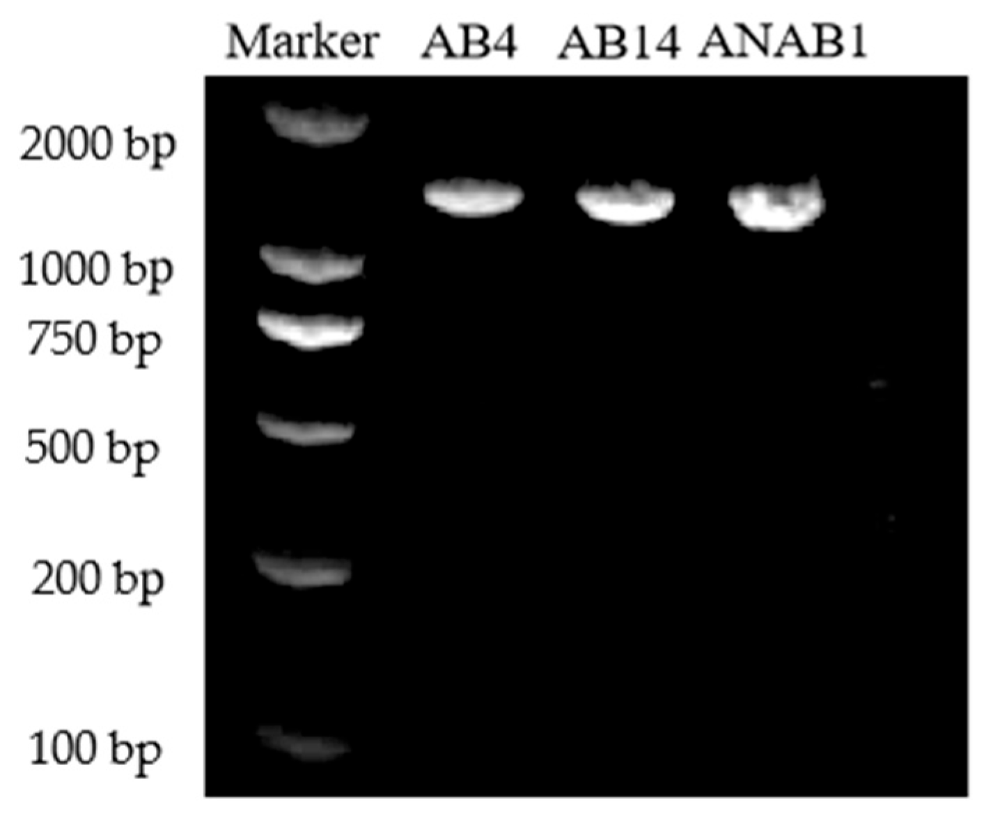
The genomic DNA of strains AB4, AB14, and ANAB1 were used as templates for PCR amplification with universal primers 27F and 1492R. This amplification yielded specific 16S rRNA fragments approximately 1.5 kb in length (Figure 6). Following gel electrophoresis and purification, sequencing analysis revealed distinct fragment sizes for each strain: 1466 bp for AB4, 1476 bp for AB14, and 1510 bp for ANAB1 (Supplementary Materials).

Figure 6.
16S rRNA PCR electrophoresis analysis of strains AB4, AB14, and ANAB1.
According to 16S rRNA phylogenetic analysis, phylogenetic trees were constructed for strains AB4, AB14, and ANAB1, respectively.
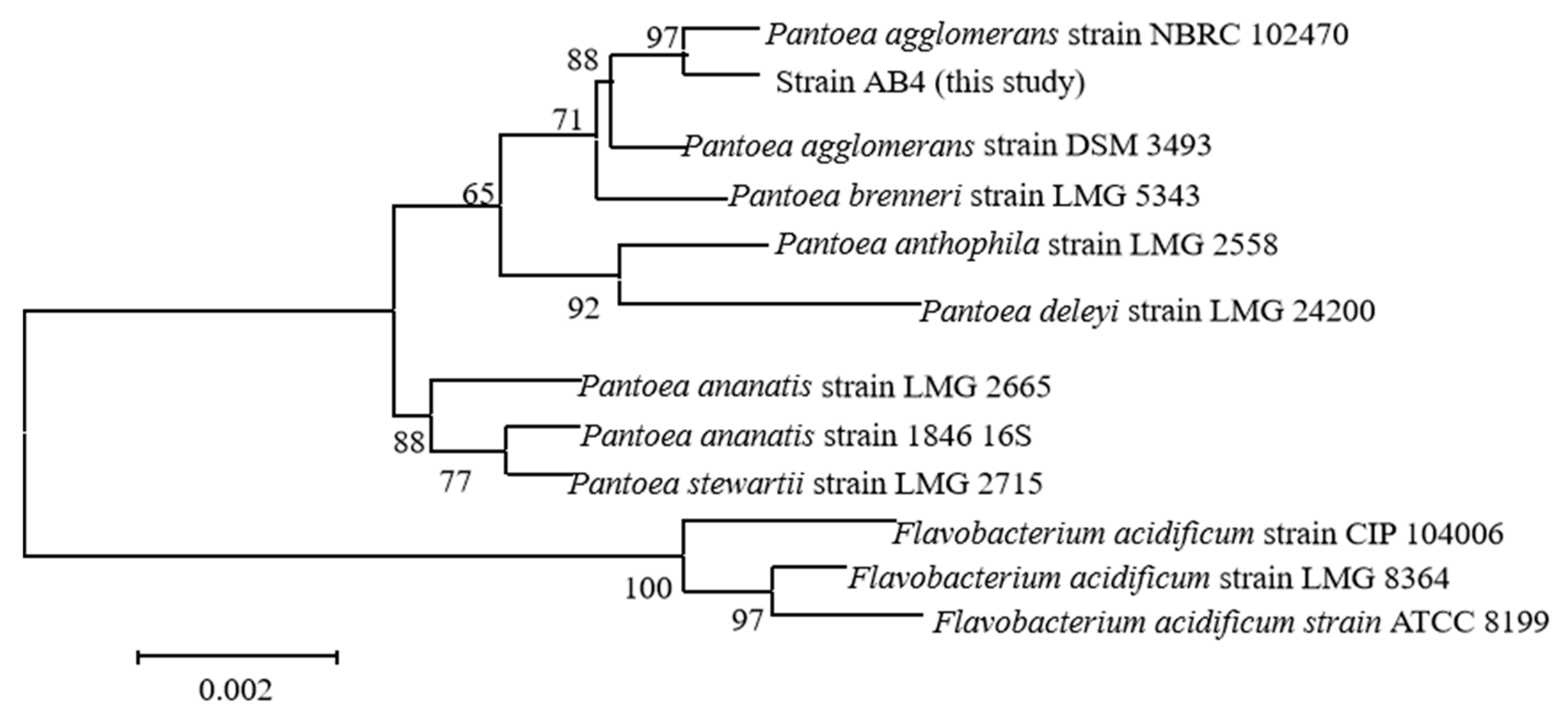
Phylogenetic analysis revealed that strain AB4 sharing high sequence similarity and forming a clade within the genus Pantoea, was most closely similar to Pantoea agglomerans strain NBRC (bootstrap value = 90%). In total, five closely related strains were identified in this clade, including P. agglomerans DSM 3493, P. brenneri LMG 5343, P. anthophila LMG 2558, and P. deleyi LMG 24200 (Figure 7). Among them, strain AB4 was most closely related to P. agglomerans NBRC 102470.

Figure 7.
Phylogenetic tree of strain AB4 based on 16S rRNA gene sequence (self-expansion value > 50%).
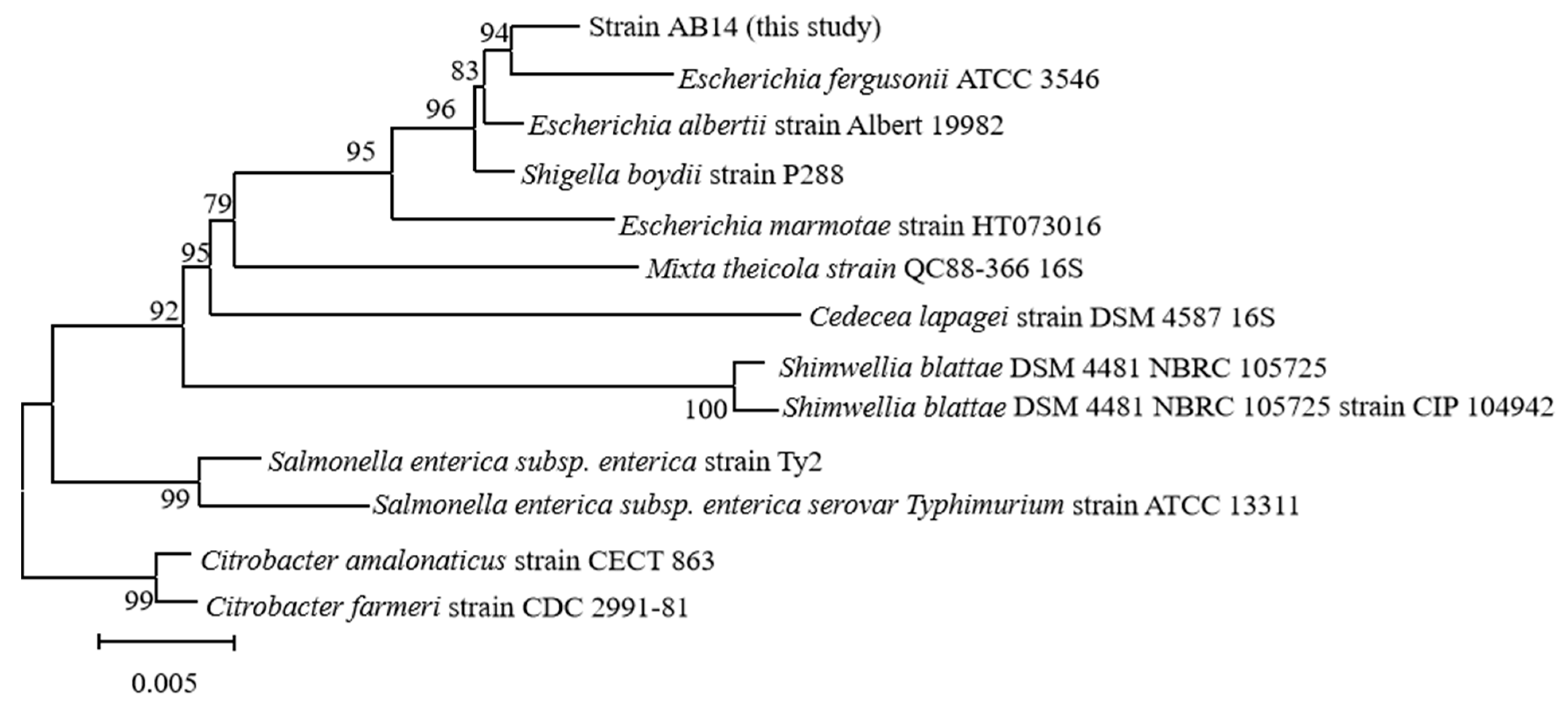
Strain AB14 was phylogenetically affiliated with the genus Escherichia, forming a well-supported monophyletic cluster with Escherichia albertii strain Albert 19982 (bootstrap value = 94%). In total, five closely related strains were identified in this clade, including Escherichia fergusonii ATCC 3546, Shigella boydii P288, Escherichia marmotae HT073016, and Mixta theicola QC88-366 16S (Figure 8). Among them, strain AB14 was most closely related to E. albertii Albert 19982.

Figure 8.
Phylogenetic tree of strain AB14 based on 16S rRNA gene sequence (self-expansion value > 50%).
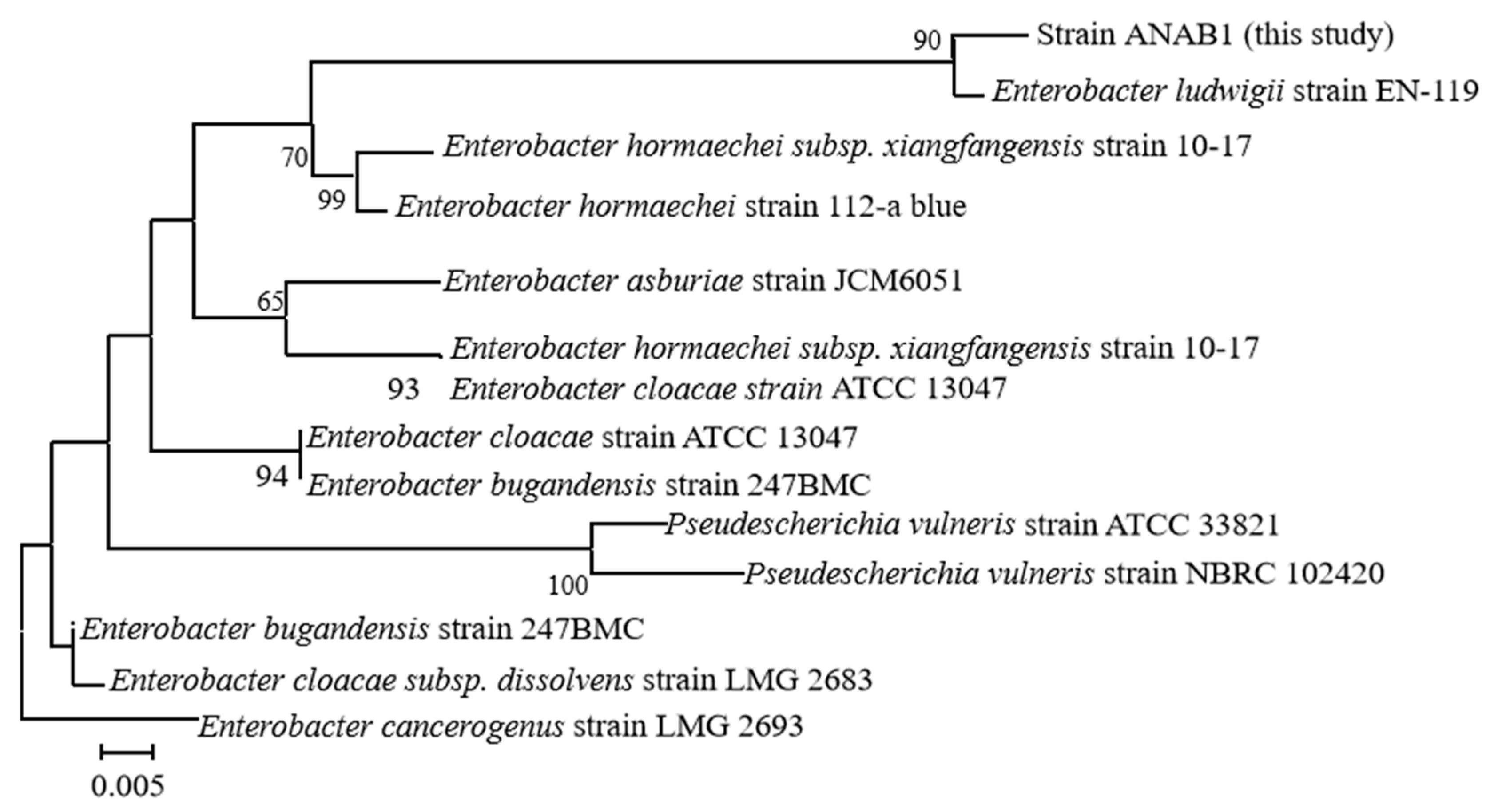
Similarly, strain ANAB1 displayed high sequence similarity to Enterobacter ludwigii strain EN-119, grouping within the Enterobacter genus (bootstrap value = 90%). A total of seven related strains were identified within this cluster, including Enterobacter hormaechei subsp. xiangfangensis 10–17, Enterobacter hormaechei 112-a blue, Enterobacter asburiae JCM6051, Enterobacter cloacae ATCC 13047, Enterobacter bugandensis 247BMC, and Pseudescherichia vulneris ATCC 33821 and NBRC 102420 (Figure 9). Among them, strain ANAB1 was most closely related to Enterobacter ludwigii EN-119.
- (2)
- Physiological and biochemical identification

Figure 9.
Phylogenetic tree of strain ANAB1 based on 16S rRNA gene sequence (self-expansion value > 50%).
Molecular identification revealed that the isolated strains were classified as follows: AB4 as Pantoea sp., AB14 as Escherichia sp., and ANAB1 as Enterobacter sp., all belonging to the Enterobacteriaceae family. Physiological and biochemical characterization showed that strain AB4 exhibited characteristics highly consistent with Pantoea agglomerans (Table 4). Similarly, strain AB14 displayed biochemical profiles matching those of Escherichia fergusonii, while strain ANAB1 demonstrated physiological and biochemical properties aligning with Enterobacter hormaechei. All of them belong to the Enterobacteriaceae family.

Table 4.
Physiological and biochemical comparison of three strains with model strains.
4. Discussion
Using the pathogen tissue isolation method, we successfully isolated and purified 28 aerobic and 7 anaerobic bacterial strains from I-69 poplar trees infected with wetwood disease. These findings demonstrate the considerable microbial diversity of pathogenic microorganisms associated with poplar wetwood, which aligns with the results reported by Song []. In subsequent inoculation tests, most of these isolated strains were able to significantly induce wetwood symptoms, while the control group showed no apparent symptoms of the disease.
P. agglomerans has attracted significant attention in botanical research as both a beneficial and pathogenic microorganism []. Belonging to the same genus as Erwinia herbicola, it is a carotenoid-producing, yellow-pigmented bacterium classified within the Enterobacteriaceae family. Initially isolated from plants, seeds, and fruits, it has since been identified in diverse environments. Studies have demonstrated its dual ecological role: while Ke [] confirmed its protective and antagonistic effects against tomato yellow leaf curl virus disease, Cruz [] identified it as a causative agent of wheat yellow wilt. Similarly, Enterobacter hormaechei, a facultative anaerobic member of the Enterobacteriaceae, exhibits ligninolytic and carotenoid-degrading capabilities. It has been implicated in root and leaf decay across multiple crop species [,]. Notably, this bacterium demonstrates high environmental adaptability, colonizing animal hosts and disseminating via fecal contamination. In contrast, Escherichia fergusonii, a reclassified species within the Enterobacteriaceae, remains poorly documented in plant pathology, with limited reports linking it to plant diseases [].
The pathogen inoculation experiments revealed that stem injection induced the most severe wetwood symptoms, primarily affecting the wood pith. In contrast, root infection and leaf infection showed negligible effects. These findings are consistent with the results reported by Wang [], indicating that the primary route of infection for poplar wetwood pathogens is through the stem, while leaf scratches or root damage rarely lead to wetwood formation. Furthermore, this aligns with field observations by Zheng [], which demonstrated that mechanical damage to poplar trunks frequently triggers wetwood disease.
Post-inoculation growth analysis demonstrated that pathogen infection had no statistically significant impact (p > 0.05) on either plant height increment or ground diameter growth in one-year-old seedlings. Stem inoculation induced distinct localized responses, including pronounced swelling at the inoculation site and subsequent scab formation at the wound, while these pathological symptoms were completely absent in non-inoculated control specimens. These findings collectively indicate that these observations likely represent an active physiological defense response against pathogenic invasion rather than a mere wound reaction. The variation in response intensity among individuals may reflect clonal or genotype-specific differences []. Pathogen inoculation consistently induced characteristic wetwood symptoms, including heartwood darkening and water-soaked appearance []. Notably, under identical conditions, different bacterial strains exhibited varying degrees of pathogenicity, demonstrating significant strain-specific effects on seedling physiology.
In this study, we isolated three highly pathogenic bacterial strains. Initial characterization through physiological and biochemical tests revealed distinct profiles across 15 different biochemical reactions, enabling preliminary identification using standard bacteriological manuals. However, given the rapidly expanding diversity of known microorganisms and the limitations of conventional biochemical methods, we complemented these analyses with molecular techniques for more precise identification []. We employed 16S rRNA gene sequencing, targeting the highly conserved regions (V1–V9) that are particularly suitable for prokaryotic taxonomy due to their stable evolutionary characteristics []. PCR amplification with universal primers and products sequencing yielded the expected around 1500 bp fragment, which was then used for phylogenetic analysis. By constructing a neighbor-joining phylogenetic tree and combining these molecular data with our physiological and biochemical profiles, we achieved accurate strain-level identification of the pathogenic isolates. However, while 16S rRNA sequencing provided useful phylogenetic placement, it has limited discriminatory power for differentiating closely related members of the Enterobacteriaceae []. Future studies employing multi-locus sequence analysis (MLSA) or whole genome sequencing (WGS) would yield more robust and definitive species-level identification [,]. In addition, we acknowledge that some pathogenic bacteria may be non-culturable under the current experimental conditions, which represents a further limitation of this study.
In previous literature, Escherichia fergusonii and Enterobacter hormaechei were primarily documented as opportunistic pathogens in clinical or environmental contexts, with limited evidence in plant pathogenesis. For example, E. fergusonii has been mainly reported in human infections and has received growing attention due to antibiotic resistance concerns []. E. hor-maechei is also predominantly considered a nosocomial pathogen, although some strains have shown plant growth-promoting traits under stress conditions [,]. Our study, however, demonstrates that under controlled inoculation conditions, these bacteria induce characteristic wetwood symptoms in poplar (Populus I-69) with statistical significance. Similar results were reported in Populus by Song [], who also identified Pantoea and Enterobacter species as wetwood pathogens. Moreover, microbial colonization of wetwood has long been linked to internal gas accumulation and methane formation, further emphasizing the ecological significance of bacterial infection in trees []. This suggests that, at least in experimental settings, they can behave as genuine pathogens rather than incidental contaminants. Further study is warranted to determine whether they act as primary pathogens under natural infection conditions.
5. Conclusions
Isolation of pathogenic bacteria from poplar wetwood yielded 28 aerobic and 7 anaerobic bacterial strains. Pathogenicity screening through stem injection of poplar seedlings identified two aerobic bacterial (Pantoea agglomerans, Escherichia fergusonii) and one anaerobic bacterial (Enterobacter hormaechei) strain capable of inducing characteristic wetwood symptoms. All three pathogenic strains were classified within the Enterobacteriaceae family. The study demonstrated that wetwood is not attributable to a single bacterial or fungal species, but rather to a complex interplay of microbial factors.
Pathogenicity assays revealed that stem injection induced the most severe wetwood symptoms. In contrast, root infection and leaf infection methods showed negligible effects. Notably, inoculation of 1-year-old seedlings with these pathogens did not significantly affect plant height or ground diameter growth.
This work provides clear evidence that E. fergusonii and E. hormaechei can act as true pathogens of poplar wetwood, thereby broadening the current understanding of the bacterial diversity associated with this disease.
These results provide valuable insights for forest health management and can inform future poplar breeding programs aimed at enhancing resistance to wetwood formation.
Supplementary Materials
The following supporting information can be downloaded at: https://www.mdpi.com/article/10.3390/f16091414/s1.
Author Contributions
Q.Z., Y.J. and X.H. performed the study, analyzed the data, and edited the manuscript. H.T. and Z.R. collected and analyzed the data. K.D. designed the experiments and revised the manuscript. All authors have read and agreed to the published version of the manuscript.
Funding
This work was funded by the Central Financial Forestry Science and Technology Extension Demonstration Subsidy Project (Hubei[2024]TG38), the National Natural Science Foundation of China (31570665), and the Hubei Provincial Natural Science Foundation of China (2021CFB446).
Data Availability Statement
The original contributions presented in this study are included in the article/Supplementary Material. Further inquiries can be directed to the corresponding author.
Conflicts of Interest
The authors declare no conflicts of interest.
References
- Fan, J.; Wang, S.; Ding, C.; Ma, C.; Chen, X.; Wang, J.; Yang, M.; Su, X. Correlation Analysis of the Bacterial Community and Wood Properties of Populus × Euramericana Cv. “74/76” Wet Heartwood. Front. Microbiol. 2022, 13, 868078. [Google Scholar] [CrossRef]
- Zu, B.S. Foreign studies on wet heart wood of poplars. Sci. Silvae Sin. 2000, 36, 85–91. [Google Scholar]
- Worrall, J.J.; Parmeter, J.R., Jr. Formation and Properties of Wetwood in White Fir. Phytopathology 1982, 72, 1209–1212. [Google Scholar] [CrossRef]
- Zhang, Q.; Hu, X.; Hu, Q.; Yang, J.; Zhao, T.; Du, K. Factors affecting poplar wetwood characteristics. J. For. Res. 2023, 34, 1615–1626. [Google Scholar] [CrossRef]
- Ward, J.; Schink, B. Microaerobic and anaerobic bacterial activities involved in formation of wetwood and discoloured wood. IAWA J. 1984, 5, 105–109. [Google Scholar] [CrossRef]
- Krause, C.; Gagnon, R. Wet heartwood distribution in the stem, stump, and root wood of black spruce in the Quebec boreal forest, Canada. North. J. Appl. For. 2005, 22, 12–18. [Google Scholar] [CrossRef]
- Jiang, X.; Zhang, L.; Xu, B.; Zheng, S. Study on the Occurrence, Distribution and Properties of Wet Heartwood in I-69 Poplar. For. Res. 1993, 6, 480–485. [Google Scholar]
- Xu, H.M.; Hu, X.Y.; Zheng, L.Y.; Fan, X.P. Research progress on wetwood. Hubei For. Sci. Technol. 2010, 4, 39–42. [Google Scholar] [CrossRef]
- Yu, X.; Hu, X.; Peng, Y.; Wu, Z.; Zhang, Q.; Li, Z.; Shi, C.; Du, K. Amplicon Sequencing Reveals Different Microbial Communities in Living Poplar Wetwood and Sapwood. Trees 2019, 33, 851–865. [Google Scholar] [CrossRef]
- Ward, J.C.; Pong, W.Y. Wetwood in Trees: A Timber Resource Problem; U.S. Department of Agriculture, Forest Service, Pacific Northwest Forest and Range Experiment Station: Portland, OR, USA, 1980. [Google Scholar] [CrossRef]
- Zeikus, J.G.; Ward, J.C. Methane formation in living trees: A microbial origin. Science 1974, 184, 1181–1183. [Google Scholar] [CrossRef]
- Zhu, Y.X.; Shu, D.Y.; Liu, S.Q. pH value, buffer content and the content of chemical components between heartwood and sapwood in poplar 72 and poplar 69. J. Anhui Agric. Univ. 2005, 32, 360–364. [Google Scholar] [CrossRef]
- Martin, L.; Brunel-Michac, N.; Conchon, P.; Cochard, H.; Badel, E. Mechanism of Wetwood Formation in Silver Fir (Abies alba Mill.). Trees 2024, 38, 1581–1592. [Google Scholar] [CrossRef]
- Shaw, D.; Edmonds, R.; Littke, W. Incidence of wetwood and decay in precommercially thinned western hemlock stands. Can. J. For. Res. 1995, 25, 1269–1277. [Google Scholar] [CrossRef]
- Murdoch, C. Stem and branch distribution of wetwood and relationship of wounding to bleeding in American elm trees. Plant Dis. 1984, 68, 890–892. [Google Scholar] [CrossRef]
- Guan, L.H.; Pan, H.X.; Huang, M.R.; Shi, J.S. Genetic variation of new clones of Populus deltoides (I-69) × Populus euramericana (I-45). J. Zhejiang For. Coll. 2004, 21, 376–381. [Google Scholar]
- Anguita-Maeso, M.; Olivares-García, C.; Haro, C.; Imperial, J.; Navas-Cortés, J.A.; Landa, B.B. Culture-Dependent and Culture-Independent Characterization of the Olive Xylem Microbiota: Effect of Sap Extraction Methods. Front. Plant Sci. 2020, 10, 1708. [Google Scholar] [CrossRef]
- Warshaw, J.E.; Leschine, S.B.; Canale-Parola, E. Anaerobic cellulolytic bacteria from wetwood of living trees. Appl. Environ. Microbiol. 1985, 50, 807–811. [Google Scholar] [CrossRef] [PubMed]
- Schink, B.; Ward, J.C.; Zeikus, J.G. Microbiology of wetwood: Role of anaerobic bacterial populations in living trees. Microbiology 1981, 123, 313–322. [Google Scholar] [CrossRef]
- Chen, C.W.; Sun, Q.G.; Zhu, J.; Wu, X.P.; Huang, H.Q.; Bao, S.X. Isolation and Screening for Endophyte with Antitumor Activities from Three Medicinal Plants. Microbiol. China 2010, 37, 1462–1466. [Google Scholar] [CrossRef]
- Zhu, X.; Ma, K.; Sun, M.; Zhang, J.; Liu, L.; Niu, S. Isolation and Identification of Pathogens of Morchella sextelata Bacterial Disease. Front. Microbiol. 2023, 14, 1231353. [Google Scholar] [CrossRef]
- Dowarah, B.; Agarwal, H.; Krishnatreya, D.B.; Sharma, P.L.; Kalita, N.; Agarwala, N. Evaluation of seed-associated endophytic bacteria from tolerant chilli cv. Firingi Jolokia for their biocontrol potential against bacterial wilt disease. Microbiol. Res. 2021, 248, 126751. [Google Scholar] [CrossRef]
- Lin, L.-H.; Ntambo, M.S.; Rott, P.C.; Wang, Q.-N.; Lin, Y.-H.; Fu, H.-Y.; Gao, S.-J. Molecular detection and prevalence of Xanthomonas albilineans, the causal agent of sugarcane leaf scald, in China. Crop Prot. 2018, 109, 17–23. [Google Scholar] [CrossRef]
- Sanders, E.R. Aseptic laboratory techniques: Plating methods. J. Vis. Exp. 2012, 63, e3064. [Google Scholar] [CrossRef]
- Bergey, D.H.; Holt, J.G. Bergey’s Manual of Determinative Bacteriology, 8th ed.; Williams & Wilkins: Baltimore, MD, USA, 1993. [Google Scholar]
- Chi, M. The Endophyte Diversity and the Screening of Antagonistic Bacteria in Poplar Stems; Chinese Academy of Forestry: Beijing, China, 2014; pp. 1–19. [Google Scholar]
- An, X.Y.; Ma, Y.M.; Liu, X.X.; Zhou, T.N.; Yan, H.; Jia, L.H. Identification of a rare actinomycetes and screening of natural products biosynthesis genes from valerian rhizosphere soil. Acta Agric. Boreali-Occident. Sin. 2023, 7, 1080–1086. [Google Scholar]
- Hall, B.G.; Acar, H.; Nandipati, A.; Barlow, M. Growth rates made easy. Mol. Biol. Evol. 2014, 31, 232–238. [Google Scholar] [CrossRef]
- O’Mahony, F.C.; Papkovsky, D.B. Rapid high-throughput assessment of aerobic bacteria in complex samples by fluorescence-based oxygen respirometry. Appl. Environ. Microbiol. 2006, 72, 1279–1287. [Google Scholar] [CrossRef] [PubMed]
- Qin, A.J. Fundamentals of Microbiology; Hohai University Press: Nanjing, China, 2000. [Google Scholar]
- Cao, H.; Pan, J.; Zhang, Y.; Li, Y. A Method for Anaerobic Bacterial Isolation and Cultivation. China Patent CN101270334B, 20 April 2011. [Google Scholar]
- Tuttle, A.R.; Trahan, N.D.; Son, M.S. Growth and Maintenance of Escherichia coli Laboratory Strains. Curr. Protoc. 2021, 1, e20. [Google Scholar] [CrossRef] [PubMed]
- Sherald, J.L. Pathogenicity of Xylella fastidiosa in American elm and failure of reciprocal transmission between strains from elm and sycamore. Plant Dis. 1993, 77, 190–193. [Google Scholar] [CrossRef]
- Fang, Z.D. Methods of Plant Pathology Research; Agricultural Press: Beijing, China, 1979. [Google Scholar]
- Suo, Y.T.; Zhang, Y.N.; Li, Z.; Zhao, M.Z.; Shen, W.N.; Fu, Y.C.; Wang, D.L.; Pan, L.; Su, X.H.; Zhao, J.P. In vivo leaf inoculation: An alternative method to assess the disease resistance of hybrid clones in poplar breeding of stem canker disease. J. Vis. Exp. 2024, 211, e67290. [Google Scholar] [CrossRef]
- Ainsworth, G.C.; Jones, B.L. Plant Pathology, 5th ed.; Cambridge University Press: Cambridge, UK, 1995. [Google Scholar]
- Song, C.C.; Wang, K.; Mei, L.; Fan, X.P.; Zhang, S.; Zhu, Y.J. Pathogen isolation, identification and infection characteristics of wet-heartwood disease in poplar. J. Huazhong Agric. Univ. 2017, 36, 15–21. [Google Scholar] [CrossRef]
- Minas, K.; McEwan, N.R.; Newbold, C.J.; Scott, K.P. Optimization of a high-throughput CTAB-based protocol for the extraction of qPCR-grade DNA from rumen fluid, plant and bacterial pure cultures. FEMS Microbiol. Lett. 2011, 325, 162–169. [Google Scholar] [CrossRef]
- Yarza, P.; Richter, M.; Peplies, J.; Euzeby, J.; Amann, R.; Schleifer, K.-H.; Ludwig, W.; Glöckner, F.O.; Rosselló-Móra, R. The all-species living tree project: A 16S rRNA-based phylogenetic tree of all sequenced type strains. Syst. Appl. Microbiol. 2008, 31, 241–250. [Google Scholar] [CrossRef] [PubMed]
- Song, C.C.; Mei, L.; Cai, L.; Zhang, S.; Fan, X.P. Identification of pathogen bacteria and antagonistic bacteria of wet-heart wood disease in Populus. J. Southwest For. Univ. 2018, 38, 142–152. [Google Scholar]
- She, X.M.; Yu, L.; Lan, G.B.; Tang, Y.F.; Li, Z.G.; Deng, M.G.; He, Z.F. Identification of the pathogen causing Ziziphus jujuba Mill. cv. Dongzao leaf necrotic disease in Guangdong Province. Acta Phytopathol. Sin. 2021, 51, 317–324. [Google Scholar] [CrossRef]
- Ke, H.J.; Wang, Y.; Wei, T.; Gu, C.; Liu, H.X.; Guo, J.H. Suppression of tomato yellow leaf curl virus disease by the Pantoea agglomerans strain ljb-2 in the field. Acta Hortic. Sin. 2014, 41, 985–993. [Google Scholar] [CrossRef]
- Cruz, A.T.; Cazacu, A.C.; Allen, C.H. Pantoea agglomerans, a plant pathogen causing human disease. J. Clin. Microbiol. 2007, 45, 1989–1992. [Google Scholar] [CrossRef] [PubMed]
- Yang, X.P.; Wang, F.F.; Yan, J.; Ye, J.B.; Ma, K.; Mao, D.B. Optimization of medium for bacterial strain of carotenoids degradation. Sci. Technol. Food Ind. 2016, 5, 210–214+219. [Google Scholar] [CrossRef]
- O’Hara, C.M.; Steigerwalt, A.G.; Hill, B.C.; Farmer, J.J., 3rd. Enterobacter hormaechei, a new species of the family Enterobacteriaceae formerly known as Enteric group 75. J. Clin. Microbiol. 1989, 27, 2046–2049. [Google Scholar] [CrossRef]
- Srinivas, K.; Ghatak, S.; Pyngrope, D.A.; Angappan, M.; Milton, A.A.P.; Das, S.; Lyngdoh, V.; Lamare, J.P.; Prasad, M.C.B.; Sen, A. Avian strains of emerging pathogen Escherichia fergusonii are phylogenetically diverse and harbor the greatest AMR dissemination potential among different sources: Comparative genomic evidence. Front. Microbiol. 2023, 13, 1080677. [Google Scholar] [CrossRef]
- Wang, K. The Infection Way of Wetwood Disease Pathogens of Poplar and Their Effects on the Growth of Poplar; Huazhong Agricultural University: Wuhan, China, 2015. [Google Scholar]
- Zheng, H.Y.; Dai, Y.S.; Xie, C.X.; Guo, T.B.; Wang, Z.Y. Studies on the cause of formation of poplar red core wood. J. Nanjing For. Univ. (Nat. Sci. Ed.) 2006, 30, 82–84. [Google Scholar]
- Eckert, C.; Wildhagen, H.; Paulo, M.J.; Scalabrin, S.; Ballauff, J.; Schnabel, S.K.; Vendramin, V.; Keurentjes, J.J.B.; Bogeat-Triboulot, M.-B.; Taylor, G.; et al. Genotypic and tissue-specific variation of Populus nigra transcriptome profiles in response to drought. Sci. Data 2022, 9, 297. [Google Scholar] [CrossRef]
- Janda, J.M.; Abbott, S.L. Bacterial identification for publication: When is enough enough? J. Clin. Microbiol. 2002, 40, 1887–1891. [Google Scholar] [CrossRef]
- Johnson, J.S.; Spakowicz, D.J.; Hong, B.Y.; Petersen, L.M.; Demkowicz, P.; Chen, L.; Leopold, S.R.; Hanson, B.M.; Agresta, H.O.; Gerstein, M.; et al. Evaluation of 16S rRNA gene sequencing for species and strain-level microbiome analysis. Nat. Commun. 2019, 10, 5029. [Google Scholar] [CrossRef]
- Janda, J.M.; Abbott, S.L. 16S rRNA gene sequencing for bacterial identification in the diagnostic laboratory: Pluses, perils, and pitfalls. J. Clin. Microbiol. 2007, 45, 2761–2764. [Google Scholar] [CrossRef]
- Glaeser, S.P.; Kämpfer, P. Multilocus sequence analysis (MLSA) in prokaryotic taxonomy. Syst. Appl. Microbiol. 2015, 38, 237–245. [Google Scholar] [CrossRef]
- Adesina, T.; Nwinyi, O.; De, N.; Akinnola, O.; Omonigbehin, E. First Detection of Carbapenem-Resistant Escherichia fergusonii Strains Harbouring Beta-Lactamase Genes from Clinical Samples. Pathogens 2019, 8, 164. [Google Scholar] [CrossRef] [PubMed]
- De Maayer, P.; Green, T.; Jordan, S.; Smits, T.H.M.; Coutinho, T.A. Pan-Genome Analysis of the Enterobacter hormaechei Complex Highlights Its Genomic Flexibility and Pertinence as a Multidrug Resistant Pathogen. BMC Genom. 2025, 26, 11590. [Google Scholar] [CrossRef] [PubMed]
- Ranawat, B.; Bachani, P.; Singh, A.; Mishra, S. Enterobacter hormaechei as Plant Growth-Promoting Bacteria for Improvement in Lycopersicum esculentum. Curr. Microbiol. 2021, 78, 1208–1217. [Google Scholar] [CrossRef] [PubMed]
Disclaimer/Publisher’s Note: The statements, opinions and data contained in all publications are solely those of the individual author(s) and contributor(s) and not of MDPI and/or the editor(s). MDPI and/or the editor(s) disclaim responsibility for any injury to people or property resulting from any ideas, methods, instructions or products referred to in the content. |
© 2025 by the authors. Licensee MDPI, Basel, Switzerland. This article is an open access article distributed under the terms and conditions of the Creative Commons Attribution (CC BY) license (https://creativecommons.org/licenses/by/4.0/).