Abstract
The purpose of this study is to more quantitatively identify changes in body function through various bio-signal parameters. (1) Background: Forest therapy is effective in stabilizing cognitive, emotional, cardiovascular, and autonomic nervous systems. In particular, it is necessary to more quantitatively confirm changes in body functions through various bio signals. (2) Methods: As a forest therapy program (FTP) for the elderly, it consisted of strength training in the forest, respiratory aerobic exercises, and cognitive function training, and a total of 19 sessions were performed for 12 weeks. The electroencephalography (EEG) and Photoplethysmography (PPG) before and after the program were measured and compared between program participants (FTP group) and non-participants (control group). (3) Results: the FTP group showed increase in the alpha band power in EEG and a decrease in the PRV index, Tad, and Tae after the program compared to the control group; (4) Conclusions: Significant differences occurred in the physiological functioning of the elderly participants after the program. This is a result that can confirm the effectiveness of forest therapy more quantitatively. Forest therapy has a positive effect on mental stress reduction and cardiovascular function.
1. Introduction
Currently, South Korea is experiencing serious issues of aging and low birth rates. As the elderly population increases, the age of the socially active population also rises, necessitating efforts to manage and maintain their health more stably. Medically, regular health check-ups are encouraged, and various hobby-based exercises are proposed for health management, with their benefits confirmed. However, considering the physical condition of the elderly, their choice of exercise types is limited. Additionally, the economic cost of health management is increasing, placing a burden on household finances and thus increasing the need for health management that utilizes the surrounding environment. A representative example of this is forest therapy. Physical and mental forest therapy programs are being developed in the field of elderly welfare services that utilize natural environments such as forests, valleys, and mountains [1]. During forest therapy, substances such as phytoncide of the terpene system generated in nature are inhaled by human breathing, which has a positive effect on brain waves, pulse rate, and relaxation of blood pressure [2]. In addition, due to the effects of mental stability and exposure to sunlight, forest therapy also has a good effect on various diseases. For patients suffering from sleep disorders, exposure to forest therapy and natural activities has resulted in improvements in sleep duration and language agitation; in patients with anxiety, the circadian rhythm of the activity cycle is stabilized and the quality of life is improved; and in patients with cognitive impairment, the effect of performing a nature-friendly integrated program was confirmed to significantly improve brain function and cognitive function [3,4,5]. In this way, forest therapy can present an optimal environment for the management and treatment of various diseases suffered by the elderly. Various studies have been conducted that have verified the positive effects of forest therapy on physical functions. In the case of mental disorders such as stress and depression, a significant decrease in cortisol level, an indicator of stress, was confirmed when forest walking and urban walking were performed in patients with coronary artery disease and chronic obstructive pulmonary disease [6,7]. One study presented a theory of stress recovery through the practice of forest therapy [8], and another study found that forest walking in stroke patients significantly reduced BDI on the depression scale [9]. Qualitative and quantitative methods such as blood sampling tests, program satisfaction surveys, and questionnaire responses are used to confirm the effectiveness of forest therapy. Furthermore, there are various prior studies that have confirmed the effects of forest therapy through biometric signals. In general, encephalography (EEG) is used to check for depression or cognitive changes. Using EEG, it is possible to check changes in the characteristics of EEG signals according to emotional and cognitive functions. EEG asymmetry was found to have a significant correlation with depression or anxiety [10,11]. In addition, pulse rate variability (PRV, or heart rate variability (HRV)) is an evaluation method for quantifying the cardiovascular system and autonomic nervous system [12,13]. HRV is analyzed by repetitive QRS waveforms measured by electrocardiogram, and pulse rate variability (PRV) refers to the change in the interval between pulses in the signal acquired using photoplethysmography (PPG) [14]. These prior studies show that EEG and PPG can be used to evaluate the effectiveness of specific indications by a multimodal method [15,16]. In this study, the forest therapy program for the elderly consisted of strength training, respiratory aerobic exercise, and cognitive function training in the forest, and a total of 24 sessions were performed twice a week for 135 min for 12 weeks. The group of patients who performed the training program performed the program in an outdoor environment in nature, and we wanted to verify the effect of the training in the forest by comparing it with the control group who performed the same program indoors. The purpose of this study is to investigate changes in physical functions before and after participation in forest therapy programs using psychological scale parameters via EEG and autonomic nervous system parameters via PPG, comparing participants and non-participants. This study aims to identify significant variables to assess the effectiveness and impact of forest therapy programs composed of cognitive and exercise components.
2. Materials and Methods
2.1. Experimental Participants
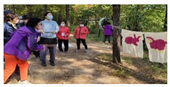
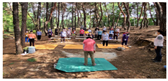
In order to compare the effectiveness of the forest therapy program, 60 elderly people registered at local health centers and dementia relief centers were recruited, and 58 people were finally selected, excluding 2 who were omitted from the collection of demographic information. Of these, 33 were divided into the experimental group and the remaining 25 into the control group. In the end, 28 people were in the experimental group (EG) and 17 were in the control group (CG). Except for some measurement errors in the measured bio-signals, the EGs used as EEG and PRV analysis data were 19 and 17, respectively, and the CGs were 14 and 11, respectively. Prior to the implementation of the program and the measurement of vital signs, all subjects were fully briefed about the experiment, and consent was obtained according to the IRB approval form (IRB No. CBNU-202004BMSBBR-0041, approval date 27 April 2020). Demographic information was collected and cognitive function tests (MMSE-K, K-MoCA, GDS, and Quality of Life Scale Tests (EQ-5D) were performed from the finally selected subjects before and after the implementation of the forest therapy program (Table 1). All participants completed all measurements and surveys at the Cheongju-city Public Health Center before starting the program. The urban forest therapy was conducted in the Maebong Mountain area of Cheongju-city, Chungcheongbuk-do, South Korea (Figure 1). The control group was managed by the research team to ensure that all measurements were taken under the same conditions and timeframes.

Table 1.
Demographic information.
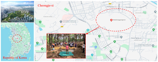
Figure 1.
Zone map of the experimental area and the urban forest site of Maebong Mountain.
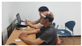
2.2. Bio-Signal Measurements
2.2.1. EEG: Electro Encephalography
A wireless EEG device was used for EEG measurement (NeuroNicle FX2, LAXTHA, Inc., Daejeon, Korea). In accordance with the standard international 10/20 electrode system, electrodes were attached to Fp1 (left prefrontal) and Fp2 (right prefrontal) of the prefrontal region. The sampling frequency was 250 Hz, 3 to 43 Hz bandpass, and 10 kΩ or less contact impedance was maintained to obtain data in a resting state (Table 2). A total of 7 EEG variables were analyzed: asymmetries of alpha band (asym_A), beta band (asym_B), and gamma band (asym_G), alpha and beta band powers (Pα and Pβ), median frequency (MEF), and ratio of alpha to theta band power (ATR). EEG measurements were taken for a total of 5 min in a stable state. After post-processing, power and frequency metrics were calculated using fast Fourier transform (FFT). The ratio of left/right brain waves for each band was computed, and statistical analysis was conducted by dividing the subjects into a control group and an experimental group.
Table 2.
Definition of EEG and PRV variables to be analyzed.
2.2.2. PPG: Photoplethysmography
PPG devices were used for HRV (PRV) analysis (Ubpulse T1, LAXTHA, Inc., Daejeon, Korea). The sampling frequency was 250 Hz, and data were obtained with a bandpass frequency of 0.3 to 10.6 Hz. PRV variables consists of a total of 8 variables: heart rate, PRV index, SDNN, VLF, Tab, Tac, Tad, and Tae (Table 2). PPG measurements were taken for a total of 5 min along with EEG. The recorded data were post-processed with filtering, and then time domain variables, frequency domain variables after FFT, and duration variables based on wave after second differentiation were calculated. Statistical analysis was conducted by dividing the subjects into a control group and an experimental group.
2.3. Experimental Methods
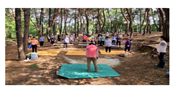
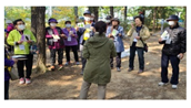
The forest therapy program consisted of fine motor exercise, breathing exercises, and cognitive function training programs using natural objects. of the participants carried out strength training; preparatory gymnastics; fine motor exercise programs such as hand pressing, shoulder rotation, tapping, and elastic band stretching; breathing exercises; knee and arm movements; loud breathing; and forest walking. Cognitive function training using natural objects consisted of memory tasks and tree puzzle solving (which is effective for memory) as well as cognitive training involving finding fruit sounds. The program consisted of a total of 19 sessions each lasting 135 min (preparatory gymnastics: 5 min, fine motor exercise: 50 min, breathing exercises: 50 min, cognitive training: 25 min, and gymnastics: 5 min) (Table 3). EEG and PPG were repeatedly measured before and after the performance of the forest therapy program using experimental measurements. In particular, there was an 8-week break after the preliminary measurement and first program due to the impact of COVID-19. Afterwards, a 12-week program was performed and post-measurements were taken.
Table 3.
Forest therapy program protocol.
3. Results
3.1. EEG Variables
Of the resting-state EEG variables, Pα and Pβ were significantly reduced in the control group (p < 0.01, effect size > 0.9 for all), and asym_B and asym_G in the experimental group increased after the forest therapy. That is, it was confirmed that there was a significant mean difference in the decrease in the degree of asymmetry (asym_B: p < 0.05, effect size: 0.57, asym_G: p < 0.01, effect size: 0.70). As a result of comparing the difference between the mean values of the experimental group and the control group, there was an average difference in the degree of change in the value of Pα. In the case of MEF (median frequency) and ATR (alpha-to-theta ratio), there was no significant difference or large effect size in the pre/post-analysis of the control group and the experimental group, and the effect size of the difference between the pre/post-change of the experimental group and control group was not significant (Table 4).
Table 4.
Results of resting-state EEG.
3.2. PRV Parameters
In the pre/post-analysis of the control group, Tad and Tae were significantly increased after the fact, and in the experimental group, the Tac and PRV index were significantly reduced after the fact. As a result of comparing the mean changes between the experimental group and the control group, the PRV index, Tad, and Tae were significantly lower in the experimental group than in the control group (Table 5).
Table 5.
Results of pulse rate variability.
4. Discussion
In this study, training was carried out to identify changes in cognition and the autonomic nervous system in physical functions, which are known to be advantages of the cognitive motor convergence forest therapy program, and this study was conducted to confirm the effect of forest therapy more quantitatively by comparing the actual performance in the forest and practice indoors.
First, EEG, which is known as an indicator of cognition and depression, is one of the factors necessary for optimizing brain function in order to improve emotional state or reduce depression [17]. In this study, it was confirmed that the degree of brain asymmetry was reduced by increasing both asym_B and asym_G values after the fact, and the asymmetry score of the experimental group was also increased compared to the control group in terms of the amount of pre/post-change. This shows that when participating in the forest therapy program, the asymmetry of the left and right brains decreases and changes to a symmetrical direction, allowing them to maintain a stable emotional state. It can be seen that this is similar to the results of previous studies [18,19]. Pα becomes lower as cognitive function deteriorates or is not in a stable state, and the decrease in Pα in the control group is expected to be a result of the accumulation of social stress caused by the COVID-19 pandemic in 2020 [20].
Second, in the PRV analysis, it was observed that the PRV index significantly decreased in the experimental group before and after the program. The PRV or HRV index refers to the degree of variation in the heartbeat, that is, the slight variation between one cardiac cycle and the next [21]. Heart rate is determined by the autonomic nervous system influencing the inherent spontaneity of the sinoatrial node and is related to the interaction between the sympathetic and parasympathetic nerves [22]. The decrease in the PRV index indicates a decrease in the body’s ability to adapt to the ever-changing environment, and the increase in this value after the implementation of the forest therapy program in the experimental group can be interpreted as an increase in the body’s ability to adapt to environmental changes. The results of comparing the average amount of change between the control group and the experimental group showed a difference in the PRV-index, indicating that the amount of change in the experimental group was larger, which means that the body’s ability to adapt to environmental changes was significantly increased compared to the control group by performing the forest therapy program. Generally, a higher PRV index indicates that the parasympathetic nervous system is dominant within the autonomic nervous system, signaling that the body is in a state of rest and digestion [23]. Conversely, lower PRV is typically interpreted as increased sympathetic nervous system activity within the autonomic system. Additionally, higher PRV signifies lower stress levels [24,25]. Therefore, it can be interpreted that the program’s influence leads to a higher level of activation of the sympathetic nervous system, which in turn indicates an increased response to stress.
The Tad and Tae variables that showed a decreasing trend in the control group are acceleration pulse wave variables, and it is easy to grasp detailed information regarding the shape of the pulse wave, such as the inflection point, which is a secondary differential waveform of the pulse wave [26]. Tad is defined as the time taken to go from the inflection point at position “a”, which is the initial peak value of the waveform that is the basis for waveform observation, to the inflection point at position “d”, which refers to the residual blood volume as a late contractile re-reduction wave, which means functional vasodilation capacity. In general, in elderly people who did not perform the program, the increase in Tad means that the time until the residual amount remains after blood flow decreases [27,28]. In the Tad results, the program group showed a significantly greater change compared to the pre/post difference of the control group, but no significant difference was observed within the program group itself. Although a decrease in Tad time was expected in the program group after program implementation, the lack of a statistically significant difference suggests that a relatively short-term program has a minimal impact on vascular elasticity or blood flow. This indicates a need for additional experiments under conditions of long-term program participation to confirm these effects. The inflection point at position “c” in Tac refers to the late contractile regrowth wave, which is associated with the elasticity of the blood vessels and the downward strength of the pulse wave just before the onset of diastolic phase. In the experimental group, the increase in this value after the implementation of the forest therapy program means an increase in the time taken to reach the onset of the diastolic phase. Tae refers to the time it takes for all the blood to flow in the first heartbeat, which means that the blood flow velocity has increased because the time has decreased significantly in the second measurement [28]. This is thought to be an effect of the correlation between blood flow velocity and vascular aging levels [28,29]. The effects of forest therapy on the autonomic nervous system have been confirmed by various previous studies [30]. Specifically, the increase in parasympathetic nerve activity and the decrease in sympathetic nerve activity related to the resting state observed in these studies are similar to the results of the current study. While many previous studies have examined the enhancement of cardiovascular function using heart rate variability frequency variables, there have been few studies investigating vascular elasticity indirectly from the perspective of second derivative PPG. It has been challenging to confirm these effects in short-term programs, but it is considered possible to confirm these effects when conducting long-term forest therapy.
In summary, relaxation or activities in forest settings can be associated with decreased blood pressure, changes in heart rate, and improvements in cardiovascular function. The natural sounds and visual elements of forests can positively impact cardiovascular or autonomous neural system control. Previous studies have confirmed that forest environments are linked to reduced levels of the stress hormone cortisol [31]. Additionally, activities in forests can influence cognitive function [32,33]. As seen in the results of our study, there was a greater tendency for left–right brain asymmetry to reduce in the group participating in the program. Thus, the reasons for engaging in forest therapy indirectly confirm the various positive impacts provided by diverse forest environments [34].
One limitation of this study is the small number of subjects. By ensuring a sufficient motivational system to encourage and manage participation in long-term training, we hope to implement a forest therapy program for more elderly people in future studies. A second limitation is that there are differences in demographic information between the two groups. Therefore, an experiment to exclude and control these effects has not been clearly carried out. To address these shortcomings, statistical comparisons were performed before and after the forest therapy program in each group. The third limitation is the break period caused by COVID-19. Measurements should be made immediately before the program is implemented, but due to the impact of COVID-19, the program was carried out after a two-month break after measurement. Another limitation is that it was difficult to identify more effective variables due to this effect. In a future study, we plan to conduct comparisons between groups in more controlled conditions. Additionally, we will conduct pre/post-measurements at several sessions, such as after the first session, and then after the fourth, eighth, and twelfth sessions.
In this study, EEG and PPG bio-signals were measured and analyzed to confirm the effect of performing forest therapy programs in the elderly, and positive effects on various parameters were confirmed. In future studies, we plan to conduct studies to determine the diversity of forest therapy programs and the effectiveness of the programs under more limited conditions.
5. Conclusions
After the implementation of the forest therapy program in the elderly, significant differences occurred in physical function. This is a result that can confirm the effect of forest therapy more quantitatively. From the results of quantitative vital signs, it was confirmed that forest therapy had a positive effect on stress and cardiovascular function. Additional research can be carried out through the small number of subjects and the adjustment of additional control variables, which is a limitation of this study and will be used as the basis of research in future large-scale studies.
Author Contributions
Conceptualization, J.U.K.; methodology, J.-W.S.; validation, W.S.; formal analysis, S.G.K.; investigation, K.K.; resources, J.C.; data curation, J.Y.; writing—original draft preparation, J.-W.S.; writing—review and editing, J.U.K.; supervision, J.U.K. All authors have read and agreed to the published version of the manuscript.
Funding
This work was supported by grant number (KSN2312022) from the KIOM (Korean Institute of Oriental Medicine) and partially supported by the R&D Program for Forest Science Technology (Project No. 2018124B10-2020-AB01) funded by the Korea Forest Service (Korea Forestry Promotion Institute).
Institutional Review Board Statement
Ethics approval was obtained from the Biological and Medical Ethics Committee of Chungbuk National University (CBNU-202004BMSBBR-0041). All participants gave informed consent to take part in this study. All methods were performed in accordance with relevant guidelines and regulations.
Informed Consent Statement
Informed consent was obtained from all subjects involved in the study.
Data Availability Statement
All data are available upon reasonable request to the corresponding author.
Conflicts of Interest
The authors declare no conflicts of interest.
References
- Baek, J.E.; Jung, J.H.; Shin, H.J.; Kim, S.H.; Sung, S.Y.; Park, S.J.; Hahm, S.C.; Cho, H.Y.; Lee, M.G. Effects of Forest Healing Anti-Aging Program on Psychological, Physiological, and Physical Health of Older People with Mild Cognitive Impairment. Int. J. Environ. Res. Public Health 2022, 19, 4863. [Google Scholar] [CrossRef] [PubMed]
- Zorić, M.; Farkić, J.; Kebert, M.; Mladenović, E.; Karaklić, D.; Isailović, G.; Orlović, S. Developing Forest Therapy Programmes Based on the Health Benefits of Terpenes in Dominant Tree Species in Tara National Park (Serbia). Int. J. Environ. Res. Public Health 2022, 19, 5504. [Google Scholar] [CrossRef] [PubMed]
- Morita, E.; Imai, M.; Okawa, M.; Miyaura, T.; Miyazaki, S. A before and after comparison of the effects of forest walking on the sleep of a community-based sample of people with sleep complaints. BioPsychoSocial Med. 2011, 5, 13. [Google Scholar] [CrossRef] [PubMed]
- Bielinis, E.; Bielinis, L.; Krupińska-Szeluga, S.; Łukowski, A.; Takayama, N. The Effects of a Short Forest Recreation Program on Physiological and Psychological Relaxation in Young Polish Adults. Forests 2019, 10, 34. [Google Scholar] [CrossRef]
- Sonntag-Öström, E.; Nordin, M.; Lundell, Y.; Dolling, A.; Wiklund, U.; Karlsson, M.; Carlberg, B.; Järvholm, L.S. Restorative effects of visits to urban and forest environments in patients with exhaustion disorder. Urban For. Urban Green 2014, 13, 344–354. [Google Scholar] [CrossRef]
- Grazuleviciene, R.; Vencloviene, J.; Kubilius, R.; Grizas, V.; Danileviciute, A.; Dedele, A.; Andrusaityte, S.; Vitkauskiene, A.; Steponaviciute, R.; Nieuwenhuijsen, M.J. Tracking restoration of park and urban street settings in coronary artery disease patients. Int. J. Environ. Res. Public Health 2016, 13, 550. [Google Scholar] [CrossRef] [PubMed]
- Jia, B.B.; Yang, Z.X.; Mao, G.X.; Lyu, Y.D.; Wen, X.L.; Xu, W.H.; Lyu, X.L.; Cao, Y.B.; Wang, G.F. Health effect of forest bathing trip on elderly patients with chronic obstructive pulmonary disease. Biomed. Environ. Sci. 2016, 29, 212–218. [Google Scholar] [CrossRef] [PubMed]
- Ulrich, R.S.; Simons, R.F.; Losito, B.D.; Fiorito, E.; Miles, M.A.; Zelson, M. Stress recovery during exposure to natural and urban environments. J. Environ. Psychol. 1991, 11, 201–230. [Google Scholar] [CrossRef]
- Chun, M.H.; Chang, M.C.; Lee, S.J. The effects of forest therapy on depression and anxiety in patients with chronic stroke. Int. J. Neurosci. 2016, 127, 199–203. [Google Scholar] [CrossRef]
- van der Vinne, N.; Vollebregt, M.A.; van Putten, M.J.A.M.; Arns, M. Frontal alpha asymmetry as a diagnostic marker in depression: Fact or fiction? A meta-analysis. Neuroimage Clin. 2017, 16, 79–87. [Google Scholar] [CrossRef]
- Blackhart, G.C.; Minnix, J.A.; Kline, J.P. Can EEG asymmetry patterns predict future development of anxiety and depression? A preliminary study. Biol. Psychol. 2006, 72, 46–50. [Google Scholar] [CrossRef]
- Song, C.; Ikei, H.; Kobayashi, M.; Miura, T.; Taue, M.; Kagawa, T.; Li, Q.; Kumeda, S.; Imai, M.; Miyazaki, Y. Effect of forest walking on autonomic nervous system activity in middle-aged hypertensive individuals: A pilot study. Int. J. Environ. Res. Public Health 2015, 12, 2687–2699. [Google Scholar] [CrossRef]
- Lee, J.; Park, B.-J.; Tsunetsugu, Y.; Ohira, T.; Kagawa, T.; Miyazaki, Y. Effect of forest bathing on physiological and psychological responses in young Japanese male subjects. Public Health 2011, 125, 93–100. [Google Scholar] [CrossRef] [PubMed]
- Yu, S.G.; Kim, S.E.; Kim, N.H.; Suh, K.H.; Lee, E.C. Pulse Rate Variability Analysis Using Remote Photoplethysmography Signals. Sensors 2021, 21, 6241. [Google Scholar] [CrossRef]
- Hong, X.-C.; Cheng, S.; Liu, J.; Dang, E.; Wang, J.-B.; Cheng, Y. The Physiological Restorative Role of Soundscape in Different Forest Structures. Forests 2022, 13, 1920. [Google Scholar] [CrossRef]
- Lu, M.; Hu, S.; Mao, Z.; Liang, P.; Xin, S.; Guan, H. Research on work efficiency and light comfort based on EEG evaluation method. Build. Environ. 2020, 183, 107122. [Google Scholar] [CrossRef]
- Nusslock, R.; Shackman, A.J.; McMenamin, B.W.; Greischar, L.L.; Davidson, R.J.; Kovacs, M. Comorbid anxiety moderates the relationship between depression history and prefrontal EEG asymmetry. Psychophysiology 2018, 55, 12953. [Google Scholar] [CrossRef]
- Yeon, P.S.; Jeon, J.Y.; Jung, M.S.; Min, G.M.; Kim, G.Y.; Han, K.M.; Shin, M.J.; Jo, S.H.; Kim, J.G.; Shin, W.S. Effect of Forest Therapy on Depression and Anxiety: A Systematic Review and Meta-Analysis. Int. J. Environ. Res. Public Health 2021, 18, 12685. [Google Scholar] [CrossRef] [PubMed]
- Perchtold-Stefan, C.M.; Fink, A.; Papousek, I. Asymmetric Activation of Frontal Brain Regions during Cognitive Reappraisal Generation—A Function of Implemented Reappraisal Strategy? Symmetry 2023, 15, 1887. [Google Scholar] [CrossRef]
- Gaber, M.M.; Hosny, H.; Hussein, M.; Ashmawy, M.A.; Magdy, R. Cognitive function and quantitative electroencephalogram analysis in subjects recovered from COVID-19 infection. BMC Neurol. 2024, 24, 60. [Google Scholar] [CrossRef]
- Mejía-Mejía, E.; Budidha, K.; Abay, T.Y.; May, J.M.; Kyriacou, P.A. Heart Rate Variability (HRV) and Pulse Rate Variability (PRV) for the Assessment of Autonomic Responses. Front. Physiol. 2020, 11, 779. [Google Scholar] [CrossRef] [PubMed]
- Kim, K.B.; Baek, H.J. Photoplethysmography in Wearable Devices: A Comprehensive Review of Technological Advances, Current Challenges, and Future Directions. Electronics 2023, 12, 2923. [Google Scholar] [CrossRef]
- Lee, S.-G.; Song, Y.D.; Lee, E.C. Experimental Verification of the Possibility of Reducing Photoplethysmography Measurement Time for Stress Index Calculation. Sensors 2023, 23, 5511. [Google Scholar] [CrossRef] [PubMed]
- Kim, H.G.; Cheon, E.J.; Bai, D.S.; Lee, Y.H.; Koo, B.H. Stress and Heart Rate Variability: A Meta-Analysis and Review of the Literature. Psychiatry Investig. 2018, 15, 235–245. [Google Scholar] [CrossRef]
- Kageyama, T.; Nishikido, N.; Kobayashi, T.; Kurokawa, Y.; Kaneko, T.; Kabuto, M. Self-reported sleep quality, job stress, and daytime autonomic activities assessed in terms of short-term heart rate variability among male white-collar workers. Ind. Health 1998, 36, 263–272. [Google Scholar] [CrossRef] [PubMed]
- Md Lazin Md Lazim, M.R.; Aminuddin, A.; Chellappan, K.; Ugusman, A.; Hamid, A.A.; Wan Ahmad, W.A.N.; Mohamad, M.S.F. Is Heart Rate a Confounding Factor for Photoplethysmography Markers? A Systematic Review. Int. J. Environ. Res. Public Health 2020, 17, 2591. [Google Scholar] [CrossRef] [PubMed]
- Takazawa, K.; Tanaka, N.; Fujita, M.; Matsuoka, O.; Saiki, T.; Aikawa, M.; Tamura, S.; Ibukiyama, C. Assessment of vasoactive agents and vascular aging by the second derivative of photoplethysmogram waveform. Hypertension 1998, 32, 365–370. [Google Scholar] [CrossRef] [PubMed]
- Shin, H.; Min, S.D. Feasibility study for the non-invasive blood pressure estimation based on ppg morphology: Normotensive subject study. BioMed. Eng. OnLine 2017, 16, 10. [Google Scholar] [CrossRef] [PubMed]
- Maier, J.A.; Andrés, V.; Castiglioni, S.; Giudici, A.; Lau, E.S.; Nemcsik, J.; Seta, F.; Zaninotto, P.; Catalano, M.; Hamburg, N.M. Aging and Vascular Disease: A Multidisciplinary Overview. J. Clin. Med. 2023, 12, 5512. [Google Scholar] [CrossRef] [PubMed]
- Kang, M.-J.; Kim, H.-S.; Kim, J.-Y. Effects of Forest-Based Interventions on Mental Health: A Meta-Analysis of Randomized Controlled Trials. Int. J. Environ. Res. Public Health 2022, 19, 4884. [Google Scholar] [CrossRef]
- Kobayashi, H.; Song, C.; Ikei, H.; Park, B.-J.; Lee, J.; Kagawa, T.; Miyazaki, Y. Population-Based Study on the Effect of a Forest Environment on Salivary Cortisol Concentration. Int. J. Environ. Res. Public Health 2017, 14, 931. [Google Scholar] [CrossRef]
- Berman, M.G.; Jonides, J.; Kaplan, S. The cognitive benefits of interacting with nature. Psychol. Sci. 2008, 19, 1207–1212. [Google Scholar] [CrossRef] [PubMed]
- Kim, J.G.; Jeon, J.; Shin, W.S. The Influence of Forest Activities in a University Campus Forest on Student’s Psychological Effects. Int. J. Environ. Res. Public Health 2021, 18, 2457. [Google Scholar] [CrossRef] [PubMed]
- Mazzoleni, E.; Donelli, D.; Zabini, F.; Meneguzzo, F.; Antonelli, M. Forest Therapy Research in Europe: A Scoping Review of the Scientific Literature. Forests 2024, 15, 848. [Google Scholar] [CrossRef]


Disclaimer/Publisher’s Note: The statements, opinions and data contained in all publications are solely those of the individual author(s) and contributor(s) and not of MDPI and/or the editor(s). MDPI and/or the editor(s) disclaim responsibility for any injury to people or property resulting from any ideas, methods, instructions or products referred to in the content. |
© 2024 by the authors. Licensee MDPI, Basel, Switzerland. This article is an open access article distributed under the terms and conditions of the Creative Commons Attribution (CC BY) license (https://creativecommons.org/licenses/by/4.0/).